DISSECTING STRATEGIES TO TUNE THE THERAPEUTIC POTENTIAL OF SARS- COV-2-SPECIFIC MONOCLONAL ANTIBODY CR3022 - JCI INSIGHT
←
→
Page content transcription
If your browser does not render page correctly, please read the page content below
Dissecting strategies to tune the therapeutic potential of SARS-
CoV-2–specific monoclonal antibody CR3022
Caroline Atyeo, … , Ralph Baric, Galit Alter
JCI Insight. 2021;6(1):e143129. https://doi.org/10.1172/jci.insight.143129.

 Research Article   COVID-19     Immunology

The rapid spread of severe acute respiratory syndrome coronavirus 2 (SARS-CoV-2), coupled with a lack of therapeutics,
has paralyzed the globe. Although significant effort has been invested in identifying antibodies that block infection, the
ability of antibodies to target infected cells through Fc interactions may be vital to eliminate the virus. To explore the role
of Fc activity in SARS-CoV-2 immunity, the functional potential of a cross–SARS-reactive antibody, CR3022, was
assessed. CR3022 was able to broadly drive antibody effector functions, providing critical immune clearance at entry and
upon egress. Using selectively engineered Fc variants, no protection was observed after administration of WT IgG1 in
mice or hamsters. Conversely, the functionally enhanced Fc variant resulted in increased pathology in both the mouse
and hamster models, causing weight loss in mice and enhanced viral replication and weight loss in the more susceptible
hamster model, highlighting the pathological functions of Fc-enhancing mutations. These data point to the critical need for
strategic Fc engineering for the treatment of SARS-CoV-2 infection.

Find the latest version:
https://jci.me/143129/pdf
RESEARCH ARTICLE

                                          Dissecting strategies to tune the
                                          therapeutic potential of SARS-CoV-2–
                                          specific monoclonal antibody CR3022
                                          Caroline Atyeo,1,2 Matthew D. Slein,1 Stephanie Fischinger,1,3 John Burke,1 Alexandra Schäfer,4
                                          Sarah R. Leist,4 Natalia A. Kuzmina,5,6 Chad Mire,5,6 Anna Honko,7,8 Rebecca Johnson,7,8
                                          Nadia Storm,7,8 Matthew Bernett,9 Pei Tong,10 Teng Zuo,10 Junrui Lin,10 Adam Zuiani,10
                                          Caitlyn Linde,11 Todd Suscovich,11 Duane R. Wesemann,10 Anthony Griffiths,7,8 John R. Desjarlais,9
                                          Boris D. Juelg,1 Jaap Goudsmit,12 Alexander Bukreyev,5,6,13 Ralph Baric,4,14,15 and Galit Alter1
                                          Ragon Institute of MGH, MIT, and Harvard, Cambridge, Massachusetts, USA. 2Program in Virology, Division of Medical
                                          1

                                          Sciences, Harvard University, Boston, Massachusetts, USA. 3Program in Immunology and Virology, University of
                                          Duisburg-Essen, Essen, Germany. 4Department of Epidemiology, Gillings School of Global Public Health, University of
                                          North Carolina at Chapel Hill, Chapel Hill, North Carolina, USA. 5Department of Pathology, University of Texas Medical
                                          Branch, Galveston, Texas, USA. 6Galveston National Laboratory, Institute for Human Infections and Immunity, University
                                          of Texas Medical Branch, Galveston, Texas, USA. 7Department of Microbiology, Boston University School of Medicine,
                                          Boston, Massachusetts, USA. 8National Emerging Infectious Diseases Laboratories, Boston University, Boston,
                                          Massachusetts, USA. 9Xencor, Monrovia, California, USA. 10Department of Medicine, Brigham and Women’s Hospital;
                                          Division of Allergy and Clinical Immunology; and Division of Genetics, Harvard Medical School, Boston, Massachusetts,
                                          USA. 11SeromYx Systems, Cambridge, Massachusetts, USA. 12Departments of Epidemiology and Immunology and
                                          Infectious Diseases, Harvard T.H. Chan School of Public Health, Boston, Massachusetts, USA. 13Department of
                                          Microbiology & Immunology, University of Texas Medical Branch, Galveston, Texas, USA. 14Departments of Microbiology
                                          and Immunology and Genetics, School of Medicine, and 15Lineberger Comprehensive Cancer Center, University of North
                                          Carolina, Chapel Hill, North Carolina, USA.

                                                  The rapid spread of severe acute respiratory syndrome coronavirus 2 (SARS-CoV-2), coupled with
                                                  a lack of therapeutics, has paralyzed the globe. Although significant effort has been invested in
                                                  identifying antibodies that block infection, the ability of antibodies to target infected cells through
                                                  Fc interactions may be vital to eliminate the virus. To explore the role of Fc activity in SARS-CoV-2
                                                  immunity, the functional potential of a cross–SARS-reactive antibody, CR3022, was assessed.
                                                  CR3022 was able to broadly drive antibody effector functions, providing critical immune clearance
                                                  at entry and upon egress. Using selectively engineered Fc variants, no protection was observed after
                                                  administration of WT IgG1 in mice or hamsters. Conversely, the functionally enhanced Fc variant
                                                  resulted in increased pathology in both the mouse and hamster models, causing weight loss in mice
                                                  and enhanced viral replication and weight loss in the more susceptible hamster model, highlighting
                                                  the pathological functions of Fc-enhancing mutations. These data point to the critical need for
                                                  strategic Fc engineering for the treatment of SARS-CoV-2 infection.

Conflict of interest: GA is a founder
of SeromYx Systems.

Copyright: © 2021, Atyeo et al. This is
an open access article published under    Introduction
the terms of the Creative Commons         The recent pandemic of coronavirus disease 2019 (COVID-19), caused by the severe acute respiratory
Attribution 4.0 International License.    syndrome coronavirus 2 (SARS-CoV-2), has resulted in millions of infections and more than 1 million
Submitted: August 10, 2020                deaths globally in a remarkably short period (1). Although most human coronavirus infections cause mild
Accepted: November 25, 2020               respiratory disease, SARS-CoV and Middle East respiratory syndrome coronavirus (MERS-CoV) resulted
Published: January 11, 2021               in fatality rates of 10% and 36%, respectively (2, 3). Although the precise death counts remain unclear for
                                          SARS-CoV-2, fatality rates appear to be lower than those of SARS-CoV and MERS (4). But its alarming
Reference information: JCI Insight.
2021;6(1):e143129.                        rates of spread, linked to transmission during the asymptomatic stage of infection, render this pathogen
https://doi.org/10.1172/jci.              particularly lethal. Although the development of vaccines against SARS-CoV-2 are underway, therapeutics
insight.143129.                           are urgently needed to support those with more severe infection. Among the therapeutics, several antivirals

                                                                                                                                                                   1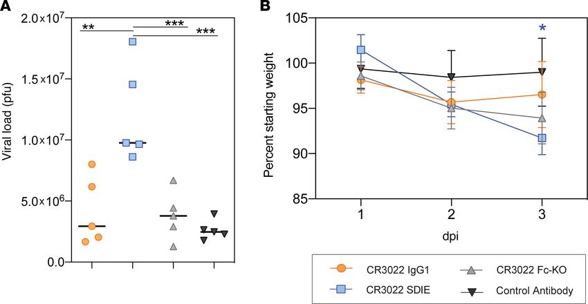
RESEARCH ARTICLE

                                  and antiinflammatories are under investigation (5, 6). In addition, anti–SARS-CoV-2 monoclonal thera-
                                  peutics also have been proposed to control and clear the infection.
                                      The coronavirus spike protein (S), found on the surface of coronaviruses, is involved in viral attach-
                                  ment and fusion (7). Treatment with monoclonal antibodies against SARS-CoV S protein has been shown
                                  to protect mice from viral pathogenesis (8). Delivery of both neutralizing and non-neutralizing antibodies
                                  against the MERS virus afforded protection (9–11), highlighting the potential importance both of block-
                                  ade of infection and targeted immune-mediated clearance of the virus/virally infected cells in protection
                                  from infection. Likewise, both neutralization and antibody-dependent cellular cytotoxicity (ADCC) have
                                  been linked to protection in SARS-infected individuals (12) and animal models (13). Given the remarkable
                                  infectiousness of SARS-CoV-2, with an estimated R0 approximately 2.5 (14–18), strategies to provide com-
                                  plete protection from infection may require both blocking and postinfection eliminating-antibody functions
                                  for maximal immunity. However, data from SARS-CoV–immunized nonhuman primates pointed to the
                                  potential role of neutralizing antibodies in enhancing disease, via the induction of inflammatory respons-
                                  es (19), suggesting that caution is warranted in the application of monoclonal antibody therapeutics for
                                  SARS-CoV-2 treatment.
                                      To begin to explore the potential immune-protective versus immunopathological role of antibodies, we
                                  focused on an antibody derived from a SARS-CoV–infected individual, CR3022, that targets a conserved
                                  epitope of the receptor binding domain (RBD) and that binds to the SARS-CoV-2 RBD (20). Because this
                                  cross-reactive antibody exhibits limited neutralization, despite binding to conserved determinants on the
                                  RBD (21), the antibody offered an opportunity to explore Fc-dependent effects on SARS-CoV-2 not con-
                                  founded by neutralization. Moreover, given that CR3022 continues to bind to RBD even in the presence of
                                  angiotensin-converting enzyme 2 (ACE2) (20–22), CR3022 has the potential to confer eradication of infect-
                                  ed cells even in the setting of high ACE2 secretion (23). Thus, here we coupled Fc functional profiling, Fc
                                  engineering, and in vivo profiling to examine the role of Fc effector function on the response to SARS-
                                  CoV-2 infection. Distinct Fc functional profiles resulted in enhancement of disease, pointing to antibody
                                  mechanisms of action that may be detrimental when developing antibody therapeutics against the virus.

                                  Results
                                  CR3022 drives innate immune activity against SARS-CoV-2. Although great effort is underway to identify potent
                                  neutralizing antibodies against SARS-CoV-2, it remains uncertain whether neutralization alone, particular-
                                  ly in the upper respiratory tract, will be sufficient to provide complete protection against this highly infec-
                                  tious pathogen. Instead, past studies in MERS infection suggest that additional antibody functions, beyond
                                  neutralization, track with protective immunity (13). Thus, the ability to clear virus or infected cells that
                                  escape restriction in the upper respiratory tract may be essential to fully prevent disease. Here we focused
                                  on a first-in-class monoclonal antibody, initially cloned from a SARS-CoV–infected individual, because of
                                  its ability to bind to a mutated neutralization-resistant form of the SARS-CoV S1-RBD (22). CR3022 binds
                                  to both SARS-CoV and SARS-CoV-2, with a binding footprint that likely provides the antibody with the
                                  ability to bind broadly across SARS-CoV mutants, such as SARS-CoV-2, and to continue to bind in the
                                  setting of ACE2 binding to the RBD (20, 21). However, whether this antibody was able to drive additional
                                  functions of potential therapeutic utility remained unclear.
                                       To begin, we confirmed the ability of CR3022 to bind to SARS-CoV and SARS-CoV-2 S as well as
                                  SARS-CoV-2 RBD and related CoV spike proteins (Figure 1A). As expected, CR3022 bound tightly to
                                  the SARS-CoV S, against which it was cloned. The antibody also bound SARS-CoV-2 RBD and S. In
                                  contrast, CR3022 did not bind to the MERS S, highlighting the specificity of this antibody for SARS-re-
                                  lated viruses. Although it was able to bind to SARS-CoV-2 RBD and potently neutralize SARS-CoV in
                                  vitro (22), limited authentic SARS-CoV-2 neutralization was observed even at very high antibody con-
                                  centrations (Figure 1B) as has been previously observed (21).
                                       Emerging data point to significant differences across monoclonal antibodies in their ability to drive Fc
                                  effector functions (24). Both the stoichiometry and geometry of binding have been implicated in modulating
                                  antibody effector function (25). Given the peculiar angle with which CR3022 interacts with the SARS-CoV-2
                                  RBD (21), we next examined the ability of CR3022 to drive Fc effector function. CR3022 bound to SARS-
                                  CoV-2 S was able to bind to Fc gamma receptors 2a and 3a (FcγR2a and FcγR3a) (Figure 1C), whereas no
                                  FcR binding was observed with the control EBOV-specific antibody (KZ52) bound to SARS-CoV-2 S, likely
                                  due to lack of EBOV monoclonal binding to the SARS-CoV-2 antigen (25). Along the same lines, CR3022

JCI Insight 2021;6(1):e143129 https://doi.org/10.1172/jci.insight.143129                                                                       2
RESEARCH ARTICLE JCI Insight 2021;6(1):e143129 https://doi.org/10.1172/jci.insight.143129 3
RESEARCH ARTICLE
Figure 1. CR3022 drives effector function against SARS-CoV-2. (A) CR3022 was serially diluted and tested for its ability to bind the spike protein of dif-
ferent coronaviruses by ELISA (left). Data are represented as the OD450 values background subtracted from the reference OD570 value. Each dot represents
the average of 2 replicates. The bar plot displays the EC50 for each antigen. MERS RBD and Ebola virus (EBOV) glycoprotein are not displayed because
their respective EC50 was each infinite. The bars represent the average of 2 replicates, and the error bars represent the standard deviation. (B) CR3022 was
serially diluted and preincubated with SARS-CoV-2 before adding to the virus and antibody to Vero E6 cells. Percentage neutralization was determined by
the percentage reduction in plaque counts compared with a vehicle control. Data represent the means of triplicates and error bars represent the standard
deviation. (C) CR3022 and a control EBOV-specific antibody binding to FcγR2a and 3a were evaluated using Luminex with a serial dilution of CR3022. The
AUC was calculated for the MFI values. The bar represents the mean of 2 replicates. (D–G) CR3022 was evaluated for its ability to drive ADCP (D), NK activi-
ty (as measured by MIP-1b activity) (E), ADCD (F), and ADNP (G). For the line graphs (left), each dot represents the mean of 2 replicates. For the bar graphs,
values are represented as the mean AUC of 8 serial dilutions run in replicate. The error bars for the dots and the AUC represent the standard deviation. For
the before-after plot (E), each dot represents the activity of 1 donor after incubation with CR3022 or serum. Significance for NK activation (E) was deter-
mined by a Wilcoxon matched pairs signed rank test, *P < 0.05. (H and I) The ability of CR3022 and a control EBOV-specific antibody (KZ52) to drive ADCP
(H) and ADCD (I) in the presence of ACE2 was analyzed. The bars indicate the average AUC of 8 serial dilutions run in 2 replicates. The error bars represent
the standard deviation. Significance was determined by a 1-way ANOVA test followed by Tukey’s multiple-comparison test. *P < 0.05.
was able to drive antibody-dependent cellular (monocyte) phagocytosis (ADCP), antibody-dependent NK
cell activation (ADNKA, as measured by macrophage inflammatory protein 1b [MIP-1b] expression), anti-
body-dependent complement deposition (ADCD), and antibody-dependent neutrophil phagocytosis (ADNP)
(Figure 1, D–G). Moreover, CR3022 was still able to drive antibody effector function, namely ADCP and
ADCD, in the presence of ACE2 (Figure 1, H and I). Thus, unlike neutralizing antibodies, many of which
compete with ACE2 binding, CR3022 may still drive antibody-effector clearance of virus or infected cells
even upon ACE2 upregulation following infection (26).
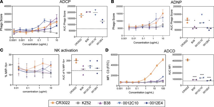
Comparison of CR3022 and other SARS-CoV-2 monoclonals. To fully probe the therapeutic potential of
CR3022, we compared the functional activity of CR3022 with recently published (27) or discovered mono-
clonal antibodies specifically cloned from SARS-CoV-2–infected individuals. Specifically, the ability of
CR3022 to mediate ADCP, NK activation, ADCD, and ADNP was compared with 2 neutralizing anti-
bodies (B38 and 0012C10) and 1 non-neutralizing antibody (0012E4) (Table 1). In addition, we used an
EBOV-targeting antibody (KZ52) as a negative control. All SARS-CoV-2 RBD-targeting antibodies drove
similar levels of ADCP and ADNP (Figure 2, A and B), with CR3022 comparable to all other SARS-
CoV-2 monoclonals. Conversely, CR3022 drove slightly more NK activation, as represented by MIP-1b
secretion (Figure 2C), and significantly more complement deposition (ADCD) compared with the other 3
SARS-CoV-2 RBD-targeting antibodies (Figure 2D). Thus, CR3022 exhibited similar if not superior anti-
body-effector function compared with neutralizing and other non-neutralizing antibodies. Moreover, given
the emerging role for ADCD in providing vaccine-mediated protection in vivo (28), the ability of CR3022
to facilitate ADCD makes CR3022 an ideal candidate for potential therapeutic applications.
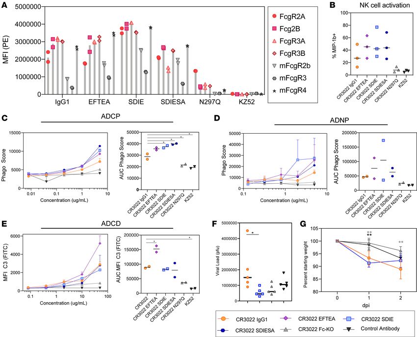
Fc engineering can tune CR3022 effector function. Although Fc effector functions have been linked to pro-
tection from MERS infection in mice (13), data have also emerged pointing to the potentially deleterious
role of Fc effector functions in enhancing SARS-CoV disease (19, 29, 30). Thus, using a simple Fc-engi-
neering approach where the variable domains of CR3022 are swapped onto the Fc domain of distinct Fc
variants with previously defined point mutations known to alter antibody interactions with Fc receptors, we
generated a small panel of CR3022 Fc variants, able to selectively augment phagocytosis, NK cell function,
and complement activity. Given clear overlap and concordance in human IgG1 performance across human
and mouse effector functions (31), we focused on known mutations in human IgG1 and compared human
and mouse Fc receptor binding as well as human antibody effector functions. These mutations in the CH2
or CH3 of the Fc of IgG1 were previously identified to alter binding to FcγRs, resulting in enhancement
or reduction of Fc effector function. Specifically, 4 mutants were explored: a mutant able to enhance all Fc
effector functions (EFTEA, ref. 32), 2 mutations known to enhance ADCC and ADCP (SDIE and SDIE-
SA, ref. 33), and a mutation that knocks out all Fc function (N297Q, ref. 34). To begin characterizing the
function of these variants, we analyzed the ability of these mutants to bind both human FcγRs and mouse
Fcγ receptors (mFcγRs) (Figure 3A). EFTEA showed similar binding to the human FcγRs as WT CR3022.
SDIE exhibited slightly higher binding to the human FcγRs compared with WT, whereas SDIESA had a
slight and selective reduction in binding to the human FcγR3A and FcγR3B. Conversely, the Fc-knockout
mutation, N297Q, resulted in a near-complete loss of Fc receptor binding.
Although the variants bound the mouse FcγRs with lower affinity than the human FcγRs, most vari-
ants retained considerable binding to mFcγR4, an activating receptor in mice that has been implicated
in antibody effector function (35). In particular, SDIE exhibited the highest binding to the mouse FcγRs
JCI Insight 2021;6(1):e143129 https://doi.org/10.1172/jci.insight.143129 4RESEARCH ARTICLE
Table 1. Neutralizing ability of antibodies that target the RBD of SARS-CoV-2
mAb Neutralization
CR3022 –
B38 +
0012C10 +
0012E4 –
The table lists the neutralizing capacity of a panel of monoclonal antibodies that bind to the SARS-CoV-2 RBD, with
B38, 0012C10, and 0012E4 cloned from infected individuals. B38 and 0012C10 are able to neutralize the virus, whereas
CR3022 and 0012E4 have limited to no neutralizing capacity.
compared with all other variants. Consistent with FcγR binding profiles, the EFTEA mutant exhibited
enhanced pan-functionality, with a selective increase in ADCD (Figure 3E). Conversely, both SDIE and
SDIESA exhibited enhanced ADCP and ADNP but not ADCD activity, with SDIE exhibiting enhanced
ADNP compared with the other mutants (Figure 3, C–E). Finally, SDIESA exhibited the highest ADCP
activity and NK activation, as measured by MIP-1b expression, of the group (Figure 3, B and C), high-
lighting the distinct functional profiles of each of the modifications. Although some residual binding was
noted for N297Q on FcγR2A (Figure 3A), near-complete loss of antibody function was observed with
this Fc variant (Figure 3, B–E), driving effector function at a similar level as the EBOV-specific control
antibody, underscoring the dominant silencing effect of this mutation. Overall, these data highlight a
range of functional variation across the Fc variants, enabling the down-selection of 3 variants for in vivo
analysis: WT, Fc-knockout (Fc-KO), and pan-functional SDIE variant, which exhibited the highest bind-
ing to mouse FcγRs (Figure 3A).
Dissecting therapeutic Fc signals of protection. Using these down-selected Fc variants, we next aimed
to probe the role of Fc effector function in protection from infection and disease (36). BALB/c mice
were treated 12 hours following SARS-CoV-2 infection to probe for therapeutic protective Fc functions.
Specifically, BALB/c mice were infected with 105 PFU of mouse-adapted SARS-CoV-2 i.n.; treated
with 200 μg of the WT, Fc-KO, or control antibody or 100 μg of the Fc-enhanced antibody i.p.; and
monitored for 2 days for lung viral titer and weight loss, with 5 mice per group (Figure 3, F and G).
Strikingly, slightly higher, but nonsignificant, differences were observed in viral replication using the WT
CR3022 IgG1 compared with the control antibody and the WT antibody (Figure 3G). Moreover, reduction
in viral load was observed with the Fc-enhanced CR3022 variant (Figure 3G). However, significant weight
loss occurred in both WT and Fc-enhanced variant-treated mice and minimal to no weight loss in mice treat-
ed with the Fc-KO variants (Figure 3G). Thus, while the WT CR3022 IgG1 and the Fc-enhanced CR3022
variants showed divergent virologic effects, both variants led to enhanced pathology. The disconnect between
viral load and weight loss for the Fc-enhanced CR3022 antibody raised the possibility that the Fc-enhanced
variant may have pathological consequences.
To further understand the role of Fc-enhanced pathology, we next tested the therapeutic benefit of
the CR3022 variants in a more pathological model of SARS-CoV-2 infection (37, 38). Syrian golden
hamsters are highly susceptible to SARS-CoV-2 infection and develop severe infection after challenge.
Thus, using this model, we assessed the panel of Fc-engineered monoclonals. Hamsters were chal-
lenged i.n. with 106 PFU/mL (107 TCID50) of SARS-CoV-2, and 1 day after infection, treated with 5
mg/kg IgG1, Fc-enhanced, Fc-KO, or a control antibody, with 5 hamsters per group. Weight was mon-
itored daily, and lung viral titers were determined 3 days after infection (Figure 4, A and B). Similar
to the results observed in the mouse model (Figure 3G), the WT CR3022 had no impact on viral load
compared with control antibody–treated animals (Figure 4A). In contrast to mice, hamsters did not
experience any benefit from the Fc-KO antibody (Figure 4A), likely due to the more severe nature of the
infection in this model. Conversely, hamsters treated with the Fc-enhanced CR3022 exhibited increased
viral load in the lung and increased weight loss (Figure 4, A and B). Despite the viral load disparity
across the mice and hamsters, both models exhibited increased weight loss upon treatment with the
Fc-enhanced CR3022 (Figure 3G and Figure 4B), suggesting similar host responses to the Fc-enhanced
monoclonal. Thus, collectively, these data point to the critical importance of balancing Fc effector func-
tion to temper pathology in susceptible populations.
JCI Insight 2021;6(1):e143129 https://doi.org/10.1172/jci.insight.143129 5RESEARCH ARTICLE
Figure 2. CR3022 possesses comparable Fc activity to neutralizing and non-neutralizing SARS-CoV-2 monoclonal antibodies. (A–D) CR3022 was
tested for its ability to drive ADCP (A), ADNP (B), NK cell activation (C), or ADCD (D) compared with other SARS-CoV-2 RBD-targeting monoclonals.
For the line graphs, each dot represents the mean of 2 replicates. For NK cell activation, 3 donors were used. For the bar graphs, the values are
represented as the AUC of serial dilutions. The dotted line represents the average AUC value for the control EBOV-targeting antibody (KZ52). The
error bars represent the standard deviation between the replicates. Significance was determined by an ordinary 1-way ANOVA followed by Tukey’s
multiple-comparison test. *P < 0.05, ****P < 0.0001.
Discussion
Given the rapid spread of SARS-CoV-2, therapeutics are urgently needed to not only prevent but also treat
COVID-19. Among the strategies, passive transfer of monoclonal antibodies, which are able to both drive
directed antiviral activity and also tune the immune system, represent an ideal class of therapeutics, poten-
tially suited for both prevention and therapy. However, emerging data pointing to the possibility of antibody
enhancement of disease following vaccination against SARS-CoV have raised the importance of carefully
considering the role of the antibody Fc in SARS-CoV-2 therapeutic design. Here we focused on a first-in-class
cross-SARS monoclonal antibody, CR3022, which interacts with a conserved region of the RBD. Although
the antibody itself was highly functional, even in the presence of ACE2 (Figure 1), additional Fc engineering
was performed, allowing us to gauge the therapeutic benefits of Fc activity (Figure 3). Surprisingly, the Fc-en-
hanced CR3022 antibody conferred some viral control when administered in mice but was accompanied by
significant morbidity in both mice and hamsters, potentially by promoting an inflammatory response.
Past reports have shown some evidence of antibody-dependent enhancement (ADE) of SARS infection.
Although many of these studies rely on in vitro systems with high levels of virus, diluted serum, or nonfunc-
tional antibodies (39, 40), a more recent study in nonhuman primates pointed to a disease-enhancing role of
modified vaccinia Ankara-induced SARS-CoV neutralizing antibodies (19). In vitro data suggested that despite
the neutralizing activity of the vaccine-induced antibodies, ADE in SARS infection was caused by FcγR2-me-
diated activation of myeloid cells (41, 42). ADE is most well documented during Dengue virus infection, in
which previous infection places an individual at risk, upon reinfection, to develop Dengue hemorrhagic fever/
Dengue shock syndrome (DHF/DSS). This disease enhancement is thought to occur due to the presence of
antibodies directed at a different Dengue serotype. Specifically, subneutralizing levels of preexisting antibod-
ies to one serotype that have limited cross-reactive neutralizing capacity to a second serotype are enriched in
children who develop DHF/DSS following exposure to a new serotype of the virus (43). Moreover, in vitro,
low levels of neutralizing antibodies have been shown to facilitate viral entry into myeloid cells following FcγR
engagement, resulting in enhanced infection and consequent inflammation (44). However, it is critical to note
that limited evidence exists for SARS-CoV-2 infection of myeloid cells (45, 46). Instead, SARS-CoV-2 target
JCI Insight 2021;6(1):e143129 https://doi.org/10.1172/jci.insight.143129 6RESEARCH ARTICLE
Figure 3. CR3022 can be enhanced through Fc engineering. (A) Each CR3022 variant was analyzed for its ability to bind the human FcγR2a, FcγR2b,
FcγR3a, and FcγR3b or the mouse FcγR2b, FcγR3, and FcγR4 by Luminex. Each bar represents the average MFI and error bars show standard devi-
ation. (B) The CR3022 variants were analyzed for their ability to drive NK activation, as measured by MIP-1b activity. The bar graphs represent the
average of 3 donors, and the error bars represent the standard deviation. For AUC, significance was determined by an ordinary 1-way ANOVA followed
by Tukey’s multiple-comparison test. *P < 0.05. (C–E) The CR3022 variants were evaluated for their ADCP (C), ADNP (D), and ADCD (E) activity. The
dots on the line graph represent the average of 2 replicates. The bar graphs represent the average AUC of 8 serial dilutions run as replicates, and the
error bars represent the standard deviation. For AUC, significance was determined by an ordinary 1-way ANOVA followed by Tukey’s multiple-com-
parison test. *P < 0.05. (F and G) BALB/c mice (5 per group) were treated with a CR3022 variant or control therapeutically. Lung viral titers were
determined 2 days postinfection (dpi) (F), and weight was monitored daily (G). For viral titer (F), significance was determined by an ordinary 1-way
ANOVA followed by Tukey’s multiple-comparison test. *P < 0.05. For weight (G), significance was determined by an ordinary 1-way ANOVA followed
by Tukey’s multiple-comparison test. *P < 0.05, **P < 0.01, between the respective antibody, indicated by the color of the asterisk, and the CR3022
WT. For weight (G), each dot represents a mouse, and the error bars represent the standard deviation.
cells do not express FcγR, and thus enhanced infection is unlikely to occur in the same manner as for Den-
gue viral infection. Conversely, SARS-CoV-2 target cells do express the neonatal Fc receptor, FcRn (47, 48).
Whether enhancement can be caused by FcRn remains unclear. Yet, enhanced FcγR engagement may deliver
virus preferentially to endosomal compartments in phagocytes, resulting in viral sensing and inflammatory
responses, that may then lead to inflammation, cellular recruitment, and potential pathology. Thus, antibodies
in SARS-CoV-2, unlike Dengue viral infection, may cause enhanced inflammation rather than enhanced infec-
tion. Importantly, here both FcγR-engaging antibodies drove pathology in both mice and hamsters but showed
differences in viral loads. These data point to a disconnect between viral load and pathogenesis that may be
dissected in the future with in-depth immunohistopathological studies across the 2 animal models.
JCI Insight 2021;6(1):e143129 https://doi.org/10.1172/jci.insight.143129 7RESEARCH ARTICLE
Figure 4. Increased Fc function causes enhancement of disease in vivo. Hamsters (5 per group) were challenged with
SARS-CoV-2 and treated with a CR3022 variant or control antibody 1 dpi. Lung viral titers were determined 3 dpi (A), and
weight was monitored daily (B). For viral titers (A), significance was determined by an ordinary 1-way ANOVA followed by
Tukey’s multiple-comparison test. **P < 0.01, ***P < 0.001. For weight (B), significance was determined by 2-way ANOVA
test followed by Tukey’s multiple-comparison test. *P < 0.05 between the respective antibody and the CR3022 WT. For
weight (B), each dot represents the average of 5 hamsters, and the error bars represent the standard deviation.
It is critical to note that antibodies elicited by infection and vaccine platforms or monoclonal
antibodies do not have the same Fc binding profiles as those induced by modified vaccinia Ankara
(MVA) vaccination that was previously associated with SARS-CoV–enhanced disease in macaques
(49). Importantly, distinct Fc binding profiles can be generated following infection and vaccination,
driven by altered Fc subclass selection and Fc glycosylation (49). Moreover, previous studies have
clearly illustrated striking differences in antibody functional profiles across MVA-, pox virus–, adeno-
virus-, and protein-based immunization strategies (50). Thus, it is plausible that polyclonal pools of
antibodies, with neutralizing properties and balancing Fc receptor binding profiles via balanced Fc gly-
cosylation, may provide protection in the absence of disease. Along these lines, recent vaccine studies
point to a positive predictive role of polyclonal Fc recruiting directed at both the whole spike and RBD
of SARS-CoV-2 (28, 51). Similarly recent monoclonal therapeutic studies with neutralizing WT IgG1
demonstrate limited evidence of disease in humans (52).
Despite the significant loss of most Fc effector functions in the Fc-KO variant (Figure 3), this variant
retained low-level binding to human FcγR2A, involved in phagocytosis in humans (53). This remaining Fc
receptor binding may have contributed to low but sufficient levels of immune complex–based activation
in the susceptible hamster model, where even the Fc-KO variant drove weight loss. These data point to
the ultrasensitive nature of the hamster model. Although mice exhibit more attenuated disease, hamsters
suffer highly pathological responses to SARS-CoV-2 (37). Whereas the Fc-KO exhibited reduced viral and
no weight loss in the mice, the administration of the Fc-enhanced variant resulted in pathology. Con-
versely, the more susceptible hamster model showed no benefit with any of the Fc variants and instead
exhibited the same enhanced pathology with the Fc-enhanced. Thus, Fc enhancement may represent a
liability across the disease spectrum. However, it is critical to note that many distinct Fc modifications
can be utilized to drive enhanced biological activity. Although the SDIE mutation used here improved all
measured Fc effector functions, other mutations exist that selectively improve NK cell activity, monocyte
phagocytosis, neutrophil activation, or complement deposition, offering potentially more precise mecha-
nisms to control infection. Moreover, recent vaccine correlates analysis in nonhuman primates highlighted
the complementary activity of monocyte phagocytosis and complement in viral control (28, 51). Whether
these functions alone may selectively clear the virus and prevent inflammation and pathology remains
unclear but could provide critical clues for the strategic engineering of monoclonal antibodies to maximize
protection and minimize pathology.
JCI Insight 2021;6(1):e143129 https://doi.org/10.1172/jci.insight.143129 8RESEARCH ARTICLE
As the number of COVID-19 cases rise globally, new therapies are urgently needed to treat this highly
infectious virus. Here, we characterized the therapeutic functions of the monoclonal antibody CR3022 that
binds to a conserved site on the RBD that is not fully blocked in the presence of ACE2, offering therapeutic
benefit even after infection has been initiated. The data presented here show no effect of CR3022 as a WT
IgG1. Surprisingly, both the Fc-enhanced and Fc-silenced variants of CR3022 showed an antiviral benefit in
mice but resulted in dichotomous treatment-associated pathology. Interestingly, the same pathological phe-
notype was observed in hamsters with the Fc-enhanced variant, highlighting the consistent disease-enhanc-
ing phenotype of highly functional non-neutralizing monoclonal variants. With the rapid discovery of novel
neutralizing antibodies and pan–cross-reactive CoV antibodies, coupled to rapid Fc engineering, enabling the
potential to deeply profile the involvement of all Fc receptors (including FcγR1, FcγR2a, FcγR2b, FcγR3a,
FcγR3b, FcRn, as well as noncanonical Fc receptors) and the generation of functionally optimized antibod-
ies with appropriate half-lives, the development of therapeutics with the highest clinical benefit is possible.
Methods
Cell lines. THP-1 cells, originally isolated from a 1-year-old male human (ATCC), were maintained
in RPMI supplemented with 10% fetal bovine serum, l-glutamine, HEPES, penicillin/streptomycin, and
0.01% β-mercaptoethanol. Vero E6 cells, from BEI Resources, National Institute of Allergy and Infectious
Diseases (NIAID) NIH: VERO C1008 (E6), African green monkey kidney, Working Bank NR-596, were
maintained in humidified incubators at 37°C and 5% CO2 in Dulbecco’s modified Eagle medium (DMEM)
with GlutaMAX and sodium pyruvate supplemented with 10% (v/v) certified US-origin heat-inactivated
fetal bovine serum (HI-FBS).
Viruses. SARS-CoV-2 USA-WA1/2020 (54) was propagated on Vero E6 cells in DMEM supplemented
with 2% HI-FBS, GlutaMAX, sodium pyruvate, nonessential amino acids, and antibiotic-antimycotic. At
62 hours, the supernatant was harvested and clarified by centrifugation. The final concentration of HI-FBS
was diluted to 10% (v/v) prior to cryopreservation at –80°C. Final passage was VERO+3, Vero E6+2 (lot
NSU-V004). The sequence of this stock was identical to the published reference consensus sequence (54).
For in vivo studies, a recombinant SARS-CoV-2 mouse-adapted variant was constructed by introduction
of 2 amino acid changes in the ACE2 binding pocket. Virus stocks were grown on Vero cells and titered by
plaque assay as previously described by our group (36).
Animals. Female 12-month-old BALB/c mice were obtained from Envigo (strain 047). All animal work
was approved by Institutional Animal Care and Use Committee at University of North Carolina at Chapel
Hill. Syrian golden hamsters, 6–7 weeks old, were from and maintained at University of Texas Medical Branch.
Plasmid design. To create the CR3022 variants, gene blocks were designed containing the Fc domain of
IgG1 and previously defined, individual Fc point-mutant backbones with known differences in binding to
Fc receptors and functional differences (32–34). These Fc domains were cloned into individual pUC donor
plasmids. In addition, 3 pUC plasmids encoding the variable heavy chain, a furin P2A sequence, or the
variable light chain were designed surrounded by BsaI sites. In addition to the 4 pUC plasmids, a destina-
tion vector was cloned with an IL-2 secretion signal, the suicide gene ccdB surrounded by BsaI sites, and
the kappa light chain sequence. The 4 pUC donor plasmids and the destination vector were combined in a
single digestion-ligation reaction, using Golden Gate cloning, to create full IgG molecules with the same
CR3022 antigen-binding (Fab) domain but different Fc domains.
Protein expression and purification. The RBD (residues 319–529) of SARS-CoV-2 S protein (Gen-
Bank: MN975262.1) were subcloned into a pVRC vector with a C-terminal SBP-tag.
The CR3022 (GenBank: DQ168569 and DQ168570), B38 (27), and 0012C10 and 0012E4 antibodies
(provided in-house) were produced in 293F suspension cells grown in FreeStyle 293 Expression media
(Gibco, Thermo Fisher Scientific). Cells were transfected with Polyethylenimine (PEI; Polysciences) at
1 μg/μL in a ratio of 3 μg PEI to 1 μg DNA. Supernatants were harvested 5 days posttransfection, and
antibody was purified using protein G magnetic beads (MilliporeSigma). For in vitro analysis, KZ52 (May-
flower Bioscience, 0260-001) was used as a negative control.
ELISA. ELISA plates were coated with 50 ng/well of antigen in PBS overnight at 4°C on a shaker at
low speed. The next day, plates were washed 5 times with PBS-0.05% Tween-20 (PBST) and blocked in 5%
BSA in PBS for 2 hours at room temperature on a shaker at low speed. Plates were washed 5 times with
PBST, and 5-fold serially diluted antibody was added and incubated for 2 hours at room temperature on a
shaker at low speed. After the incubation, plates were washed with PBST and anti–human IgG1–HRP was
JCI Insight 2021;6(1):e143129 https://doi.org/10.1172/jci.insight.143129 9RESEARCH ARTICLE
added for detection. Plates were incubated for 1 hour at room temperature on a shaker at low speed. Plates
were washed with PBST. The ELISA was developed with the addition of TMB (Invitrogen, Thermo Fisher
Scientific). The reaction was stopped with 1 M H2SO4. Signal reading was carried out at 450 nm (reference
wavelength of 570). Data were reference value and background corrected.
ADCP assay. The ADCP assay was adapted from Ackerman et al. (55). Briefly, antigen was biotinylat-
ed using sulfo-NHS (N-hydroxysulfosuccinimide, Pierce, Thermo Fisher Scientific, A39269) LC-LC biotin,
coupled to yellow-green fluorescent Neutravidin 1 μm beads (Invitrogen, Thermo Fisher Scientific, F8776)
for 2 hours at 37°C and washed 3 times in 0.1% BSA in PBS. The coupled beads were resuspended to a final
volume of 10 μg/mL. A total of 10 μL/well of coupled beads were added to 96-well plates with 100 μL/well
of antibodies at a concentration of 5 μg/mL, 1 μg/mL, 0.2 μg/mL, and 0.04 μg/mL for 2 hours at 37°C to
form immune complexes. After incubation, the immune complexes were spun down and the supernatant was
removed. THP-1 cells (ATCC) were added at a concentration of 2.5 × 10⁴ cells/well and incubated for 18 hours
at 37°C. After incubation, the plates were spun down, the supernatant was removed, and cells were fixed with
4% PFA for 20 minutes. Fluorescence was acquired with an Intellicyt iQue. Phagocytic score was calculated
using the following formula: (percentage of FITC+ cells × the geometric MFI of the FITC+ cells)/10,000.
ADNP assay. The ADNP assay was adapted from Karsten et al. (56). Antigens were coupled to beads
and immune complexes were formed as described for ADCP. Neutrophils were isolated from freshly drawn
whole blood. Erythrocytes were lysed with ammonium-chloride potassium lysis buffer (150 mM NH4Cl, 10
mM KHCO3, 0.1 mM Na2 EDTA, pH 7.4), and leukocytes were separated out by centrifugation, 500g for
5 minutes at room temperature. Leukocytes were washed with cold PBS, resuspended in R10, and added to
plates at a concentration of 5 × 10⁴ cells/well. The plates were incubated for 1 hour at 37°C. The neutrophil
marker CD66b (Pacific Blue–conjugated anti-CD66b; BioLegend, 305112) was used to stain cells. Cells were
fixed for 20 minutes in 4% paraformaldehyde (PFA). Fluorescence was acquired with an Intellicyt iQue, and
the phagocytic score was calculated as described for ADCP.
ADCD assay. The ADCD assay was adapted from Fischinger et al. (57). Antigen was coupled to red fluo-
rescent Neutravidin 1 μm beads (Invitrogen, Thermo Fisher Scientific, F8775) as described for ADCP. Immune
complexes were formed by incubating 10 μL of coupled beads with 50 μL of antibody at concentrations of
50 μg/mL, 10 μg/mL, 2 μg/mL, and 0.4 μg/mL for 2 hours at 37°C. Plated were spun down, and immune
complexes were washed with PBS. Lyophilized guinea pig complement (Cedarlane, CL4051) was resuspended
in 1 mL of cold water, diluted 1:50 in GVB++ (gelatin veronal buffer and additional Ca2+ and Mg2+, Boston
BioProducts, IBB-300X), and added to the immune complexes. The plates were incubated for 20 minutes at
37°C, and the reaction was stopped by washing the plates twice with 15 mM EDTA in PBS. To detect comple-
ment deposition, plates were incubated with fluorescein-conjugated goat anti–guinea pig complement C3 (MP
Biomedicals, 0855385) for 15 minutes in the dark. Fluorescence was acquired with an Intellicyt iQue.
ADNKA (NK activation). Human NK cells were isolated from buffy coats using RosetteSep NK cell enrich-
ment kit (StemCell Technologies) and Ficoll separation. The isolated NK cells were rested overnight at 1.5
× 106 cells/mL in IL-15 at 37°C. ELISA plates were coated with antigen at 300 ng/well and incubated for 2
hours at 37°C. Plates were blocked with 5% BSA in PBS overnight at 4°C. The next day, 100 μL of antibodies,
at a concentration of 5 μg/mL, were added to the plates. Plates were incubated for 2 hours at 37°C to form
immune complexes. After the incubation, NK cells were added to the plates at 5 × 10⁴ cells/well in R10 sup-
plemented with anti-CD107a PE-Cy5, Brefeldin A (MilliporeSigma, B7651-5MG), and GolgiStop (BD Bio-
sciences, 555802). Plates were incubated for 5 hours at 37°C. Following the incubation, NK cells were stained
for the surface markers with anti-CD56 PE-Cy7, anti-CD16 APC-Cy7, and anti-CD3 Pacific Blue (BD Biosci-
ences, 557747, 557758, 558124). NK cells were fixed and permeabilized with Fix&Perm cell permeabilization
kit (Invitrogen, Thermo Fisher Scientific). Cells were incubated with anti–MIP-1β PE and anti–IFN-γ FITC
(BD Biosciences, 550078, 340449) to stain for intracellular markers. Cells were acquired on an Intellicyt iQue.
FcR binding. A multiplex assay was used to determine FcR binding as described in Brown et al. (58,
59). A 2-step carbodiimide reaction was used to couple antigen to Magplex Luminex beads. Beads were
activated for 30 minutes at room temperature using 100 mM monobasic sodium phosphate, pH 6.2, with
5 mg/mL sulfo-NHS and 5 mg/mL ethyl dimethylaminopropyl carbodiimide hydrochloride. Beads were
then washed with 50 mM 2-(N-Morpholino)ethanesulfonic acid (MES), pH 5.0, and incubated with 25
μg of antigen in 50 mM MES, pH 5.0, for 2 hours on a rotator. The coupled beads were blocked in Block-
ing Buffer (PBS, 0.1% BSA, 0.02% Tween-20, 0.05% Azide, pH 7.4). After blocking, coupled beads were
washed in PBS-Tween, resuspended in PBS, and stored at 4°C.
JCI Insight 2021;6(1):e143129 https://doi.org/10.1172/jci.insight.143129 10RESEARCH ARTICLE
For the detection of FcR binding, FcRs with an AviTag were biotinylated using a BirA500 kit (Avidity)
per the manufacturer’s instructions. Coupled beads were diluted to a concentration of 100 microspheres per
antigen/μL in 0.1% BSA in PBS. Antibodies were serially diluted in 0.1% BSA in PBS; mixed with diluted
beads in a black, clear-bottom, 384-well plate; and incubated at 4°C for 16 hours, shaking at 900 rpm. After
the incubation, plates were washed with 0.1% BSA in PBS. FcRs were incubated with streptavidin-PE
(Prozyme, PJ31S) for 10 minutes. PE-labeled FcRs were added to plates and incubated for 1 hour at room
temperature on a shaker. Plates were washed with 0.1% BSA in PBS and resuspended in Qsol Buffer (Intel-
licyt). Fluorescence was acquired on the Intellicyt iQue.
In vitro plaque reduction neutralization assay. The day prior to assay, VeroE6 cells were seeded to a
density of 8 × 10⁵ cells/well in 6-well plates. SARS-CoV-2 was diluted in DMEM with GlutaMAX and
sodium pyruvate supplemented with 1× antibiotic-antimycotic and 2% HI-FBS to 1000 PFU/mL (tar-
get 100 PFU per well). Antibody was serially diluted in Dulbecco’s PBS, and an equal volume of dilut-
ed SARS-CoV-2 was added, mixed, and incubated for 1 hour at 37°C before plating on 6-well plates
(200 μL in triplicate). Following a 1-hour incubation at 37°C with periodic rocking, they were overlaid
with a 1:1 mixture of 2.5% (w/v) Avicel RC-591 (provided by DuPont Nutrition & Health) prepared in
distilled water and 2× Temin’s Modified Eagle Medium (Thermo Fisher Scientific) supplemented with
10% HI-FBS, 2× GlutaMAX, and 2× antibiotic-antimycotic. Following a 2-day incubation at 37°C and
5% CO2, plates were fixed with 10% neutral buffered formalin for removal from biocontainment and
stained with a solution of 0.2% Gentian Violet and 10% neutral buffered formalin. Plates were rinsed
under water and plaques were enumerated. Percentage neutralization was calculated from vehicle/
virus-only control wells.
In vivo challenge. Female 12-month-old BALB/c mice were treated prophylactically (12 hours before
infection) or therapeutically (12 hours after infection) with 200 μg or 100 μg of antibody through
the intraperitoneal route. Each group contained 5 mice. Mice were challenged i.n. with 105 PFU of
mouse-adapted SARS-CoV-2, representing 0.69 × 10⁵ TCID50, which falls clearly in the range of viral
loads observed in hospitalized patients (60). Weight was monitored on days 0, 1, and 2 after infec-
tion. Mice were sacrificed 2 days after infection, and lung viral titer was determined by plaque assay.
Although the Fc variants experience different half-lives in vivo, the studies performed here were short;
but half-life should be considered for longer studies.
Adult hamsters were microchipped a day prior to experimental challenge. On day 0, hamsters were
anesthetized with ketamine/xylazine and challenged with SARS-CoV-2 by the i.n. route using a 107
TCID50 (or 106 PFU/mL) dose in a total volume of 100 μL. The final challenge dose was 104 PFU dilut-
ed in sterile PBS. Body weight and body temperature were measured each day, starting at day 0. On day
1 postchallenge (1 dpc) hamsters were treated with 5 mg/kg of monoclonal antibodies diluted in 0.5 mL
of sterile PBS via the intraperitoneal (i.p.) route. The control group got an equal volume of sterile PBS
via the same i.p. route. On 3 dpc all animals were sacrificed. At necropsy, lungs were harvested for all
groups. Left lungs were fixed with 10 volumes of fresh 10% formalin; right lungs were frozen in 5 mL
lysogeny broth from Thermo Fisher Scientific for viral load analysis. Tissue sections were homogenized
in bead beater tubes (Thomas Scientific) and weighed, and supernatants were titrated per standard pro-
tocol. Briefly, 100 μL of a 10× dilution of supernatants was incubated in 96-well plates for 1 hour, and
supernatants were replaced by methyl cellulose overlay and incubated for 3 days at 5% CO2 and 37°C.
Plates were fixed with formalin and removed from the biosafety level 4 facility, after which the plates
were inactivated and immunostained, and the plaques were counted to obtain viral titers.
Statistics. All data were visualized and analyzed in GraphPad Prism. Nonparametric tests were
performed as described in figure legends. Where applicable, significance was determined as *P < 0.05,
**P < 0.01, ***P < 0.001.
Study approval. Primary human innate immune cells were isolated from fresh peripheral blood samples
collected by the Massachusetts General Hospital (MGH) blood bank. All subjects provided informed con-
sent, and the study was approved by the MGH Institutional Review Board. All subjects were older than 18
years of age, and samples were deidentified prior to use.
All mouse work was approved by the Institutional Animal Care and Use Committee at the University
of North Carolina at Chapel Hill. The animal protocols for the hamster models were approved by the Insti-
tutional Animal Care and Use Committee of the University of Texas Medical Branch.
JCI Insight 2021;6(1):e143129 https://doi.org/10.1172/jci.insight.143129 11RESEARCH ARTICLE
Author contributions
CA, MDS, AG, TS, BDJ, JG, DRW, AB, RB, and GA designed the study. CA, SF, MDS, JB, AS, SRL,
NAK, CM, AH, RJ, NS, PT, TZ, JL, AZ, and CL performed experiments. MB, DRW, and JRD provided
reagents. CA performed all analysis.
Acknowledgments
We thank Nancy Zimmerman, Bruce Walker, Mark and Lisa Schwartz, and Terry and Susan Ragon for
their support. We would also like to thank Bing Chen, Kizzmekia Corbett, Aaron Schmidt, Jared Feld-
man, Blake Hauser, and Tim Caradonna for protein production efforts and Sierra Downs for technical
support. The following reagent was obtained through BEI Resources, NIAID, NIH: VERO C1008 (E6),
Kidney (African green monkey), Working Cell Bank, NR-596. The SARS-CoV-2 starting material was
provided by the World Reference Center for Emerging Viruses and Arboviruses, with Natalie Thornburg
(nax3@cdc.gov) as the CDC principal investigator. Avicel RC-591 was provided by DuPont Nutrition
& Health. We would like to thank Deborah Gakpo, Jillian Bensko, Sudeshna Fisch, Meghan Travers,
Shaghayaegh Habibi, Yuezhou Chen, Adam Zuiani, and Felipe N. Lelis for organizing and collecting
human samples used for the cloning of monoclonal antibodies. We would also like to thank Massachu-
setts Consortium on Pathogen Readiness, the Samana Cay MGH Scholar program, and an anonymous
donor for financial support. This work was supported by the NIH (3R37AI080289-11S1) and the NIAID,
NIH (1U01CA260476-01, U19 AI135995, NIH AI121394, AI139538).
Address correspondence to: Galit Alter, 400 Technology Square, Cambridge, Massachusetts 02139, USA.
Phone: 857.268.7003; Email: galter@mgh.harvard.edu.
1. WHO. Weekly Epidemiological Update on COVID-19. https://www.who.int/publications/m/item/weekly-epidemiologi-
cal-update-8-december-2020. Updated December 8, 2020. Accessed December 14, 2020.
2. Peiris JS. Severe acute respiratory syndrome (SARS). J Clin Virol. 2003;28(3):245–7.
3. de Wit E, et al. SARS and MERS: recent insights into emerging coronaviruses. Nat Rev Microbiol. 2016;14(8):523–34.
4. Ruan S. Likelihood of survival of coronavirus disease 2019. Lancet Infect Dis. 2020; 20(6):630–631.
5. Devaux CA, et al. New insights on the antiviral effects of chloroquine against coronavirus: what to expect for COVID-19? Int J
Antimicrob Agents. 2020;55(5):105938.
6. Grein J, et al. Compassionate use of remdesivir for patients with severe Covid-19. N Engl J Med. 2020;382(24):2327–2336.
7. Song Z, et al. From SARS to MERS, thrusting coronaviruses into the spotlight. Viruses. 2019;11(1):E59.
8. Jin Y, et al. Human monoclonal antibodies as candidate therapeutics against emerging viruses. Front Med. 2017;11(4):462–470.
9. Widjaja I, et al. Towards a solution to MERS: protective human monoclonal antibodies targeting different domains and func-
tions of the MERS-coronavirus spike glycoprotein. Emerg Microbes Infect. 2019;8(1):516–530.
10. Corti D, et al. Prophylactic and postexposure efficacy of a potent human monoclonal antibody against MERS coronavirus. Proc
Natl Acad Sci U S A. 2015;112(33):10473–10478.
11. Li Y, et al. A humanized neutralizing antibody against MERS-CoV targeting the receptor-binding domain of the spike protein.
Cell Res. 2015;25(11):1237–1249.
12. Du L, et al. The spike protein of SARS-CoV--a target for vaccine and therapeutic development. Nat Rev Microbiol.
2009;7(3):226–236.
13. Zhao J, et al. Passive immunotherapy with dromedary immune serum in an experimental animal model for Middle East respira-
tory syndrome coronavirus infection. J Virol. 2015;89(11):6117–6120.
14. Li Q, et al. Early transmission dynamics in Wuhan, China, of Novel coronavirus-infected pneumonia. N Engl J Med.
2020;382(13):1199–1207.
15. Zhang S, et al. Estimation of the reproductive number of novel coronavirus (COVID-19) and the probable outbreak size on the
Diamond Princess cruise ship: a data-driven analysis. Int J Infect Dis. 2020;93:201–204.
16. Cheng ZJ, Shan J. 2019 Novel coronavirus: where we are and what we know. Infection. 2020;48(2):155–163.
17. Maimuna M, Mandi KD. Early transmissibility assessment of a novel coronavirus in Wuhan, China [preprint]. https://
doi:10.2139/ssrn.3524675. Posted on SSRN January 24, 2020.
18. Sanche S, et al. High contagiousness and rapid spread of severe acute respiratory syndrome coronavirus 2. Emerg Infect Dis.
2020;26(7):1470–1477.
19. Liu L, et al. Anti-spike IgG causes severe acute lung injury by skewing macrophage responses during acute SARS-CoV infec-
tion. JCI Insight. 2019;4(4):e123158.
20. Tian X, et al. Potent binding of 2019 novel coronavirus spike protein by a SARS coronavirus-specific human monoclonal anti-
body. Emerg Microbes Infect. 2020;9(1):382–385.
21. Yuan M, et al. A highly conserved cryptic epitope in the receptor-binding domains of SARS-CoV-2 and SARS-CoV [preprint].
https://doi:10.1101/2020.03.15.992883. Posted on bioRxiv March 17, 2020.
22. ter Meulen J, et al. Human monoclonal antibody combination against SARS coronavirus: synergy and coverage of escape
mutants. PLoS Med. 2006;3(7):e237.
23. Hamming I, et al. Tissue distribution of ACE2 protein, the functional receptor for SARS coronavirus. A first step in understand-
JCI Insight 2021;6(1):e143129 https://doi.org/10.1172/jci.insight.143129 12RESEARCH ARTICLE
ing SARS pathogenesis. J Pathol. 2004;203(2):631–637.
24. Gunn BM, et al. A role for Fc function in therapeutic monoclonal antibody-mediated protection against Ebola virus. Cell Host
Microbe. 2018;24(2):221–233.e5.
25. Saphire EO, et al. Systematic analysis of monoclonal antibodies against Ebola virus GP defines features that contribute to pro-
tection. Cell. 2018;174(4):938–952.e13.
26. Ziegler CGK, et al. SARS-CoV-2 receptor ACE2 is an interferon-stimulated gene in human airway epithelial cells and is detect-
ed in specific cell subsets across tissues. Cell. 2020;181(5):1016–1035.e19.
27. Wu Y, et al. A noncompeting pair of human neutralizing antibodies block COVID-19 virus binding to its receptor ACE2. Sci-
ence. 2020;368(6496):1274–1278.
28. Yu J, et al. Bibliometric analysis of Ebola research indexed in web of science and scopus (2010-2020). Biomed Res Int.
2020;2020:5476567.
29. Takada A, Kawaoka Y. Antibody-dependent enhancement of viral infection: molecular mechanisms and in vivo implications.
Rev Med Virol. 2003;13(6):387–398.
30. Taylor A, et al. Fc receptors in antibody-dependent enhancement of viral infections. Immunol Rev. 2015;268(1):340–364.
31. Overdijk MB, et al. Crosstalk between human IgG isotypes and murine effector cells. J Immunol. 2012;189(7):3430–3438.
32. Moore GL, et al. Engineered Fc variant antibodies with enhanced ability to recruit complement and mediate effector functions.
MAbs. 2010;2(2):181–189.
33. Lazar GA, et al. Engineered antibody Fc variants with enhanced effector function. Proc Natl Acad Sci U S A. 2006;103(11):4005–4010.
34. Leabman MK, et al. Effects of altered FcγR binding on antibody pharmacokinetics in cynomolgus monkeys. MAbs.
2013;5(6):896–903.
35. Nimmerjahn F, et al. FcgammaRIV: a novel FcR with distinct IgG subclass specificity. Immunity. 2005;23(1):41–51.
36. Dinnon KH, et al. A mouse-adapted SARS-CoV-2 model for the evaluation of COVID-19 medical countermeasures [preprint].
https://doi:10.1101/2020.05.06.081497. Published on bioRxiv May 7, 2020.
37. Sia SF, et al. Pathogenesis and transmission of SARS-CoV-2 in golden hamsters. Nature. 2020;583(7818):834–838.
38. Imai M, et al. Syrian hamsters as a small animal model for SARS-CoV-2 infection and countermeasure development. Proc Natl
Acad Sci U S A. 2020;117(28):16587–16595.
39. Wang SF, et al. Antibody-dependent SARS coronavirus infection is mediated by antibodies against spike proteins. Biochem Bio-
phys Res Commun. 2014;451(2):208–214.
40. Yang ZY, et al. Evasion of antibody neutralization in emerging severe acute respiratory syndrome coronaviruses. Proc Natl Acad
Sci U S A. 2005;102(3):797–801.
41. Yip MS, et al. Antibody-dependent infection of human macrophages by severe acute respiratory syndrome coronavirus. Virol J.
2014;11:82.
42. Jaume M, et al. Anti-severe acute respiratory syndrome coronavirus spike antibodies trigger infection of human immune cells
via a pH- and cysteine protease-independent FcγR pathway. J Virol. 2011;85(20):10582–10597.
43. Katzelnick LC, et al. Antibody-dependent enhancement of severe dengue disease in humans. Science. 2017;358(6365):929–932.
44. Ayala-Nunez NV, et al. How antibodies alter the cell entry pathway of dengue virus particles in macrophages. Sci Rep.
2016;6:28768.
45. Puelles VG, et al. Multiorgan and renal tropism of SARS-CoV-2. N Engl J Med. 2020;383(6):590–592.
46. Sungnak W, et al. SARS-CoV-2 entry factors are highly expressed in nasal epithelial cells together with innate immune genes.
Nat Med. 2020;26(5):681–687.
47. Latvala S, et al. Distribution of FcRn across species and tissues. J Histochem Cytochem. 2017;65(6):321–333.
48. Kuo TT, et al. Neonatal Fc receptor: from immunity to therapeutics. J Clin Immunol. 2010;30(6):777–789.
49. Mahan AE, et al. Antigen-specific antibody glycosylation is regulated via vaccination. PLoS Pathog. 2016;12(3):e1005456.
50. Chung AW, et al. Dissecting polyclonal vaccine-induced humoral immunity against HIV using systems serology. Cell.
2015;163(4):988–998.
51. Mercado NB, et al. Single-shot Ad26 vaccine protects against SARS-CoV-2 in rhesus macaques. Nature. 2020;586(7830):583–588.
52. Eli Lilly. Lilly’s Neutralizing Antibody Bamlanivimab (LY-CoV555) Receives FDA Emergency Use Authorization for the Treatment
of Recently Diagnosed COVID-19. https://investor.lilly.com/news-releases/news-release-details/lillys-neutralizing-antibody-
bamlanivimab-ly-cov555-receives-fda. Updated November 9, 2020. Accessed November 12, 2020.
53. Barnhart BC, Quigley M. Role of Fc-FcγR interactions in the antitumor activity of therapeutic antibodies. Immunol Cell Biol.
2017;95(4):340–346.
54. Harcourt J, et al. Severe acute respiratory syndrome coronavirus 2 from patient with coronavirus disease, United States. Emerg
Infect Dis. 2020;26(6):1266–1273.
55. Ackerman ME, et al. A robust, high-throughput assay to determine the phagocytic activity of clinical antibody samples. J Immu-
nol Methods. 2011;366(1-2):8–19.
56. Karsten CB, et al. A versatile high-throughput assay to characterize antibody-mediated neutrophil phagocytosis. J Immunol
Methods. 2019;471:46–56.
57. Fischinger S, et al. A high-throughput, bead-based, antigen-specific assay to assess the ability of antibodies to induce comple-
ment activation. J Immunol Methods. 2019;473:112630.
58. Brown EP, et al. Multiplexed Fc array for evaluation of antigen-specific antibody effector profiles. J Immunol Methods.
2017;443:33–44.
59. Brown EP, et al. High-throughput, multiplexed IgG subclassing of antigen-specific antibodies from clinical samples. J Immunol
Methods. 2012;386(1-2):117–123.
60. Wolfel R, et al. Virological assessment of hospitalized patients with COVID-2019. Nature. 2020;581(7809):465–469.
JCI Insight 2021;6(1):e143129 https://doi.org/10.1172/jci.insight.143129 13You can also read