The Na+, K+-ATPase β1 subunit regulates epithelial tight junctions via MRCKα - JCI Insight
←
→
Page content transcription
If your browser does not render page correctly, please read the page content below
RESEARCH ARTICLE

                                         The Na+, K+-ATPase β1 subunit regulates
                                         epithelial tight junctions via MRCKα
                                         Haiqing Bai,1,2 Rui Zhou,1 Michael Barravecchia,1 Rosemary Norman,1 Alan Friedman,1,3 Deborah Yu,1
                                         Xin Lin,1 Jennifer L. Young,1 and David A. Dean1
                                         Department of Pediatrics and 2Department of Pathology, School of Medicine and Dentistry, University of Rochester,
                                         1

                                         Rochester, New York, USA. 3Department of Materials Design and Innovation, School of Engineering and Applied Sciences,
                                         University at Buffalo, Buffalo, New York, USA.

                                                 An intact lung epithelial barrier is essential for lung homeostasis. The Na+, K+-ATPase (NKA),
                                                 primarily serving as an ion transporter, also regulates epithelial barrier function via modulation of
                                                 tight junctions. However, the underlying mechanism is not well understood. Here, we show that
                                                 overexpression of the NKA β1 subunit upregulates the expression of tight junction proteins, leading
                                                 to increased alveolar epithelial barrier function by an ion transport–independent mechanism.
                                                 Using IP and mass spectrometry, we identified a number of unknown protein interactions of the
                                                 β1 subunit, including a top candidate, myotonic dystrophy kinase–related cdc42-binding kinase
                                                 α (MRCKα), which is a protein kinase known to regulate peripheral actin formation. Using a
                                                 doxycycline-inducible gene expression system, we demonstrated that MRCKα and its downstream
                                                 activation of myosin light chain is required for the regulation of alveolar barrier function by the NKA
                                                 β1 subunit. Importantly, MRCKα is expressed in both human airways and alveoli and has reduced
                                                 expression in patients with acute respiratory distress syndrome (ARDS), a lung illness that can be
                                                 caused by multiple direct and indirect insults, including the infection of influenza virus and SARS-
                                                 CoV-2. Our results have elucidated a potentially novel mechanism by which NKA regulates epithelial
                                                 tight junctions and have identified potential drug targets for treating ARDS and other pulmonary
                                                 diseases that are caused by barrier dysfunction.

                                         Introduction
                                         An intact alveolar epithelial barrier is essential for normal gas exchange and alveolar liquid reabsorption.
                                         Damage of the barrier is associated with serious lung diseases, such as acute respiratory distress syndrome
                                         (ARDS), a life-threatening condition that affects over 190,000 people each year in the United States and
                                         accounts for 74,500 deaths (1). Current therapies for ARDS rely on supportive care, rather than targeting
                                         the underlying pathophysiology of the disease, which is characterized by a leaky lung barrier. Although
                                         various mechanisms that lead to its disruption have been identified (2), pathways that may restore the nor-
                                         mal lung barrier function remain underexplored.
                                              The Na+, K+-ATPase (NKA) is a multifunctional transmembrane protein that is expressed in all mam-
Conflict of interest: The authors have   malian cells. In the alveolar epithelium, it is located at the basolateral surface of alveolar epithelial type I
declared that no conflict of interest    cells (ATI) and alveolar epithelial type II cells (ATII) and produces a vectorial ion gradient that is required
exists.                                  for fluid balance in the lung air space. The whole pump is composed of a catalytic α subunit and a noncat-
Copyright: © 2021, Bai et al. This is    alytic β subunit, which facilitates maturation and membrane targeting of the α subunit. Both subunits have
an open access article published under   decreased expression and/or activity in lung illnesses resulting from respiratory viral infection or lung ede-
the terms of the Creative Commons        ma (3–6). Overexpression of these subunits in ARDS animal models accelerated alveolar fluid clearance,
Attribution 4.0 International License.   reduced lung edema, and improved disease outcome (7–13). Surprisingly, the noncatalytic β1 subunit was
Submitted: November 8, 2019              found to provide a protective role in the alveolar epithelial barrier, as demonstrated by increased expression
Accepted: January 13, 2021               of tight junction proteins and decreased alveolar barrier permeability (8, 13). However, whether the effect
Published: January 28, 2021              is due to pump activity or other signaling pathways remains elusive.
                                              In this study, we aimed to identify the signaling pathway by which the NKA β1 subunit regulates
Reference information: JCI Insight.
2021;6(4):e134881.                       alveolar tight junctions. We first demonstrated that the barrier-enhancing effect is specific to the β1 subunit
https://doi.org/10.1172/jci.             and appears to be independent of the ion transport activity. Using IP and mass spectrometry (MS), we
insight.134881.                          identified a number of previously unreported interacting partners of the β1 subunit. Among them, MRCKα

                                                                                                                                                                 1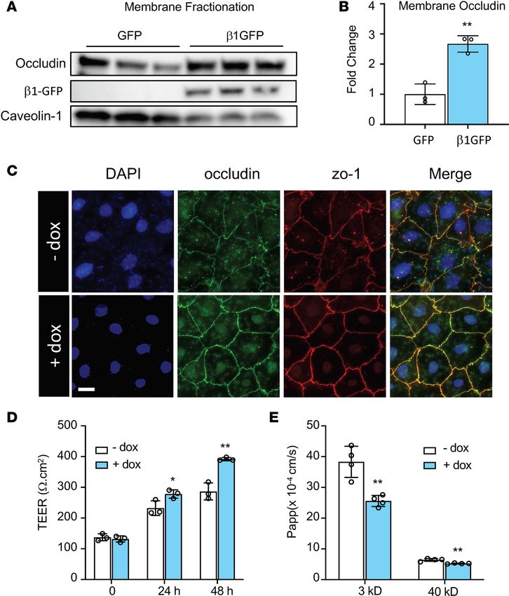
RESEARCH ARTICLE

                                 is a serine/threonine protein kinase that may regulate the tight junction assembly pathway. Using loss-of-
                                 function, chemical inhibition, and gain-of-function experiments, we revealed a potentially novel molecular
                                 pathway by which the β1 subunit binds and activates MRCKα, thereby phosphorylating non–muscle myo-
                                 sin II and increasing tight junction levels at the membrane. Interestingly, the expression of MRCKα in lungs
                                 from ARDS patients is significantly decreased. Together, our findings have elucidated a molecular pathway
                                 by which NKA regulates alveolar barrier function by interacting with and activating MRCKα.

                                 Results
                                 β1 subunit overexpression increases expression of alveolar tight junctions. Rat primary alveolar epithelial cells were
                                 used to investigate the role of the β1 subunit on tight junctions. To characterize these cells, quantitative
                                 PCR (qPCR) analysis was performed for genes that are specific for ATI (CAV1 and PDPN) or ATII (SPC
                                 and LAMP3) cells (Figure 1A). SPC and LAMP3 levels were high at 24 hours after isolation but decreased
                                 after 72 hours while CAV1 and PDPN levels gradually arise, suggesting differentiation from an ATII phe-
                                 notype to an ATI phenotype. When cultured in transwell plates coated with 20 μg/mL fibronectin, these
                                 cells developed high transepithelial electrical resistance (TEER) (Supplemental Figure 1A; supplemental
                                 material available online with this article; https://doi.org/10.1172/jci.insight.134881DS1) and displayed
                                 membrane staining of occludin and zo-1 (Supplemental Figure 1B). In addition, when treated with 1 μg/
                                 mL LPS, a bacterial endotoxin, these cells showed decreased TEER and increased permeability to 4 kD
                                 dextran (Supplemental Figure 1, C and D), indicating barrier damage. Thus, the rat primary ATI culture
                                 system can serve as a relevant model to study the alveolar epithelial barrier.
                                      Next, we aimed to overexpress the β1 subunit in ATI cells in order to examine its role in epithelial
                                 barrier function. We used electroporation to transfect ATI cells with a plasmid encoding the rat β1 sub-
                                 unit, and we measured expression of tight junction proteins 24 hours later. Transfection of the β1 subunit
                                 had no effect on the mRNA abundance of occludin or zo-1 (Figure 1B). However, at the protein level, we
                                 observed significant increases of both proteins (Figure 1, C and D). We also observed increased levels of
                                 zo-2 and claudin-18 (Supplemental Figure 1E), a tight junction component that recently has been found
                                 to play an essential role in alveolar barrier properties (14–17). Notably, treatment with the NKA-specific
                                 inhibitor ouabain at concentrations that block the enzymatic and ion transport activity of the NKA α
                                 subunit (18) failed to block the upregulation of occludin (Figure 1, E and F). In parallel, when cells were
                                 transfected with the β2 or the β3 subunit, 2 other NKA β isoforms that can also form functional heterod-
                                 imers with the α subunit, no upregulation of tight junction proteins was observed (Supplemental Figure
                                 2, A and B). These data demonstrate that overexpression of the β1 subunit of the NKA increases the
                                 expression of many tight junction proteins, and this effect is independent of its regulatory role in pump
                                 activity of NKA αβ heterodimers.
                                      Overexpression of the β1 subunit increases ATI barrier integrity. Given that tight junctions components
                                 are localized to the apical membrane of cells in a mature epithelial layer, we aimed to investigate if the
                                 β1 subunit increases tight junctions at cell membranes and enhances alveolar barrier function. West-
                                 ern blot of the plasma membrane fraction shows that the β1 subunit significantly increases the level
                                 of membrane-associated occludin (Figure 2, A and B). To explore the relevance to barrier function,
                                 we developed a doxycycline-inducible system to control β1 subunit expression by cloning the rat β1
                                 subunit into a Tet-on plasmid. To characterize the system, human bronchial epithelial cells 16HBE14o-
                                 were cotransfected with pCMV-tet regulator plasmids and pTet3G-human β1 subunit expressing plas-
                                 mids, followed immediately by addition of doxycycline (0, 1, 10, 100, and 1000 ng/mL). Immunoblot
                                 analysis showed that doxycycline caused a dose-dependent upregulation of the β1 subunit at 24 hours
                                 after transfection with maximal induction at 1000 ng/mL (Supplemental Figure 3A). Consequently, the
                                 expression of occludin and zo-1 were also increased proportionally, confirming previous findings using
                                 the pCMV-rat β1 plasmid. Cotransfection of the regulator plasmid and a luciferase reporter plasmid
                                 confirmed robust transgene expression for up to 7 days after transfection (Supplemental Figure 3B).
                                 qPCR for SPC and Cav1 showed that doxycycline does not affect the differentiation from ATII to ATI
                                 (Supplemental Figure 3C). Next, we sought to examine the role of the β1 subunit in epithelial barrier
                                 function. Immunostaining at 48 hours after induction of β1 transgene expression showed increased
                                 localization of occludin and zo-1 at cell-to-cell junctions, indicating more mature epithelial barriers
                                 than controls (Figure 2C). TEER was significantly higher at 24 hours and 48 hours after doxycycline
                                 induction (Figure 2D), suggesting an increase in barrier integrity. In accordance, the permeabilities to

JCI Insight 2021;6(4):e134881 https://doi.org/10.1172/jci.insight.134881                                                                             2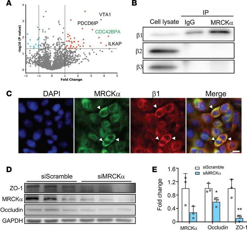
RESEARCH ARTICLE

Figure 1. Overexpression of the β1 subunit increases expression of tight junction proteins. (A) Rat primary ATII cells differentiate into ATI-like cells when
cultured in vitro. Cells were lysed for qPCR analysis of ATII markers (SPC and LAMP3) and ATI markers (CAV1 and PDPN) at different days after culture. Data
represents n = 3 biological replicates. (B) Relative mRNA levels for occludin and zo-1 in control and the β1 subunit–transfected cells. (C) Cells were transfected
with plasmid expressing the rat β1 subunit or pCDNA3 empty plasmid as control at day 3 after isolation. Cells were lysed for Western blot 24 hours later. (D)
Quantification of the Western blots in C. Data are representative of 3 independent experiments. Data are presented as mean ± SD. Statistical analysis was
performed by 2-tailed Student’s t test. *P < 0.05; **P < 0.01; ***P < 0.001. (E) Ouabain treatment at the indicated concentrations does not inhibit the β1
subunit–mediated occludin upregulation. AT1 cells were transfected with the pCDNA3 plasmid or the pCMV-β1 plasmid, and 4 hours later, the DMSO control or
ouabain at 10 nM, 100 nM, or 1 μM was added to cells. (F) Western blots were performed at 24 hours after transfection and were quantified. Data are represen-
tative of 3 independent experiments. Data are presented as mean ± SD. Statistical analysis was performed by 2-tailed Student’s t test. *P < 0.05; **P < 0.01.

                                    3 kD dextran and 40 kD dextran decreased by 33.2% and 18.5%, respectively, following doxycycline
                                    treatment for 48 hours (Figure 2E). Taken together, our data demonstrate that overexpression of the
                                    NKA β1 subunit leads to improved alveolar epithelial barrier function.
                                         Identification of the β1 subunit interactome. Since our findings suggest that the β1 subunit–mediated epi-
                                    thelial barrier tightening is independent of its role in ion transport activity of the NKA, we reasoned that
                                    this regulation may act through protein-to-protein interactions. So far, a limited number of protein inter-
                                    actions of the β1 subunit have been reported in the literature. To overcome this, we used MS to systemat-
                                    ically identify binding partners of the β1 subunit. Cell lysates from untransfected 16HBE14o- cells were

JCI Insight 2021;6(4):e134881 https://doi.org/10.1172/jci.insight.134881                                                                                         3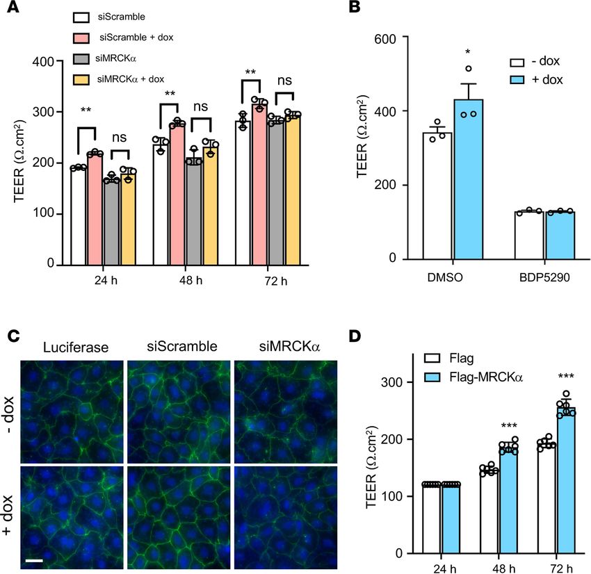
RESEARCH ARTICLE

                                 Figure 2. Overexpression of the β1 subunit increases alveolar type I barrier function. (A) Overexpression of the β1
                                 subunit in HEK293T cells increased occludin expression at the plasma membrane. Caveolin-1 was used as membrane
                                 loading control. (B) Densitometry of gels in A, with analysis by Student’s t test, **P < 0.01. (C) ATII cells were cotrans-
                                 fected with 4 mg/mL pCMV-Tet3G plasmid and 16 mg/mL pTet3G-human β1 plasmid day 1 after isolation. Cells were
                                 then plated on fibronectin-coated coverslips. Doxycycline (1 μg/mL) was added 48 hours later. Representative immu-
                                 nofluorescence staining of ATI cells shows that doxycycline-induced expression of the β1 subunit induces more mature
                                 tight junctions, as indicated by occludin (green) and zo-1 (red) staining. Images represent 3 independent experiments.
                                 Scale bar: 20 mm. (D) ATII cells were cotransfected as in C, but cells were plated on fibronectin-coated 12-well transwell
                                 plates. Twenty-four hours later at day 2, 1 μg/mL of doxycycline (dox) was added to induce β1 gene expression. TEER
                                 was measured every 24 hours. ANOVA followed by Bonferroni’s post hoc test was used for statistical analysis, *P <
                                 0.05, **P < 0.01. (E) After TEER measurement at day 4, permeability to 3 kD Texas Red–dextran and 40 kD FITC-dex-
                                 tran was measured for a duration of 2 hours. Data are presented as mean ± SD. ANOVA followed by Bonferroni’s post
                                 hoc test was used for statistical analysis, **P < 0.01.

                                 immunoprecipitated using an antibody against the β1 subunit or an antibody against GFP as a negative
                                 control. The resulting protein complexes were separated by SDS-PAGE, and the gels were cut into 10 seg-
                                 ments to increase resolution of protein identification. The proteins were extracted from the gel segments
                                 and subjected to trypsin digestion (Supplemental Figure 4A). After database searching for the spectrums,
                                 we identified 2936 unique proteins from 3 independent experiments (Supplemental Table 1). We then
                                 quantified their relative abundance using normalized spectrum abundance factor (NSAF), a label-free
                                 quantification method based on counting the number of unique peptides assigned to each protein (19). A
                                 total of 138 proteins passed the criteria for potential interactions (P < 0.05, Student’s t test) (Figure 3A).

JCI Insight 2021;6(4):e134881 https://doi.org/10.1172/jci.insight.134881                                                                                   4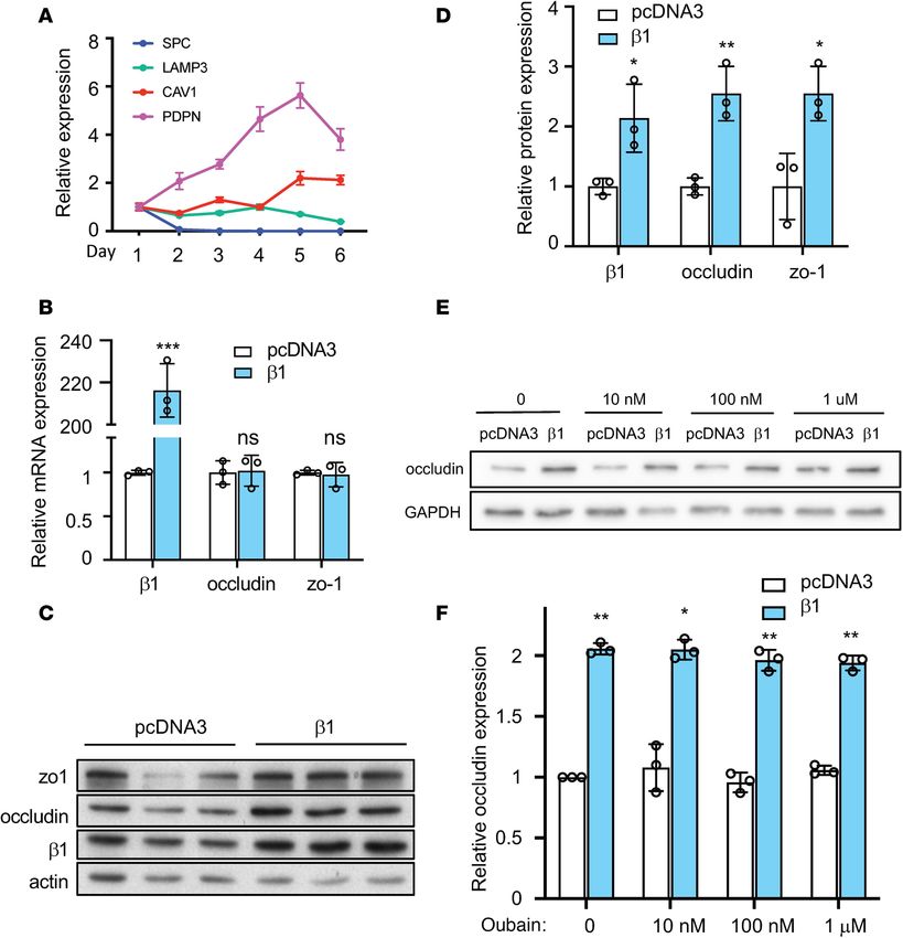
RESEARCH ARTICLE

                                 Figure 3. MRCKα interacts with the β1 subunit and stabilizes tight junction. (A) Volcano plot of proteins identified
                                 from triplicate mass spectrometry experiments. CDC42BPA (MRCKα) is labeled on the graph. Dashed line indicates the
                                 P value threshold of 0.05. (B) The interaction of MRCKα with the β1 subunit was confirmed using co-IP. A total of 5% of
                                 total cell lysate was used for input. The β2 or β3 subunit did not coimmunoprecipitate with MRCKα. (C) The β1 subunit
                                 (red) and MRCKα (green) colocalize in ATI cells. Scale bar: 20 μm. (D) ATI cells were transfected with a scrambled siRNA
                                 (siScramble) or a siRNA against MRCKα (siMRCKα). (E) Twenty-four hours later, cells were lysed for immunoblot analy-
                                 sis and quantified. Data represent 3 biological replicates and Error bars show SD. Student’s t test, *P < 0.05, **P < 0.01.

                                 Top candidates include CDC42BPA (serine/threonine-protein kinase MRCKα), ILKAP (integrin-linked
                                 kinase–associated serine/threonine phosphatase 2), and VTA1 (vacuolar protein sorting–associated pro-
                                 tein VTA1 homolog) (Table 1). Gene Ontology (GO) enrichment analysis (20) of the identified protein
                                 interactors revealed significant enrichment for biological processes, including endosomal sorting complex
                                 required for transport (ESCRT) disassembly and multivesicular body organization, 2 processes involved in
                                 the endosomal sorting of ubiquitylated membrane proteins (Table 2).
                                      MRCKα regulates epithelial barrier integrity. One of the top proteins identified from our MS experiment is
                                 MRCKα, a serine/threonine-protein kinase and a downstream effector of Cdc42 in cytoskeletal reorganization.
                                 In its native state, MRCKα forms a homodimer that blocks its kinase activity (21). Once activated, it phosphory-
                                 lates substrates including myosin light chain 2 (MLC2) and LIM kinase, thereby modulating actin-myosin con-
                                 traction (22). The dissociation of the autoinhibitory homodimerization complex is a prerequisite for MRCKα
                                 activation, which can be induced by a number of factors, such as Rap1 (23) and PDK1 (24). By regulating the
                                 cytoskeleton, activated MRCKα is involved in many cellular processes, such as cell migration (24, 25), cell polar-
                                 ity (26), and endothelial junction formation (23, 27). We hypothesized that the β1 subunit may increase alveolar
                                 epithelial barrier integrity through MRCKα.
                                      MRCKα had more than 40% sequence coverage from our MS analysis (Supplemental Figure 4B).
                                 To further confirm its interaction with the β1 subunit, we performed a co-IP experiment in untransfected
                                 16HBE14o- cells. Among the 3 endogenous β isoforms, only the β1 subunit was detected in the MRCKα
                                 pulldown complex, suggesting the specificity of the interaction (Figure 3B). Further immunofluorescence
                                 staining in ATI cells showed that β1 colocalizes with MRCKα on the cell membrane (Figure 3C). To inves-
                                 tigate the functional role of MRCKα in the epithelial barrier, we knocked down its expression in ATI cells
                                 and evaluated protein levels of tight junctions. Cells transfected with small interfering RNA (siRNA) against

JCI Insight 2021;6(4):e134881 https://doi.org/10.1172/jci.insight.134881                                                                                  5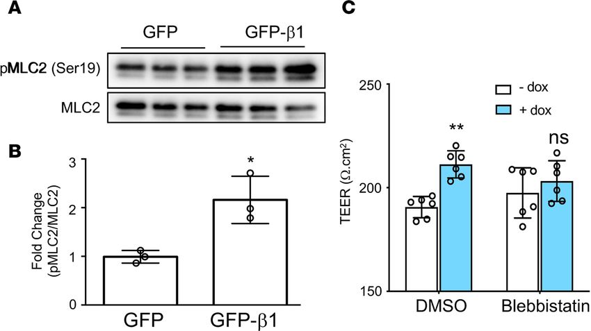
RESEARCH ARTICLE
Table 1. Top 10 interacting proteins of the NKA β1 subunit
Protein Name Gene p_NSAF Diff_NSAF
Serine/threonine-protein kinase MRCK alpha CDC42BPA 0.009 4.33
Integrin-linked kinase-associated serine/threonine phosphatase 2C ILKAP 0.032 4.17
Vacuolar protein sorting-associated protein VTA1 homolog VTA1 0.001 3.76
Programmed cell death 6-interacting protein PDCD6IP 0.022 3.76
Zinc finger and BTB domain-containing protein 1 ZBTB1 0.010 3.61
Serine/threonine-protein phosphatase 6 regulatory ankyrin repeat ANKRD28 0.003 3.11
subunit A
Spastin SPAST 0.035 2.94
Vacuolar protein sorting-associated protein 4A VPS4A 0.0003 2.54
Mannosyl-oligosaccharide glucosidase MOGS 0.022 2.44
NADH-ubiquinone oxidoreductase 75 kDa subunit, mitochondrial NDUFS1 0.0134 2.42
MRCKα showed significantly lower levels of both occludin and zo-1 (Figure 3, D and E), suggesting that
MRCKα may stabilize the expression of tight junction proteins.
Since MRCKα loss of function impairs tight junctions, we hypothesized that the NKA β1 subunit
enhances alveolar barrier function through its interaction with MRCKα. To test this hypothesis, we first
depleted MRCKα using siRNA and subsequently induced β1 overexpression using doxycycline. TEER was
significantly higher in ATI monolayers at 24, 48, and 72 hours after doxycycline treatment, but it was abol-
ished when cells were transfected with siRNA against MRCKα (Figure 4A). To further confirm this, cells
were treated with 2 μM BDP5290, a potent inhibitor of MRCKα (28), and barrier integrity was evaluated
by TEER. Consistent with siRNA silencing, baseline TEER was decreased upon MRCKα inhibition. More
importantly, inhibitor treatment prevented the β1 subunit–induced increase of barrier integrity (Figure 4B).
Immunofluorescence staining also confirmed that the β1 subunit increased intensity and membrane local-
ization of zo-1. Again, this phenomenon was abolished when MRCKα was knocked down (Figure 4C).
Collectively, our data indicate a critical role of the β1 subunit in improving alveolar barrier function through
activation of MRCKα. We next tested whether overexpressing MRCKα directly was able to enhance alveolar
barrier function. After ATII cells were transfected with MRCKα plasmids, TEER was measured to monitor
barrier function. Twenty-four hours after transfection, we detected no significant differences in TEER; how-
ever, at both 48 and 72 hours after transfection, we observed significantly higher resistance in cells transfected
with MRCKα compared with those transfected with empty plasmid (Figure 4D). These results demonstrate
that overexpression of MRCKα alone is sufficient to promote alveolar epithelial barrier integrity.
Activation of non–muscle myosin II mediates β1 subunit stabilization of tight junctions. Our results support the
hypothesis that the β1 subunit interacting protein MRCKα is both necessary and sufficient to promote the
formation of alveolar epithelial cell tight junctions. To further substantiate this conclusion, we examined
the activation of MLC2, a downstream effector of MRCKα (23). Western blot showed that overexpres-
sion of the β1 subunit induced the phosphorylation of MLC2 at Ser19 by 2-fold (Figure 5, A and B). The
activation of actin-myosin via MLC2 regulates the assembly of tight junction complexes and their steady
state level through endocytic degradation (29–31). In addition, the activation of MLC2 is associated with
junctional recruitment, formation of circumferential actin bundles, and barrier maturation (23, 26, 27, 32,
33). Therefore, we investigated whether β1 subunit–mediated activation of MLC2 is responsible for the
increased barrier integrity seen upon β1 overexpression. Pretreatment of cells with 20 μM blebbistatin, a
specific inhibitor of MLC2, prevented the increase in TEER induced by overexpression of the β1 subunit
(Figure 5C). Taken together, these results suggest that the activation of MLC2 is required for β1-mediated
tight junction stabilization and alveolar epithelial barrier potentiation.
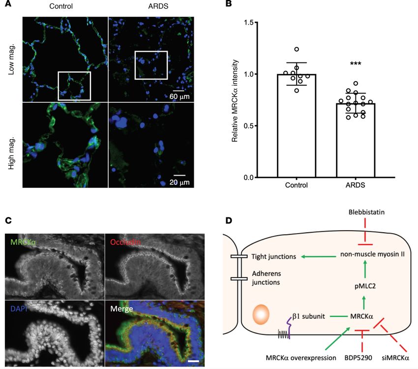
Human ARDS patients show decreased expression of MRCKα. Given that MRCKα regulates alveolar barrier
integrity, we investigated whether its expression altered in ARDS. Immunofluorescence staining demonstrat-
ed that lungs from ARDS patients (n = 5) express much lower levels of MRCKα compared with lungs from
control donors (n = 3) (Supplemental Figure 5A and Figure 6A), with an average of 30% less relative fluores-
cent staining intensities (Figure 6B). In addition to alveoli, small airways also express high levels of MRCKα,
especially in the cilia where apical occludin is expressed, and in basal cells (Figure 6C). Importantly, staining
JCI Insight 2021;6(4):e134881 https://doi.org/10.1172/jci.insight.134881 6RESEARCH ARTICLE
Table 2. Gene Ontology (GO) analysis of biological process for β1-interacting proteins
GO biological process Fold Enrichment P value
ESCRT III complex disassembly > 100 1.47 × 10⁻²
ESCRT complex disassembly > 100 1.47 × 10⁻²
nuclear envelope reassembly 89.54 4.92 × 10⁻²
multivesicular body assembly 59.69 6.02 × 10⁻³
multivesicular body organization 57.77 6.85 × 10⁻³
cytoskeleton-dependent cytokinesis 27.64 9.72 × 10⁻³
cytokinesis 25.44 1.45 × 10⁻²
protein-containing complex subunit 4.12 1.92 × 10⁻³
organization
intensities in these tissues are also decreased in ARDS patients (Supplemental Figure 5B). Taken together,
these data imply that lower levels of MRCKα in the lung may be associated with ARDS pathology.
Discussion
NKA is well known for its transport activity — moving Na+ out of the cell and importing K+. Our
results have identified a potentially new function of this enzyme. Specifically, we have found that the
small, non–catalytic β1 subunit promotes alveolar epithelial barrier integrity through a transport-inde-
pendent mechanism that involves protein interaction and activation of MRCKα (Figure 6D). Inhibition
of MRCKα using either siRNA or pharmacological inhibitors prevents the upregulation of occludin
and the increase of TEER induced by β1 subunit overexpression; on the other hand, overexpression
MRCKα alone is sufficient to enhance barrier function. Consistent with an activation of MRCKα,
overexpression of the β1 subunit increases the phosphorylation of MLC2 at Ser19 (25). Blebbistatin, a
specific inhibitor of myosin II, abrogates the increase of TEER by β1 subunit overexpression. Togeth-
er, these data demonstrate that the β1 subunit increases epithelial tight junction function by controlling
MRCKα activation and myosin-actin activity.
During our investigation to decipher the signaling pathway, we have established a cellular model
of the alveolar epithelial barrier using ATI-like cells that enables efficient and dose-dependent induc-
tion of gene expression. Using this model, we demonstrated that overexpression the β1 subunit leads
to improved barrier integrity, as demonstrated by the upregulation of tight junction proteins occludin,
zo-1, zo-2, and claudin-18; increased electrical resistance; and decreased permeability to fluorescent
tracers. This study supplements previous findings in mice and pigs (11, 13, 34), and it provides a mecha-
nistic basis to apply ARDS gene therapy approaches for potential human clinical use. The cellular mod-
el that we established here can also be used to study other lung diseases characterized by barrier defects,
such as asthma. Importantly, claudin-18 is the most abundant claudin in ATI cells (14). Deficiency in
claudin-18 results in both alveolar and airway barrier dysfunction (14–16). However, the claudin-18–KO
mouse has increased levels of the NKA β1 subunit but decreased occludin expression (15). This sug-
gests a possible compensatory effect between the β1 subunit and claudin-18 during development. Our
unpublished data also suggest a functional interaction between occludin and claudin-18, indicating that
the proper expression and localization of tight junction proteins may depend on each other.
Our data suggest a β1 subunit–specific effect in regulating alveolar barrier integrity among all NKA
β subunits. Surprisingly, overexpression of the β3 subunit, but not the β2 subunit, decreased expres-
sion of the β1 subunit, occludin, and zo-1 (Supplemental Figure 2, A and B). It is worth mentioning
that such a competing mechanism between β1 and β3 subunits, but not with the β2 subunit, has been
reported previously in the literature (35). β1 Subunit–KO mice show higher β3 subunit expression (36).
However, overexpression of the β2 subunit in WT mice did not decrease β1 subunit levels (37). Future
experiments to compare the effect of the 3 subunits in treating LPS-induced lung injury will further sub-
stantiate our findings. The conclusion that β1 subunit–mediated tight junction upregulation is a process
independent of the ion transport activity of the NKA is consistent with previous findings from our lab-
oratory (10, 11, 13) and others (7–9, 38) that only the β1 subunit, but not the α subunit or the epithelial
sodium channel, decreases lung permeability and treats mice with existing ARDS.
JCI Insight 2021;6(4):e134881 https://doi.org/10.1172/jci.insight.134881 7RESEARCH ARTICLE
Figure 4. MRCKα is required for the β1 subunit–mediated alveolar barrier tightening. (A) ATII cells were cotransfected
with siRNA (scramble control or against MRCKα) and plasmids (CMV-tet and Tet- β1) 24 hours after isolation. A total
of 1 μg/mL doxycycline was added to induce gene expression at day 2. TEER was then measured every 24 hours from
day 3 to day 5. ANOVA followed by Bonferroni’s post hoc test was used for statistical analysis, **P < 0.01. (B) ATII cells
were cotransfected with plasmids (CMV-tet and Tet-β1) 24 hours after isolation and treated immediately with 2 μM
MRCKα inhibitor BDP5290. TEER was measured 24 hours later. Data are presented as mean ± SD. ANOVA followed by
Bonferroni’s post hoc test was used for statistical analysis, *P < 0.05. (C) Immunofluorescence staining of zo-1 in cells
treated with or without doxycycline for 48 hours after transfection with luciferase plasmid alone, β1 plasmid and siS-
cramble, or β1 plasmid and siMRCKα. Images represent 3 separate experiments. Scale bar: 20 um.(D) Overexpression of
MRCKα increases TEER. Data represent n = 6 biological replicates. Data are presented as mean ± SD. ANOVA followed
by Bonferroni’s post hoc test was used for statistical analysis, ***P < 0.001.
Our results have confirmed some known protein interactions of the β1 subunit, including the NKA
α1 subunit, the ER protein Wolframin (39), coatomer subunit β (40), and lethal giant larvae protein
(41). Some proteins previously reported to interact with the β1 subunit (42–45) were not detected in our
analysis, likely because these proteins — which are mainly expressed in the neural system — are not
expressed in the lung. More importantly, many binding partners have been identified. To our knowl-
edge, this is the first proteomic analysis of the β1 subunit interaction network. The interactome of many
integral membrane proteins has remained unknown or is only poorly characterized due to their hydro-
phobicity, low expression, and lack of trypsin cleavage sites in their transmembrane segments (46, 47).
The current MS analysis greatly enriches our knowledge of the protein interactome of the β1 subunit.
The binding partners identified from this study can be confirmed by future experiments and will provide
important insight regarding the activity and cellular functions of NKA.
A previous study using siRNA injection into mouse embryos proposed that the β1 subunit is
required for proper distribution of tight junctions, likely via regulation of the actin cytoskeleton (48).
Our data suggest that MRCKα appears to be involved in these processes. MRCKα is involved in cell
migration, polarization, and junction formation by regulating actin-myosin activity (23, 24, 26). In
accordance with our finding, a previous study in endothelial cells suggests that MRCKα mediates the
JCI Insight 2021;6(4):e134881 https://doi.org/10.1172/jci.insight.134881 8RESEARCH ARTICLE
Figure 5. The MRCKα downstream pathway is activated upon overexpression of the β1 subunit. (A) Cells were
electroporated with plasmid expressing the rat β1 subunit or pCDNA3 empty plasmid as control at day 3 after isolation.
(B) Cells were lysed for Western blot 24 hours later and quantified. Data represent n = 3 biological replicates. Statis-
tical analysis is by Student’s t test, *P < 0.05. (C) At day 1 after isolation, cells were cotransfected with pCMV-tet and
pTet-β1 and treated with 20 μM Blebbistatin or DMSO as control. After another 24 hours, 1 μg/mL doxycycline was
added to induce gene expression. TEER was measured 24 hours later. ANOVA followed by Bonferroni’s post hoc test
was used for statistical analysis. **P < 0.001.
activation of non–muscle myosin at cell-to-cell contacts and the formation of circumferential actin
bundles, which is essential for cell junctions (23). The mechanism of how MRCKα is activated upon
interacting with the β1 subunit is unknown. It is possible that the interaction promotes the plasma
membrane localization of MRCKα, similar to that seen for the NKA β1 subunit and the sodium calci-
um exchanger 1 (49) or megalencephalic leukoencephalopathy with subcortical cysts 1 (44). Another
possibility is that the β1 association with MRCKα abolishes the autoinhibition of MRCKα by binding
to its 2 distal CC domains, which interact intramolecularly with the kinase domain and negatively
regulate its activity (21). These 2 events may also happen concurrently. Future investigation is needed
to test these possibilities.
One striking finding from our results is that lungs from patients with ARDS tend to express lower
amounts of MRCKα. No genetic susceptibility of ARDS has been linked to MRCKα so far. However,
one of its downstream targets, MLC kinase, is associated with ARDS susceptibility and outcomes
(50). Additionally, a recent study suggested that MRCKα is involved in epithelial extrusion following
apoptosis (51). Epithelial extrusion is a process by which dying or unwanted cells are removed from
an epithelium while preserving the barrier function of the layer (52). To date, no study has explored
the physiological and pathological roles of MRCKα in the lung. It will be interesting to investigate
whether decreased MRCKα results in a defect of epithelial extrusion, thereby predisposing the lung to
injuries that ultimately lead to ARDS.
The reason lungs from ARDS patients express significantly lower amounts of MRCKα is unclear.
One possibility is lower basal transcription of MRCKα due to genetic causes (such as reduced gene
copy numbers or epigenetic modification). Another possibility is that risk factors for ARDS, such
as inflammation, may downregulate MRCKα levels. Regardless, MRCKα could be a drug target for
treating ARDS or other human diseases characterized by barrier defects. Currently, only inhibitors of
MRCKα have been identified (28, 53). Activation of MRCKα may be achieved by using a peptide that
corresponds to the interacting domains on the NKA β1 subunit. Such a peptide modulator could be a
promising drug to enhance epithelial barrier function and could ultimately lead to a simple pharmaco-
logical treatment of ARDS.
In conclusion, our data have supported a nontransport associated role of the NKA β1 subunit in the
regulation of tight junctions. This work enhances our understanding of the NKA and defines a role for
MRCKα in the homeostasis of lung epithelial barrier properties.
JCI Insight 2021;6(4):e134881 https://doi.org/10.1172/jci.insight.134881 9RESEARCH ARTICLE
Figure 6. Decreased MRCKα levels in the alveolar epithelium of human ARDS patients. (A) Representative images of immunofluorescence staining
for MRCKα (green) in lung sections of a control donor and a patient with ARDS. Upper panel shows images taken at 20× objective magnification, and
lower panel shows images taken at 63× objective magnification for the boxed region in the upper panel. (B) Quantification of MRCKα expression in
the alveoli. ROI (region of interest) were drawn in the alveoli region, and the ratio of integrated pixel intensity for MRCKα and DAPI was calculated for
each ROI. Three normal donors and 5 ARDS patients were used for quantification, with 3 random fields chosen for each sample. Data are expressed
as mean ± SEM, with n = 9 (3 patients) for normal control and n = 15 (5 patients) for ARDS. Statistical analysis was by 2-tailed Student’s t test, ***P
< 0.001. (C) Costaining of MRCKα (green) and occludin (red) in the small airway from control donor. Scale bar: 20 µm. (D) Working model of the β1
subunit increases alveolar epithelial barrier integrity. The β1 subunit of the NKA interacts with MRCKα, assists in its activation, leads to higher myo-
sin phosphorylation, and eventually stabilize tight junctions.
Methods
Plasmids and siRNA. pCDNA3 and pCMV-EGFP plasmids were purchased from Invitrogen. Mouse
NKA β2 subunit and mouse NKA β3 subunit with Myc-DDK tag were obtained from OriGene. The
Tet-On 3G drug-inducible gene expression system was purchased from Clontech. The human NKA
β1 subunit–coding sequence was inserted into the pTRE3G vector at the SalI and BamHI restriction
enzyme sites. The human MRCKα plasmid was a gift from Paolo Armando Gagliardi at the University
of Bern (Bern, Switzerland) (24). Knockdown was carried out using the TriFECTa DsiRNA Kit (IDT)
according to manufacturer’s instructions. siRNA duplexes at a final concentration of 100 nM were
transfected in 4 mm cuvettes (Bio-Rad) using a GenePulser Xcell (Bio-Rad) instrument.
Antibodies and inhibitors. Primary antibodies for Western blot include anti–NKA β1 subunit (Upstate,
05-382), anti-occludin (Invitrogen, 71-1500), anti–zo-1 (Invitrogen, 61-7300), anti–zo-2 (Invitrogen, 71-1400),
JCI Insight 2021;6(4):e134881 https://doi.org/10.1172/jci.insight.134881 10RESEARCH ARTICLE
anti-actin (MilliporeSigma, A2066), anti-GAPDH (MilliporeSigma, CB1-001), anti–NKA β2 subunit (Abcam,
ab185210), anti-DDK (OriGene, TA50011-100), anti-MRCKα (Santa Cruz Biotechnology Inc., sc-374568),
anti-MYPT1 (Cell Signaling Technology, 2634S), anti–phospho-MYPT1 (Thr696, Cell Signaling Technology,
5163S), anti-MLC2 (Cell Signaling Technology, 3672S), and anti–phospho-MLC2 (Ser19, Cell Signaling Tech-
nology, 3671S). The primary antibodies for immunofluorescence include anti–occludin Alexa Fluor594 (Invi-
trogen, 331594), anti–zo-1-Alexa Fluor594 (Invitrogen, 339194), and anti-MRCKα (Thermo Fisher Scientific,
PA1-10038). The inhibitor for MRCKα BDP5290 was purchased from Aobious. Myosin inhibitor blebbistatin
was purchased from Abcam.
Primary cell isolation and cell culture. Primary rat ATII cells were isolated using an IgG-panning
approach as described by Dobbs et al. (54). Briefly, lungs from Sprague Dawley rats (Charles River
Laboratories) (200–250 g) were surgically removed and perfused, lavaged, and treated with 1 mg/mL
elastase (Worthington Biochemical) to release the epithelial cells. Next, lung lobes were separated, cut,
minced, filtered, and spun down at 1500 rpm for 15 minutes. The cells were resuspended in DMEM
without FBS and transferred to 2 IgG plates. After incubation at 37˚C for 1 hour, nonadhered cells (pre-
dominately ATII cells) were transferred to a new tube and centrifuged at 250g at room temperature for
15 minutes. The cells were resuspended in DMEM containing 10% FBS and plated on fibronectin coat-
ed plates. To coat the plates with fibronectin, 20 μg/mL fibronectin from bovine plasma (F1141, Mil-
liporeSigma) was added to 100 mm culture plates (using 3 mL) or the upper chamber of the transwell
plates (using 400 μL/well). Plates were left at 37°C for 3 hours. Residual solution was removed, and
plates were dried in a tissue culture hood for at least 30 minutes before cells were added. 16HBE14o-
human bronchial epithelial cells were cultured in DMEM as previously reported (13).
Transfection. Transfection was carried out by electroporation using the Gene Pulser MXcell electropora-
tion system (Bio-Rad). The electroporation conditions for ATI cells was 1 square wave pulse at 300 V, 1000
Ω, and 20 milliseconds. A total of 10 μg plasmid DNA was used for 1 × 10⁶ cells.
Western blot. Cells were lysed with reporter lysis buffer (1×, Promega) supplemented with protease
inhibitor (cOmplete, Mini, EDTA-free tablets; Roche) and phosphatase inhibitor (PhosStop Phosphatase
Inhibitor Cocktail; Roche). Proteins were separated on 10% SDS-PAGE gels, transferred to PVDF mem-
brane, and probed with primary antibodies at room temperature for 2 hours or at 4°C overnight. After
incubation with secondary antibodies and development, bands were detected on film (Biomax MR film;
Carestream Health) or using the ChemiDoc Imaging System (Bio-Rad) and quantified using Image Studio
Lite software (Li-COR) or Image Lab software (Bio-Rad).
Plasma membrane isolation. HEK293 cells were transfected with plasmids expressing GFP, or plasmids
expressing GFP-β1 using Lipofectamine 2000 (Invitrogen). Two days after transfection, 6 × 10⁶ cells were
subjected to membrane isolation using the Minute Plasma Membrane Protein Isolation and Cell Fraction-
ation Kit (Invent Biotechnologies), and proteins were analyzed by SDS-PAGE and Western blots.
qPCR. Total RNA was isolated using the RNeasy Mini Kit (Qiagen). After determining RNA concen-
trations by spectrophotometry, 100–1000 ng of total RNA was used for cDNA synthesis. Reverse transcrip-
tion was conducted using the Reverse Transcription System (Promega). A total of 10 μL of the reaction was
diluted to 100 μL, from which 1 μL was taken for qPCR using iTaq Universal SYBR Green Supermix (Bio-
Rad). The specificity of primers was confirmed by melting curve analysis and gel electrophoresis. qPCR
was performed on a CFX Connect Real Time PCR Detection System (Bio-Rad). Samples were assayed in
triplicate. Relative RNA level was quantified using the ΔΔCt method and normalized to the endogenous
control GAPDH unless specified otherwise.
Immunofluorescence. Cells were washed 3 times before fixation with 4% paraformaldehyde in PBS for
15 minutes at room temperature. Fixed cells were washed with PBS and permeabilized with 0.2% Tri-
ton X-100 in PBS for 10 minutes. After washing with PBS, transwell inserts were blocked with blocking
reagent (Dako Protein Block Serum Free, Agilent) for 1 hour and incubated with primary antibody at 4°C
overnight. Nuclei were stained with 2.5 μg/mL DAPI for 5 minutes and then washed twice with PBS. The
transwell membrane was then carefully cut out using a clean razor blade and mounted on a glass slide
with ProLong antifade mounting media (Thermo Fisher Scientific). Slides were examined under a Leica
DMI6000 microscope, and photos were captured using the open source software μManager or Volocity
software (Velocity Inc.). Tissue sections of human lungs from patients with ARDS were provided by
Zhongren Zhou in the Department of Pathology at the University of Rochester using an IRB-approved
protocol. All samples were taken at autopsy. In total, 16 sections from 6 ARDS patients and 7 sections
JCI Insight 2021;6(4):e134881 https://doi.org/10.1172/jci.insight.134881 11RESEARCH ARTICLE
from 3 control patients without ARDS were obtained. The H&E staining of each corresponding section
shows varying degrees of lung injury and edema content. For immunofluorescence staining, tissue sec-
tions were deparaffinized and rehydrated. Then, an antigen retrieval step was performed to expose epi-
topes for subsequent antibody binding and immunofluorescence.
TEER. Prior to measuring TEER, cells cultured on 12-well transwell plates (12 mm transwell with 0.4
μm pore polyester membrane insert; Corning) were moved to the tissue culture hood for 15 minutes to
allow the medium to equilibrate to room temperature. TEER was measured using an epithelial voltmeter
(EVOM2; World Precision Instruments). Three to 6 wells were measured for each condition, and 3 read-
ings were recorded and averaged for each well. To calculate TEER, the resistance of the fibronectin-coated
insert without cells (blank resistance) was subtracted from the measured resistance and then multiplied by
1.12 cm2 to account for the surface area of the insert.
Permeability. Permeability to fluorescent tracers was measured using a modified protocol previously
described (55). After TEER measurement, the upper and lower transwell chamber were washed twice
with P buffer (10 mM HEPES at pH 7.4, 1 mM sodium pyruvate, 10 mM glucose, 3 mM CaCl2, and
145 mM NaCl) (Invitrogen). A total of 500 μL of freshly prepared solution containing 100 μg/mL of 40
kD FITC-dextran and 100 μg/mL of 3kD Texas Red–dextran was added to the apical compartment. A
total of 1000 μL of P buffer was added to the bottom chamber. After 2 hours incubation at 37°C, 100 μL
of the basal medium was collected, and the fluorescence of the transported dextran was measured with
a SpectraMax M5 multimode microplate reader (Molecular Devices). The excitation wavelength and
emission wavelength are 492 nm and 520 nm for FITC and 596 nm and 615 nm for Texas-red, respec-
tively. The quantity of tracer was calculated by comparison with a standard curve. A permeability coef-
ficient was determined using the following equation (56): Pc (cm/min) = V/(A × Co) × (C/T), where
V is volume in the lower compartment (1 mL), A is the surface area of the membrane (1.12 cm2 for the
12-well transwell used here), Co is the dextran concentration in the upper compartment at time 0 (0.1
mg/mL), and C is the dextran concentration in the lower compartment at time T of sampling (2 hours).
IP and MS. Cells from one 100 mm plate were lysed with 1 mL of IP lysis buffer (1% NP-40, 50
mM Tris HCl at pH 8.0) and homogenized 10 times with a 25-gauge syringe. IP was performed using
the μMACS Protein G Kit according to the manufacturer’s instructions (Miltenyi Biotec). The precleared
samples were incubated with anti-MRCKα antibody (PA1-10038, 1:50 dilution; Thermo Fisher Scientific),
anti-β1 antibody (Upstate, 05-382, 1:250 dilution), or IgG as control at 4°C overnight. The elute was ana-
lyzed by SDS-PAGE Gradient Gels (4%–20%). Each lane was cut into 10 pieces of approximately the same
size. The gel bands were then destained, reduced, and digested with trypsin overnight. The digested peptide
mixtures were then subjected to LC-MS/MS analysis using the Orbitrap system.
Label-free quantification of proteins interacting with the β1 subunit. Thermo raw data were transformed
into mgf format. The resulting peak lists were searched using Protein Prospector (v5.22.0) with the fol-
lowing settings: Trypsin as protease with a maximum of 1 missed cleavage site, 10 ppm mass tolerance
for MS, 0.5 Da (ion trap), and 0.05 Da (Orbitrap), respectively, for MS/MS, carbamidomethylation
(C) as fixed, oxidation (M), and phosphorylation (S/T/Y) as variable modifications. Results from Pro-
tein Prospector were retrieved and cleaned up using in-house python script. Protein quantitation using
the NSAF measurement was described previously (19). Data normalization, annotation, and statistical
analysis were performed using Perseus (57). Two-tailed Student’s t test was used for statistical analysis
of NSAF (58). The proteomics data have been deposited to the ProteomeXchange Consortium via
MassIVE (Mass Spectrometry lnteractive Virtual Environment) with the accession no. MSV000084881.
Statistics. Each experiment was repeated at least 3 times. The data of each series is displayed as mean
values ± SD unless otherwise noted. Graphing and statistical comparison of the data were performed using
Prism 7 (GraphPad Software). Measurements for 2 groups were analyzed using the Student’s t test. Mea-
surements for more than 2 groups were analyzed by 1-way ANOVA and multiple comparisons. P values
less than 0.05 were considered to be statistically significant.
Study approval. All animal studies were approved by the University of Rochester Committee on Animal
Resources, and experimental procedures were carried out under the institutional guidelines for the care and
use of laboratory animals in an American Association for the Accreditation of Laboratory Animal Care–
approved facility. Human lung tissues from patients with and without ARDS was obtained at autopsy at the
University of Rochester using and IRB approved protocol.
JCI Insight 2021;6(4):e134881 https://doi.org/10.1172/jci.insight.134881 12RESEARCH ARTICLE
Author contributions
HB and DAD conceived, designed, and analyzed experiments and wrote the manuscript. HB and RZ performed
experiments. MB, RN, and RZ performed primary cell isolation. AF assisted in MS. DY was involved in staining
of human tissues. XL and JLY advised and conceived experiments.
Acknowledgments
We would like to thank Zhongren Zhou (currently at the Robert Wood Johnson Medical School and the
New Jersey Medical School of Rutgers University) for patient samples. We would also like to thank Paolo
Armando Gagliardi from the University of Bern for MRCKα plasmids. This research was supported by the
NIH grants HL120521, HL131143, and HL148825.
Address correspondence to: David A. Dean, Department of Pediatrics, University of Rochester, 601
Elmwood Avenue BOX 850, Rochester, New York 14642, USA. Phone: 585.276.3933; Email: david_
dean@urmc.rochester.edu.
HB’s present address: Wyss Institute for Biologically Inspired Engineering, Harvard University, Boston,
Massachusetts, USA.
1. Rubenfeld GD, et al. Incidence and outcomes of acute lung injury. N Engl J Med. 2005;353(16):1685–1693.
2. Bhattacharya J, Matthay MA. Regulation and repair of the alveolar-capillary barrier in acute lung injury. Annu Rev Physiol.
2013;75:593–615.
3. Peteranderl C, et al. Macrophage-epithelial paracrine crosstalk inhibits lung edema clearance during influenza infection. J Clin
Invest. 2016;126(4):1566–1580.
4. Nieto-Torres JL, et al. Severe acute respiratory syndrome coronavirus envelope protein ion channel activity promotes virus fit-
ness and pathogenesis. PLoS Pathog. 2014;10(5):1004077.
5. Helenius IT, et al. Role of ubiquitination in Na,K-ATPase regulation during lung injury. Proc Am Thorac Soc. 2010;7(1):65–70.
6. Comellas AP, et al. Endothelin-1 impairs alveolar epithelial function via endothelial ETB receptor. Am J Respir Crit Care Med.
2009;179(2):113–122.
7. Factor P, et al. Adenoviral-mediated overexpression of the NA,K-ATPase beta1 subunit gene increases lung edema clearance
and improves survival during acute hyperoxic lung injury in rats. Chest. 1999;116(1 Suppl):24S–25S.
8. Factor P, et al. Adenovirus-mediated transfer of an Na+/K+-ATPase beta1 subunit gene improves alveolar fluid clearance and
survival in hyperoxic rats. Hum Gene Ther. 2000;11(16):2231–2242.
9. Adir Y, et al. Na,K-ATPase gene transfer increases liquid clearance during ventilation-induced lung injury. Am J Respir Crit Care
Med. 2003;168(12):1445–1448.
10. Machado-Aranda D, et al. Gene transfer of the Na+,K+-ATPase beta1 subunit using electroporation increases lung liquid clear-
ance. Am J Respir Crit Care Med. 2005;171(3):204–211.
11. Mutlu GM, et al. Electroporation-mediated gene transfer of the Na+,K+ -ATPase rescues endotoxin-induced lung injury. Am J
Respir Crit Care Med. 2007;176(6):582–590.
12. Emr BM, et al. Electroporation-mediated gene delivery of Na+,K+ -ATPase, and ENaC subunits to the lung attenuates acute
respiratory distress syndrome in a two-hit porcine model. Shock. 2015;43(1):16–23.
13. Lin X, et al. β1-Na(+),K(+)-ATPase gene therapy upregulates tight junctions to rescue LPS-induced acute lung injury. Gene Ther.
2016;23(6):489–499.
14. LaFemina MJ, et al. Claudin-18 deficiency results in alveolar barrier dysfunction and impaired alveologenesis in mice. Am J
Respir Cell Mol Biol. 2014;51(4):550–558.
15. Li G, et al. Knockout mice reveal key roles for claudin 18 in alveolar barrier properties and fluid homeostasis. Am J Respir Cell
Mol Biol. 2014;51(2):210–222.
16. Sweerus K, et al. Claudin-18 deficiency is associated with airway epithelial barrier dysfunction and asthma. J Allergy Clin Immu-
nol. 2017;139(1):72–81.
17. Zhou B, et al. Claudin-18-mediated YAP activity regulates lung stem and progenitor cell homeostasis and tumorigenesis. J Clin
Invest. 2018;128(3):970–984.
18. Hiyoshi H, et al. Quiescence and γH2AX in neuroblastoma are regulated by ouabain/Na,K-ATPase. Br J Cancer.
2012;106(11):1807–1815.
19. Paoletti AC, et al. Quantitative proteomic analysis of distinct mammalian Mediator complexes using normalized spectral abun-
dance factors. Proc Natl Acad Sci U S A. 2006;103(50):18928–18933.
20. The Gene Ontology Consortium. Expansion of the gene ontology knowledgebase and resources. Nucleic Acids Res.
2017;45(D1):D331–D338.
21. Tan I, et al. Intermolecular and intramolecular interactions regulate catalytic activity of myotonic dystrophy kinase-related
Cdc42-binding kinase alpha. Mol Cell Biol. 2001;21(8):2767–2778.
22. Unbekandt M, Olson MF. The actin-myosin regulatory MRCK kinases: regulation, biological functions and associations with
human cancer. J Mol Med (Berl). 2014;92(3):217–225.
23. Ando K, et al. Rap1 potentiates endothelial cell junctions by spatially controlling myosin II activity and actin organization. J Cell Biol.
2013;202(6):901–916.
JCI Insight 2021;6(4):e134881 https://doi.org/10.1172/jci.insight.134881 13RESEARCH ARTICLE
24. Gagliardi PA, et al. PDK1-mediated activation of MRCKα regulates directional cell migration and lamellipodia retraction. J Cell Biol.
2014;206(3):415–434.
25. Wilkinson S, et al. Cdc42-MRCK and Rho-ROCK signalling cooperate in myosin phosphorylation and cell invasion. Nat Cell
Biol. 2005;7(3):255–261.
26. Zihni C, et al. An apical MRCK-driven morphogenetic pathway controls epithelial polarity. Nat Cell Biol. 2017;19(9):1049–1060.
27. Marston DJ, et al. MRCK-1 drives apical constriction in C. elegans by linking developmental patterning to force generation.
Curr Biol. 2016;26(16):2079–2089.
28. Unbekandt M, et al. A novel small-molecule MRCK inhibitor blocks cancer cell invasion. Cell Commun Signal. 2014;12:54.
29. Shen L, Turner JR. Actin depolymerization disrupts tight junctions via caveolae-mediated endocytosis. Mol Biol Cell.
2005;16(9):3919–3936.
30. Nighot PK, Blikslager AT. Chloride channel ClC-2 modulates tight junction barrier function via intracellular trafficking of
occludin. Am J Physiol Cell Physiol. 2012;302(1):C178–C187.
31. Marchiando AM, et al. Caveolin-1-dependent occludin endocytosis is required for TNF-induced tight junction regulation in
vivo. J Cell Biol. 2010;189(1):111–126.
32. Smutny M, et al. Myosin II isoforms identify distinct functional modules that support integrity of the epithelial zonula adher-
ens. Nat Cell Biol. 2010;12(7):696–702.
33. Itoh M, et al. Rho GTP exchange factor ARHGEF11 regulates the integrity of epithelial junctions by connecting ZO-1 and
RhoA-myosin II signaling. Proc Natl Acad Sci U S A. 2012;109(25):9905–9910.
34. Emr BM, et al. Electroporation mediated gene delivery of Na+,K+-ATPase and ENaC subunits to the lung attenuates acute
respiratory distress syndrome in a two-hit porcine model. Shock. 2014;43(1):16–23.
35. Yoshimura SH, et al. Fast degradation of the auxiliary subunit of Na+/K+-ATPase in the plasma membrane of HeLa cells. J Cell Sci.
2008;121(Pt 13):2159–2168.
36. Flodby P, et al. Knockout mice reveal a major role for alveolar epithelial type I cells in alveolar fluid clearance. Am J Respir Cell
Mol Biol. 2016;55(3):395–406.
37. Clifford RJ, Kaplan JH. Regulation of Na,K-ATPase subunit abundance by translational repression. J Biol Chem.
2009;284(34):22905–22915.
38. Factor P, et al. Augmentation of lung liquid clearance via adenovirus-mediated transfer of a Na,K-ATPase beta1 subunit gene.
J Clin Invest. 1998;102(7):1421–1430.
39. Zatyka M, et al. Sodium-potassium ATPase 1 subunit is a molecular partner of Wolframin, an endoplasmic reticulum protein
involved in ER stress. Hum Mol Genet. 2008;17(2):190–200.
40. Morton MJ, et al. Association with {beta}-COP regulates the trafficking of the newly synthesized Na,K-ATPase. J Biol Chem.
2010;285(44):33737–33746.
41. Hatzold J, et al. Tumor suppression in basal keratinocytes via dual non-cell-autonomous functions of a Na,K-ATPase beta sub-
unit. Elife. 2016;5:14277.
42. Mao H, et al. MONaKA, a novel modulator of the plasma membrane Na,K-ATPase. J Neurosci. 2005;25(35):7934–7943.
43. Brignone MS, et al. The beta1 subunit of the Na,K-ATPase pump interacts with megalencephalic leucoencephalopathy with sub-
cortical cysts protein 1 (MLC1) in brain astrocytes: new insights into MLC pathogenesis. Hum Mol Genet. 2011;20(1):90–103.
44. Lanciotti A, et al. Megalencephalic leukoencephalopathy with subcortical cysts protein 1 functionally cooperates with the
TRPV4 cation channel to activate the response of astrocytes to osmotic stress: dysregulation by pathological mutations. Hum
Mol Genet. 2012;21(10):2166–2180.
45. de Juan-Sanz J, et al. Na+/K+-ATPase is a new interacting partner for the neuronal glycine transporter GlyT2 that downregu-
lates its expression in vitro and in vivo. J Neurosci. 2013;33(35):14269–14281.
46. Pankow S, et al. Deep interactome profiling of membrane proteins by co-interacting protein identification technology. Nat Pro-
toc. 2016;11(12):2515–2528.
47. Vit O, Petrak J. Integral membrane proteins in proteomics. How to break open the black box? J Proteomics. 2017;153:8–20.
48. Madan P, et al. Na/K-ATPase beta1 subunit expression is required for blastocyst formation and normal assembly of trophecto-
derm tight junction-associated proteins. J Biol Chem. 2007;282(16):12127–12134.
49. Balasubramaniam SL, et al. Sodium-calcium exchanger 1 regulates epithelial cell migration via calcium-dependent extracellular
signal-regulated kinase signaling. J Biol Chem. 2015;290(20):12463–12473.
50. Acosta-Herrera M, et al. Assessing the quality of studies supporting genetic susceptibility and outcomes of ARDS. Front Genet.
2014;5:20.
51. Gagliardi PA, et al. MRCK alpha is activated by caspase cleavage to assemble an apical actin ring for epithelial cell extrusion.
J Cell Biol. 2018;217(1):231–249.
52. Gudipaty SA, Rosenblatt J. Epithelial cell extrusion: pathways and pathologies. Semin Cell Dev Biol. 2017;67:132–140.
53. Unbekandt M, et al. Discovery of potent and selective MRCK inhibitors with therapeutic effect on skin cancer. Cancer Res.
2018;78(8):2096–2114.
54. Dobbs LG, et al. An improved method for isolating type II cells in high yield and purity. Am Rev Respir Dis. 1986;134(1):141–145.
55. Larre I, et al. Ouabain modulates epithelial cell tight junction. Proc Natl Acad Sci U S A. 2010;107(25):11387–11392.
56. Strengert M, Knaus UG. Analysis of epithelial barrier integrity in polarized lung epithelial cells. Methods Mol Biol.
2011;763:195–206.
57. Tyanova S, et al. The Perseus computational platform for comprehensive analysis of (prote)omics data. Nat Methods.
2016;13(9):731–740.
58. Morris JH, et al. Affinity purification-mass spectrometry and network analysis to understand protein-protein interactions. Nat
Protoc. 2014;9(11):2539–2554.
JCI Insight 2021;6(4):e134881 https://doi.org/10.1172/jci.insight.134881 14You can also read