The Extracellular Superoxide Dismutase Sod5 From Fusarium oxysporum Is Localized in Response to External Stimuli and Contributes to Fungal ...
←
→
Page content transcription
If your browser does not render page correctly, please read the page content below
ORIGINAL RESEARCH
                                                                                                                                          published: 02 March 2021
                                                                                                                                     doi: 10.3389/fpls.2021.608861

                                             The Extracellular Superoxide
                                             Dismutase Sod5 From Fusarium
                                             oxysporum Is Localized in Response
                                             to External Stimuli and Contributes
                                             to Fungal Pathogenicity
                                             Qiang Wang † , Ambika Pokhrel and Jeffrey J. Coleman*
                                             Department of Entomology and Plant Pathology, Auburn University, Auburn, AL, United States

                         Edited by:          Reactive oxygen species (ROS) produced by hosts serve as a general defense
                       Harold Meijer,
Wageningen University and Research,
                                             mechanism against various pathogens. At the interaction site between the host
                        Netherlands          and pathogen, host cells rapidly accumulate high concentrations of ROS, called the
                        Reviewed by:         oxidative burst, that damage and kill the invading microbes. However, successful
                          David Turra,       pathogens usually survive in a high ROS environment and have evolved strategies
       Università degli Studi di Napoli
                      Federico II, Italy     to overcome these detrimental effects. Here we characterized the biological function
                        Ken Komatsu,         of the extracellular superoxide dismutase (SOD) FoSod5 from Fusarium oxysporum f.
       Tokyo University of Agriculture
              and Technology, Japan
                                             sp. vasinfectum. FoSOD5 is strongly up-regulated during infection of cotton, and a
                    *Correspondence:
                                             1FoSOD5 mutant was significantly reduced in virulence on cotton. Purified 6 × His-
                      Jeffrey J. Coleman     FoSod5 could significantly inhibit the reduction of NBT and WST-1, indicating that
               jjcoleman@auburn.edu;         FoSod5 was a functional SOD protein. Based on CRISPR/Cas9 technology, several
                   jjc0032@auburn.edu
                  † Present
                                             different FoSod5 variants were generated and used to assess the secretion, expression,
                             address:
                          Qiang Wang,        and subcellular localization of FoSod5 in F. oxysporum. The subcellular localization
 State Key Laboratory of Crop Stress         of FoSod5 is altered under different environmental conditions. During normal growth
   Biology for Arid Areas and College
  of Plant Protection, Northwest A&F
                                             conditions, FoSod5 was primarily localized to the phialides; however, in a nutrient-limited
           University, Xianyang, China       environment, FoSod5 was localized to a wide array of fungal structures including the
                                             septum and cell wall. FoSod5 is an alkaline-induced glycosylphosphatidylinositol (GPI)
                   Specialty section:
         This article was submitted to
                                             protein and the GPI anchor was required for proper protein subcellular localization.
         Plant Pathogen Interactions,        The multiple mechanisms fungi utilize to tolerate the oxidative burst is indicative of
               a section of the journal
                                             the importance of this plant defense response; however, the presence of a conserved
            Frontiers in Plant Science
                                             extracellular SOD in many phytopathogenic fungi suggests tolerance to ROS is initiated
      Received: 21 September 2020
       Accepted: 08 February 2021            prior to the ROS entering the fungal cell.
         Published: 02 March 2021
                                             Keywords: cell wall protein, cotton, Fusarium wilt, glycosylphosphatidylinositol anchor, reactive oxygen species,
                             Citation:       superoxide dismutase, virulence factor
              Wang Q, Pokhrel A and
  Coleman JJ (2021) The Extracellular
   Superoxide Dismutase Sod5 From
    Fusarium oxysporum Is Localized
                                            INTRODUCTION
      in Response to External Stimuli
            and Contributes to Fungal
                                            The ascomycete fungus Fusarium oxysporum is an important pathogen that can infect and cause
                       Pathogenicity.       disease on a wide range of hosts including plants, animals, and humans (Gordon, 2017). Over 100
         Front. Plant Sci. 12:608861.       formae speciales have been described within the F. oxysporum species complex based on their ability
      doi: 10.3389/fpls.2021.608861         to cause disease on different host plants (Michielse and Rep, 2009). F. oxysporum f. sp. vasinfectum

Frontiers in Plant Science | www.frontiersin.org                                  1                                         March 2021 | Volume 12 | Article 608861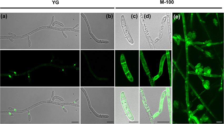
Wang et al.                                                                                       Characterization of the Extracellular FoSod5 in F. oxysporum

(Fov) is responsible for Fusarium wilt of cotton, and is a                a Cu-only SOD (Sod5) which contributes to pathogen tolerance
significant disease found worldwide in all growing areas (Davis           to ROS (Gleason et al., 2014). The C. albicans Sod5 rapidly binds
et al., 2006). This soil-borne fungus invades the vascular tissue         and sequesters a copper co-factor from the host, compromising
via the roots and rapidly spreads to the aboveground portion              copper toxicity to C. albicans (Li et al., 2015a). Similarly,
of the host. Typical field symptoms of Fusarium wilt include              an extracellular SOD in the dimorphic fungus Histoplasma
yellowing, chlorotic leaves, dark-brown/necrotic xylem, and               capsulatum contributes to resistance to host-derived oxidative
wilting, eventually leading to plant death (Davis et al., 2006).          stress in yeast cells (Youseff et al., 2012). In Puccinia striiformis,
   Reactive oxygen species (ROS) are produced by host cells               the causative agent of stripe rust on wheat, a secreted extracellular
and play an important role in defense against various pathogens.          Zn-only SOD contributes to enhanced resistance to oxidative
These small ROS molecules are highly toxic to the infective agents        stress during the interaction between the wheat host and the
and are able to directly eliminate them. At the interaction site          fungus (Liu et al., 2016). Collectively, these studies indicate
between the host and pathogen, plant cells rapidly synthesize             extracellular SODs play an important role in pathogenicity on
and accumulate large quantities of ROS via a membrane-                    various hosts and confer tolerance to the host derived oxidative
bound NADPH oxidase during a process called the oxidative                 stress caused by ROS.
burst (Lamb and Dixon, 1997; Heller and Tudzynski, 2011).                    Extracellular SODs are predicted to have evolved from the
In plants, the oxidative burst may result in the hypersensitive           canonical SOD1 and are phylogenetically conserved throughout
response (HR) which inhibits the spread of the pathogen to                fungi (Robinett et al., 2018), and therefore are postulated to
surrounding tissue (Zurbriggen et al., 2010), and can serve               share a similar biological function. In this study, an extracellular
as an important signal that initiates a series of other plant             SOD (FoSod5) from Fov was important for tolerance to
defense responses or the production of plant hormones (Sauer              multiple ROS and was required for full virulence on cotton.
et al., 2001). These processes stimulate host plants to alter the         The subcellular localization of FoSod5 was dependent on the
expression of genes involved in defense response, leading to              environmental condition, where FoSod5 was primarily localized
the production of phytoalexins, callose deposition, and systemic          to the fungal phialides, but when exposed to a high ROS
acquired resistance; thereby impeding further pathogen spread             environment the enzyme was localized to the septum and cell
and disease development (Sauer et al., 2001; Forman et al., 2010).        wall. This demonstrates that some phytopathogenic fungi utilize
   Despite the extensive production of ROS during the plant               extracellular SODs to tolerate the oxidative burst during the plant
defense response, successful pathogens are usually able to survive        defense response.
in an environment with a high ROS concentration and have
evolved mechanisms to overcome the detrimental effects of this
defense response (Aguirre et al., 2006; Fones and Preston, 2012).         MATERIALS AND METHODS
The family of enzymes known as superoxide dismutases (SOD)
participates in catalyzing the partitioning of superoxide radicals.       Fusarium oxysporum Isolate and Growth
SOD enzymes are divided into several classes based on the                 Conditions
structure of the enzymes and the specific binding of a metal              The race 7 Fov isolate was obtained from the Fungal Genetics
cofactor, and include the copper and zinc SODs (Cu/Zn SOD),               Stock Center (FGSC #10442) (McCluskey et al., 2010), and
the iron or manganese SODs (Fe/Mn SOD), and the nickel                    the wild-type, mutants, and complemented strains (listed in
SODs (Miller, 2012). The cytosolic Cu/Zn SODs are the most                Supplementary Table 1) were grown and maintained at room
common SOD enzymes in eukaryotic cells and there is at least              temperature (25◦ C) on Potato Dextrose agar (PDA; Difco,
one gene encoding a Cu/Zn SOD in a fungal genome (Miller,                 Franklin Lakes, NJ, United States) medium or minimum nutrient
2012). In Saccharomyces cerevisiae, loss of SOD1 results in slow          medium (M-100) (Stevens, 1974).
growth and increased sensitivity to H2 O2 and ROS-generating                 Fungal sensitivity to various chemical stressors was evaluated
compounds (Moradas-Ferreira and Costa, 2000). Additionally,               by placing a 5 mm diameter mycelial plug from the edge of a
the loss of function of SOD1 can alter normal fungal physiology.          4-day-old culture grown on M-100 agar medium and put onto
For instance, a SOD1 mutant of the ericoid mycorrhizal fungus             the new agar medium containing the chemical. The diameter
Oidiodendron maius has reduced production of conidia and the              of the colony was evaluated after 7 days. Each treatment was
capacity for mycorrhization (Abba et al., 2009).                          repeated three times.
   Extracellular SODs have recently been described in various
pathogens (Robinett et al., 2018). In fungi, extracellular                Bioinformatic Analysis
SODs typically contain an N-terminal secretion signal peptide             The SOD protein family in F. oxysporum was identified by
and a C-terminal glycosylphosphatidylinositol (GPI) anchor                BLASTp against the EnsembleFungi database1 using several well
attachment site. The mature GPI anchor enables the protein to             characterized SOD proteins as query sequences (S. cerevisiae
be localized to the cell membrane and/or cell wall by covalent            SOD1: YJR104C and SOD2: YHR008C; C. albicans SOD5:
attachment (Mayor and Riezman, 2004; Robinett et al., 2018).              XP_719507). Protein domains were predicted using PFAM (El-
Some fungal extracellular SODs are secreted out of the cell and           Gebali et al., 2019) and BLASTp software. The subcellular
participate in catalyzing the partitioning of superoxide radicals         localization peptide sequences were identified with SeqNLS and
produced during a host defense response enabling survival in a
high ROS environment. The genome of Candida albicans encodes              1
                                                                              https://fungi.ensembl.org/index.html

Frontiers in Plant Science | www.frontiersin.org                      2                                              March 2021 | Volume 12 | Article 608861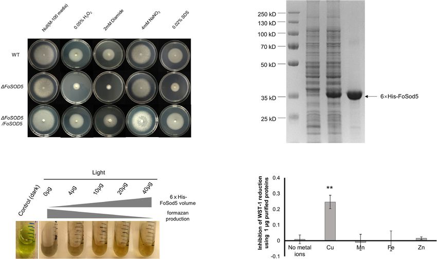
Wang et al.                                                                                Characterization of the Extracellular FoSod5 in F. oxysporum

WoLF PSORT (Horton et al., 2007; Lin and Hu, 2013), and                  respectively. The SalI restriction enzyme was used to confirm the
secreted signal peptides were predicted with SignalP 4.1 (Petersen       hygromycin cassette copy number, while the BamHI restriction
et al., 2011). Multiple sequence alignments were conducted with          enzyme was used to detect FoSOD5 cassette copy number. All the
T-Coffee (Notredame et al., 2000), and a phylogenetic tree was           primers used in this study are listed in Supplementary Table 2.
generated with PhyML 3.1 (Guindon et al., 2010). The 3D protein
structure of FoSod5 was predicted using the SWISS-MODEL
server (Gleason et al., 2014; Waterhouse et al., 2018).
                                                                         6 × His-FoSod5 Protein Purification and
                                                                         SOD Activity Assay
Gene Disruption and Complementation                                      The 6 × His-FoSod5 protein purification was conducted
of FoSOD5                                                                using a previously published method (Gleason et al., 2014).
The target gene, FoSOD5, was disrupted with the hygromycin               Briefly, the middle sequence of FoSOD5 encoding the SOD
B resistance cassette using a previously described split-                domain (lacking the regions encoding the N-terminus signal
marker approach with a few modifications (Goswami, 2012).                peptide and the C-terminus GPI-anchor site) was inserted
Generation of protoplasts was conducted as previously                    into the pHis-Parallel1 vector (Sheffield et al., 1999), and
described (Coleman et al., 2011; Wang et al., 2018) using                the recombinant plasmid was transformed into Escherichia
a mixture of 10 mg/mL DriselaseTM (Sigma, St. Louis,                     coli RosettaTM (DE3) competent cells. Protein production was
MO, United States), 15 mg/mL β-Glucanase (Sigma),                        induced with the addition of 0.5 mM IPTG to a culture
and 5 mg/mL Lyzing enzymes (Sigma) for 2-3 h. DNA                        with an OD600 value between 0.5 and 0.6 and allowed to
fragments were generated by overlapping fusion PCR with the              grow for 4 h at 37◦ C. Protein purification was performed
oligomer pairs, olFoSOD51F/olFoSOD51R, HYGF1/HYGR2,                      under denaturing conditions with prepared denaturing buffer
olFoSOD53F/olFoSOD54R, and HYGBF3/HYGBR4 (Goswami,                       (50 mM Tris–HCl (pH 8.0), 8 M urea, and 1.5 mM reduced
2012). Transformants were selected on 150 µg/mL hygromycin B,            glutathione) using a Ni-NTA Purification System (Thermo
and colonies appearing after 3–5 days were transferred to M-100          Fisher Scientific, Waltham, MA, United States). A series of
medium with hygromycin B (150 µg/mL) for further evaluation.             dialysis buffers containing a decreasing gradient of concentration
   An Agrobacterium-mediated transformation method was                   of urea (50 mM Tris–HCl (pH 8.0), 6/4/2/0 M urea, and
used to generate the FoSOD5 gene complementation strain as               0.25 mM oxidized glutathione) were sequentially used for
described in a previous publication with a few modifications             FoSod5 protein refolding with a 10 kDa dialysis tube. SOD
(Mullins Chen et al., 2001). The FoSOD5 gene complementation             proteins usually require different metals to fold into the correct
plasmid was constructed using pCAMBIA1302 (Cambia,                       structure for function, and different metal ions (20mM of ZnCl2 ,
Canberra, Australia) as the background plasmid where the                 FeCl3 , CuSO4 , and MnSO4 ) were provided individually to the
35S promoter and hygromycin phosphatase gene located                     6 × His-FoSod5 during the final overnight dialysis. The final
between the XhoI and KpnI restriction sites was replaced                 6 × His-FoSod5 protein was concentrated to 2 mg/mL with
with the neomycin phosphotransferase II cassette (trpC                   a 3 K protein concentrator tube (MilliporeSigma, Burlington,
promoter) amplified from the pII99 plasmid (Namiki                       MA, United States).
et al., 2001) using primers NeoF_KpnI and NeoR_XhoI.                        Two methods, the nitro-blue tetrazolium (NBT) method and
An approximately 4 kb locus containing the FoSOD5                        the SOD-WST assay, were used for assessing the enzymatic
gene was amplified from Fov genomic DNA using the                        activity of the purified 6 × His-FoSod5. The NBT method reflects
primers FoSOD5cassF and FoSOD5cassR, and ligated                         SOD enzyme efficiency through inhibition of the reduction of
into the above constructed plasmid between the LB- and                   NBT when superoxide is present. When NBT is reduced by
RB-T-DNA sequences using the NEBuilder HiFi DNA    R
                                                                         superoxide, formazan is produced resulting in a proportional
Assembly Cloning Kit (New England Biolabs, Ipswich, MA,                  dark blue color change that can be monitored. Three mL of the
United States), generating the desired complementation plasmid,          freshly prepared reaction buffer (10 µM riboflavin, 45 µM EDTA,
pCom-G418R -FoSOD5.                                                      350 µM NBT, 60 µM methionine, and potassium phosphate with
   The 1FoSOD5 strain was co-cultivated with the                         a pH value of 7.8) was added to different concentrations (0, 4, 10,
A. tumefaciens strain AGL-1 with plasmid pCom-G418R -FoSOD5              20, and 40 µg) of the purified 6 × His-FoSod5 protein, mixed,
and placed in the dark at 28◦ C for 2 days as described (Mullins         and immediately incubated under light (∼4,000 Lx) for 20 min at
Chen et al., 2001). Selection of the desired complemented strain         28◦ C (Durak et al., 1993). Presented data is a representative set of
was accomplished by plating on M-100 medium containing                   samples based on three independent experiments.
60 µg/mL G418 and 300 µg/mL cefotaxime, and grown at room                   The SOD-WST assay was conducted using the EnzyChromTM
temperature for 5–7 days. G418 resistant transformants were              SOD Assay Kit (BioAssay Systems, Hayward, CA, United States)
isolated to new selective medium for screening by PCR and                according to the manufacturer introductions. One µg of the
Southern blot. The Southern blot was conducted according to the          FoSod5 protein that was reconstituted in the presence of the
manufacturer’s instructions (DIG-High Prime DNA Labeling and             different metal cations was added to the WST-1 reaction buffer
Detection Starter Kit I, Roche). PCR DIG probes for detecting the        and incubated at room temperature for 1 h. The result was
hygromycin cassette and FoSOD5 cassette were generated with              detected by measuring the absorbance at 440 nm (Cytation 3,
the primer pairs HyBF/HyBR and olFoSOD55F/olFoSOD56R,                    BioTek, Winooski, VT, United States). Two independent

Frontiers in Plant Science | www.frontiersin.org                     3                                        March 2021 | Volume 12 | Article 608861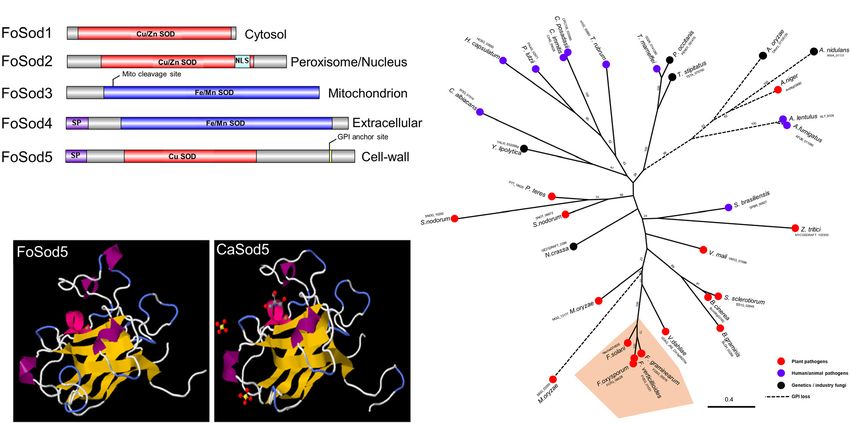
Wang et al.                                                                               Characterization of the Extracellular FoSod5 in F. oxysporum

experiments were conducted and the average and standard                  for the different time points of fungal infection using primers
deviation presented.                                                     qFoSOD5F and qFoSOD5R (Supplementary Table 2) and the
                                                                         F. oxysporum elongation factor 1 alpha gene (EF1α) was used as
Pathogenicity Assay                                                      the internal reference gene using primers qEF1αF and qEF1αR
For the cotton root infection assay, cotton seeds (cultivar              (Supplementary Table 2). The relative expression levels were
FM1944) were planted in sterile soil and placed in a growth              calculated using the 11Ct method (Livak and Schmittgen, 2001).
chamber with a 16 h 28◦ C light/8 h 24◦ C dark cycle. After              Two biological replicates (each with three technical replicates)
2 weeks, cotton plants at the two-to-three true leaf stage were          were performed in this experiment.
used in the infection assay. The cotton seedlings were carefully
uprooted and immersed in sterile water to remove adhering soil           Plasmid Construction for Generation of
from the root system. A set of eight seedlings were transplanted
to a water-culturing box containing nutrient solution (1/10
                                                                         GFP and LacZ Reporter FoSod5 Strains
Murashige and Skoog medium (MS, PhytoTechnology                          The HITI Cas9 RNP transformation plasmid, pUC19-HITI-
Laboratories, Shawnee Mission, KS, United States) supplemented           FoSOD5C2, was constructed using the NEBuilder HiFi                 R

with 0.1% sucrose) to sustain the growth of the cotton plant and         DNA Assembly Cloning Kit and four PCR fragments were
allowed to acclimate for 4 days in the growth chamber.                   simultaneously assembled into the pUC19 plasmid between
    The wild-type and mutant strains of Fov inoculum were                the BamHI and HindIII restriction sites. The first fragment
prepared by growing on a rotary shaker in 100 mL PDB at                  contained a ∼1 kb upstream sequence and partial amino
room temperature for 5 days. The conidia were collected from             acid coding region (AA: 1–53; primers: NA_FoSod5C1hitiF
the cultures, washed in sterile water, and suspended at a final          and NA_FoSod5C2uphitiR); the second fragment was the
concentration of 1 × 10⁷ conidia/mL. Two mL of the conidia               sGFP coding sequence (primers: NA_FoSod5C2midsGFPF
were pipetted into the water-culturing box containing the cotton         and NA_FoSod5C2midsGFPF); the third fragment contained
plants and mixed well with gentle shaking. Each week the                 a partial amino acid coding region (AA: 186-end containing
nutrient solution in the plant culturing box was replaced with           the GPI site) and the predicted native terminator of the
fresh nutrient solution. After 5–8 weeks, disease symptoms were          FoSOD5 gene (∼800 bp) (primers: NA_FoSod5C2downF
recorded by photo and the vascular tissue of the cotton plant was        and NA_FoSod5C2downR); and the forth fragment was the
evaluated. Based on disease symptoms of F. oxysporum on cotton,          hygromycin cassette amplified from plasmid pUC19-HDRI-
four disease index categories were defined: 0 – no symptoms, 1 –         FoSSO1-mCherry using primers: NA_FoSod5C2HYGBF
light yellowing of the leaves and black roots present, 2 – heavy         and Universal_NA_forHITITR. The plasmid pUC19-HITI-
chlorosis of the leaves, wilting evident, and brownish discolored        FoSOD5C2 contained most of the FoSOD5 gene with the
vascular tissue, 3 – wilted plant with dark discolored vascular          exception that the catalytic SOD domain was replaced by
tissue or death of the plant. Pathogenicity assays for each strain       sGFP. The plasmids, pUC19-HITI-FoSOD5C1 and pUC19-
were conducted at least twice.                                           HITI-FoSOD5C3 were generated in a similar method. Plasmid
                                                                         pUC19-HITI-FoSOD5C1 lacked all the amino acid sequences
                                                                         after the SOD domain, while in pUC19-HITI-FoSOD5C3 the
RNA Extraction of Fov-Infected Cotton                                    entire ORF of FoSOD5 gene was replaced by the β-galactosidase
Roots and qRT-PCR                                                        (lacZ) coding region cloned from plasmid, pCYC-lacZ (gift
Preparation and inoculation of the cotton plants (FM1944)                from Dr. Paul Cobine, Auburn University). The three different
was slightly different than the pathogenicity assay. Instead of          plasmids were used to transform the WT Fov isolate to generate
geminating seeds in soil, they were germinated in 1/2 strength           the FoSod5 variants enabling the study of the FoSod5 function
MS medium polymerized with 2.6 g/L of phytagel. After 1 week             including protein secretion, protein subcellular localization, and
of germination, the plants were transferred to a water-culturing         protein expression (Wang and Coleman, 2019). All PCR primers
box containing 1/2 strength MS nutrient solution. When the               used are listed in Supplementary Table 2.
plants were 2 weeks old, the MS medium in the box was
replaced with 1/10 MS medium supplemented with 0.1% sucrose
and inoculated with ∼2 × 10⁷ conidia in each box. The three              In vitro Cas9 Nuclease Assay and Cas9
randomly selected Fov-infected cotton roots from three plants            RNP Transformation
were pooled for each time point and samples were collected               Before Cas9 RNP transformation, an in vitro Cas9 nuclease
at multiple time-points (0, 4, 12, 24, and 36 h, 2, 3, 5, and            assay was conducted to confirm the Cas9 RNP cleavage activity
7 days post inoculation) for total RNA extraction. Total RNA             (Wang et al., 2018). The Cas9 RNP mediated transformation
was extracted using the E.Z.N.A. Total RNA Kit I (Omega Bio-
                                           R
                                                                         was conducted as previously described (Wang and Coleman,
Tek, Norcross, GA, United States) according to the manufacturer          2019), and the amount of donor plasmid did not exceed
instructions, and the total RNA was treated with DNase I                 6 µg per transformation plate. After transformation, positive
(New England Biolabs). Reverse transcription was immediately             colonies were isolated on M-100 selective medium containing
conducted to generate cDNA using the QuantiTect Reverse                  150 µg/mL hygromycin. Three pairs of PCR primers were
Transcription Kit (Qiagen). Transcript expression of FoSOD5              used to confirm the location of the integrated plasmids. Since
was determined by qRT-PCR on a Bio-Rad CFX96 instrument                  four different FoSod5 variants were generated using the HITI

Frontiers in Plant Science | www.frontiersin.org                     4                                       March 2021 | Volume 12 | Article 608861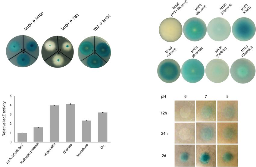
Wang et al.                                                                              Characterization of the Extracellular FoSod5 in F. oxysporum

strategy, the cleavage site at the 50 -terminus (SS, Supplementary       Chicago, IL) using the Mini Trans-Blot Electrophoretic Transfer
                                                                                                                    R

Figure 3) was sequenced.                                                 Cell (Bio-Rad, Hercules, CA, United States). Anti-GFP antibody
                                                                         (Rockland Immunochemicals, Gilbertsville, PA, United States)
β-Galactosidase (LacZ) Activity Assay                                    and an ECL chemiluminescent detection kit (GE Healthcare)
The expression and regulation of FoSOD5 was investigated                 were used to detect the resulting proteins.
under various conditions. For different pH values, M-100
agar medium with a final X-gal concentration of 200 ng/mL                Confocal Microscopy
was adjusted with a pH ranging from 6 to 8. FoSOD5                       Confocal microscopy was conducted with a Nikon A1 Confocal
expression under the influence of different carbon sources               Microscope with an excitation wavelength of 488 nm for
was investigated using M-100 medium as a base where the                  detection of sGFP fluorescence as previously described (Wang
glucose component was replaced with 1% (w/v) of various                  and Coleman, 2019). For different chemical treatments, a
carbon sources (glycerol, carboxymethylcellulose (CMC), starch,          mixture of hyphae and conidia was inoculated into YG medium
sucrose, sorbitol, and mannitol). ∼1 × 10⁵ conidia were                  and cultured for 3 days. One mL of the hyphae-conidial
dropped onto the center of the agar plates with or without the           mixture was transferred into fresh YG medium and cultured
X-gal. For quantification of β-Galactosidase enzymatic activity,         on a rotary shaker at 18◦ C, 220 rpm for 16 h. Assayed
an O-Nitrophenyl-B-D-Galactopyranoside (ONPG) assay was                  chemicals (0.03% H2 O2 and 20 µg/mL xanthine oxidase/0.1 mM
conducted according to two previous protocols (Pardee et al.,            hypoxanthomine) were added to the YG medium and cultured
1959; Smale, 2010). The proFoSod5:LacZ strain was cultured               for an additional 2 h. After this time, samples were aliquoted,
to the exponential stage in YG liquid medium. Different                  fixed with 4% paraformaldehyde, and observed by confocal
chemicals (0.05% H2 O2 , 0.5 mM diamide, 0.2 mM menadione,               microscopy using an inverted agar method (Hickey and Read,
20 µg/mL xanthine oxidase/0.1 mM hypoxanthomine, and                     2009). All microscopy experiments were repeated at least three
150 µM CuSO4 ) were added to the YG medium and the                       independent times.
fungi were cultured for an additional 3 h. Total protein was
extracted with liquid nitrogen, and 5 µg of total protein
(in 75 µL) from each treatment and 40 µL of 4 mg/mL                      RESULTS
ONPG were added to 700 µL reaction buffer (8.5 mg/mL
Na2 HPO4 , 5.5 mg/mL Na2 H2 PO4 .H2 O, 750 ng/mL KCl,                    Identification of an Extracellular GPI
and 246 ng/mL MgSO4 .7H2 O, pH 7.0) and incubated at                     Anchored SOD Protein (FoSod5) From
30◦ C for 25 min. 100 µL of 1M Na2 CO3 was used to
stop the above reaction, and the β-Galactosidase enzymatic
                                                                         F. oxysporum
activity was measured by absorbance at 420 nm (Cytation                  Several Sod proteins from various fungal species were used as
3, BioTek, Winooski, VT, United States). All assays were                 a query and identified five SOD proteins in the F. oxysporum
repeated in triplicate.                                                  genome. These SOD encoding genes included three Cu/Zn
                                                                         SOD genes (FoSOD1: FOTG_01421; FoSOD2: FOTG_16882;
                                                                         FoSOD5: FOTG_08628) and two Fe/Mn SOD genes (FoSOD3:
Secreted Protein Extraction and Western                                  FOTG_10379; FoSOD4: FOTG_02058) (Figure 1A). Prediction
Blotting                                                                 of the subcellular localization indicated FoSod1 (a Cu/Zn
A trichloroacetic acid (TCA) precipitation method was used               SOD) was localized to the fungal cytosol while FoSod3
to extract the fungal secreted proteins. The fungal isolates             (a Fe/Mn SOD) was localized to the mitochondrion, in
were cultured in YG liquid medium for 3 days at room                     agreement with the subcellular localization of homologous
temperature. The resulting fungal culture was centrifuged                SOD proteins from other fungi (Luk et al., 2003; Yao et al.,
at 13,000 g for 10 min and filtered through a 0.22 µM                    2016). However, FoSod2 (a Cu/Zn SOD) was predicted to be
filter to remove hyphae and conidia. TCA was added to                    localized to either the peroxisome and/or the nucleus while
a final concentration of 10%, and the mixture chilled at                 FoSod4 (a Fe/Mn SOD) is predicted to have a secretion
4◦ C overnight. The solution was centrifuged at 13,000 g for             signal peptide and could be a secreted SOD enzyme. FoSod5,
10 min, and the resulting pellets were washed with ice-                  contains an N-terminal secretion signal (AA: 1–21) and a
cold acetone at least twice. The final protein pellets were              C-terminal glycophosphatidyl inositol (GPI) attachment site
dissolved in 50 mM Tris–HCl buffer (pH = 7.5), and the                   (AA: 240), suggesting this protein is secreted and anchored
protein concentration was determined using a Qubit 3.0                   to the fungal cell wall or membrane (Figure 1A). Sequence
Fluorometer with the QubitTM Protein Assay Kit (Thermo                   alignment of selected Cu/Zn SODs revealed that FoSod5 lacks
Fisher Scientific).                                                      two histidine residues involving in zinc binding implicating
    Western blotting was conducted with a total of 5 µg of the           FoSod5 is a Cu-specific SOD enzyme (Supplementary Figure 1;
secreted proteins which were separated by 10% SDS-PAGE gel               Gleason et al., 2014). Although the amino acid sequence
electrophoresis and stained with Coomassie staining solution             similarity between CaSod5 and FoSod5 was only 33%, the
(0.1% Coomassie Brilliant Blue R-250, 50% methanol, and 10%              predicted structure of FoSod5 was highly similar to CaSod5,
glacial acetic acid). The proteins in the SDS-PAGE gel were              suggesting functional similarities between the two proteins
transferred to a nitrocellulose filter membrane (GE Healthcare,          (Figure 1B). Phylogenetic analysis indicated many fungi

Frontiers in Plant Science | www.frontiersin.org                     5                                      March 2021 | Volume 12 | Article 608861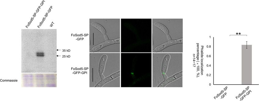
Wang et al.                                                                               Characterization of the Extracellular FoSod5 in F. oxysporum

in the Ascomycota contain a single ortholog of SOD5                      complemented (1FoSOD5/FoSOD5) strains on whole cotton
(Figure 1C), and isolates within Fusarium carried a single               plants to assess the role of FoSOD5 in infection. While the
phylogenetically conserved copy of the SOD5 gene (Figure 1C).            wild-type isolate was able to colonize the xylem tissue in the
Interestingly, all the genomes from the Aspergillus species              roots and cause necrosis, the 1FoSOD5 strain exhibited reduced
included in this analysis appear to have lost the GPI anchor             necrosis (Figures 3B,C). Overall, the cotton plants inoculated
site during their evolution, but it had been retained in the             with the FoSOD5 mutant displayed less wilting symptoms, had
closely related Penicillium spp. indicating the loss of the              less browning of the leaves, and there was less necrosis of the
GPI anchor occurred after the divergence between these two               root xylem when compared to cotton plants inoculated with the
genera (Figure 1C).                                                      WT isolate and the 1FoSOD5/FoSOD5 complemented strains
                                                                         (Figures 3B,C).
FoSod5 Is a Functional SOD Protein
A mutant of FoSOD5 was generated to assess the contribution of           Expression of FoSOD5 Is Regulated by
the SOD to oxidative stress tolerance (Supplementary Figure 2).
The 1FoSOD5 mutant had increased sensitivity to hydrogen
                                                                         the Carbon Source and Is Repressed in a
peroxide and diamide, a ROS-generating compound, in a radial             Nutrient Rich-Medium
growth ROS stress assay (Figure 2A). Functional SOD activity             The function of cell wall proteins can vary and be species-
for FoSod5 was confirmed using two in vitro methods, the                 specific, contributing to the diverse properties of the cell
NBT method and the SOD-WST1 assay. The middle amino                      wall and enabling fungi to adapt to adverse environments
acid sequence harboring the SOD domain was fused with a                  (Gow et al., 2017). In order to investigate the expression of
N-terminal 6 × His tag for protein purification (Figure 2B). This        FoSOD5 in various environmental conditions, a lacZ reporter
soluble purified 6 × His-FoSod5 protein was used for functional          construct was generated under the control of the native FoSOD5
assessment of SOD enzymatic activity and determination of the            promoter (Supplementary Figure 3). The lacZ gene, encoding
specificity for different metal ions to serve as a cofactor.             a β-galactosidase, is frequently used as a reporter gene for
    The traditional NBT-riboflavin method indicated the                  the in vivo analysis of gene regulation in various organisms.
6 × His-FoSod5 protein was a functional SOD enzyme. Under                After transformation, eight independent colonies were selected
illumination, the riboflavin-methionine mixture produces                 for analysis, and seven of these transformants produced the
superoxide causing NBT to be reduced to the blue colored                 blue pigment when grown on M-100 medium containing
formazan, and this reaction can be inhibited by SOD activity.            X-gal, indicating the expression of lacZ is a suitable reporter
A concentration series of the 6 × His-FoSod5 protein (0                  for the in vivo analysis of gene regulation in F. oxysporum
to 40 µg) were added to the reaction buffer, and as the                  (Supplementary Figure 4). Sequencing revealed that DNA repair
concentration of the 6 × His-FoSod5 protein increased, a                 occurred without errors at the sgRNA cleavage site in six of the
decrease in the production of formazan was evident (Figure 2C),          seven transformants (Supplementary Figure 5, SS site), while
indicating that FoSod5 is a SOD enzyme able to inhibit the               the remaining transformant had a 77 bp nucleotide deletion
reduction of NBT. Using the SOD-WST1 assay to determine                  (Supplementary Figure 5).
heavy metal cofactor specificity, of all the heavy metals tested            A proFoSOD5:lacZ reporter strain was used to investigate the
only copper was able to confer SOD activity, demonstrating               expression pattern of FoSOD5 when grown in various carbon
that FoSod5 was a copper-specific SOD (Figure 2D), and is                sources. LacZ activity was evident when the fungus was grown on
consistent with previous findings on the SOD5 class of enzymes           a minimal nutrient medium (M-100) for 4 days. When an agar
(Gleason et al., 2014).                                                  plug (5 mm in diameter) was transferred from the lacZ inducing
                                                                         M-100 plate to a rich nutrient medium (TB3), the hyphae that
                                                                         grew on the TB3 medium had reduced LacZ activity after 4 days
FoSOD5 Is Up-Regulated During                                            and the hyphae were primarily white in color (Figure 4A).
Infection and Is Required for Full                                       Even after 1 week, there was no significant color change of the
Virulence of Fov on Cotton                                               mycelia on TB3 media. When an agar plug of the proFoSOD5:lacZ
Many fungal virulence factors have increased expression during           reporter mycelia that was grown on TB3 was placed on M-100
infection of host plants. As SOD enzymes are known to be                 medium, the mycelia had LacZ activity. This regulation of LacZ
involved in virulence in other fungal pathogens (Heller and              activity by the FoSOD5 promoter indicates that the expression of
Tudzynski, 2011), the expression profile of FoSOD5 during the            the SOD is dependent on the available nutrient(s).
infection process of cotton was investigated by qRT-PCR. The                As the carbon sources for the M-100 and TB3 media were
expression of FoSOD5 was relatively low during the initial 12 h of       different, it was hypothesized that the carbon source in the
infection, but the gene was significantly increased in expression        medium may influence the expression FoSOD5. To evaluate
from 24 h to 7 day after infection with the peak being at                this possibility, the 1% (w/v) glucose content in the M-100
2 days (Figure 3A). From 24 h after infection, the average               agar medium was replaced with 1% (w/v) of various other
increase in the number of FoSOD5 transcripts was at least >100-          carbon sources (sucrose, mannitol, glycerol, CMC, starch, and
fold (Figure 3A).                                                        sorbitol). Four days after inoculation, LacZ activity was evident
   As Fov is responsible for wilting symptoms, a virulence assay         for mycelia grown on all carbon sources investigated, but there
was conducted with the WT, FoSOD5 mutant (1FoSOD5), and                  was a clear difference between nutrients. Interestingly, the LacZ

Frontiers in Plant Science | www.frontiersin.org                     6                                       March 2021 | Volume 12 | Article 608861
Wang et al. Characterization of the Extracellular FoSod5 in F. oxysporum
FIGURE 1 | Domain, structure, and phylogenic analysis of FoSod5. (A) Identification of the Sod protein family in F. oxysporum and predicted subcellular localization
of FoSod proteins. NLS: nuclear localization sequence; SP: secreted peptide. (B) The predicted protein structure of FoSod5 and comparison with CaSod5. (C) The
maximum-likelihood phylogenetic tree of Sod5 from ascomycete fungi. The tree was generated with a bootstrap value of 500.
activity of the mycelia in the presence of CMC and starch were FoSod5 variants under the control of the native promoter
higher than those for the other carbon sources (Figure 4B), were generated to assess the N-terminal secretion signal
suggesting that plant-derived carbon sources may favor FoSOD5 peptide of FoSod5 (Supplementary Figure 3b). A homologous-
expression. In addition, FoSOD5 gene regulation was investigated independent targeted integration (HITI) approach was used to
in the presence of different chemical stimuli including hydrogen insert an entire plasmid at the FoSOD5 locus (Supplementary
peroxide and the ROS generating compounds diamide and Figure 3a), generating a functional FoSOD5 gene and the
menadione. The LacZ activity of the mycelia was increased by desired FoSOD5 GFP reporter variant in close proximity to one
these compounds, in particular hypoxanthomine and diamide another. The FoSod5-SP-GFP variant included the sequence
which had a four fold higher level of LacZ activity than encoding the first 53 amino acids of FoSod5 containing the
background (Figure 4C). signal peptide and sGFP, removing the nucleotides encoding
the SOD domain and the GPI site (AA: 53 – 263). The second
FoSOD5 Is More Rapidly Expressed variant, FoSod5-SP-GFP-GPI, replaced the internal catalytic
SOD domain (AA: 53 – 186) with sGFP maintaining the GPI
Under an Alkaline Environment
anchor attachment site (SP + sGFP + GPI) (Supplementary
Many virulence factors of phytopathogenic fungi, including those
Figure 3b). After transformation, the region of interest of three
in F. oxysporum, are dependent on the pH of the surrounding
FoSod5-SP-GFP and two FoSod5-SP-GFP-GPI transformants
environment, and therefore it was hypothesized that the pH of the
were sequenced to confirm they did not contain indels or
medium may play a role in the regulation of FoSOD5. Conidia of
other undesired alterations (Supplementary Figure 5). These
the proFoSOD5:lacZ reporter strain were placed on M-100 plates
FoSod5 variants and the wild type isolate were grown in liquid
that were adjusted to a pH ranging from 6 to 8 and monitored
culture and after 4 days the supernatant was evaluated for the
over time. In a neutral or alkaline environment (pH = 7 or 8),
presence of sGFP by western blot using an anti-GFP antibody.
FoSOD5 was induced within the first 12 h; however, mycelia at a
sGFP was only detected in the liquid culture medium from
pH 6 did not display LacZ activity until 48 h (Figure 4D).
FoSod5-SP-GFP (Figure 5A), confirming FoSod5 contained a
functional N-terminal secretion peptide. When the GPI anchor
The SP and GPI Anchor of FoSod5 Are was present (FoSod5-SP-GFP-GPI) the protein was unable to be
Required for Subcellular Localization detected in liquid culture medium, even when ∼25 µg of the
Glycosylphosphatidylinositol proteins may contain an extracted protein was used for the western blot and the exposure
N-terminus secretion signal peptide in addition to the time for detection was increased.
C-terminus GPI site (Mayor and Riezman, 2004), which Comparison of the two sGFP constructs, FoSod5-SP-GFP
enables attachment to either the fungal cell wall or the plasma and FoSod5-SP-GFP-GPI, which only differed with the presence
membrane (De Groot et al., 2005; Ouyang et al., 2013). Two of the GPI site, allows the influence of the GPI anchor on
Frontiers in Plant Science | www.frontiersin.org 7 March 2021 | Volume 12 | Article 608861Wang et al. Characterization of the Extracellular FoSod5 in F. oxysporum
FIGURE 2 | FoSod5 is a functional SOD protein and confers tolerance to ROS. (A) Evaluation of fungal sensitivity to different chemical stressors. The indicated
strains were inoculated on minimal medium (M-100). The assay was repeated three times. (B) SDS-PAGE gel depicting protein expression and purification
conditions. Lane 1 represents total protein before inducing; Lane 2 total protein after addition of 0.5 mM IPTG for 4 h; Lane 3 represents purified 6 × His-FoSod5
protein. (C) Assessment of purified 6 × His-FoSod5 enzymatic activity using the nitro-blue tetrazolium (NBT) method. The production of formazan decreases as the
6 × His-FoSod5 protein increases. Three independent replicates per treatment were conducted. (D) Determination of metal ion co-factor preference for
6 × His-FoSod5 using the SOD-WST assay. Only copper ions were able to confer SOD activity. The average inhibition and standard deviation is depicted in the
histogram and is based on two independent experiments.
cellular localization to be evaluated. When the FoSod5-SP- Since the FoSod5-SP-GFP-GPI variant did not contain the
GFP-GPI strain was grown in YG medium, a vast majority SOD domain, it was uncertain if this domain had an influence on
of the fluorescent signal was observed at the phialides by the subcellular localization of FoSod5. To address this concern,
confocal microscopy while no fluorescence was observed for another FoSod5 variant was generated (FoSod5-SP-GFP-SOD-
the FoSod5-SP-GFP variant (Figures 5B,C). This finding, taken GPI) which contained 2 × sGFP in front of the SOD domain
together with the previous finding that sGFP was detected (Supplementary Figure 3b). Under confocal microscopy, the
by western blotting in the supernatant of the FoSod5-SP-GFP FoSod5-SP-GFP-SOD-GPI variant had a similar localization
transformant, indicates the GPI site of FoSod5 is required for pattern as FoSod5-SP-GFP-GPI indicating the functional SOD
proper physiological localization at the septum of the phialides domain was not required for proper subcellular localization
when grown in YG medium. (Supplementary Figure 6).
External Environmental Conditions
DISCUSSION
Influence the Subcellular Localization of
FoSod5 Host cells impede invading fungal pathogens through the rapid
FoSod5 was primarily localized to the fungal phialides in YG production of ROS by membrane-bound NADPH oxidases
media (Figures 5B, 6a); however, a weak fluorescent signal (Marino et al., 2012); and successful fungal pathogens must
could also be observed in conidia, hyphal tips, septum, and evolve strategies to overcome the cellular damage from ROS
hyphae (Figures 6a,b). When this isolate was grown in minimal during infection (Heller and Tudzynski, 2011). For instance,
nutrient M-100 medium, FoSod5 was distributed within an Magnaporthe oryzae has a robust anti-oxidant defense system
extensive array of fungal structures including the conidia, hyphae, conferring high tolerance to the host oxidative burst generated
and septa, indicating a significant alteration of the subcellular by the host rice plant (Samalova et al., 2014). It has
localization (Figures 6c–e). been well established that fungal SOD proteins, in particular
Frontiers in Plant Science | www.frontiersin.org 8 March 2021 | Volume 12 | Article 608861Wang et al. Characterization of the Extracellular FoSod5 in F. oxysporum
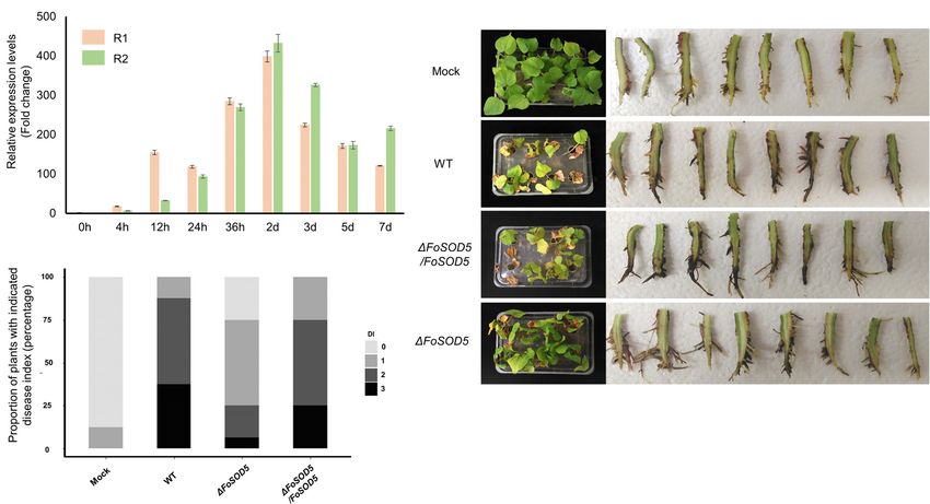
FIGURE 3 | FoSOD5 is up-regulated in planta and involved in virulence on cotton. (A) The expression of FoSOD5 during infection as quantified by qRT-PCR using
cDNA from infected plant tissue. FoSOD5 transcripts were significantly increased 24 h post inoculation. The R1 and R2 data sets represent two independent
experiments with three technical replicates. (B) Virulence assay depicting cotton symptoms caused by an infection of WT, 1FoSOD5, and 1FoSOD5/FoSOD5
strains and control group (Mock). Whole plant symptoms are shown on the left side of the panel, and necrotic development on the root on the right side of the panel.
Two independent experiments were conducted. (C) The disease index on cotton for the WT, 1FoSOD5, and 1FoSOD5/FoSOD5 strains. Ratings were scored from
0 to 3, where 0 was a healthy plant with no symptoms and three indicated a wilted plant with discolored vascular tissue or plant death.
members of the Cu/Zn family of SOD proteins, contribute to to alternative sites under stressful conditions. In S. cerevisiae, the
ROS tolerance by detoxification of reactive superoxide radical Cu/Zn SOD enzyme Sod1 is mainly distributed in the cytosol, but
anions (Veluchamy et al., 2012; Li et al., 2015b). Collectively, rapidly relocates to the nucleus in response to high endogenous
multiple studies have indicated that fungal SOD proteins and/or exogenous ROS, aiding in the maintenance of genomic
play an important role as an antioxidant and contribute to stability and serving as a nuclear transcription factor by binding
fungal pathogenicity. DNA promoter regions of oxidative resistance and repair genes
In fungi, the number of SOD encoding genes in a genome (Tsang et al., 2014).
varies greatly. The genome of S. cerevisiae only encodes two Most extracellular Sod proteins only use copper as the
SOD genes (SOD1 and SOD2) while some filamentous fungi, heavy metal co-factor and function under Zn-limited conditions.
including F. oxysporum, encode five or more SOD encoding A majority of fungi contain 1 to 3 genes encoding extracellular
genes, eluding to a complex, selective process during the Sod proteins (Broxton and Culotta, 2016), and some of these
expansion of the SOD gene family. Usually expansion of a protein Cu extracellular Sods have been shown to be involved in
family leads to divergence of protein function. Bioinformatic fungal pathogenicity in the clinically relevant fungi C. albicans
analysis predicts that the five SOD enzymes of F. oxysporum and H. capsulatum; however, none have been characterized
are localized to different organelles or the cytosol. FoSod2 and in phytopathogenic fungi. The extracellular Sod FoSod5 from
FoSod5 are predicted to reside in the perioxisome/nucleus and F. oxysporum is a virulence factor for cotton, as an FoSOD5
cell wall/membrane, respectively; however, phylogenetic analysis mutant caused less severe wilt symptoms and had less xylem
suggests these two SODs may have evolved from the cytosolic colonization in infected cotton plants when compared to the
FoSod1 (a Cu/Zn SOD). Another example is FoSod4, which wild-type. FoSOD5 was gradually up-regulated during infection,
is similar to the mitochondrial localized FoSod3, although suggesting FoSod5 may participate in ROS scavenging during
FoSod4 is predicted to contain a secretion signal peptide at the infection process, and mutation of FoSOD5 led to increased
its N-terminus. These results indicate that Sod proteins are sensitivity to ROS.
translocated to different organelles, which likely influences their While extracellular Sod5-like proteins are widely distributed
physiological function. Increasing evidence has shown that Sod among most pathogenic fungi and may contribute to virulence
proteins have various subcellular locations and could be localized during infection, there are exceptions. Deletion of the
Frontiers in Plant Science | www.frontiersin.org 9 March 2021 | Volume 12 | Article 608861Wang et al. Characterization of the Extracellular FoSod5 in F. oxysporum FIGURE 4 | In vivo expression analysis of FoSOD5 under different environmental conditions via the lacZ reporter strain. (A) Production of β-galactosidase in the lacZ reporter strain of FoSOD5 is dependent on the medium that the fungus is grown. M-100 is a minimal nutrient medium and TB3 is a rich medium. (B) The FoSOD5 lacZ reporter strain produced a varying intensity of blue pigment when grown on M-100 medium supplemented with various sources of carbon (1% w/v). CMC = carboxymethylcellulose. (C) β-galactosidase activity of LacZ under different ROS and ROS-generating chemical stimuli. (D) The effect of pH on FoSOD5 gene expression. In a neutral pH or slightly alkaline environment, expression was evident within the first 12 h after treatment. All the assays were replicated three times. FIGURE 5 | The SP and GPI anchor are required for FoSod5 subcellular localization within Fov. (A) Western blot analysis with anti-GFP of total protein found in the culture medium of FoSod5 derivatives and the WT. Coomassie protein staining indicated the same amount of loaded protein samples. (B) Phialide localization of FoSod5-SP-GFP and FoSod5-SP-GFP-GPI by fluorescence microscopy. (C) Statistical analysis of phialide localization among FoSod5-SP-GFP and FoSod5-SP-GFP-GPI, where n represents the number of phialides counted at a given time. Three counts from independent biological samples were used for the statistical analysis. Scale bars represents 10 µm. orthologous GPI-anchored SOD from Fusarium graminearum, where F. oxysporum is mainly responsible for damage to host a close relative to F. oxysporum and the main causal agent roots and colonization of the xylem while F. graminearum of Fusarium head blight on wheat, failed to have any effect infects the wheat spikelets, the leaf sheath, and the culm. In on fungal pathogenicity (Rittenour and Harris, 2013). While addition, FoSOD5 from Fov was gradually upregulated during the discrepancy between the role of the SOD5 orthologs in infection of cotton, indicating a host-induced expression profile; virulence on plant hosts could be due to multiple factors, it is whereas the SOD5 ortholog from F. graminearum did not important to note that these fungi infect different host tissues, have a host-induced expression profile and had low expression Frontiers in Plant Science | www.frontiersin.org 10 March 2021 | Volume 12 | Article 608861
Wang et al.                                                                                        Characterization of the Extracellular FoSod5 in F. oxysporum

  FIGURE 6 | FoSod5-SP-GFP-GPI subcellular localization under confocal microscopy (a,b) FoSod5-SP-GFP-GPI subcellular localization in YG medium (c–e)
  FoSod5-SP-GFP-GPI subcellular localization in M-100 medium. (e) The Z-projection of FoSod5-SP-GFP-GPI on M-100 medium under confocal microscopy. Scale
  bars represent 10 µm.

in planta (Yao et al., 2016). Therefore, the regulation of the SOD5              enzyme during conidiogenesis. However, there was no significant
orthologs is different between these two pathogens, and whether                  difference in the number of conidia produced between the
this is due to the regulatory mechanisms at the species level or                 wild-type and FoSOD5 mutant in YG medium (data not
the in planta environment remains unknown.                                       shown), indicating there are likely other factors involved in ROS
    Expression of FoSOD5 in F. oxysporum was highly dependent                    scavenging during conidiogenesis or FoSod5 has an alternative
on environmental conditions, as in a nutrient-limited (M-100)                    role at the fungal phialides. In support of additional factors,
medium it was highly expressed. In addition, different carbon                    increased expression of the MnSOD encoding SOD2 gene
resources or stresses also influenced the expression of FoSOD5,                  in Colletotrichum graminicola is evident during generation of
a phenomenon also observed in C. albicans when yeast cells were                  conidia (Fang et al., 2002). Additionally, regulation of FoSod5
treated with osmotic or oxidative stress conditions (Martchenko                  appears to be more complex in M-100 medium as it was found
et al., 2004). All these results suggest Sod5 plays an important role            in multiple locations including the cell wall/membrane, septum,
in fungal adaptation to different environments.                                  conidia, and phialides. Collectively these results indicates FoSod5
    Fusarium oxysporum is known to secrete peptides (F-RALF)                     alters its subcellular localization based on environmental cues
that induce alkalization in plants and enhance fungal virulence                  and might facilitate adaptation to different environments.
during infection (Masachis et al., 2016; Fernandes et al., 2017),                   The critical role the oxidative burst plays in plant defense
and MAPK-mediated fungal growth on cellophane is more                            has been well established and pathogens have utilized multiple
invasive at pH 7 than that at pH 5 (Masachis et al., 2016).                      mechanisms to overcome this defense mechanism. Several
Plants inoculated with a F. oxysporum f-ralf mutant display                      members of the Cu/Zn Sod family that are predicted to reside
ROS accumulation (Masachis et al., 2016), an indication of                       in the cytosol serve as virulence factors on plant hosts; however
programmed cell death due to the pathogen. As FoSOD5                             this study indicates that tolerance to ROS begins before the toxic
was induced in the presence of high ROS conditions and an                        compounds even enter the fungal cell though extracellular SODs.
alkaline environment, FoSOD5 could play a critical role in                       Given the conserved nature of these SODs, this mechanism
fungal infection.                                                                may potentially serve as a target for development of alternative
    Previous studies indicated fungal cell wall associated proteins              management strategies for several fungal diseases, including
are highly dynamic, dependent on the surrounding environment,                    Fusarium wilt.
and species-specific (De Groot et al., 2005; Klis et al., 2010).
The localization of FoSod5 was dependent on the culture
environment, where FoSod5 accumulated at the fungal phialides                    DATA AVAILABILITY STATEMENT
in YG medium. ROS production has been implicated as a signal
for fungal differentiation and increases during conidiogenesis                   The original contributions presented in the study are included
(Heller and Tudzynski, 2011). The localization of FoSod5 at                      in the article/Supplementary Material, further inquiries can be
the phialides indicates it might act as a ROS scavenging                         directed to the corresponding author/s.

Frontiers in Plant Science | www.frontiersin.org                            11                                        March 2021 | Volume 12 | Article 608861
Wang et al. Characterization of the Extracellular FoSod5 in F. oxysporum
AUTHOR CONTRIBUTIONS ACKNOWLEDGMENTS
QW and JC conceived and designed the experiments and The authors would like to thank Drs. Seogchan Kang (Penn
composed the manuscript. QW and AP conducted the State University), Gillian Turgeon (Cornell University), and Paul
experiments. All authors contributed to the article and approved Cobine (Auburn University) for providing the Agrobacterium-
the submitted version. mediated transformantion plasmid, the geneticin resistance
cassette in pII99, and the LacZ reporter gene, respectively.
FUNDING SUPPLEMENTARY MATERIAL
This research was supported by the Alabama Agricultural The Supplementary Material for this article can be found
Experiment Station and the Hatch program of the National online at: https://www.frontiersin.org/articles/10.3389/fpls.2021.
Institute of Food and Agriculture, USDA. 608861/full#supplementary-material
REFERENCES Goswami, R. S. (2012). Targeted gene replacement in fungi using a split-marker
approach. Methods Mol. Biol. 835, 255–269. doi: 10.1007/978-1-61779-501-5_
Abba, S., Khouja, H. R., Martino, E., Archer, D. B., and Perotto, S. (2009). SOD1- 16
targeted gene disruption in the ericoid mycorrhizal fungus Oidiodendron maius Gow, N. A. R., Latge, J. P., and Munro, C. A. (2017). The fungal cell wall: structure,
reduces conidiation and the capacity for mycorrhization. Mol. Plant-Microbe biosynthesis, and function. Microbiol. Spectr. 5.
Interact. 22, 1412–1421. doi: 10.1094/mpmi-22-11-1412 Guindon, S., Dufayard, J. F., Lefort, V., Anisimova, M., Hordijk, W., and Gascuel,
Aguirre, J., Hansberg, W., and Navarro, R. (2006). Fungal responses to reactive O. (2010). New algorithms and methods to estimate maximum-likelihood
oxygen species. Med. Mycol. 44, S101–S107. phylogenies: assessing the performance of PhyML 3.0. Syst Biol. 59, 307–321.
Broxton, C. N., and Culotta, V. C. (2016). SOD enzymes and microbial pathogens: Heller, J., and Tudzynski, P. (2011). Reactive oxygen species in phytopathogenic
surviving the oxidative storm of infection. PLoS Pathog 12:e1005295. doi: 10. fungi: signaling, development, and disease. Annu. Rev. Phytopathol. 49, 369–
1371/journal.ppat.1005295 390. doi: 10.1146/annurev-phyto-072910-095355
Coleman, J. J., White, G. J., Rodriguez-Carres, M., and VanEtten, H. D. (2011). Hickey, P. C., and Read, N. D. (2009). Imaging living cells of Aspergillus in vitro.
An ABC transporter and a cytochrome P450 of Nectria haematococca MPVI Med. Mycol. 47(Suppl. 1), S110–S119.
are virulence factors on pea and are the major tolerance mechanisms to the Horton, P., Park, K. J., Obayashi, T., Fujita, N., Harada, H., Adams-Collier, C. J.,
phytoalexin pisatin. Mol. Plant-Microbe Interact. 24, 368–376. doi: 10.1094/ et al. (2007). WoLF PSORT: protein localization predictor. Nucleic Acids Res.
mpmi-09-10-0198 35, W585–W587.
Davis, R. M., Colyer, P. D., Rothrock, C. S., and Kochman, J. K. (2006). Fusarium Klis, F. M., Brul, S., and De Groot, P. W. J. (2010). Covalently linked wall proteins
wilt of cotton: population diversity and implication for management. Plant Dis. in ascomycetous fungi. Yeast 27, 489–493. doi: 10.1002/yea.1747
90, 692–703. doi: 10.1094/pd-90-0692 Lamb, C., and Dixon, R. A. (1997). The oxidative burst in plant disease resistance.
De Groot, P. W. J., Ram, A. F., and Klis, F. M. (2005). Features and functions of Annu. Rev. Plant Physiol. Plant Mol. Biol. 48, 251–275. doi: 10.1146/annurev.
covalently linked proteins in fungal cell walls. Fungal. Genet Biol. 42, 657–675. arplant.48.1.251
doi: 10.1016/j.fgb.2005.04.002 Li, C. X., Gleason, J. E., Zhang, S. X., Bruno, V. M., Cormack, B. P., and Culotta,
Durak, I., Yurtarslanl, Z., Canbolat, O., and Akyol, O. (1993). A methodological V. C. (2015a). Candida albicans adapts to host copper during infection by
approach to superoxide dismutase (SOD) activity assay based on inhibition of swapping metal cofactors for superoxide dismutase. Proc. Natl. Acad. Sci. U S
nitroblue tetrazolium (NBT) reduction. Clin. Chim. Acta 214, 103–104. doi: A. 112, E5336–E5342.
10.1016/0009-8981(93)90307-p Li, F., Shi, H. Q., Ying, S. H., and Feng, M. G. (2015b). Distinct contributions of
El-Gebali, S., Mistry, J., Bateman, A., Eddy, S. R., Luciani, A., Potter, S. C., et al. one Fe- and two Cu/Zn-cofactored superoxide dismutases to antioxidation. UV
(2019). The Pfam protein families database in 2019. Nucleic Acids Res. 47, tolerance and virulence of Beauveria bassiana. Fungal Genet Biol. 81, 160–171.
D427–D432. doi: 10.1016/j.fgb.2014.09.006
Fang, G. C., Hanau, R. M., and Vaillancourt, L. J. (2002). The SOD2 gene, encoding Lin, J. R., and Hu, J. (2013). SeqNLS: nuclear localization signal prediction based
a manganese-type superoxide dismutase, is up-regulated during conidiogenesis on frequent pattern mining and linear motif scoring. PLoS One 8:e76864. doi:
in the plant-pathogenic fungus Colletotrichum graminicola. Fungal Genet Biol. 10.1371/journal.pone.0076864
36, 155–165. doi: 10.1016/s1087-1845(02)00008-7 Liu, J., Guan, T., Zheng, P., Chen, L., Yang, Y., Huai, B., et al. (2016). An
Fernandes, T. R., Segorbe, D., Prusky, D., and Di Pietro, A. (2017). How extracellular Zn-only superoxide dismutase from Puccinia striiformis confers
alkalinization drives fungal pathogenicity. PLoS Pathog 13:e1006621. doi: 10. enhanced resistance to host-derived oxidative stress. Environ. Microbiol. 18,
1371/journal.ppat.1006621 4118–4135. doi: 10.1111/1462-2920.13451
Fones, H., and Preston, G. M. (2012). Reactive oxygen and oxidative stress Livak, K. J., and Schmittgen, T. D. (2001). Analysis of relative gene expression data
tolerance in plant pathogenic Pseudomonas. FEMS Microbiol. Lett. 327, 1–8. using real-time quantitative PCR and the 2(T)(-Delta Delta C) method. Methods
doi: 10.1111/j.1574-6968.2011.02449.x 25, 402–408. doi: 10.1006/meth.2001.1262
Forman, H. J., Maiorino, M., and Ursini, F. (2010). Signaling functions of reactive Luk, E., Carroll, M., Baker, M., and Culotta, V. C. (2003). Manganese activation
oxygen species. Biochem 49, 835–842. doi: 10.1021/bi9020378 of superoxide dismutase 2 in Saccharomyces cerevisiae requires MTM1, a
Gleason, J. E., Galaleldeen, A., Peterson, R. L., Taylor, A. B., Holloway, S. P., member of the mitochondrial carrier family. Proc. Natl. Acad. Sci. U S A. 100,
Waninger-Saroni, J., et al. (2014). Candida albicans SOD5 represents the 10353–10357. doi: 10.1073/pnas.1632471100
prototype of an unprecedented class of Cu-only superoxide dismutases required Marino, D., Dunand, C., Puppo, A., and Pauly, N. (2012). A burst of plant NADPH
for pathogen defense. Proc. Natl. Acad. Sci. U S A. 111, 5866–5871. doi: 10.1073/ oxidases. Trends Plant Sci. 17, 9–15. doi: 10.1016/j.tplants.2011.10.001
pnas.1400137111 Martchenko, M., Alarco, A. M., Harcus, D., and Whiteway, M. (2004). Superoxide
Gordon, T. R. (2017). Fusarium oxysporum and the Fusarium wilt syndrome. dismutases in Candida albicans: transcriptional regulation and functional
Annu. Rev. Phytopathol. 55, 23–39. doi: 10.1146/annurev-phyto-080615- characterization of the hyphal-induced SOD5 gene. Mol. Biol. Cell 15, 456–467.
095919 doi: 10.1091/mbc.e03-03-0179
Frontiers in Plant Science | www.frontiersin.org 12 March 2021 | Volume 12 | Article 608861You can also read