Apoplasmic Barriers and Oxygen Transport Properties of Hypodermal Cell Walls in Roots from Four Amazonian Tree Species1
←
→
Page content transcription
If your browser does not render page correctly, please read the page content below
Apoplasmic Barriers and Oxygen Transport Properties of
Hypodermal Cell Walls in Roots from Four Amazonian
Tree Species1
Oliviero De Simone, Karen Haase, Ewald Müller, Wolfgang J. Junk, Klaus Hartmann, Lukas Schreiber,
and Wolfgang Schmidt*
Max-Planck Institute for Limnology, Tropical Ecology Workgroup, P.O. Box 165, D–24302 Plön, Germany
(O.D.S., K. Haase, E.M., W.J.J.); University of Bonn, Ecophysiology of Plants, Institute of Botany, Kirschallee
1, D–53115 Bonn, Germany (K. Hartmann, L.S.); and University of Oldenburg, Department of Biology, P.O.
Box 2503, D–26111 Oldenburg, Germany (W.S.)
The formation of suberized and lignified barriers in the exodermis is suggested to be part of a suite of adaptations to flooded
or waterlogged conditions, adjusting transport of solutes and gases in and out of roots. In this study, the composition of
apoplasmic barriers in hypodermal cell walls and oxygen profiles in roots and the surrounding medium of four Amazon tree
species that are subjected to long-term flooding at their habitat was analyzed. In hypodermal cell walls of the deciduous tree
Crateva benthami, suberization is very weak and dominated by monoacids, 2-hydroxy acids, and -hydroxycarboxylic acids.
This species does not show any morphological adaptations to flooding and overcomes the aquatic period in a dormant state.
Hypodermal cells of Tabernaemontana juruana, a tree which is able to maintain its leaf system during the aquatic phase, are
characterized by extensively suberized walls, incrusted mainly by the unsaturated C18 -hydroxycarboxylic acid and the
␣,-dicarboxylic acid analogon, known as typical suberin markers. Two other evergreen species, Laetia corymbulosa and Salix
martiana, contained 3- to 4-fold less aliphatic suberin in the exodermis, but more than 85% of the aromatic moiety of suberin
are composed of para-hydroxybenzoic acid, suggesting a function of suberin in pathogen defense. No major differences in
the lignin content among the species were observed. Determination of oxygen distribution in the roots and rhizosphere of
the four species revealed that radial loss of oxygen can be effectively restricted by the formation of suberized barriers but
not by lignification of exodermal cell walls.
Suberin is a heterogeneous extracellular biopoly- Bouquin et al., 2001). The assembly of the aromatic
mer closely attached to the inner primary cell wall moiety of suberin, in most cases cinnamic acid deriv-
(Schreiber et al., 1999). On the basis of chemical anal- atives, proceeds via the general phenylpropanoid
ysis of enzymatically isolated cell walls, the compo- pathway with Phe ammonia-lyase as the central en-
sition of suberin in the exodermis was shown to zyme (Kolattukudy, 2001). Similar to suberin, lignin is
consist of long-chain aliphatic monomers esterified a highly variable biopolymer synthesized in a complex
with aromatic compounds like ferulic and coumaric pathway. The basic lignin molecule is derived
acids and cell wall carbohydrates (Zeier and Schre- from the oxidative polymerization of the monolignols
iber, 1997; Kolattukudy, 2001). Recently, glycerol has p-coumaryl alcohol, coniferyl alcohol and sinapyl al-
been identified as a new important structural ele- cohol, bearing the three aromatic residues p-hydroxy-
ment in the suberin macromolecule, which is sup- phenyl, guaiacyl, and syringyl (Freudenberg, 1965;
posed to cross-link the aliphatic and aromatic suberin Boudet, 1998).
domains (Moire et al., 1999; Graça and Pereira, 2000a, The root peripheral cell layers separates the plant
2000b). The aliphatic monomers of suberin are synthe- from the subterranean environment and plays a cru-
sized via the fatty acid biosynthetic pathway, cata- cial role in root-rhizosphere interactions. The occur-
lyzed by fatty acid elongases in the root cells (Dome- rence of an exodermis, a hypodermis with Casparian
regue et al., 1998; Schreiber et al., 2000). Hydroxylation bands and suberized cell walls located underneath
is mediated by cytochrome P450-dependent enzymes, the epidermis of the root (Perumalla et al., 1990), is
converting -hydroxyacids to either 1,-dicarboxylic widespread among both herbaceous and woody
acids or alcohols (Agrawal and Kolattukudy, 1978; Le plant species (Peterson and Perumalla, 1990). Depo-
sition of suberin in anticlinal, including Casparian
1
bands, and tangential cell walls of the exodermis
This work was supported by the Deutsche Forschungsgemein-
equips the root with a hydrophobic barrier that con-
schaft.
* Corresponding author; e-mail wolfgang.schmidt@uni- tributes to the plant’s overall resistance under unfa-
oldenburg.de; fax 494417983331. vorable growth conditions, such as low oxygen levels
Article, publication date, and citation information can be found or high salinity. A suberized exodermis seems to be
at www.plantphysiol.org/cgi/doi/10.1104/pp.102.014902. well designed to prevent loss of water and stored
206 Plant Physiology, May 2003, Vol. 132, pp. 206–217, www.plantphysiol.org © 2003 American Society of Plant Biologists
Downloaded on February 1, 2021. - Published by https://plantphysiol.org
Copyright (c) 2020 American Society of Plant Biologists. All rights reserved.Apoplasmic Barriers of Amazonian Tree Species
solutes into the rhizosphere during drought periods, et al., 2000). The drastic changes of the soil chemistry
which may represent another important protective during inundation pose extreme constraints for plant
feature (Hose et al., 2001). The physiological function survival and reproductivity. The oxygen depletion of
of lignin was attributed to mechanical support and the flooded soil by microbial activity is followed by a
compressive strength, providing a prerequisite for rapid decrease of the soil redox potential, leading to
the development of plants adapted to a terrestrial a build up of high levels of reduced and potentially
habitat. In addition, its resistance to degradation may harmful compounds (Kozlowski, 1984; Armstrong
contribute to plant defense (Campbell and Sederoff, and Armstrong, 1999; Čížeková et al., 1999). Different
1996; Önnerud et al., 2002). The role of suberin may strategies of adaptation to the long flooding period
be multiple. Investigations on rice (Miyamoto et al., during the aquatic phase becomes obvious in the
2001) and corn roots (Zimmermann et al., 2000) leaf-shedding behavior of the species inhabiting
showed that increased amounts of suberin in the these forests. Várzea tree species can be divided into
hypodermal cell layers of aeroponically grown roots two groups; deciduous tree species, which reduce the
reduced the hydraulic conductivity for radial water transpiring area by complete defoliation during the
flow. In addition, suberin acts as a component of the aquatic phase, and evergreen trees, which are able to
wound- and pathogen-induced plant defense re- maintain their leaf system during this period (Worbes
sponse, preventing infection by microbial pathogens et al., 1992; Parolin et al., 1998). Shedding of leaves
(Mohan et al., 1993a, 1993b). It is supposed that a can be considered as a visible symptom of down-
heavily suberized exodermis limits radial oxygen regulating metabolism during conditions of pro-
loss (ROL) from the root to the rhizosphere, support- longed flooding, which aids in preventing loss of root
ing root growth in oxygen-depleted soils under energy reserves and water by reducing energy con-
flooded conditions (Colmer et al., 1998; Armstrong et suming fermentation processes and leaf-transpiration.
al., 2000; De Simone et al., 2002a). It also appears that The aim of the present study was to evaluate the
suberization prevents the entry of reduced phytotoxic function of a suberized and lignified exodermis in the
compounds into the roots, suggesting an important adaptation of plants to low oxygen levels. Conse-
function in adapting roots to waterlogged or tempo- quently, the suberin and lignin composition of rhizo-
rarily flooded soils. However, neither the physiologi- dermal cell walls (RHCWs) was analyzed and quan-
cal function nor the composition and quantity of tified using gas chromatography/mass spectrometry
suberin deposited in cells of species occurring in hab- and was related to the distribution of oxygen in and
itats with temporarily suboptimal oxygen supply has around the roots of four species inhabiting floodplain
been clearly demonstrated up to now. forests in Central Amazonia. It is shown that oxygen
Covering more than 300,000 square kilometers, the profiles in and around roots correlate with the degree
Central Amazon floodplain represents one of the of suberization in peripheral cell layers of the roots.
largest inundation areas in the world (Junk, 1997). Analysis of the suberin compounds further suggests
The Amazon river and its large tributaries are accom- a function of the exodermis in pathogen defense in
panied by adjacent species-rich and highly adapted some of the species and supports a multifunctional
floodplain forest communities, which are subjected role of the exodermis in adapting plants to an ex-
to a monomodal floodpulse for up to 10 months with tremely fluctuating environment.
extremely high water levels, reaching an amplitude
of about 10 m (Junk, 1997; Junk et al., 1989). Due to
the chemical composition of the flood water, these RESULTS
floodplains have been classified as nutrient-rich Amazon Tree Species Differ in Their
white-water river floodplains (várzea) and nutrient- Leaf-Shedding Behavior
poor black-water river floodplains (igapó; Sioli, 1968;
Prance, 1979). With more than 250 tree species, an Four tree species typical of Amazon floodplains
uncommonly high species diversity was recorded for that are subjected to a similar water regime at their
white-water floodplains of the Amazon basin (Junk natural habitat were chosen for the present investi-
Table I. Morphological characteristics of the four Central Amazon floodplain tree species
With the exception of adventitious roots, which are formed in response to low oxygen conditions,
morphological traits were not affected by the oxygen level in the growth medium. The intensity of
suberization and adventitious rooting is indicated by ⫹ and ⫺ symbols using the following order:
⫺ ⬍ ⫹ ⬍ ⫹⫹ ⬍ ⫹⫹⫹.
Adventitious Intercellular
Species Leaf-Shedding Suberization Aerenchyma
Roots Spaces
T. juruana ⫺ ⫹⫹⫹ ⫹ ⫺ ⫹
L. corymbulosa ⫺ ⫹⫹ ⫺ ⫺ ⫺
S. martiana ⫺ ⫹ ⫹⫹ ⫹ ⫹
C. benthami ⫹ ⫺ ⫺ ⫺ ⫹
Plant Physiol. Vol. 132, 2003 207
Downloaded on February 1, 2021. - Published by https://plantphysiol.org
Copyright (c) 2020 American Society of Plant Biologists. All rights reserved.De Simone et al.
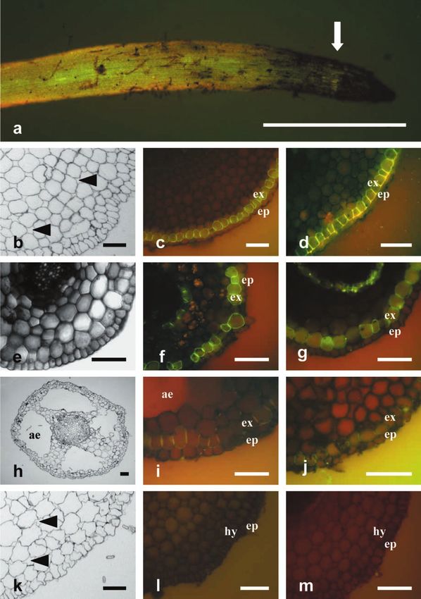
Figure 1. Transverse sections of young root segments (30 mm) from Central Amazon floodplain tree species. For fluores-
cence microscopical investigations of suberin deposits, root sections were stained with neutral red after quenching of
autofluorescence with toluidine blue. a, Aerobically grown root tips from T. juruana. Note that the suberization initiates
(Legend continues on facing page.)
208 Plant Physiol. Vol. 132, 2003
Downloaded on February 1, 2021. - Published by https://plantphysiol.org
Copyright (c) 2020 American Society of Plant Biologists. All rights reserved.Apoplasmic Barriers of Amazonian Tree Species
gations. On the basis of in situ observations in a conditions among the four examined tree species
várzea forest near Manaus, Brazil, these four trees (Fig. 1).
either are deciduous or are able to maintain their leaf Roots of T. juruana are characterized by small, in-
system during the aquatic phase (for a survey, see tercellular spaces in the cortex of schizogenous ori-
Table I). Tabernaemontana juruana is a late succes- gin, which are present throughout the root (Fig. 1b),
sional and shade-tolerant shrub that typically inhab- and by a heavily suberized hypodermal cell layer
its the understory of forest communities. The leaf- (Fig. 1, c and d). Suberization includes anticlinal and
system of T. juruana is not affected by the rise of the tangential cell walls and initiates about 2 mm behind
water table during the aquatic phase. Laetia corymbu- the root tip (Fig. 1a). No intercellular spaces are
losa is one of the most abundant várzea tree species, developed in the root cortex of L. corymbulosa (Fig.
reaching a stem height of up to 25 m. The leaf-system 1e). Suberization is distributed along walls of the
of L. corymbulosa is continuously renewed during the hypodermal cell layer with numerous passage cells
year. Complete leaf-shedding does not occur. Salix devoid of suberin (Fig. 1, f and g). S. martiana shows
martiana is a light-demanding and fast growing pio- large air spaces in the root cortex, arising from lysig-
neer species that inhabits open sites along the Ama- enous degeneration of cortical cells (Fig. 1h). In roots
zon river. The stem reaches heights of up to 12 m, of S. martiana, suberization is most abundant in radial
and the leaf-system is well developed during the cell walls of the hypodermis, suggesting Casparian
flooding period. The formation of adventitious roots bands (Fig. 1, i and j). Roots of C. benthami were
allows S. martiana to tolerate high sedimentation found to form intercellular spaces in the cortex (Fig.
rates on sand banks. Crateva benthami is a leaf- 1k), but this species completely lack a visible suber-
shedding species from the lower canopy inhabiting ized lamella in the hypodermis (Fig. 1, l and m).
low elevation sites. The rise of the water table results The morphological characteristics were reflected
in complete defoliation of submerged individuals. by the measurements of the root porosity of the
investigated species (Table II). As expected, the aer-
enchymatous species S. martiana showed the highest
The Formation of Suberin Barriers in Young Root root porosity, reaching values that are 7-fold higher
Zones Is Restricted to Evergreen Species than those determined for roots from T. juruana. Due
to the lack of air spaces, roots from L. corymbulosa
Suberization in the root exodermis has been attrib- exhibited almost no root porosity. In all species, no
uted to the adaptation to low oxygen availability. To plasticity toward the formation of air spaces or lignin
elaborate whether maintenance of the leaf system and suberin deposits was observed among the
during the aquatic phase is associated with changes treatments.
in the root anatomy, cross sections from young root
segments (30 mm behind the root tip) were analyzed
by light and fluorescence microscopy, and suberin Evergreen Species Show Higher Contents of
incrustations were visualized by staining with the Suberin Components
fluorescent dye neutral red. To discover possible
morphological changes induced by flooding, plants For a qualitative analysis of peripheral cell walls
were grown either aerobically or under hypoxic con- (RHCWs), cell wall samples were enzymatically iso-
ditions. T. juruana and S. martiana responded to hy- lated from root segments derived 0 to 3 cm from the
poxic growth conditions by inducing the formation apex. Because walls of hypodermal cells cannot be
of a new root type from the stem basis, which can be separated from those of the rhizodermis, analysis of
referred to as adventitious roots. L. corymbulosa and suberin and lignin compounds always comprises
C. benthami were not able to form such roots. Micro- both cell types. RHCWs of all investigated tree spe-
scopic examinations did not reveal any anatomical cies released detectable amounts of suberin mono-
differences between aerobically grown roots, hypoxi- mers after chemical degradation by transesterifica-
cally treated roots, and roots induced by hypoxic tion with BF3 in methanol. A survey over the
Figure 1. (Legend continued from facing page.)
shortly behind the root tip. Arrow shows initiation of suberization. b through d, Transverse sections of aerobically (b and c)
and hypoxically (d) grown roots. Roots of T. juruana are characterized by the presence of small intercellular spaces in the
root cortex (b) and a strongly suberized hypodermis (c and d). e through g, Transverse sections of aerobically (e and f) and
hypoxically (g) grown roots of L. corymbulosa. e, No air spaces in the root cortex are evident. Suberin staining pattern reveals
an incompletely suberized hypodermis with a high number of passage cells. h through j, Transverse sections of aerobically
(h and i) and hypoxically (j) grown roots of S. martiana. Cross sections show the formation of large aerenchymatous air
spaces in the root cortex (h) and a weak suberin staining of the hypodermal cell layer (i and j). k through m, Transverse
sections of aerobically (k and l) and hypoxically (m) grown roots of C. benthami. k, Small intercellular air spaces are present
in the root cortex. Staining reveals no suberin deposits in young root segments. Bars ⫽ 5 mm (a) and 50 m (b–m). Eight root
segments were analyzed per species and growth type (C. benthami, n ⫽ 4).
Plant Physiol. Vol. 132, 2003 209
Downloaded on February 1, 2021. - Published by https://plantphysiol.org
Copyright (c) 2020 American Society of Plant Biologists. All rights reserved.De Simone et al.
Table II. Root porosity as a percentage of the total volume of roots from four Central Amazon tree
species, either grown in aerated nutrient solution or for at least 4 weeks under hypoxic conditions
Root porosity was determined for the apical 5 cm of the roots. Data are means of three replicates.
N.d., Not determined.
⫹O2 ⫺O2 ⫺O2 Adventitious Roots
T. juruana 2.8 ⫾ 0.7 3.3 ⫾ 0.2 3.5 ⫾ 0.1
L. corymbulosa 0.2 ⫾ 0.3 0.0 ⫾ 0.0 n.d.
S. martiana 20.4 ⫾ 6.9 21.4 ⫾ 1.1 21.2 ⫾ 1.2
C. benthami 3.9 ⫾ 0.5 4.4 ⫾ 0.3 n.d.
chemical composition of the suberin compounds is lignin in T. juruana and L. corymbulosa was dominated
given in Table III. In all four species, the aromatic by syringyl units, RHCWs from S. martiana exhibited
domain is composed of ester-linked cis/trans-ferulic higher contents of guaiacyl. The lignin monomer
acid and a syringyl-derived lignin monomer. Para- p-hydroxyphenyl was not detectable in the species
hydroxybenzoic acid was dominating in RHCWs of under investigation.
L. corymbulosa and S. martiana, whereas it was not
detectable in T. juruana and C. benthami (Table III). In Suberin Functions as a Permeability Barrier for
all species, the aliphatic moiety of suberin consists of Gas Exchange
long-chain (C16-C28) monoalcohols, monocarboxylic
acids, ␣,-dicarboxylic acids, -hydroxyacids, and The barrier function of suberin for gas-exchange
2-hydroxyacids (Table III), which were detected as between the root and the rhizosphere was investi-
their monomethylesters or trimethylsilyl-derivatives. gated by using oxygen microelectrodes controlled by
The aliphatic suberin composition of the RHCWs a mechanical micromanipulator. Oxygen measure-
from the evergreen species T. juruana, L. corymbulosa, ments were performed both on the root surface and
and S. martiana is dominated by dicarboxylic acids inside the root after radial penetration of the micro-
and -hydroxyacids. In contrast, aliphatic suberin of electrode in roots of agar-embedded plants. No ana-
RHCWs from C. benthami is mainly composed of tomical differences were observed between hydro-
␣,-dicarboxylic acids, monocarboxylic acids, and ponically grown and agar exposed roots. The species
2-hydroxyacids (Table III). under investigation showed marked differences in
Quantitatively, the suberin content of the isolated ROLs and root cortex oxygen concentration. Highest
RHCWs differed considerably among the species. ROLs were observed in adventitious roots of S. mar-
The aliphatic suberin content was 3.3- and 4-fold tiana, with rates remaining almost equal along the
higher in RHCWs from T. juruana compared with whole root (Fig. 5). A slight increase in O2 levels was
RHCWs from L. corymbulosa and S. martiana, respec- noted in areas where laterals emerged (e.g. at a dis-
tively, and 6.7-fold higher than in RHCWs from C. tance of 50 mm from the apex in Fig. 5). Adventitious
benthami (Fig. 2). These differences are mainly attrib- roots of T. juruana showed only a thin oxygen layer in
utable to the characteristic C18-unsaturated suberin a narrow (approximately 5 mm) zone at the root tip.
markers -hydroxycarboxylic acid and ␣,-dicarb- However, the oxygen level around the root within
oxylic acid. Quantitative analysis of these two com- this zone did not by far reach the values characteris-
pounds in RHCWs of the four species is shown in tic of root tips from S. martiana. Behind the apex, no
Figure 3. For both monomers RHCWs from T. juruana oxygen was detectable on the surface of roots from T.
exhibit about 6-fold higher values than those of L. juruana, indicating the presence of an effective gas
corymbulosa and S. martiana, which show similar con- barrier. Surface measurements on roots of L. corym-
tents of these components in the peripheral cell walls. bulosa and C. benthami did not exhibit any detectable
Compared with RHCWs from T. juruana, lowest leakage of oxygen into the rhizosphere, irrespective
amounts of C18-unsaturated -hydroxycarboxylic of the root zone.
acid and ␣,-dicarboxylic acid were observed in C. The findings described above were confirmed by
benthami. determination of the oxygen levels within the roots.
Lignin monomers, corresponding to the three typ- Root cortex oxygen concentration was only measur-
ical lignin units p-hydroxyphenyl, syringyl, and guai- able in adventitious roots from T. juruana and S.
acyl, were characterized by thioacidolysis of RHCWs martiana (Fig. 6). Oxygen concentration in the cortex
from T. juruana, L. corymbulosa, and S. martiana. All of adventitious root from T. juruana did not exceed
investigated species revealed amounts of total lignin 2.0 mg L⫺1 (Fig. 6a). The steep increase in the oxygen
between 22 and 28 nmol cm⫺2 (Fig. 4), which were level behind the root periphery indicates that the
90% lower in T. juruana and 50% lower in L. corym- suberized exodermis can effectively prevent the ef-
bulosa and S. martiana than the aliphatic suberin flux of oxygen from the root into the rhizosphere.
amounts. In all species, the lignin biopolymer was The decrease in oxygen concentration in the stele is
composed of syringyl and guaiacyl units. Whereas related to the absence of air spaces. The oxygen con-
210 Plant Physiol. Vol. 132, 2003
Downloaded on February 1, 2021. - Published by https://plantphysiol.org
Copyright (c) 2020 American Society of Plant Biologists. All rights reserved.Apoplasmic Barriers of Amazonian Tree Species
Table III. Suberin composition of rhizodermal cell walls (RHCWs) from root tip segments (0 –30 mm) as a percentage of all identified
suberin monomers
SDs of three replicates are given. n.d., Not detectable.
T. juruana L. corymbulosa S. martiana C. benthami
Aromatics
cis-Ferulic acid 0.65 ⫾ 0.21 0.29 ⫾ 0.14 n.d. 1.35 ⫾ 0.29
trans-Ferulic acid 6.56 ⫾ 0.55 6.64 ⫾ 0.65 4.89 ⫾ 0.13 18.46 ⫾ 1.99
p-OH-Benzoic acid n.d. 47.13 ⫾ 1.15 52.82 ⫾ 4.05 n.d.
s-Lignin 0.66 ⫾ 0.22 0.39 ⫾ 0.09 0.75 ⫾ 0.04 3.11 ⫾ 0.61
Total 7.86 ⫾ 0.98 54.45 ⫾ 2.03 58.46 ⫾ 4.22 22.92 ⫾ 2.89
Aliphatics
Monocarboxylic acids
C16 n.d. n.d. n.d. 3.54 ⫾ 1.05
C18 1.25 ⫾ 0.37 1.12 ⫾ 0.11 n.d. 5.27 ⫾ 1.22
C18-9-en 2.07 ⫾ 0.37 3.00 ⫾ 0.50 3.15 ⫾ 0.39 8.27 ⫾ 2.64
C18-9,12-dien 2.92 ⫾ 0.28 3.25 ⫾ 0.24 2.38 ⫾ 0.42 9.27 ⫾ 2.63
C20 0.42 ⫾ 0.05 1.01 ⫾ 0.20 n.d. n.d.
C22 1.64 ⫾ 0.33 0.66 ⫾ 0.13 0.66 ⫾ 0.17 2.52 ⫾ 2.27
C24 0.98 ⫾ 0.36 0.42 ⫾ 0.02 0.77 ⫾ 0.08 3.26 ⫾ 3.97
C26 1.54 ⫾ 0.18 n.d. 0.53 ⫾ 0.14 n.d.
C28 n.d. n.d. n.d. 0.30 ⫾ 0.03
Total 10.82 ⫾ 1.94 9.46 ⫾ 1.20 7.49 ⫾ 1.21 32.44 ⫾ 13.80
␣,-Dicarboxylic acids
C16 7.74 ⫾ 1.35 7.97 ⫾ 1.67 4.03 ⫾ 0.71 1.80 ⫾ 0.02
C18 1.09 ⫾ 0.33 0.55 ⫾ 0.15 1.32 ⫾ 0.33 1.30 ⫾ 0.04
C18-9-en 20.94 ⫾ 3.57 4.71 ⫾ 1.14 4.97 ⫾ 0.79 3.23 ⫾ 0.23
C22 0.89 ⫾ 0.31 0.43 ⫾ 0.14 n.d. n.d.
C24 1.17 ⫾ 0.09 n.d. n.d. n.d.
Total 31.83 ⫾ 5.63 13.65 ⫾ 3.09 10.32 ⫾ 1.84 6.33 ⫾ 0.29
-Hydroxyacids
C16 3.64 ⫾ 0.82 5.45 ⫾ 0.98 4.50 ⫾ 0.59 3.15 ⫾ 0.12
C18 n.d. 0.96 ⫾ 0.24 1.51 ⫾ 0.23 n.d.
C18⫺9-en 26.56 ⫾ 5.31 7.11 ⫾ 1.50 8.29 ⫾ 0.98 11.09 ⫾ 0.30
C20 n.d. 0.81 ⫾ 0.24 0.70 ⫾ 0.06 1.09 ⫾ 0.01
C22 4.74 ⫾ 1.26 3.42 ⫾ 0.69 2.46 ⫾ 0.17 1.68 ⫾ 0.15
C24 4.18 ⫾ 1.33 0.50 ⫾ 0.11 1.35 ⫾ 0.25 n.d.
C26 0.62 ⫾ 0.18 n.d. 1.51 ⫾ 0.55 n.d.
C28 0.68 ⫾ 0.90 n.d. n.d. n.d.
Total 40.41 ⫾ 9.79 18.26 ⫾ 3.76 20.32 ⫾ 2.82 17.02 ⫾ 0.58
2-Hydroxyacids
C18 n.d. n.d. n.d. 1.67 ⫾ 0.06
C22 1.24 ⫾ 0.32 0.44 ⫾ 0.07 n.d. n.d.
C24 1.75 ⫾ 0.09 0.83 ⫾ 0.05 1.17 ⫾ 0.03 12.14 ⫾ 1.57
C26 n.d. 0.30 ⫾ 0.02 n.d. 3.83 ⫾ 0.67
Total 2.99 ⫾ 0.41 1.57 ⫾ 0.14 1.17 ⫾ 0.03 17.63 ⫾ 2.30
Alcohols
C18 0.63 ⫾ 0.09 0.37 ⫾ 0.10 n.d. 1.69 ⫾ 0.15
C20 2.09 ⫾ 0.54 0.23 ⫾ 0.01 n.d. n.d.
C22 1.47 ⫾ 1.03 1.29 ⫾ 0.10 0.91 ⫾ 0.15 1.48 ⫾ 0.02
C24 0.85 ⫾ 0.52 0.14 ⫾ 0.02 0.42 ⫾ 0.10 n.d.
C26 0.35 ⫾ 0.26 0.34 ⫾ 0.00 0.90 ⫾ 0.27 n.d.
C28 0.69 ⫾ 0.41 0.23 ⫾ 0.03 n.d. 0.49 ⫾ 0.16
Total 6.08 ⫾ 2.84 2.61 ⫾ 0.26 2.24 ⫾ 0.52 3.65 ⫾ 0.33
centration in the cortex of adventitious roots from S. DISCUSSION
martiana was about 3-fold higher than that in adven-
titious roots of T. juruana (Fig. 6c). The gradual de- The response of plants to flooding is complex, in-
cline of the oxygen levels in the rhizosphere with volving an array of physiological, biochemical, and
increasing distance from the root indicates radial O2 morphological adaptations. Anatomical changes,
diffusion, which corresponds to the measurements such as the development of aerenchyma and the
along the whole root surface. No significant amount formation of apoplastic barriers in the peripheral cell
of oxygen was detectable in roots of L. corymbulosa layers, are thought to be part of complex strategies to
(Fig. 6b) and C. benthami (Fig. 6d). withstand periods of low oxygen availability. Depo-
Plant Physiol. Vol. 132, 2003 211
Downloaded on February 1, 2021. - Published by https://plantphysiol.org
Copyright (c) 2020 American Society of Plant Biologists. All rights reserved.De Simone et al.
Figure 2. Aliphatic and aromatic suberin mono-
mers released from RHCWs of young roots seg-
ments (0–30 mm) from the evergreen species T.
juruana (Tj), L. corymbulosa (Lc), and S. marti-
ana (Sm) and the deciduous species C. benthami
(Cb). Data are given in nanomoles per square
centimeter. Three samples per species out of
pooled root tips from 30 plants were analyzed
(C. benthami, n ⫽ 3 of 5 plants).
sition of suberin and lignin compounds in the outer resembles the suberin composition in roots of Picea
cell layers of roots has been described for many wet- abies (Matzke and Riederer, 1991). The detection of a
land species, either as being induced by flooding syringyl-derived monomer in the analyzed samples
(Colmer et al., 1998) and/or phytotoxins (Armstrong confirms the findings of Zeier and Schreiber (1997),
and Armstrong, 1999) or as being constitutively showing that aromatic lignin-related monomers can
present. A comparison of the suberin content of the be released from the suberin polymer by transesteri-
investigated species with those of other taxa is ham- fication, and indicates the presence of lignin in the
pered by the lack of available data. In comparison isolated RHCWs of the investigated tree species.
with herbaceous species, the várzea tree species On the basis of their anatomical distribution, the
reached multifold amounts of aliphatic suberin function of suberized cell walls has been primarily
monomers (Schreiber et al., 1999; Zimmermann et al., attributed to water retention and to a restriction of
2000). Qualitatively, the aliphatic moiety of RHCWs solute transport through the apoplast. In wetland
is dominated by -hydroxycarboxylic acid and diac- plants, the hypodermis was assumed to act as a gas
ids, representing typical suberin substance classes diffusion barrier, restricting the radial loss of oxygen
(Kolattukudy, 2001). Only in C. benthami, the compo- into the rhizosphere (Armstrong et al., 1994). Plants
sition of the suberin lamellae is dominated by mono- subjected to flooding rely on transport of oxygen
acids, 2-hydroxy acids, and -hydroxy acids, which from aerial plant parts to the roots. The effectiveness
Figure 3. Released amounts of unsaturated C18
␣, -9en-dicarboxylic acid and -OH-9-en-
carboxylic acid, characteristic suberin markers
from enzymatically isolated RHCW. Data are
given in nanomoles per square centimeter. (Tj,
T. juruana; Lc, L. corymbulosa; Sm, S. martiana;
and Cb, C. benthami). Three samples per spe-
cies out of pooled root tips from 30 plants were
analyzed (C. benthami, n ⫽ 3 of 5 plants). Error
bars indicate SD among the three samples.
212 Plant Physiol. Vol. 132, 2003
Downloaded on February 1, 2021. - Published by https://plantphysiol.org
Copyright (c) 2020 American Society of Plant Biologists. All rights reserved.Apoplasmic Barriers of Amazonian Tree Species
Figure 4. Total lignin and lignin monomer
amounts of RHCWs (0–30 mm) from T. juruana
(Tj), L. corymbulosa (Lc), and S. martiana (Sm)
identified by thioacidolysis. Data are given in
nanomoles per square centimeter. Three sam-
ples per species out of pooled root tips from 30
plants were analyzed. Error bars indicate SD
among the three samples.
of internal gas transport depends on the resistance to matic material in plant cell walls are not always
diffusion within the plant, the plant’s respiratory sensitive enough to allow for a qualitative estimation
demand for oxygen, and the rate of O2 diffusion out of the substances involved in forming such barriers.
of the roots. The presence of a barrier to ROL is Although, in general, quantitative differences in
obviously of advantage in terms of maintenance of suberin content among the investigated species were
energy metabolism. On the other hand, the diffusion revealed by microscopic analysis, deviations between
of oxygen out of the roots restores ion uptake (Enge- the intensity of morphological staining and the
laar, 1993) and helps to re-oxidize toxic compounds suberin amount of isolated RHCWs from roots of L.
of flooded soils to nontoxic compounds. Although corymbulosa and S. martiana imply that quantitative
the restriction of gas exchange by cell wall deposits in chemical analysis of root sections is more reliable and
wetland plant roots has been demonstrated polaro- more sensitive than microscopic techniques. The in-
graphically using Clark-type microelectrodes (Arm- accuracy of the suberin staining also becomes obvi-
strong et al., 2000; Visser et al., 2000), the nature of ous in roots of C. benthami. No suberin was visualized
potential barrier-biopolymers in hypodermal walls of by the staining procedure, but suberin monomers
such species has yet not been determined. Staining could be detected in isolated RHCWs, implying that
techniques developed to localize aliphatic and aro- the suberin amount in RHCWs from C. benthami does
Figure 5. Surface oxygen concentration of roots
at various distances from the root apex embed-
ded in stagnant and oxygen-free agar. Measure-
ments on T. juruana and S. martiana were per-
formed on 10- to 12-d-old adventitious roots. In
the cases of L. corymbulosa and C. benthami,
where cultivation in anoxic agar did not induce
adventitious roots, pre-existing roots were used
for the determinations. ROL was only observed in
roots of T. juruana and S. martiana. Adventitious
roots of T. juruana exhibited oxygen leakage to
the rhizosphere only in a small (approximately 5
mm wide) zone at the root tip. No outward dif-
fusion of oxygen was detectable from roots of L.
corymbulosa and C. benthami. Lengths of the
roots were 12 cm (S. martiana), 5 cm (T. juruana),
or 6 cm (L. corymbulosa and C. benthami). De-
terminations were performed on 20 different
roots from at least six individual plants without
major deviations in the overall pattern. Data are
from a representative experiment.
Plant Physiol. Vol. 132, 2003 213
Downloaded on February 1, 2021. - Published by https://plantphysiol.org
Copyright (c) 2020 American Society of Plant Biologists. All rights reserved.De Simone et al.
Figure 6. Oxygen profiles in the rhizosphere
and in roots of the investigated species. Vertical
axes indicate points of vertical penetration of
the microelectrode into and out of the roots.
Profiles were taken 10 to 15 mm behind the
apex. a, Oxygen profile through a 12-d-old and
5-cm-long adventitious root from T. juruana.
The low oxygen concentration indicates hy-
poxic conditions in the root cortex and stele. No
ROL was detectable. b, Measurement of the
oxygen distribution revealed the absence of ox-
ygen in the rhizosphere and in roots of L. corym-
bulosa. c, The oxygen profiles through 10-d-old
and 12-cm-long adventitious roots from S. mar-
tiana revealed high oxygen concentrations in-
side the roots and a several-millimeter-thick ox-
ygenated zone around the roots. d, Roots from
C. benthami did not exhibit any oxygen inside
or outside the roots. Lengths of the roots were 12
cm (S. martiana), 5 cm (T. juruana), or 6 cm (L.
corymbulosa and C. benthami). Determinations
were performed on 20 different roots from at
least six individual plants without major devia-
tions in the overall pattern. Data are from a
representative experiment.
not reach the threshold value for becoming detect- spite the strong resistance to oxygen, water transport
able by histochemical probing. may not be severely restricted by suberin. Water
Combining the data collected from the chemical transfer across the root peripheral cell layers may
analysis of RHCWs with the ROL- measurements, we occur via the middle lamellae, as suggested for the
are able to confirm the postulated role of suberin as a outermost phellems in various tree species (Groh et
permeability barrier for gas-exchange between the al., 2002). A 3-fold lower aliphatic suberin amount
root and the rhizosphere. The results validate the was recorded in roots of S. martiana in which high
presence of such a barrier in roots of T. juruana, ROLs were evident. This suggests that suberin in
consisting of a heavily suberized hypodermis with these concentrations is not an absolute air-tight bar-
depositions in tangential and radial cell walls. De- rier and should rather be described as a resistor
214 Plant Physiol. Vol. 132, 2003
Downloaded on February 1, 2021. - Published by https://plantphysiol.org
Copyright (c) 2020 American Society of Plant Biologists. All rights reserved.Apoplasmic Barriers of Amazonian Tree Species
whose permeability depends on its chemical compo- transferred into the experimental aerobic and hypoxic treatments in 1.5-L
plastic pots with four cuttings per container for 1 to 4 weeks. Hypoxic
sition and on the root oxygen concentration. In gen-
conditions were induced by continuous flushing of the nutrient solution
eral, a higher content of suberin components was with nitrogen through a gas permeable S6/2 Accurel tube (Membrana,
observed in evergreen species (this study; De Simone Wuppertal, Germany). Oxygen concentration did not exceed 0.1 mg L⫺1 in
et al., 2002b), suggesting that suberin barriers are of the plastic pots during the whole experimental period. Aerobic controls
advantage to prevent leaf shading during the aquatic were grown in 1.5-L pots in aerated nutrient solution for the same period
of time.
phase. No major difference in lignin content was
observed between the two species, suggesting that
lignin does not contribute to the resistance to oxygen Measurement of Root Porosity
loss in the species under investigation.
Porosity of roots was determined after the method of Raskin (1983), using
Suberin polymers have been suggested to be in- the equations modified by Thomson et al. (1990). The apical 0 to 5 cm of 20
volved in pathogen defense, either by a breakdown to 40 roots from each species, grown for 4 weeks under aerobic or hypoxic
of polymers by enzymes of microbial origin and sub- growth conditions, were detached with a razor blade and cut into segments
sequent release of toxic phenols or by acting as a of approximately 1.5 cm. The root systems of S. martiana and T. juruana were
divided into hypoxic roots and adventitious roots which emerged in the
mechanical barrier (Peterson, 1997). A point of major flood water. The fresh weight of the samples was determined after carefully
deviation in the composition of suberin among the removing surface water by blotting with tissue paper. Buoyancy of root
species is the high percentage of aromatics in RHCWs samples before and after vacuum infiltration with water was measured
of L. corymbulosa and S. martiana, which is dominated using a balance with a water-filled flask containing the root samples at-
tached to the under-carriage and submerged in a beaker of water under the
by para-hydroxybenzoic acid (⬎ 85% of total aromat- balance. The porosity was calculated from the difference in weight of
ics). Similar to salicylic acid, para-hydroxybenzoic acid the samples before and after infiltration (volume of airspace), divided by the
was shown to be involved in pathogen defense, either difference between fresh weight and weight under water before infiltration
by their direct antimicrobial action or by induction of (volume of the segments).
a subset of pathogenesis-related genes (Ryals et al.,
1996; Smith-Becker et al., 1998). In roots of L. corym- Light and Fluorescence Microscopy
bulosa and S. martiana, suberization did not start
shortly behind the root tip, emphasizing the adaptive After 4 weeks of cultivation, roots were detached and prepared for
morphological investigations. Root structure was investigated by light mi-
value of this feature to defend the roots from micro- croscopy and fluorescence microscopy of root segments. For light micros-
biological attack during the aquatic period. copy analysis, root segments were fixed in 3.7% (w/v) paraformaldehyde in
In summary, our data show that the composition of 100 mm phosphate buffer, pH 7.0, dehydrated in an upgrading ethanol
the hypodermis can contribute significantly to the series, infiltrated with London Resin White (London Resin Co. Ltd. Lon-
adaptation of plants to long-term flooding condi- don), and polymerized for 24 h at 55°C and 250 mbar. Cross sections (1 m)
were cut with a microtome (Ultracut E, Reichert, Vienna) stained with 0.05%
tions. Suberin barriers may determine important pro- (w/v) toluidine blue O for 1 min, and viewed in dark field. Fluorescence
cesses that in turn aid in withstanding low oxygen microscopy investigations were carried out on transverse free-hand sec-
availability such as pathogen defense, re-oxidation of tions. To visualize suberization of the epidermal and subepidermal cell
reduced phytotoxins, and the restriction of oxygen walls, sections were stained with 0.1% (w/v) neutral red in 100 mm phos-
phate buffer, pH 6.0, for 1 min and washed twice with tap water. The
loss to the rhizosphere. We show here for the first specificity of the neutral red technique for the hydrophobic/lipid domain of
time, to our knowledge, that suberin, and not lignin, suberin was shown previously (Lulai and Morgan, 1992). For quenching of
represents the barrier for the diffusion of oxygen. autofluorescence, sections were previously incubated for 2 h in 0.05% (w/v)
Our data also show that different adaptive strategies toluidine blue O in 100 mm phosphate buffer, pH 6.0. Blue-violet excitation
can be realized in plant species facing similar envi- (exciter filter EX 420–490, dichromatic beamsplitter DM 505, barrier filter BA
520, Nikon, Tokyo) was used for all investigations of suberin deposits.
ronmental constraints. Photographs were taken with a digital camera (Nikon Coolpix 990, 3.34
megapixels).
MATERIALS AND METHODS
Qualification and Quantification of Suberin
Plants and Growth Conditions
Isolation and Purification of Cell Wall Materials
Experiments were carried out with 3- to 4-month-old cuttings of Taber-
naemontana juruana [Markgr.] Schumann ex J.F. Macbride (Apocynaceae), Cell walls from root tip segments (0–30 mm) were isolated enzymatically
Laetia corymbulosa Spruce ex Bent. (Flacourtiaceae), Salix martiana Leyb. similar to the procedure described by Schreiber et al. (1994). The segments
(Salicaceae), and Crateva benthami Eichl. In Mart.(Capparaceae), which were were vacuum infiltrated with a solution containing cellulase (Onozuka,
grown hydroponically in a climate-controlled greenhouse under the follow- R-10, Serva, Heidelberg) and pectinase (Macerozyme R-10, Serva) dissolved
ing conditions: 70% to 90% relative humidity, day/night regime of 16 h/8 h in 0.01 m acetate buffer at pH 4.5. After 8 weeks of maceration in the enzyme
(150 mol m⫺2 s⫺1 supplied by IP 23 lamps [Philips, Eindhoven, The solution, the hypodermal cylinder and the central cylinder were separated
Netherlands]) and 30°C/22°C day/night temperature. The cuttings were mechanically using two precision forceps and a stereo microscope. Because
derived from 1- to 2-year-old young trees. Basal shoot ends were spread rhizodermal and the attached hypodermal/exodermal cell walls could not
with 2% (w/v) Rhizopon AA (Rhizopon bv, Hazerswonde, The Nether- be separated from each other, the isolated cell wall fraction was termed
lands) and incubated in peat to induce root formation. After initiation of RHCWs. The isolated RHCWs fraction was washed several times with
rooting, the cuttings were transferred to aerated nutrient solution contain- borate buffer (0.01 m Na2B4O7, pH 9.0) and distilled water, followed by
ing 3.00 mm NH4NO3, 0.50 mm MgSO4, 1.50 mm CaCl2, 1.50 mm K2SO4, 1.50 subsequent extraction of the dry material with chloroform:methanol (1:1;
mm NaH2PO4, 25.0 m H3BO3, 1.00 m MnSO4, 0.50 m ZnSO4, 0.05 m v/v) to remove soluble lipids. Finally, samples were dried again and stored
(NH4)6Mo7O24, 0.30 m CuSO4, and 40.0 m FeEDTA, pH 6.0, and grown over silica gel until further use. To determine the RHCWs dry weight-root
for an additional 2 months. Nutrient solution was changed weekly, pH surface relation, RHCWs of root segments with known surface area were
values ranged from 5.5 to 6.0 within this period. The cuttings were then isolated, dried, and weighted separately.
Plant Physiol. Vol. 132, 2003 215
Downloaded on February 1, 2021. - Published by https://plantphysiol.org
Copyright (c) 2020 American Society of Plant Biologists. All rights reserved.De Simone et al.
Chemical Degradation and Chromatographic Analyses of surface—by means of a precision magnifying glass—the root was placed
Isolated Cell Walls close to the glass wall of the basin. The temperature during the measure-
ments was about 30°C. Oxygen microsensors (MasCom, Bremen, Germany)
Purified cell wall material was subjected to chemical degradation meth- were used to measure oxygen profiles in the agar medium approaching to
ods specific for the detection of the biopolymers suberin and lignin. For the root surface and in the outer tissues of the root. These sensors corre-
suberin analysis, the extracted RHCWs were depolymerized using a BF3 spond to a miniaturized Clark oxygen electrode (Revsbech, 1989) which is
catalyzed methanolic transesterification (Kolattukudy and Agrawal, 1974) sealed with a thin silicon membrane. The permeating oxygen reacts at the
as described in detail by Zeier and Schreiber (1997, 1998). Extracted RHCWs negatively charged microcathode (⫺0.8 V), and causes a current signal that
(0.5–1 mg) were added to 1 mL of a 10% (w/v) BF3/methanol solution is proportional to the oxygen concentration in solution. These oxygen mi-
(Fluka, Deisenhofen, Germany) and heated to 70°C for 16 h. After cooling, croelectrodes operate with a response time lower than 2 s, extremely low
solid residues were removed, and the remaining solution was extracted oxygen consumption, a very stable signal, and a detection limit of 0.1 to 0.2
three times with 1 mL of CHCl3 containing 20 g of dotriacontane (Fluka) mg L⫺1oxygen. Due to the high linearity in response to oxygen concentra-
as an internal standard. The combined extracts were washed with 2 mL of tion, an easy 2-point calibration in nitrogen-bubbled water and air-saturated
a saturated sodium chloride solution and 1 mL of distilled water. The water at the measuring temperature was possible. The microelectrodes were
organic phase of chloroform was separated and dried over Na2SO4. calibrated once a day. The current output of the employed microelectrodes
Thioacidolysis was used for the detection of lignin according to Lapierre varied between 1 and 5 pA in nitrogen-bubbled water and between 80 and
et al. (1991). Forty microliters of BF3 etherate (Merck, Darmstadt, Germany) 200 pA in air-saturated stagnant water. Air in the laboratory (relative
and 160 L of ethanethiol (Fluka) were dissolved under argon in 300 L of humidity 50%) gave maximum 5% higher readings depending on the re-
dioxane in a tube fitted with a Teflon-lined screw cap. The solution was spective electrode. Water-saturated air would minimize these differences
adjusted to a volume of 1.6 mL with 1.1 mL of dioxane. The sample (0.5–1.0 (Revsbech and Ward, 1983). Thus, the error caused by the change between
mg) was added, and the mixture was stirred at 100°C. After 4 h, the reaction liquid phase (root cells) and gas phase (aerenchyma) in the cortex of the
mixture was ice-cooled, diluted with 2 mL of water, and extracted three times adventitious root is negligible. The tip diameters of the electrodes were
with 3 mL of CHCl3, containing 20 g of dotriacontane (Fluka) as an internal smaller than 20 m. The microelectrodes were gradually driven into the
standard. The combined organic phases were dried over Na2SO4. agar or into the root by means of a mechanical micromanipulator in steps of
Gas chromatography and mass spectroscopy were used for quantification 50 m in the rhizosphere and 20 m in the roots. The direction of the
and identification of the released suberin and lignin monomers. Before injec- electrode tip was right-angled to the position of the root. The current signals
tion, samples were derivatized by N,N-bis-trimethylsilyltrifluoroacetamide were converted to voltage signals by the electronic sensor, digitized in the
(Machery-Nagel, Düren, Germany) catalyzed with pyridine to convert free computer, and saved as an ASCII file.
hydroxyl and carboxyl groups to their respective trimethylsilyl esters and
ethers. Qualitative sample analyses were performed by gas chromatography
(Agilent 6890N gas chromatograph, Agilent Technologies, Böblingen, Ger- ACKNOWLEDGMENTS
many) combined with a quadropole mass selective detector (Agilent 5973N
mass selective detector, Agilent Technologies). Quantitative sample analysis The study is part of the research which is carried out jointly by the
was carried out with a HP 5890 Series II gas chromatograph (Hewlett Pack- tropical Ecology Group of the Max-Planck-Institute of Limnology (Plön,
ard, Palo Alto, CA), equipped with a flame ionization detector. Germany) and the Instituto Nacional de Pesquisas da Amazônia (Manaus,
Brazil).
Received September 19, 2002; returned for revision November 6, 2002;
Root Surface Determination accepted January 14, 2003.
The suberin content of rhizodermal cell walls (micrograms per milligram)
was related to the root surface (micrograms per square centimeter). Fresh LITERATURE CITED
transversal sections, cut 5 and 20 mm behind the root apex, were prepared
for light microscopy and transferred with a camera system on a Mini-MOP Agrawal VP, Kolattukudy PE (1978) Purification and characterization of a
picture digitizer (Kontron, Munich). The circumference was related to the wound-induced -hydroxy fatty acid: NADP oxidoreductase from potato
segment length (30 mm) for calculation of the root surface. Five root seg- tuber disks. Arch Biochem Biophys 191: 452–465
ments from five individual plants were analyzed. Armstrong J, Armstrong W (1999) Phragmites die-back: toxic effects of
propionic, butyric, and caproic acids in relation to pH. New Phytol 142:
201–217
Oxygen Measurements Armstrong W, Brandle R, Jackson MB (1994) Mechanisms of flood toler-
ance in plants. Acta Bot Neerl 43: 307–358
Measurements on the oxygen distribution in and around roots were Armstrong W, Cousins D, Armstrong J, Turner DW, Beckett PM (2000)
carried out with oxygen microelectrodes on 2-month-old cuttings that were Oxygen distribution in wetland plant roots and permeability barriers to
previously grown in customary potting soil to induce root formation. The gas-exchange with the rhizosphere: a microelectrode and modelling
roots of the cuttings were rinsed with tap water to remove soil residues and study with Phragmites australis. Ann Bot 86: 687–703
immersed in a stagnant agar (0.5%, w/v) nutrient solution in a narrow glass Boudet A-M (1998) A new view of lignification. Trends Plant Sci 3: 67–71
basin (width 20 cm, height 8 cm, depth 5 cm). To simulate natural conditions Campbell MM, Sederoff RR (1996) Variations in lignin content and com-
of flooding and to prevent drying of the agar, a layer of nutrient solution position: mechanisms of control and implications for the genetic im-
(about 1 cm) covered the agar surface. The plants were cultivated in a provement of plants. Plant Physiol 110: 3–13
climate chamber under the following conditions: 70% to 80% relative hu- Čížeková H, Brix H, Kopeckı J, Lukavská J (1999) Organic acids in the
midity, day/night regime of 12 h/12 h, 32°C/28°C day/night temperature. sediments of wetlands dominated by Phragmites australis: evidence of
The agar medium was changed once a week. Illumination of the roots was phytotoxic concentrations. Aquat Bot 64: 303–315
prevented by wrapping the glass basin in black paper. For oxygen measure- Colmer TD, Gibberd MR, Wiengweera A, Tinh TK (1998) The barrier to
ments plants were immersed into a fresh agar medium that first had been radial oxygen loss from roots of rice (Oryza sativa L.) is induced by
flushed and subsequently overflown with nitrogen for at least 20 h before growth in stagnant solution. J Exp Bot 49: 1431–1436
insertion of the plantlets. During the measurements, the basin was sealed De Simone O, Haase K, Müller E, Junk WJ, Gonsior G, Schmidt W (2002a)
with a thin foil, and water-saturated nitrogen flew above the agar surface Impact of root morphology on metabolism and oxygen distribution in
during. The agar medium for the oxygen measurements consisted of two roots and rhizosphere from two Central Amazon floodplain tree species.
layers: a lower layer of solid agar (2%, w/v) to build up a horizontal overlay Funct Plant Biol 29: 1025–1035
to fix the roots by means of thin glass hook at the surface, and an upper layer De Simone O, Müller E, Junk WJ, Schmidt W (2002b) Adaptations of
of liquid, but stagnant agar (0.1%, w/v) in which the roots were immersed Central Amazon tree species to prolonged flooding: root morphology
and the oxygen profiles were recorded. The upper layer was 5 to 6 mm thick. and leaf-longevity. Plant Biol 4: 515–522
A hole in the lower solid agar filled with liquid agar contained the bulk Domeregue F, Bessoule J-J, Moreau P, Lessire R, Cassagne C (1998) Recent
roots. For observing the apex of the microelectrode approaching the root advances in plant fatty acid elongation. In JL Harwood, ed, Plant Lipid
216 Plant Physiol. Vol. 132, 2003
Downloaded on February 1, 2021. - Published by https://plantphysiol.org
Copyright (c) 2020 American Society of Plant Biologists. All rights reserved.Apoplasmic Barriers of Amazonian Tree Species
Biosynthesis, Vol 67. Cambridge University Press, Cambridge, pp Parolin P, Ferreira LV, Junk WJ (1998) Central Amazonian floodplains:
185–220 effect of two water types on the wood density of trees. Int Assoc Theor
Engelaar WMHG (1993) Root porosities and radial oxygen losses of Rumex Appl Limnol 26: 1106–1112
and Plantago species as influenced by soil pore diameter and soil aeration. Perumalla CJ, Peterson CA, Enstone DE (1990) A survey of angiosperm
New Phytol 125: 565–574 species to detect hypodermal Casparian bands: I. Roots with a uniserate
Freudenberg K (1965) Lignin: its constitution and formation from hypodermis or exodermis. Bot J Linn Soc 103: 93–112
p-hydroxycinnamyl alcohols. Science 148: 595–600 Peterson CA (1997) The exodermis and its interactions with the environ-
Graça J, Pereira H (2000a) Methanolysis of bark suberin: analysis of glycerol ment. In HE Flores, JP Lynch, D Eissenstadt, eds, Radial Biology: Ad-
and acid monomers. Phytochem Anal 11: 45–51 vances and Perspectives on the Function of Plant Roots. American Society
Graça J, Pereira H (2000b) Suberin in potato periderm: glycerol, long-chain of Plant Physiologists, Rockville, MD, pp 131–138
monomers, and glyceryl and feruloyl dimers. J Agric Food Chem 48: Peterson CA, Perumalla CJ (1990) A survey of angiosperm species to detect
5476–5483 hypodermal Casparian bands: II. Roots with a multseriate hypodermis or
Groh B, Hübner C, Lendzian KJ (2002) Water and oxygen permeance of exodermis. Bot J Linn Soc 103: 113–125
phellems isolated from trees: the role of waxes and lenticels. Planta 215: Prance GT (1979) Notes on the vegetation of Amazonia: III. Terminology of
794–801 Amazonian forest types subjected to inundation. Brittonia 31: 26–38
Hose E, Schreiber L, Clarskon D, Steudle E, Hartung W (2001) The exo- Raskin I (1983) A method for measuring leaf volume, density, thickness,
dermis: a variable apoplastic barrier in roots. J Exp Bot 52: 2245–2264 and internal gas volume. Hortscience 18: 698–699
Junk WJ (1997) Distribution and size of neotropical floodplains. In WJ Junk, Revsbech NP (1989) An oxygen microsensor with a guard cathode. Limnol
ed, The Central Amazon Floodplain. Ecological Studies 126. Springer, Oceanogr 34: 474–478
Berlin, pp 12–16 Revsbech NP, Ward DM (1983) Oxygen microelectrode that is insensitive to
Junk WJ, Bayley PB, Sparks RE (1989) The flood pulse concept in river- medium chemical composition: use in an acid microbial mat dominated
floodplain systems. Can J Fish Aquat Sci 106: 110–127 by Cyanidium caldarium. Appl Environ Microbiol 45: 755–759
Junk WJ, Ohly JJ, Piedade MTF, Soares MGM (2000) The Central Amazon Ryals JA, Neuenschwander UH, Willits MG, Mollina A, Steiner H-Y,
Floodplain: Actual Use and Options for a Sustainable Management. Hunt MD (1996) Systemic acquired resistance. Plant Cell 8: 1809–1819
Backhys Publishers, Leiden, The Netherlands Schreiber L, Breiner HW, Riederer M, Düggelin M, Guggenheim R (1994)
Kolattukudy PE (2001) Polyesters in higher plants. Adv Biochem Eng The Casparian band of Clivia miniata Reg.: isolation, fine structure and
Biotechnol 71: 1–49 chemical nature. Bot Acta 107: 353–361
Kolattukudy PE, Agrawal VP (1974) Structure and composition of aliphatic Schreiber L, Hartmann K, Skrabs M, Zeier J (1999) Apoplastic barriers in
constituents of potato tuber skin (suberin). Lipids 9: 682–691 roots: chemical composition of endodermal and hypodermal cell walls. J
Kozlowski TT (1984) Plant responses to flooding of soil. Bioscience 34: Exp Bot 50: 1267–1280
162–167 Schreiber L, Skrabs M, Hartmann K, Becker D, Cassagne C, Lessire R
Lapierre C, Pollet B, Monties B (1991) Thioacidolysis of spruce lignin: (2000) Biochemical and molecular characterization of corn (Zea mays L.)
GC-MS analysis of the main dimers recovered after Raney nickel desul- root elongases. Biochem Soc Trans 28: 647–649
phuration. Holzforschung 45: 61–68 Sioli H (1968) Hydrochemistry and geology in the Brasilian Amazon region.
Le Bouquin R, Skrabs M, Kahn R, Beneviste I, Salaün J-P, Schreiber L, Amazoniana 1: 267–277
Durst F, Pinot F (2001) CYP94A5, a new cytochrome P450 from Nicotiana Smith-Becker J, Marois E, Huguet EJ, Midland SL, Sims JJ, Keen NT (1998)
tabacum is able to catalyze the oxidation of fatty acids to the -alcohol and Accumulation of salicylic acid and 4-hydroxybenzoic acid in phloem
to the corresponding diacid. Eur J Biochem 268: 3083–3090 fluids of cucumber during systemic acquired resistance is preceded by a
Lulai EC, Morgan WC (1992) Histochemical probing of potato periderm transient increase in phenylalanine ammonia-lyase activity in petioles
with neutral red: a sensitive cytofluorochrome for the hydrophobic do- and stems. Plant Physiol 116: 231–238
main of suberin. Biotechnol Histochem 67: 185–195 Thomson CJ, Armstrong W, Waters I, Greenway H (1990) Aerenchyma
Matzke K, Riederer M (1991) A comparative study into the chemical formation and associated oxygen movement in seminal and nodal roots
constitution of cutins and suberins from Picea abies (L.) Karst., Quercus of wheat. Plant Cell Environ 13: 395–403
robur L., and Fagus sylvatica L. Planta 185: 233–245 Visser EJW, Colmer TD, Blom CWPM, Voesenek LACJ (2000) Changes in
Miyamoto N, Steudle E, Hirasawa T, Lafitte R (2001) Hydraulic conduc- growth porosity and radial oxygen loss from adventitious roots of se-
tivity of rice roots. J Exp Bot 52: 1835–1846 lected mono- and dicotyledonous wetland species with contrasting types
Mohan R, Bajar MA, Kolattukudy PE (1993a) Induction of a tomato anionic of aerenchyma. Plant Cell Environ 23: 1237–1245
peroxidase gene (tap1) by wounding in transgenic tobacco and activation Worbes M, Klinge H, Revilla JD, Martius C (1992) On the dynamics,
of tap1/GUS and tap2/GUS chimeric gene fusions in transgenic tobacco floristic subdivision and geographical distribution of Várzea forests in
by wounding and pathogen attack. Plant Mol Biol 21: 341–354 Central Amazonia. J Veg Sci 3: 553–564
Mohan R, Vijayan P, Kolattukudy PE (1993b) Developmental and tissue- Zeier J, Schreiber L (1997) Chemical composition of hypodermal and
specific expression of a tomato anionic peroxidase (tap1) gene by a endodermal cell walls and xylem vessels isolated from Clivia miniata.
minimal promoter with wound and pathogen induction by an additional Plant Physiol 113: 1223–1231
5⬘-flanking region. Plant Mol Biol 22: 475–490 Zeier J, Schreiber L (1998) Comparative investigation of primary and ter-
Moire L, Schmutz A, Buchala A, Yan B, Stark R, Ryser U (1999) Glycerol tiary endodermal cell walls isolated from the roots of five monocotyle-
is a suberin monomer: new experimental evidence for an old hypothesis. doneous species: chemical composition in relation to fine structure.
Plant Physiol 119: 1137–1146 Planta 206: 349–361
Önnerud H, Zhang L, Gellerstedt G, Henriksson G (2002) Polymerization Zimmermann HM, Hartmann K, Schreiber L, Steudle E (2000) Chemical
of monolignols by redox shuttle-mediated enzymatic oxidation: a new composition of apoplastic barriers in relation to radial hydraulic conduc-
model in lignin biosynthesis I. Plant Cell 14: 1953–1962 tivity of corn roots (Zea mays L.). Planta 210: 302–311
Plant Physiol. Vol. 132, 2003 217
Downloaded on February 1, 2021. - Published by https://plantphysiol.org
Copyright (c) 2020 American Society of Plant Biologists. All rights reserved.You can also read