PED in 2021: a major update of the protein ensemble database for intrinsically disordered proteins
←
→
Page content transcription
If your browser does not render page correctly, please read the page content below
D404–D411 Nucleic Acids Research, 2021, Vol. 49, Database issue                                                  Published online 10 December 2020
doi: 10.1093/nar/gkaa1021

PED in 2021: a major update of the protein ensemble
database for intrinsically disordered proteins
Tamas Lazar 1,2 , Elizabeth Martı́nez-Pérez 3,4 , Federica Quaglia 5 , András Hatos 5 ,
Lucı́a B. Chemes 6 , Javier A. Iserte 3 , Nicolás A. Méndez 6 , Nicolás A. Garrone 6 ,
Tadeo E. Saldaño 7 , Julia Marchetti 7 , Ana Julia Velez Rueda 7 , Pau Bernadó 8 ,
Martin Blackledge 9 , Tiago N. Cordeiro 8,10 , Eric Fagerberg 11 , Julie D. Forman-Kay 12,13 ,
Maria S. Fornasari 7 , Toby J. Gibson 4 , Gregory-Neal W. Gomes 14,15 ,

                                                                                                                                                                  Downloaded from https://academic.oup.com/nar/article/49/D1/D404/6030232 by guest on 29 September 2021
Claudiu C. Gradinaru 14,15 , Teresa Head-Gordon 16 , Malene Ringkjøbing Jensen 9 ,
Edward A. Lemke 17,18 , Sonia Longhi 19 , Cristina Marino-Buslje 3 , Giovanni Minervini 5 ,
Tanja Mittag 20 , Alexander Miguel Monzon 5 , Rohit V. Pappu 21 , Gustavo Parisi 7 ,
Sylvie Ricard-Blum 22 , Kiersten M. Ruff21 , Edoardo Salladini 19 , Marie Skepö 11,23 ,
Dmitri Svergun 24 , Sylvain D. Vallet 22 , Mihaly Varadi 25 , Peter Tompa 1,2,26,* ,
Silvio C.E. Tosatto 5,* and Damiano Piovesan 5
1
  VIB-VUB Center for Structural Biology, Flanders Institute for Biotechnology, Brussels 1050, Belgium, 2 Structural
Biology Brussels, Bioengineering Sciences Department, Vrije Universiteit Brussel, Brussels 1050, Belgium,
3
  Bioinformatics Unit, Fundación Instituto Leloir, Buenos Aires, C1405BWE, Argentina, 4 Structural and Computational
Biology Unit, European Molecular Biology Laboratory, Heidelberg 69117, Germany, 5 Dept. of Biomedical Sciences,
University of Padua, Padova 35131, Italy, 6 Instituto de Investigaciones Biotecnológicas “Dr. Rodolfo A. Ugalde”,
IIB-UNSAM, IIBIO-CONICET, Universidad Nacional de San Martı́n, CP1650 San Martı́n, Buenos Aires, Argentina,
7
  Laboratorio de Quı́mica y Biologı́a Computacional, Departamento de Ciencia y Tecnologı́a, Universidad Nacional de
Quilmes, Bernal B1876BXD, Buenos Aires, Argentina, 8 Centre de Biochimie Structurale (CBS), CNRS, INSERM,
University of Montpellier, Montpellier 34090, France, 9 Univ. Grenoble Alpes, CNRS, CEA, IBS, Grenoble, F-38000,
France, 10 Instituto de Tecnologia Quı́mica e Biológica António Xavier, Universidade Nova de Lisboa, Av. da República,
Oeiras 2780-157, Portugal, 11 Theoretical Chemistry, Lund University, Lund, POB 124, SE-221 00, Sweden,
12
   Molecular Medicine Program, Hospital for Sick Children, Toronto, M5G 1X8, Ontario, Canada, 13 Department of
Biochemistry, University of Toronto, Toronto, M5S 1A8, Ontario, Canada, 14 Department of Physics, University of
Toronto, Toronto, M5S 1A7, Ontario, Canada, 15 Department of Chemical and Physical Sciences, University of Toronto
Mississauga, Mississauga, L5L 1C6, Ontario, Canada, 16 Departments of Chemistry, Bioengineering, Chemical and
Biomolecular Engineering University of California, Berkeley, CA 94720, USA, 17 Biocentre, Johannes
Gutenberg-University Mainz, Mainz 55128, Germany, 18 Institute of Molecular Biology, Mainz 55128, Germany,
19
   Aix-Marseille University, CNRS, Architecture et Fonction des Macromolécules Biologiques (AFMB), Marseille
13288, France, 20 Department of Structural Biology, St. Jude Children’s Research Hospital, Memphis, TN 38105,
USA, 21 Department of Biomedical Engineering, Center for Science & Engineering of Living Systems (CSELS),
Washington University in St. Louis, MO 63130, USA, 22 Univ Lyon, University Claude Bernard Lyon 1, CNRS, INSA
Lyon, CPE, Institute of Molecular and Supramolecular Chemistry and Biochemistry (ICBMS), UMR 5246,
Villeurbanne, 69629 Lyon Cedex 07, France, 23 LINXS - Lund Institute of Advanced Neutron and X-ray Science, Lund
223 70, Sweden, 24 European Molecular Biology Laboratory, Hamburg Unit, Hamburg 22607, Germany, 25 European
Molecular Biology Laboratory, European Bioinformatics Institute (EMBL-EBI), Wellcome Genome Campus, Hinxton,
CB10 1SD, UK and 26 Institute of Enzymology, Research Centre for Natural Sciences, Budapest, 1117, Hungary

Received September 14, 2020; Revised October 13, 2020; Editorial Decision October 14, 2020; Accepted December 08, 2020

* To
   whom correspondence should be addressed. Tel +32 473 785386; Email: peter.tompa@vub.be
Correspondence may also be addressed to Silvio C. E. Tosatto. Tel: +39 049 827 6269; Email: silvio.tosatto@unipd.it

C The Author(s) 2020. Published by Oxford University Press on behalf of Nucleic Acids Research.
This is an Open Access article distributed under the terms of the Creative Commons Attribution-NonCommercial License
(http://creativecommons.org/licenses/by-nc/4.0/), which permits non-commercial re-use, distribution, and reproduction in any medium, provided the original work
is properly cited. For commercial re-use, please contact journals.permissions@oup.com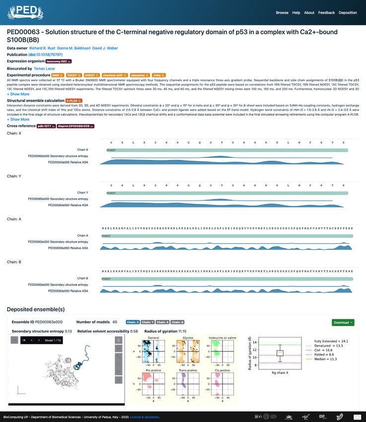
Nucleic Acids Research, 2021, Vol. 49, Database issue D405

ABSTRACT                                                        their extreme conformational dynamics, ensemble descrip-
                                                                tion is often applied for structural modeling of IDPs. Con-
The Protein Ensemble Database (PED) (https://                   formational ensembles are representative sets of conform-
proteinensemble.org), which holds structural ensem-             ers reflecting on the structural dynamics of IDPs sampling
bles of intrinsically disordered proteins (IDPs), has           the space. Ensemble modeling usually relies on experimen-
been significantly updated and upgraded since its               tal data originating from NMR spectroscopy (10–13) and
last release in 2016. The new version, PED 4.0, has             small-angle X-ray scattering (SAXS) data (14–18), Förster
been completely redesigned and reimplemented with               resonance energy transfer (FRET) (19,20) circular dichro-
cutting-edge technology and now holds about six                 ism (CD) spectroscopy data (21) or a combination thereof
times more data (162 versus 24 entries and 242 ver-             (22–25). These measurements are then used to define lo-
sus 60 structural ensembles) and a broader represen-            cal or nonlocal structural constraints for the computational
tation of state of the art ensemble generation meth-            modeling of the conformational ensemble, such as for the
                                                                restraining or reweighting of a pool of statistical random

                                                                                                                                  Downloaded from https://academic.oup.com/nar/article/49/D1/D404/6030232 by guest on 29 September 2021
ods than the previous version. The database has a
                                                                coils, or of molecular dynamics (MD) trajectories (22,26–
completely renewed graphical interface with an inter-           28).
active feature viewer for region-based annotations,                Solving structural ensembles, however, is fraught with un-
and provides a series of descriptors of the qualita-            certainties, because the number of degrees of freedom is
tive and quantitative properties of the ensembles.              inherently much larger than the number of experimentally
High quality of the data is guaranteed by a new sub-            determined structural restraints. As a result, determining
mission process, which combines both automatic                  an ensemble is a mathematically ‘ill-posed’ or ‘underdeter-
and manual evaluation steps. A team of biocura-                 mined’ problem that has more than one solution. We don’t
tors integrate structured metadata describing the en-           yet know how to select the ‘best’ ensemble from multiple
semble generation methodology, experimental con-                alternatives, neither can we be sure if an actual ensemble
straints and conditions. A new search engine allows             is a faithful representation of the real physical state of the
                                                                IDP/IDR, nor is only a reasonable fit to experiment obser-
the user to build advanced queries and search all en-
                                                                vations. To help address these issues, IDP/IDR ensembles
try fields including cross-references to IDP-related            solved at the time were collected and made available in the
resources such as DisProt, MobiDB, BMRB and SAS-                dedicated Protein Ensemble database (published as pE-DB
BDB. We expect that the renewed PED will be useful              in 2014 (26), renamed as PED in later versions).
for researchers interested in the atomic-level under-              This first version was an ambitious attempt to fill the
standing of IDP function, and promote the rational,             niche in the deposition of ensembles of fully disordered pro-
structure-based design of IDP-targeting drugs.                  teins and proteins with IDRs. At the time of the publica-
                                                                tion, it only stored data for a few dozens of ensembles for
                                                                a limited set of proteins, which increased very slowly in the
INTRODUCTION
                                                                following years. Manual deposition and validation of entry
Valuable mechanistic and functional information can be ob-      submissions used to hinder the smooth maintenance and
tained from protein structures modeled at atomistic reso-       increment of the database. A lot has happened, however,
lution (1–3). Due to the growth of experimentally deter-        in the structural–functional characterization of IDPs/IDRs
mined structures deposited in the Protein Data Bank (PDB)       since the inception of PED. For example, it has been proven
(4), currently there are >160 000 3D structures of macro-       that structural ensembles can predict independent struc-
molecules available in the database (4). As structural bi-      tural data (24,26), i.e. they are realistic and do have predic-
ology has mainly focused on determining the structure of        tive power. Based on novel, better suited force-fields (29–
globular proteins until the recent past, the presence of in-    32) the computational simulation of IDPs has also signifi-
trinsically disordered protein (IDP) regions (IDRs) have        cantly advanced (33,34). Influential IDP-related databases
mostly been inferred either from unresolved or proteolyt-       have been either updated (e.g. DisProt (35) or MobiDB (36))
ically digested tails or loops of these globular structures     or created anew (e.g. MFIB (37) or DIBS (38)). Successful
solved by X-ray crystallography, or from shorter regions        targeting of IDPs/IDRs by small molecules offers hope for
yielding few structural constraints in nuclear magnetic res-    a new class of effective drugs (39,40). A superposition-free
onance (NMR) spectroscopy measurements (5). The de-             method for comparing alternative ensembles has also been
pletion of long IDRs (LDRs) in PDB has been known               worked out (41), and allosteric regulatory mechanisms op-
for a long time, and the tightening of the gap in this re-      erating in the heterogeneous ensemble of IDPs/IDRs (mul-
gard has only become practical very recently (6). However,      tistery) have been elaborated (42,43). The appreciation of
this recent abundance of LDRs is predominantly due to           the importance of structural disorder in the novel field of
the context-dependent folding of proteins with conditional      liquid–liquid phase separation (LLPS) is on the rise (44) and
disorder, such as pH sensitivity, PTM-dependent folding,        persistent structural disorder of phase-separating proteins
localization-dependent disorder and folding upon binding        even in the condensed state has been reported (45,46). Last,
to a partner (7–9).                                             but not least, many ensembles have been solved (24,47) but
   Although conditionally folded IDRs provide important         not made publicly available.
structural insights, in-depth understanding of mechanis-           This rapid progress in the protein disorder field mandates
tic details of how IDPs function also requires knowledge        a basic upgrade and significant update of PED. To meet this
about the dynamic structures in the free state. By virtue of    goal, PED 4.0 was completely redesigned and extended with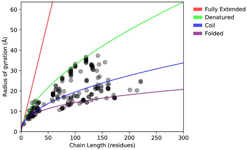
D406 Nucleic Acids Research, 2021, Vol. 49, Database issue

several new functionalities. To set a higher standard for the
quality of data, a new submission process is now carried out
through a web interface that enables automated validation
of the ensemble deposited by the authors and manual cu-
ration steps with the assistance of the database biocurators.
PED is now better cross-referenced with other IDP-related
databases such as BMRB (48), SASBDB (49), DisProt (35)
and MobiDB (36), and has a well-documented RESTful
API for programmatic access, search and download. In all,
the new PED has about six times more data than the previ-
ous version.

                                                                                                                                               Downloaded from https://academic.oup.com/nar/article/49/D1/D404/6030232 by guest on 29 September 2021
PROGRESS
Database structure and implementation                             Figure 1. PED 4.0 entry statistics. Stacked histogram of models per ensem-
                                                                  ble for different measurement methods in PED 4.0, binned based on the
One of the major changes since the previous version is the        number of the consisting conformer models.
whole new deposition process, which includes an automatic
data validation step and a curation step. The validation
has been introduced to standardize the data and improve
its quality by providing a number of structural indicators,
while the manual curation step provides metadata for bet-
ter data accessibility. A team of biocurators standardize the
description of the experimental methodology using terms
from a controlled vocabulary and identify cross-references
to third-party databases. Curators also scan the literature
to collect ensembles that have not yet been deposited into
PED.
    In PED, an entry is identified by the PED prefix and 5 dig-
its (e.g. PED00001), which corresponds to an experiment
on a protein (or protein complex), while a PED ensemble
(e.g. PED00001e001) is the set of conformations (or mod-          Figure 2. Chain compactness of PED 4.0 entries. Radius of gyration of
els) generated to fit the experimental data. Different ensem-     protein chains plotted against their chain length. Each dot represents a
bles generated using the same proteoform (same sequence           given chain in a given ensemble. The reference curves (54) represent val-
construct and PTMs), the same experimental conditions             ues specific for folded proteins (purple), random coils (blue), denatured
and the same experimental and computational methodol-             proteins (green) and fully extended chains (red). Four long folded pro-
                                                                  teins (PED00007, PED00010, PED00014 and PED00162) with over 300
ogy, represent different replicas of the same experiment (al-     residues are omitted, but fit well to the purple trend line.
ternative solutions to the same set of structural restraints
determined) and are grouped together in the same PED en-
try.                                                              DATABASE CONTENT
    PED also stores conformation weights as provided by the
                                                                  New entries
authors. Weights represent the probability for each confor-
mation to populate the ensemble, however, since these are         The number of entries in PED 4.0 has increased six-fold
not yet standardized and provided only for a limited num-         compared to the previous release. Some entries have been
ber of entries, they have not been considered in the calcu-       deposited after literature curation, while others have been
lation of ensemble descriptors such as Rg, accessibility and      directly provided by the experimentalists who generated
secondary structure propensities.                                 the data (data owners). Previous entries were manually re-
    The backend of the PED server processes each entry sub-       viewed and re-annotated. Old entries that included differ-
mission. The server executes a collection of scripts devel-       ent experiments were split up. The mapping from old to
oped in-house that generate summary statistics (solvent ac-       new identifiers is reported on the website (URL: https://
cessibility, secondary structure populations, radius of gy-       proteinensemble.org/help#mapping).
ration and maximum dimension). Secondary structure and               For new entries, PED curators focused on biologically
solvent accessibility are calculated by DSSP (50,51), while       interesting protein regions with conformational ensembles,
MolProbity (52) provides quality descriptors (torsion-angle       or more often, a set of ensembles determined under differ-
outliers, covalent bond-length and angle outliers, beta-          ent conditions (different construct or mutant, different pH,
carbon deviations and steric clashes). For each entry, the        denaturants etc.) or using different types of experimental
pipeline generates a report, which can be used to assess a        datasets and modeling methodology. As sensitivity to con-
submission. Since the same approach is used for all entries,      ditions is well-known for IDPs, these alternate ensembles
it is possible to make comparisons across the entire database     might provide very valuable insights into the conditional
and generate meaningful statistics. The report is available       disorder of these proteins (41). Furthermore, multiple en-
for download as a PDF document for all entries.                   sembles for a region measured under very similar condi-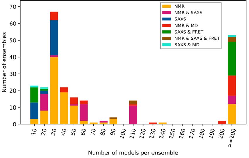
Nucleic Acids Research, 2021, Vol. 49, Database issue D407
Downloaded from https://academic.oup.com/nar/article/49/D1/D404/6030232 by guest on 29 September 2021
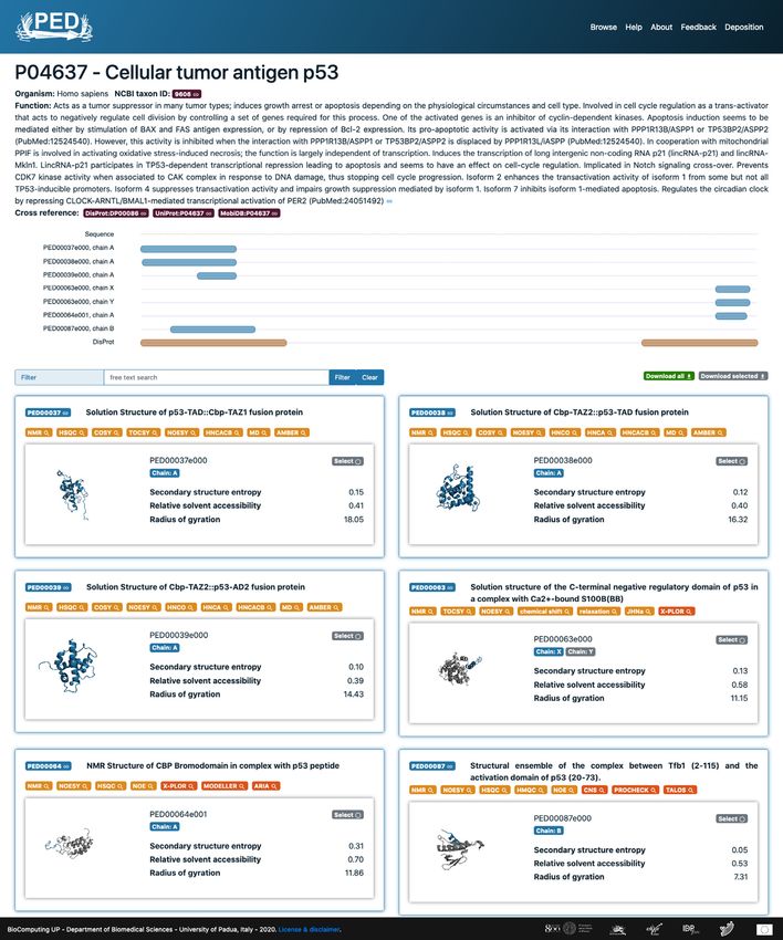
Figure 3. Example for PED’s Protein page. Protein page P04637 summarizes the human p53 ensembles currently stored in PED for both the N-terminal
and C-terminal disordered region. The feature viewer also integrates intrinsic disorder evidence from DisProt.
tions may highlight the biases in the modeling protocols ensembles of usually 50) determined by SAXS only are absent in PED,
IDRs determined using experimental constraints. The Dis- as currently most SAXS-based ensemble modeling tools are
Prot database was harnessed to make sure many of the ad- known to represent ensembles through several equivalent
ditions correspond to bona fide experimentally determined data sets, reducing the number of representative models in
IDPs/IDRs. This was complemented by an analysis of the each set to a range of 10–50 (15,53). However, larger ensem-
radii of gyration (Rg ) of the protein chains. bles generated by a combination of SAXS, smFRET and
molecular dynamics methods are present.
Protein compactness is often characterized by the Rg as a
Statistics
function of the length of the polypeptide chain (Figure 2).
Statistical analysis of the PED entries (Figure 1) shows an The Rg of folded proteins scales with chain length by follow-
increment in all classes of determination methods and in all ing a scaling law while, trivially, rigid rod-like chains follow
sizes of ensembles (i.e. number of models ranging from a a linear trend. Disordered proteins, however, fall in between
dozen to thousands). It also highlights that while NMR re- these two extremes due to their propensities to form local
mains as the most highly represented method used to model or nonlocal transient secondary (or tertiary) structure ele-D408 Nucleic Acids Research, 2021, Vol. 49, Database issue
Downloaded from https://academic.oup.com/nar/article/49/D1/D404/6030232 by guest on 29 September 2021
Figure 4. Example for PED’s Entry page. Entry page is shown for the C-terminal disordered region of p53 in a tetrameric complex with Ca2+ -bound S100B
(PED00063). The feature viewer shows chain-specific information, while molecular graphics, Ramachandran maps and Rg distribution are presented below.
ments. Figure 2 shows that the disordered proteins of PED The ‘Entry’ page provides details about the experimental
largely exhibit an Rg ranging from that of random coils to design and shows information on the complete make-up of
that of denatured proteins (54) across a wide range of IDP the ensemble, i.e. describes if a protein complex includes
protein lengths (10–200 residues), implying that the IDPs in nonpeptidic molecules or protein chains not mapping to
PED represent the known variety of IDP compaction be- UniProt (55).
havior. The points lying on the folded line correspond to Figure 3 shows the ‘Protein’ page for human p53 with
globular binding partners present in the ensembles that rep- multiple available ensembles for both the N-terminal and
resent complexes of IDPs and folded proteins/domains. C-terminal regions. By clicking on PED identifiers, it is
possible to open the corresponding Entry pages. For ex-
ample, PED00063 (Figure 4) corresponds to the p53 C-
NOVEL FEATURES terminal region folding upon binding to S100B. In sharp
contrast, PED00064 (not shown) is a disordered complex
Now PED 4.0 has both a protein-centric and an experimen-
of p53 binding to the CBP bromodomain.
tal entry-centric view. In the protein-centric view (Protein
page), ensembles from different PED entries are grouped
based on their UniProt accession. In this way, it is possi-
Entry views
ble to appreciate the differences between ensembles corre-
sponding to the same region on a single page, which may The Entry page (Figure 4) provides the title of the exper-
arise from the use of different techniques and conditions. iment, authors, and the corresponding publication whenNucleic Acids Research, 2021, Vol. 49, Database issue D409
available. PED does not include primary data, like struc- velopment of novel approaches––experimental and com-
tural constraints, but instead provides cross-references to putational tools––for developing and depositing ever more
primary databases; when available (PDB (4), BMRB (48) accurate ensembles. Second, it has been significantly ex-
and SASBDB (49)). MobiDB (36) and DisProt (35) are tended in size and has a greatly improved representation
cross-referenced in order to link evidence about the intrinsic of ensemble-generation methodologies and of functionally
disorder of the protein region. validated ‘bona fide’ IDRs, thanks to a community-wide cu-
For each entry, the PED biocurators generate a detailed ration effort. The number of entries has increased from 24
description of the ensemble determination. This description to 152, whereas the number of ensembles has grown from 60
about experimental and computational protocols is orga- to 215. In all, the total number of ‘conformers’ stored in the
nized into three different blocks (experimental procedure, database now exceed 290,532 PDB models (versus 24 615 in
structural ensemble calculation and, if applicable, MD cal- the old PED).
culations), each including a narrative and a set of terms se- PED has also been profoundly upgraded in a quest for
lected from a controlled vocabulary (CV). The CV ensures better consistency. The most important novel feature is the
Downloaded from https://academic.oup.com/nar/article/49/D1/D404/6030232 by guest on 29 September 2021
advanced accessibility and searchability and is constantly implementation of a new deposition process aimed at im-
updated to capture new developments of the field. The cur- proving the quality of the entire database. PED now in-
rent CV is available on the ‘About’ page of the PED website. cludes a web submission system. Each deposition is sub-
The rest of the Entry page provides a graphical view jected to an automatic validation step, which generates a
of structural features of the ensemble. The Feature-Viewer report on model quality, and a manual curation step, in
(56) component summarizes the make-up at the chain which a submission is manually evaluated and integrated
level. It shows the protein construct, solvent accessibility, with structured metadata. The automatic validation step in-
secondary-structure populations and the respective vari- cludes statistics on bond angles and lengths, backbone tor-
ability (entropy or standard deviation) across ensemble sion angles and steric clashes. Whereas statistics on ‘out-
models. For each chain of the ensemble, the distribution liers’ in the various geometric categories do not entail the re-
of the radii of gyration (Rg ) is shown as a box plot, along jection of deposition, it gives the user the option of selecting
with the corresponding theoretical values (dashed lines) for only ensembles that meet certain preset quality criteria. The
a protein chain of the same length if it was folded, random biocurator submission interface will soon be made available
coil-like or denatured, and expected Rg value for a rod-like to the public with the idea of providing a tool similar to the
or fully extended chain of the same length. Torsion angles OneDep system of the wwPDB (4) in the near future. Con-
are mapped to a Ramachandran plot to evaluate the struc- tributing new ensembles is highly encouraged, and for that,
tural preferences of the ensemble of the entire protein com- information about submission inquiries are available on the
plex (not chains) and the quality of backbone modeling. A Deposition page.
Quick view on the ensemble conformations (models) is pro- Additional novel features of PED 4.0 include a com-
vided by the MOL* structure viewer (57). The metadata, pletely new implementation of the website and database
ensemble coordinates and validation report are all down- schema. PED stores ensemble weights representing confor-
loadable. mational probabilities. Even though these are not taken into
account in the calculation of ensemble properties due to a
lack of standardization, they will be extensively integrated
Browse and search
in the future. The web interface has both a protein- and
Browse and advance search are implemented on the same experiment-centric view, an advanced search engine and a
page. A customizable table lists all entries with informa- well-documented API for programmatic access.
tion about the protein, types of measurements, number of The quick and significant growth of PED is due to the
ensembles and conformers. Each row represents a chain steady activity of experimentalists generating disordered en-
of an ensemble or a fragment in cases when the ensemble sembles that accumulated large amounts of data in the past
is calculated on an engineered construct. The correspond- years, and the perseverance of database curators. This sig-
ing UniProt accessions are provided for the majority of the nals the vitality of the concept of protein disorder and the
PED entries. A search box allows the user to look up specific strength of the disorder community working on integrat-
words in a free-text form or to search PED and all cross- ing structural, functional and medical aspects of structural
referenced identifiers. Moreover, it is possible to search all disorder. We expect that the new database will foster a sig-
the terms from the controlled vocabulary and to build com- nificant conceptual leap in the field. Even today, after more
plex queries or exploit regular expressions. Simple search is than two decades of research that has brought solidification
also available on the Main page, while programmatic search of the basic concept, we still tend to perceive structural dis-
and data access (or download) is implemented via a REST- order as a binary classifier, thinking of proteins or protein
ful API. An extended documentation and examples are pro- regions as either ordered or disordered. Structural disorder,
vided on the Help page. however, is not a simple, homogeneous structural state, it
rather represents a continuum of states from fully ordered
to fully disordered (58). PED is currently the only database
CONCLUSION
focused on representing the diversity of IDP protein ensem-
After several years of steadily diminishing activity, PED bles, which are not stored in databases focused on the de-
has finally come to new life. First, it has been trans- position of primary data (SASDB, BMRB, PDB), creating
ferred to a stable location that ensures continuous main- an extremely valuable resource for the IDP community. The
tenance and regular updates, hopefully stimulating the de- analysis of ensembles in PED 4.0 will enable us to better un-D410 Nucleic Acids Research, 2021, Vol. 49, Database issue
derstand determinants of these various sub-states in terms 2. Sillitoe,I., Dawson,N., Lewis,T.E., Das,S., Lees,J.G., Ashford,P.,
of compactness, secondary structure content and dynam- Tolulope,A., Scholes,H.M., Senatorov,I., Bujan,A. et al. (2019)
CATH: expanding the horizons of structure-based functional
ics, which will definitely help correctly interpret the func- annotations for genome sequences. Nucleic Acids Res., 47,
tional consequences of intrinsic structural disorder. Given D280–D284.
the prevalence of structural disorder in disease (59), the in- 3. Mitchell,A.L., Attwood,T.K., Babbitt,P.C., Blum,M., Bork,P.,
sight expected from structural ensembles in PED 4.0 will Bridge,A., Brown,S.D., Chang,H.-Y., El-Gebali,S., Fraser,M.I. et al.
also give a new impetus to efforts of structure-based drug (2019) InterPro in 2019: improving coverage, classification and access
to protein sequence annotations. Nucleic Acids Res., 47, D351–D360.
discovery against IDPs. 4. wwPDB,consortium. (2019) Protein Data Bank: the single global
The renewal and relocation of the database reflects on archive for 3D macromolecular structure data. Nucleic Acids Res., 47,
the ambition of the IDP community to actively maintain D520–D528.
the database and, more ambitiously, also to integrate it 5. Tompa,P. (2010) In: Structure and function of intrinsically disordered
proteins. Chapman & Hall/CRC Press, Boca Raton.
into DisProt’s IDP-specific complex ecosystem of databases 6. Monzon,A.M., Necci,M., Quaglia,F., Walsh,I., Zanotti,G.,
and computational tools (60). Significant further develop-
Downloaded from https://academic.oup.com/nar/article/49/D1/D404/6030232 by guest on 29 September 2021
Piovesan,D. and Tosatto,S.C.E. (2020) Experimentally determined
ments in the near future, such as mirroring the database long intrinsically disordered protein regions are now abundant in the
among multiple locations and contacting journals to rec- Protein Data Bank. Int. J. Mol. Sci., 21, 143–143.
ommend ensemble deposition into PED, are also planned. 7. Bugge,K., Brakti,I., Fernandes,C.B., Dreier,J.E., Lundsgaard,J.E.,
Olsen,J.G., Skriver,K. and Kragelund,B.B. (2020) Interactions by
Continuous maintenance and implementation of these and Disorder - A matter of context. Front. Mol. Biosci., 7, 110.
other future plans are ensured by the IDPcentral, MSCA- 8. Hausrath,A.C. and Kingston,R.L. (2017) Conditionally disordered
RISE IDPfun and ELIXIR IDP community groups. To en- proteins: bringing the environment back into the fold. Cell. Mol. Life
sure communication with users about recent growth of the Sci., 74, 3149–3162.
9. Jakob,U., Kriwacki,R. and Uversky,V.N. (2014) Conditionally and
database and new features, PED now will have a more active transiently disordered proteins: awakening cryptic disorder to
social media presence on Twitter with the original @Pro- regulate protein function. Chem. Rev., 114, 6779–6805.
teinEnsemble Twitter account. 10. Ozenne,V., Schneider,R., Yao,M., Huang,J., Salmon,L.,
Zweckstetter,M., Jensen,M.R. and Blackledge,M. (2012) Mapping
the potential energy landscape of intrinsically disordered proteins at
ACKNOWLEDGEMENTS amino acid resolution. J. Am. Chem. Soc., 134, 15138–15148.
11. Kosol,S., Contreras-Martos,S., Cedeño,C. and Tompa,P. (2013)
PED is maintained as a service of the ELIXIR IDP com- Structural characterization of intrinsically disordered proteins by
munity (URL: elixir-europe.org/communities/intrinsically- NMR spectroscopy. Mol. Basel Switz., 18, 10802–10828.
disordered-proteins). This project has received funding 12. Jensen,M.R., Zweckstetter,M., Huang,J. and Blackledge,M. (2014)
from the European Union’s Horizon 2020 research and in- Exploring free-energy landscapes of intrinsically disordered proteins
at atomic resolution using NMR spectroscopy. Chem. Rev., 114,
novation programme under grant agreement No 778247. 6632–6660.
13. Salvi,N., Salmon,L. and Blackledge,M. (2017) Dynamic descriptions
FUNDING of highly flexible molecules from NMR dipolar Couplings: Physical
basis and limitations. J. Am. Chem. Soc., 139, 5011–5014.
Italian Ministry of University and Research (MIUR) to 14. Cordeiro,T.N., Herranz-Trillo,F., Urbanek,A., Estaña,A., Cortés,J.,
SCET [2017483NH8]; European Union’s Horizon 2020 Sibille,N. and Bernadó,P. (2017) Small-angle scattering studies of
intrinsically disordered proteins and their complexes. Curr. Opin.
to SCET [778247]; Hungarian Scientific Research Fund Struct. Biol., 42, 15–23.
(OTKA) to PT [K124670, K131702]; Universidad Nacional 15. Tria,G., Mertens,H.D.T., Kachala,M. and Svergun,D.I. (2015)
de Quilmes to GP [PUNQ 1309/19]; National Agency Advanced ensemble modelling of flexible macromolecules using
for the Promotion of Science and Technology (ANPCyT) X-ray solution scattering. IUCrJ, 2, 207–217.
16. Gräwert,T.W. and Svergun,D.I. (2020) Structural modeling using
to GP [PICT-2014-3430] and to LBC [PICT-2017-1924]; solution Small-Angle X-ray Scattering (SAXS). J. Mol. Biol., 432,
Fondation pour la Recherche Médicale to SRB and SDV 3078–3092.
[DBI20141231336]; Natural Sciences and Engineering Re- 17. Vallet,S.D., Miele,A.E., Uciechowska-Kaczmarzyk,U., Liwo,A.,
search Council of Canada to CCG [RGPIN 2017-06030]; Duclos,B., Samsonov,S.A. and Ricard-Blum,S. (2018) Insights into
Agence Nationale de la Recherche (ANR) to PB [ANR- the structure and dynamics of lysyl oxidase propeptide, a flexible
protein with numerous partners. Sci. Rep., 8, 11768.
10-LABX-12-01]; National Institutes of Health (NIH) to 18. Hamdi,K., Salladini,E., O’Brien,D.P., Brier,S., Chenal,A., Yacoubi,I.
JFK and THG [5R01GM127627-03]; German Ministry of and Longhi,S. (2017) Structural disorder and induced folding within
Science and Education (SAS-BSOFT) to DS [16QK10A]; two cereal, ABA stress and ripening (ASR) proteins. Sci. Rep., 7,
EU Horizon 2020 programme (iNEXT-Discovery) to DS 15544.
19. Holmstrom,E.D., Holla,A., Zheng,W., Nettels,D., Best,R.B. and
[871037]; Vrije Universiteit Brussel (VUB) to PT [SRP51]; Schuler,B. (2018) Accurate transfer efficiencies, distance distributions,
EM-P, NG, NM, JM are PhD students, AJVR, TES are and ensembles of unfolded and intrinsically disordered proteins from
Postdocs and GP, CM-B, JI, LBC and MSF are researchers Single-Molecule FRET. Methods Enzymol., 611, 287–325.
of the National Research Council (CONICET) of Ar- 20. Fuertes,G., Banterle,N., Ruff,K.M., Chowdhury,A., Mercadante,D.,
gentina. Funding for open access charge: Vrije Universiteit Koehler,C., Kachala,M., Estrada Girona,G., Milles,S., Mishra,A.
et al. (2017) Decoupling of size and shape fluctuations in
Brussel (VUB) [SRP51]. heteropolymeric sequences reconciles discrepancies in SAXS vs.
Conflict of interest statement. None declared. FRET measurements. Proc. Natl. Acad. Sci. U.S.A., 114,
E6342–E6351.
21. Nagy,G., Igaev,M., Jones,N.C., Hoffmann,S.V. and Grubmüller,H.
REFERENCES (2019) SESCA: Predicting circular dichroism spectra from protein
1. PDBe-KB,consortium. (2020) PDBe-KB: a community-driven molecular structures. J. Chem. Theory Comput., 15, 5087–5102.
resource for structural and functional annotations. Nucleic Acids
Res., 48, D344–D353.Nucleic Acids Research, 2021, Vol. 49, Database issue D411
22. Krzeminski,M., Marsh,J.A., Neale,C., Choy,W.-Y. and 41. Lazar,T., Guharoy,M., Vranken,W., Rauscher,S., Wodak,S.J. and
Forman-Kay,J.D. (2013) Characterization of disordered proteins with Tompa,P. (2020) Distance-Based metrics for comparing
ENSEMBLE. Bioinformatics, 29, 398–399. conformational ensembles of intrinsically disordered proteins.
23. Sterckx,Y.G.J., Volkov,A.N., Vranken,W.F., Kragelj,J., Jensen,M.R., Biophys. J., 118, 2952–2965.
Buts,L., Garcia-Pino,A., Jové,T., Van Melderen,L., Blackledge,M. 42. Tompa,P. (2014) Multisteric regulation by structural disorder in
et al. (2014) Small-angle X-ray scattering- and nuclear magnetic modular signaling proteins: an extension of the concept of allostery.
resonance-derived conformational ensemble of the highly flexible Chem. Rev., 114, 6715–6732.
antitoxin PaaA2. Struct. Lond. Engl. 1993, 22, 854–865. 43. Wodak,S.J., Paci,E., Dokholyan,N.V., Berezovsky,I.N., Horovitz,A.,
24. Schwalbe,M., Ozenne,V., Bibow,S., Jaremko,M., Jaremko,L., Li,J., Hilser,V.J., Bahar,I., Karanicolas,J., Stock,G. et al. (2019)
Gajda,M., Jensen,M.R., Biernat,J., Becker,S., Mandelkow,E. et al. Allostery in its many Disguises: From theory to applications. Struct.
(2014) Predictive atomic resolution descriptions of intrinsically Lond. Engl. 1993, 27, 566–578.
disordered hTau40 and ␣-synuclein in solution from NMR and small 44. Brangwynne,C.P., Tompa,P. and Pappu,R.V. (2015) Polymer physics
angle scattering. Struct. Lond. Engl. 1993, 22, 238–249. of intracellular phase transitions. Nat. Phys., 11, 899–904.
25. Ibáñez de Opakua,A., Merino,N., Villate,M., Cordeiro,T.N., 45. Murthy,A.C., Dignon,G.L., Kan,Y., Zerze,G.H., Parekh,S.H.,
Ormaza,G., Sánchez-Carbayo,M., Diercks,T., Bernadó,P. and Mittal,J. and Fawzi,N.L. (2019) Molecular interactions underlying
Downloaded from https://academic.oup.com/nar/article/49/D1/D404/6030232 by guest on 29 September 2021
Blanco,F.J. (2017) The metastasis suppressor KISS1 is an intrinsically liquid-liquid phase separation of the FUS low-complexity domain.
disordered protein slightly more extended than a random coil. PLoS Nat. Struct. Mol. Biol., 26, 637–648.
One, 12, e0172507. 46. Murthy,A.C. and Fawzi,N.L. (2020) The (un)structural biology of
26. Varadi,M., Kosol,S., Lebrun,P., Valentini,E., Blackledge,M., biomolecular liquid-liquid phase separation using NMR
Dunker,A.K., Felli,I.C., Forman-Kay,J.D., Kriwacki,R.W., spectroscopy. J. Biol. Chem., 295, 2375–2384.
Pierattelli,R. et al. (2014) pE-DB: a database of structural ensembles 47. Delaforge,E., Kragelj,J., Tengo,L., Palencia,A., Milles,S.,
of intrinsically disordered and of unfolded proteins. Nucleic Acids Bouvignies,G., Salvi,N., Blackledge,M. and Jensen,M.R. (2018)
Res., 42, D326–D35. Deciphering the dynamic interaction profile of an intrinsically
27. Rangan,R., Bonomi,M., Heller,G.T., Cesari,A., Bussi,G. and disordered protein by NMR exchange spectroscopy. J. Am. Chem.
Vendruscolo,M. (2018) Determination of structural ensembles of Soc., 140, 1148–1158.
Proteins: Restraining vs Reweighting. J. Chem. Theory Comput., 14, 48. Romero,P.R., Kobayashi,N., Wedell,J.R., Baskaran,K., Iwata,T.,
6632–6641. Yokochi,M., Maziuk,D., Yao,H., Fujiwara,T., Kurusu,G. et al. (2020)
28. Köfinger,J., Stelzl,L.S., Reuter,K., Allande,C., Reichel,K. and BioMagResBank (BMRB) as a Resource for Structural Biology.
Hummer,G. (2019) Efficient ensemble refinement by reweighting. J. Methods Mol. Biol. Clifton NJ, 2112, 187–218.
Chem. Theory Comput., 15, 3390–3401. 49. Kikhney,A.G., Borges,C.R., Molodenskiy,D.S., Jeffries,C.M. and
29. Huang,J., Rauscher,S., Nawrocki,G., Ran,T., Feig,M., de Groot,B.L., Svergun,D.I. (2020) SASBDB: Towards an automatically curated and
Grubmüller,H. and MacKerell,A.D. (2017) CHARMM36m: an validated repository for biological scattering data. Protein Sci. Publ.
improved force field for folded and intrinsically disordered proteins. Protein Soc., 29, 66–75.
Nat. Methods, 14, 71–73. 50. Kabsch,W. and Sander,C. (1983) Dictionary of protein secondary
30. Song,D., Luo,R. and Chen,H.-F. (2017) The IDP-Specific force field structure: pattern recognition of hydrogen-bonded and geometrical
ff14IDPSFF improves the conformer sampling of intrinsically features. Biopolymers, 22, 2577–2637.
disordered proteins. J. Chem. Inf. Model., 57, 1166–1178. 51. Touw,W.G., Baakman,C., Black,J., te Beek,T.A.H., Krieger,E.,
31. Robustelli,P., Piana,S. and Shaw,D.E. (2018) Developing a molecular Joosten,R.P. and Vriend,G. (2015) A series of PDB-related databanks
dynamics force field for both folded and disordered protein states. for everyday needs. Nucleic Acids Res., 43, D364–D368.
Proc. Natl. Acad. Sci. U.S.A., 115, E4758–E4766. 52. Williams,C.J., Headd,J.J., Moriarty,N.W., Prisant,M.G., Videau,L.L.,
32. Rahman,M.U., Rehman,A.U., Liu,H. and Chen,H.-F. (2020) Deis,L.N., Verma,V., Keedy,D.A., Hintze,B.J., Chen,V.B. et al. (2018)
Comparison and evaluation of force fields for intrinsically disordered MolProbity: More and better reference data for improved all-atom
proteins. J. Chem. Inf. Model., 60, 4912–4923. structure validation. Protein Sci. Publ. Protein Soc., 27, 293–315.
33. Chong,S.-H., Chatterjee,P. and Ham,S. (2017) Computer simulations 53. Bernadó,P., Mylonas,E., Petoukhov,M.V., Blackledge,M. and
of intrinsically disordered proteins. Annu. Rev. Phys. Chem., 68, Svergun,D.I. (2007) Structural characterization of flexible proteins
117–134. using small-angle X-ray scattering. J. Am. Chem. Soc., 129,
34. Shrestha,U.R., Juneja,P., Zhang,Q., Gurumoorthy,V., 5656–5664.
Borreguero,J.M., Urban,V., Cheng,X., Pingali,S.V., Smith,J.C., 54. Hofmann,H., Soranno,A., Borgia,A., Gast,K., Nettels,D. and
O’Neill,H.M. et al. (2019) Generation of the configurational Schuler,B. (2012) Polymer scaling laws of unfolded and intrinsically
ensemble of an intrinsically disordered protein from unbiased disordered proteins quantified with single-molecule spectroscopy.
molecular dynamics simulation. Proc. Natl. Acad. Sci. U.S.A., 116, Proc. Natl. Acad. Sci. U.S.A., 109, 16155–16160.
20446–20452. 55. UniProt Consortium (2019) UniProt: a worldwide hub of protein
35. Hatos,A., Hajdu-Soltész,B., Monzon,A.M., Palopoli,N., Álvarez,L., knowledge. Nucleic Acids Res., 47, D506–D515.
Aykac-Fas,B., Bassot,C., Benı́tez,G.I., Bevilacqua,M., Chasapi,A. 56. Paladin,L., Schaeffer,M., Gaudet,P., Zahn-Zabal,M., Michel,P.-A.,
et al. (2020) DisProt: intrinsic protein disorder annotation in 2020. Piovesan,D., Tosatto,S.C.E. and Bairoch,A. (2020) The
Nucleic Acids Res., 48, D269–D276. Feature-Viewer: a visualization tool for positional annotations on a
36. Piovesan,D., Tabaro,F., Paladin,L., Necci,M., Micetic,I., sequence. Bioinforma. Oxf. Engl., 36, 3244–3245.
Camilloni,C., Davey,N., Dosztányi,Z., Mészáros,B., Monzon,A.M. 57. Sehnal,D., Rose,A.S., Koča,J., Burley,S.K. and Velankar,S. (2018)
et al. (2018) MobiDB 3.0: more annotations for intrinsic disorder, Mol*: towards a common library and tools for web molecular
conformational diversity and interactions in proteins. Nucleic Acids graphics. In: Proceedings of the Workshop on Molecular Graphics and
Res., 46, D471–D476. Visual Analysis of Molecular Data, MolVA ’18. Eurographics
37. Fichó,E., Reményi,I., Simon,I. and Mészáros,B. (2017) MFIB: a Association, Goslar, DEU, pp. 29–33.
repository of protein complexes with mutual folding induced by 58. Sormanni,P., Piovesan,D., Heller,G.T., Bonomi,M., Kukic,P.,
binding. Bioinforma. Oxf. Engl., 33, 3682–3684. Camilloni,C., Fuxreiter,M., Dosztanyi,Z., Pappu,R.V., Babu,M.M.
38. Schad,E., Fichó,E., Pancsa,R., Simon,I., Dosztányi,Z. and et al. (2017) Simultaneous quantification of protein order and
Mészáros,B. (2018) DIBS: a repository of disordered binding sites disorder. Nat. Chem. Biol., 13, 339–342.
mediating interactions with ordered proteins. Bioinforma. Oxf. Engl., 59. Uversky,V.N., Oldfield,C.J. and Dunker,A.K. (2008) Intrinsically
34, 535–537. disordered proteins in human diseases: introducing the D2 concept.
39. Tsafou,K., Tiwari,P.B., Forman-Kay,J.D., Metallo,S.J. and Annu. Rev. Biophys., 37, 215–246.
Toretsky,J.A. (2018) Targeting intrinsically disordered transcription 60. Davey,N.E., Babu,M.M., Blackledge,M., Bridge,A.,
Factors: Changing the paradigm. J. Mol. Biol., 430, 2321–2341. Capella-Gutierrez,S., Dosztanyi,Z., Drysdale,R., Edwards,R.J.,
40. Ruan,H., Sun,Q., Zhang,W., Liu,Y. and Lai,L. (2019) Targeting Elofsson,A., Felli,I.C. et al. (2019) An intrinsically disordered
intrinsically disordered proteins at the edge of chaos. Drug Discov. proteins community for ELIXIR. F1000Res., 8, ELIXIR-1753.
Today, 24, 217–227.You can also read