PROCESS DEVELOPMENT AND SCALE-UP OPTIMIZATION OF THE SARS-COV-2 RECEPTOR BINDING DOMAIN-BASED VACCINE CANDIDATE, RBD219-N1C1
←
→
Page content transcription
If your browser does not render page correctly, please read the page content below
Applied Microbiology and Biotechnology
https://doi.org/10.1007/s00253-021-11281-3

    BIOTECHNOLOGICAL PRODUCTS AND PROCESS ENGINEERING

Process development and scale-up optimization of the SARS-CoV-2
receptor binding domain–based vaccine candidate, RBD219-N1C1
Jungsoon Lee 1,2 & Zhuyun Liu 1,2 & Wen-Hsiang Chen 1,2 & Junfei Wei 1,2 & Rakhi Kundu 1,2 & Rakesh Adhikari 1,2 &
Joanne Altieri Rivera 1,2 & Portia M. Gillespie 1,2 & Ulrich Strych 1,2 & Bin Zhan 1,2 & Peter J. Hotez 1,2,3,4,5 &
Maria Elena Bottazzi 1,2,3,4

Received: 9 January 2021 / Revised: 31 March 2021 / Accepted: 6 April 2021
# The Author(s) 2021

Abstract
A SARS-CoV-2 RBD219-N1C1 (RBD219-N1C1) recombinant protein antigen formulated on Alhydrogel® has recently been
shown to elicit a robust neutralizing antibody response against SARS-CoV-2 pseudovirus in mice. The antigen has been
produced under current good manufacturing practices (cGMPs) and is now in clinical testing. Here, we report on process
development and scale-up optimization for upstream fermentation and downstream purification of the antigen. This includes
production at the 1-L and 5-L scales in the yeast, Pichia pastoris, and the comparison of three different chromatographic
purification methods. This culminated in the selection of a process to produce RBD219-N1C1 with a yield of >400 mg per liter
of fermentation with >92% purity and >39% target product recovery after purification. In addition, we show the results from
analytical studies, including SEC-HPLC, DLS, and an ACE2 receptor binding assay that were performed to characterize the
purified proteins to select the best purification process. Finally, we propose an optimized upstream fermentation and downstream
purification process that generates quality RBD219-N1C1 protein antigen and is fully scalable at a low cost.

Key points
• Yeast fermentation conditions for a recombinant COVID-19 vaccine were determined.
• Three purification protocols for a COVID-19 vaccine antigen were compared.
• Reproducibility of a scalable, low-cost process for a COVID-19 vaccine was shown.

Keywords COVID-19 . Spike protein . Pichia pastoris . Fermentation . Purification

* Jungsoon Lee
                                                                             Introduction
  jslee@bcm.edu
                                                                             After the first report of coronavirus disease 2019 (COVID-19)
* Maria Elena Bottazzi
  bottazzi@bcm.edu                                                           in December 2019 (Li et al. 2020; Lu et al. 2020), the number
                                                                             of cases is now at 124 million with over 2.3 million deaths
1
     National School of Tropical Medicine, Department of Pediatrics,         worldwide (JHU 2021). As of March 23, 2021, several vac-
     Baylor College of Medicine, One Baylor Plaza, BCM113,                   cines have been approved for emergency use in multiple coun-
     Houston, TX 77030, USA
                                                                             tries and are currently deployed for mass vaccinations globally
2
     Texas Children’s Hospital Center for Vaccine Development, Baylor        (Zimmer et al. 2021). The development and production of the
     College of Medicine, 1102 Bates Street, Houston, TX 77030, USA
                                                                             COVID-19 vaccines relies on several technologies or plat-
3
     National School of Tropical Medicine, Department of Molecular           forms, mainly nucleic acid and viral vector vaccines as novel
     Virology & Microbiology, Baylor College of Medicine, One Baylor
                                                                             technologies and whole inactivated, live attenuated viral or
     Plaza, BCM113, Houston, TX 77030, USA
4
                                                                             recombinant protein subunit or virus-like particle vaccines as
     Department of Biology, College of Arts and Sciences, Baylor
                                                                             conventional platforms (Chakraborty et al. 2021). Among the
     University, Waco, TX, USA
5
                                                                             first to have received approval for use were two messenger
     James A. Baker III Institute for Public Policy, Rice University,
                                                                             RNA (mRNA)-based vaccines from Pfizer/BioNTech and
     Houston, TX, USA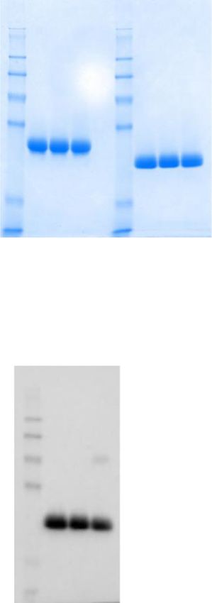
Appl Microbiol Biotechnol

Moderna, both produced in record time, but posing the chal-         like its close relative, SARS-CoV that had caused an outbreak
lenges that are relatively expensive to manufacture and diffi-      of severe acute respiratory disease in 2002. The receptor bind-
cult to scale and require transportation and storage at ultra-low   ing domain (RBD) of the S protein binds to a cellular receptor,
temperatures. The viral vector–based vaccines developed by          angiotensin-converting enzyme 2 (ACE2), that mediates
Oxford-AstraZeneca, Johnson & Johnson (J&J), Gamaleya,              membrane fusion during viral entry into the cell (Li et al.
and CanSino have also now been approved for use in a variety        2003; Yan et al. 2020). S proteins for both viruses have served
of countries and bring the added advantage that do not have         as vaccine antigens that could elicit antibodies to prevent virus
these ultra-low cold-chain requirements providing easier de-        entry by blocking the binding of RBD to ACE2 (Hoffmann
livery and, in the case of the J&J vaccine, the advantage of        et al. 2020), and all the leading COVID-19 vaccines currently
being used as a single dose. Furthermore, several whole             in clinical trials, including the mRNA vaccines, use the S
inactivated viral vaccines produced by the Chinese manufac-         protein to elicit immunity (Haynes et al. 2020).
turers Sinopharm and Sinovac and by Bharat Biotech, an              Overwhelmingly, such vaccines protect through their induc-
India-based manufacturer, have added to the list of approved        tion of virus-neutralizing antibodies, together with T cell re-
vaccines (Craven 2020; Eccleston-Turner and Upton 2021).            sponses (Jiang et al. 2020).
However, formidable challenges still remain to produce much            Building on our experience with the RBD of the SARS-
larger scales and distribute COVID-19 vaccines within low-          CoV spike protein (Chen et al. 2014, 2017, 2020), the SARS-
and middle-income countries (LMICs) (Lancet Commission              CoV-2 RBD was cloned and expressed in the yeast Pichia
on and Therapeutics Task Force 2021). Data from                     pastoris. Yeast has a track record of serving as a host organism
Duke’s Global Health Innovation Center and others                   for the production of multiple regulatory-approved and
clearly highlight the continued procurement and                     prequalified recombinant subunit vaccines, including vaccines
manufacturing challenges, leading to enormous inequity              for hepatitis B, influenza B, human papillomavirus, as well as
in the access for COVID-19 vaccines in these regions of             for diphtheria and tetanus (Bill 2015; Kumar and Kumar
the world (Duke Global Health Innovation Center 2021;               2019). Eukaryotic expression in yeast shows advantages over
Ritchie et al. 2021).                                               the prokaryote, Escherichia coli, with respect to the produc-
   Therefore, this situation is leaving LMICs bereft of low-        tion of recombinant protein vaccines. Proper protein folding,
cost COVID-19 vaccines suitable for their modest or depleted        disulfide bridge formation, post-translational modifications,
health systems (Lancet Covid-19 Commissioners and                   and secretory cleavage are better supported in yeast,
Commission 2020). In response, there is an urgent need that         while also allowing for robust production with low costs
the LMIC vaccine developers and manufacturers from the              and full scalability, features that distinguish this plat-
Developing Country Vaccine Manufacturers Network                    form from other eukaryotic systems, such as insect
(DCVMN) (DCVMN 2021) accelerate the development of                  cells, mammalian cells, and plants.
additional vaccines employing the traditional platforms, espe-         The RBD219-N1C1 antigen is derived from residues 332–
cially additional vaccines based on recombinant proteins            549 of the SARS-CoV-2 RBD with a single mutation of a free
(Craven 2020; Eccleston-Turner and Upton 2021). These vac-          cysteine residue (Cys538) to alanine to prevent intermolecular
cine platforms are less demanding with respect to transport         disulfide bond formation and therefore unwanted oligomeri-
and storage and often come with a long history of successful        zation during process development (Chen et al. 2021). In ad-
global large-scale production capabilities and affordable use       dition, N1 refers to the deletion of Asn331 to avoid
for other infectious diseases (Hotez and Bottazzi 2020).            hyperglycosylation observed in previous studies with the
Particularly attractive in this aspect are recombinant protein      SARS-CoV RBD219-N1 antigen (Chen et al. 2014). In initial
antigens (Pollet et al. 2021a), in particular those produced        studies, the modifications used to express RBD219-N1C1 re-
through microbial fermentation in yeast. For instance, recom-       combinant protein did not affect the in vitro binding to its
binant hepatitis B vaccine has been administered to adults and      receptor ACE2, when compared to the yeast-expressed re-
children for decades (World Health Organization 2017).              combinant wild-type RBD (Chen et al. 2021). Additionally,
Currently, only a few protein-based COVID-19 vaccine can-           when RBD219-N1C1 was adjuvanted with Alhydrogel®, the
didates have advanced to phase 3 trials, namely from                vaccine formulation has been shown to elicit a robust neutral-
Novavax, Vector Institute in Russia, and Cuba’s Finlay              izing antibody response against SARS-CoV-2 pseudovirus in
Vaccine Institute (Pollet et al. 2021a), but in aggregate, it is    mice (Pollet et al. 2021b).
promising to see more than 30 protein-based vaccines are               With any COVID-19 vaccine candidate, the ability to pro-
actively in development (Zimmer et al. 2021), which will            duce billions of doses efficiently is crucial to satisfy the po-
greatly enable future access and distribution of safe, effective,   tential global vaccine demand. We, therefore, have been de-
and affordable COVID-19 vaccines for the world.                     veloping and optimizing a scalable production process of the
   The SARS-CoV-2 virus, the pathogen that causes COVID-            RBD219-N1C1 vaccine candidate at a low cost to support its
19, uses its surface spike (S) protein for host cell entry, just    technology transfer. Initial fermentation runs scouting for
Appl Microbiol Biotechnol

growth media, induction time, and glycerol fed-batch condi-         g/L potassium hydroxide, 0.93 g/L calcium sulfate de-
tions were executed in a 1-L bioreactor and resulted in a ~10-      hydrate, 26.7 mL/L of 85% phosphoric acid, and 40 g/L
fold increase in RBD219-N1C1 expression levels. Further             glycerol) or low-salt medium (LSM) (pH 5.0; LSM:
scale-up experiments in a 5-L bioreactor established the repro-     4.55 g/L potassium sulfate, 3.73 g/L magnesium sulfate
ducibility of the selected conditions. Simultaneously, a purifi-    heptahydrate, 1.03 g/L potassium hydroxide, 0.23 g/L
cation scheme was developed based on the process used for           calcium sulfate dehydrate, 10.9 mL/L of 85% phospho-
the 70% homologous SARS-CoV RBD antigen (Chen et al.                ric, and 40 g/L glycerol) to a starting cell density
2017) and further optimized to allow full scalability and lower     (OD600) of 0.5. Fermentation was conducted using a
the cost. Taking into consideration yield, purity, functionality,   Biostat Qplus bioreactor with a 1-L vessel (Sartorius
and removal of host cell contaminants, we have developed an         Stedim, Guxhagen, Germany). For 5-L runs, the seed
optimized fermentation at the 5-L scale and purification (pro-      culture (125–250 mL) was inoculated into 2.5 L of
cess 2) suitable for production and manufacturing of a high-        LSM, and fermentation was conducted in a CelliGen
yield (and therefore, potentially low-cost) COVID-19 vaccine        310 bioreactor with a 7.5-L vessel (Eppendorf, New
antigen candidate. The developed process has already been           York, USA), controlled by the Eppendorf Bio
transferred to Biological E, an industrial vaccine manufacturer     Command software. Cell expansion was continued at
in India and is currently undergoing further production matu-       30 °C with a dissolved oxygen (DO) set point of
rity while the vaccine candidate has recently completed a           30%. After 19 ± 2 h of growth, a dissolved oxygen
combined phase 1 and 2 clinical trial. We expect the results        spike was observed on the trend chart, which indicates
from this trial to be published and available during the second     glycerol depletion. A fed-batch was initiated with 50%
quarter of 2021.                                                    glycerol at a feed rate of 15 mL/L/h for 6 h to further
                                                                    expand biomass. During the last hour of the fed-batch
                                                                    phase, pH was adjusted to 6.5 using 14% NH 4OH,
Materials and methods                                               while the temperature was adjusted to 25 °C. When a
                                                                    glycerol fed-batch was not included in the fermentation
Generation of research cell bank                                    process, the pH and temperature were adjusted to the
                                                                    desired value during the first hour of induction. After
To generate a research cell bank (RCB), P. pastoris X33 strain      the fed-batch phase, methanol induction was initiated;
was transformed with expression plasmid pPICZαA contain-            the total induction time was approximately 68–72 h.
ing RBD219-N1C1 coding DNA, and one transformed colo-               Biomass was removed by centrifugation at 12,227×g
ny with high expression of recombinant RBD219-N1C1 pro-             for 30 min at 4 °C before the supernatant was filtered
tein (Chen et al. 2021) was selected and streaked on yeast          through 0.45-μm polyethersulfone (PES) filters stored at
extract peptone dextrose (YPD) plates containing 100 μg/            −80 °C until purification.
mL Zeocin to make single colonies. The plates were incubated
at 30 °C for approximately 3 days until single colonies were        Purification overview of three processes
observed. Subsequently, 200-mL plant-derived phytone YPD
medium was inoculated with a single colony from the                 The fermentation supernatant (FS) was removed from −80 °C
respective plate and incubated at 30 °C with constant               and thawed at 22 °C for 4–6 h. Three purification processes
shaking (225 rpm) until the OD600 reached 9.3. Finally,             were performed with 1-L FS aliquots (Fig. 1b). In process 1,
the cell culture was mixed with plant-derived glycerol              the RBD219-N1C1 protein was captured from the FS using
to a final concentration of 20% and aseptically aliquoted           hydrophobic interaction chromatography (HIC), concentrated
(1 mL each) into 1.2-mL cryovials. For long-term stor-              by ultrafiltration/diafiltration (UFDF), and polished using size
age, the cryovials were stored at −80 °C.                           exclusion chromatography (SEC). In process 2, the RBD219-
                                                                    N1C1 protein was captured using HIC, buffer-exchanged
Fermentation                                                        (UFDF), and polished using anion-exchange chromatography
                                                                    (AEX). Finally, in process 3, the FS was buffer-exchanged
One vial of the SARS-CoV-2 RBD219-N1C1 RCB was                      using UFDF before the target protein was captured using
used to inoculate a 0.5-L buffered minimal glycerol                 cation-exchange chromatography (CEX), buffer-exchanged
(BMG) medium in a 2-L baffled shake flask. The shake                (UFDF), and polished using AEX.
flask culture was grown at 30 °C and 225 rpm until an
OD600 of 5–10. For 1-L fermentations, this seed culture             UFDF (ultrafiltration and diafiltration)
(20–40 mL) was inoculated into 0.4 L of sterile basal-
salt medium (BSM) (pH 5.0; BSM: 18.2 g/L potassium                  Two types of devices were used for UFDF, a centrifu-
sulfate, 14.9 g/L magnesium sulfate heptahydrate, 4.13              gal concentrator, and a flat sheet membrane, depending
Appl Microbiol Biotechnol
Fig. 1 Fermentation (a) and a b
Fermentation Purification
purification (b) flow diagrams.
Three purification processes
performed are shown in different Research cell bank Thaw fermentation supernatant
colors. The color scheme remains
consistent throughout all figures. Inoculate shake flask
UFDF, ultrafiltration and Process-1 Process-2 Process-3
diafiltration; HIC, hydrophobic
Inoculate bioreactor TFF
interaction chromatography; UFDF
SEC, size exclusion
chromatography; TFF, tangential
Glycerol batch phase Capture
flow filtration; CEX, cation HIC HIC CEX
exchange chromatography; AEX,
anion exchange chromatography Methanol induction phase
UFDF Centrifugation TFF TFF
Harvest after 68-70 hours of induction
AEX AEX
Polish SEC
(negative) (negative)
Filter supernatant and freeze at -80°C
on the target volume. For process 1, Amicon centrifugal Performance column (4.4 cm diameter and 7.4 cm bed height)
concentrator, with a 10 kDa molecular weight cutoff at a 20 mL/min flow rate. The column was washed with 1 M
(MWCO) (MilliporeSigma, Burlington, USA) was used ammonium sulfate in 30 mM Tris-HCl (pH 8.0). Bound pro-
to concentrate the HIC elution pool (2050×g at 4 °C). teins were eluted with 0.4 M ammonium sulfate in 30 mM
This allowed concentration to the small volume needed Tris-HCl (pH 8.0).
for SEC. For process 2, a flat sheet Pellicon XL
Cassette with a Biomax 5 membrane (5 kDa MWCO) Size exclusion chromatography
and a Labscale TFF System (MilliporeSigma,
Burlington, USA) were used to concentrate the HIC Five milliliters of the concentrated HIC elution pool was load-
elution pool 8-fold, followed by diafiltration with 4 ed on a HiLoad 16/600 Superdex 75 prep-grade column
diavolumes of 20 mM Tris-HCl (pH 7.5) and 100 mM (Cytiva, Marlborough, USA), pre-equilibrated with 20 mM
NaCl. A crossflow was kept at 25 mL/min over a Tris-HCl (pH 7.5) and 150 mM NaCl, and eluted at a flow
0.005-m2 membrane area throughout the entire process rate of 1 mL/min. The SEC elution pool was aseptically fil-
with an average transmembrane pressure (TMP) of ~15 tered using a 0.2-μm PES filter in a biosafety cabinet and
psi. For process 3, a flat sheet Pellicon 2 Mini Cassette stored at −80 °C until usage.
with a Biomax 5 membrane (MilliporeSigma,
Burlington, USA) was used for the first UFDF
(UFDF-1) to concentrate the FS 4-fold followed by Ion exchange chromatography
diafiltration with 4 diavolumes of 20 mM sodium citrate
(pH 4.2) and 10 mM NaCl. A crossflow was kept con- In process 3, RBD219-N1C1 was captured using CEX. The
stant at 200 mL/min over a 0.1-m 2 membrane area Pellicon 2 retentate pool in 20 mM sodium citrate (pH 4.2) and
throughout the entire process with an average TMP of 10 mM NaCl was loaded on a 50-mL CM Sepharose Fast
~8 psi. For the UFDF-2, the CEX elution pool was Flow column (2.6 cm diameter and 9.3 cm bed height)
concentrated 4-fold followed by diafiltration with 4 at a 10 mL/min flow rate. The column was washed with
diavolumes of 25 mM Tris-HCl (pH 7.2) and 5 mM 20 mM sodium citrate (pH 4.2) and 10 mM NaCl.
NaCl using the Pellicon XL Cassette as described for Bound proteins were eluted in 20 mM sodium citrate
process 2. (pH 6.6) and 10 mM NaCl.
In processes 2 and 3, RBD219-N1C1 was polished using a
negative capture AEX. The Pellicon XL retentate pool was
Hydrophobic interaction chromatography loaded on a HiPrep Q Sepharose XL 16/10 column (Cytiva,
Marlborough, USA) that was pre-equilibrated with 20 mM
In processes 1 and 2, HIC was used to capture RBD219-N1C1 Tris-HCl (pH 7.5) and 100 mM NaCl for process 2, and
proteins from the FS. Ammonium sulfate salt was added to the 25 mM Tris-HCl (pH 7.2) and 5 mM NaCl for process 3.
FS to a final concentration of 1 M (w/v), and the pH was The flowthrough from AEX was collected, aseptically filtered
adjusted to 8.0. The FS was filtered through a 0.45-μm PES using 0.2-μm PES filters in a biosafety cabinet, and stored at
filter unit and loaded on a 112-mL Butyl Sepharose High- −80 °C until usage. NaCl (95 mM) was added to the finalAppl Microbiol Biotechnol
purified proteins from process 3 prior to storage in 25 mM mg/mL with TBS, and approximately 40 μL of protein was
Tris-HCl (pH 7.2) and 100 mM NaCl. then loaded into a clear bottom 384-well plate in four repli-
cates to evaluate the hydrodynamic radius and molecular
Protein yield and purity determination by weight using the cumulant fitting on a Wyatt Technology
quantitative SDS-PAGE DynaPro Plate Reader II.
In-process samples taken at each purification step were loaded Host cell protein quantification by ELISA
on either 14% Tris-glycine gels or 4–12% Bis-Tris gels to
determine the concentration and purity of the various Yeast-expressed RBD219-N1C1 is N-glycosylated (Chen
RBD219-N1C1 samples. Purified RBD219-N1C1 proteins et al. 2021). To avoid any cross-reactivity from anti-
of known concentrations were used as standards. After SDS- P. pastoris HCP antibodies that recognize the N-glycans,
PAGE, gels were stained with Coomassie blue and scanned which could result in an over-estimation of true HCP, we
with a GS-900 densitometer (Bio-Rad, Hercules, USA). Gel performed quantitative ELISAs with a second-generation an-
images were processed with Image Lab software (Bio-Rad, ti-Pichia pastoris HCP ELISA Kit (Cygnus, Southport, USA;
Hercules, USA) to create a standard curve and determine pro- Cat#: F640) following the manufacturer’s instructions. This
tein concentration and purity. kit provides strips pre-coated with anti-P. pastoris HCP anti-
bodies. Serially diluted RBD219-N1C1 was loaded onto the
Western blot strips (HCP standards range from 0 to 250 ng/mL) in the
presence of HRP-conjugated anti-P. pastoris antibodies. The
Western blot analysis was performed to detect RBD219- strips were then incubated for approximately 3 h at room tem-
N1C1 as well as P. pastoris host cell protein (HCP). Five perature followed by 4 washes. Finally, 100 μL of 3,3′,5,5′-
micrograms of purified protein was run on 14% Tris- tetramethylbenzidine (TMB) solution was added to react with
Glycine gels under non-reducing and reducing conditions to the HRP-conjugated antibodies that were presented in the strip
detect RBD219-N1C1 and HCP, respectively. Proteins in gel for 30 min prior to the addition of 100 μL of 1 M HCl to stop
were transferred to PVDF membranes and blocked with 5% the reaction. The absorbance of 450 nm was measured in each
dry milk in PBST (1× PBS with 0.05% Tween-20). RBD219- well of the strip, and a linear standard curve was generated by
N1C1 was detected using a rabbit monoclonal antibody plotting an “absorbance vs concentration” graph with the HCP
against the SARS-CoV-2 spike S1 protein (Sino Biological, standards to further calculate the HCP concentration present in
Beijing, China; Cat#: 40150-R007) and goat anti-rabbit IgG the RBD219-N1C1 proteins.
secondary antibodies conjugated with horseradish peroxidase
(Invitrogen, Carlsbad, USA; Cat#: G21234). HCPs were de- Endotoxin test
tected using an anti-P. pastoris:HRP conjugate (2G) solution
(Cygnus, Southport, USA; Cat#: F641-12). The blots were Endotoxin levels in the purified RBD219-N1C1 samples were
developed using ECL Prime Substrate System (Cytiva, measured using the Endosafe Portable Testing System
Marlborough, USA). (Charles River Laboratory, Wilmington, USA). The purified
protein was diluted 10-fold with Endosafe LAL reagent water,
Size Exclusion Chromatography-High Performance and 25 μL of diluted protein was loaded to each of the four
Liquid Chromatography wells of PTS20 Limulus amebocyte lysate Reagent Cartridge
for the measurement as described in the literature (Charles
Waters® Alliance HPLC Separations Modules and River Laboratory, Wilmington, USA) (Jimenez et al. 2010).
Associated PDA Detectors were operated to analyze the size
and purity of purified RBD219-N1C1 proteins. Fifty micro- In vitro ACE2 binding ELISA
grams of the RBD219-N1C1 protein was injected into a Yarra
SEC-3000 column (300 mm × 7.8 mm; catalog #: 00H-4513- The binding of RBD219-N1C1 to recombinant human
K0) and was eluted in 20 mM Tris-HCl (pH 7.5) and 150 mM ACE2 was evaluated using an ELISA procedure de-
NaCl, at the flow rate of 0.6 mL/min. The elution of protein scribed previously (Chen et al. 2021). In short, 96-well
was confirmed by detecting the absorbance at 280 nm. ELISA plates were coated with 100 μL of 2 μg/mL
RBD219-N1C1 overnight at 4 °C followed by blocking
Dynamic light scattering with PBST/0.1% BSA. One hundred microliters of seri-
ally diluted ACE2-hFc (LakePharma, San Carlos, USA;
The size of the purified RBD219-N1C1 proteins was also Cat#: 46672) was added to the wells and incubated at
analyzed by dynamic light scattering (DLS) (Chen et al. room temperature for 2 h, and the binding was detected
2017, 2020). In short, RBD219-N1C1 was first diluted to 1 by adding 100 μL 1:10,000 diluted HRP conjugatedAppl Microbiol Biotechnol
anti-human IgG antibodies (GenScript, Piscataway, Fermentation scalability and reproducibility
USA; Cat#: A00166) with a 1-h incubation period at
room temperature. Finally, 100 μL TMB substrate was The fermentation process with 6 h of glycerol feed was then
provided to each well to react with HRP and the reac- scaled up from 1 to 5 L to test scalability and reproducibility
tion was terminated with 100 μL of 1 M HCl. (Table 1, run 4). The induction time was extended to 87 h until
Absorbance at 450 nm was measured using an Epoch biomass started to drop. This suggested that the cells were no
2 microplate reader (BioTek, Winooski, USA). longer actively dividing. Since excessive methanol feeding
may lead to cell death thus leading to a loss of protein yield,
it was decided to stop the methanol feeding after 87 h of
induction. The peak yield of RBD219-N1C1 was 449 ± 8
Results mg/L at 70 h after induction (day 3), after which the yield
slightly dropped to 408 ± 9 mg/L at 87 h after induction.
Fermentation optimization The fermentation process without the 6-h glycerol fed-
batch phase was also scaled up from 1 to 5 L for comparison
When BSM and LSM were compared for the production (Table 1, run 5). After 70 ± 2 h of induction, the yield of
of the RBD219-N1C1 protein, no differences were ob- RBD219-N1C1 reached 479 ± 15 mg/L (a 128% increase
served in the growth profiles and the final biomass. compared to the 1-L scale). This yield was close to the yield
However, the salt concentration appeared to have a sig- of the fermentation run with the 6-h glycerol fed-batch phase
nificant effect on the yield. The yield of the RBD219- (Table 1, run 4). Since there was no significant increase in
N1C1 protein using BSM was only 52 mg/L, while yield by the glycerol fed-batch at the 5-L scale, we decided
using LSM, 237 mg/L was achieved (Table 1, runs 1 to proceed without this step (Fig. 1a). To establish reproduc-
and 2). Therefore, LSM was used for the further devel- ibility, this fermentation process (Fig. 1a) was repeated four
opment of the fermentation process. times (runs 5–8). The average yield of four reproducibility
The baseline fermentation process consisted of two runs was 428 ± 36 mg/L, with a coefficient of variance of
phases: a glycerol-batch phase and a methanol fed- 8.3%. The SDS-PAGE gel analysis of fermentation superna-
batch phase. In glycerol-batch mode, LSM contained tants of a representative run (run 5) with the lockdown process
40 g/L of glycerol. At the time of glycerol depletion, is shown in Fig. 2. RBD219-N1C1 (a dominant protein band
the initial induction biomass was 110 ± 10 g/L (WCW). of ~28 kDa) was secreted and accumulated in the fermentation
In this study, a glycerol fed-batch phase was then added supernatant through the course of methanol induction.
before methanol induction to test the efficiency of pro-
tein expression based on the initial induction biomass. Three purification schemes
After a 6-h glycerol-fed-batch phase, the initial induc-
tion biomass doubled to 210 ± 20 g/L (WCW). The In parallel with the fermentation optimization, three different
methanol feed strategies were kept the same. At harvest, processes were performed to purify RBD219-N1C1 from the
the final OD600 and the biomass were determined to be FS (Fig. 1b). Process 1 was developed by adapting the purifi-
260 AU and 417 g/L, respectively. By adding the glyc- cation method of our SARS-CoV RBD219-N1 antigen that
erol fed-batch, the yield of RBD219-N1C1 was in- shares 70% homology with the SARS-CoV-2 RBD (Chen
creased about 120% to 533 mg/L (Table 1, run 3). et al. 2017, 2021; Shang et al. 2020). In this process, the target
Table 1 Summary of the
development fermentation runs Run # Fermentation conditions Endpoint analysis
Volume (L) Medium Glycerol Total induction Biomass OD600 Peak yield
fed-batch time (g/L) (AU) (mg/L of FS)
1 1 BSM No 70 ± 1 417 ± 3 260 52 ± 2
2 1 LSM No 70 ± 1 437 ± 4 257 237 ± 7
3 1 LSM 6h 70 ± 1 434 ± 3 254 533 ± 3
4 5 LSM 6h 87 ± 1 413 ± 2 230 449 ± 8
5–8* 5 LSM No 70 ± 2 394 ± 20 232 ± 11 428 ± 36
FS fermentation supernatant
*Four reproducibility runs (runs 5–8) were performed. The averages of biomass and yields from four runs are
shownAppl Microbiol Biotechnol
PI D1 D2 D3 Pepstats (Madeira et al. 2019), RBD219-N1C1 was pre-
kDa dicted to be positively charged at a pH below its theo-
retical pI. After screening buffer conditions, consisting
250 of Tris-HCl at different pH values ranging from 7.0 to
148 8.0 and various concentrations of NaCl, the buffer
98 consisting of 20 mM Tris-HCl (pH 7.5) and 100 mM
NaCl showed that the RBD219-N1C1 did not bind to
64 the Q XL column while non-specific HCPs were bound
to the column and removed effectively. The step recov-
50
ery during AEX was 78%, which is lower than the 89%
of the step recovery seen from SEC in process 1. The
36 final purity of the purified protein from process 2 was
95.1%, which is lower than the 98.3% purity seen in
process 1 but still highly pure. However, the overall
recovery in process 2 was only 39%, much lower than
22 the 50% for process 1. This is due to the lower recov-
ery during HIC, 45%, that lags the 67% recovery seen
16 for the equivalent step in process 1. This lower recovery
may offer the opportunity for improvement, but overall,
it is fair to conclude that AEX can successfully replace
SEC for the polishing step.
6
In process 3, we further optimized process 2 to utilize CEX for
Fig. 2 Timepoint SDS-PAGE analysis of pre- and post-induction fermenta-
the capture step instead of HIC. After the first UFDF step (UFDF-
tion samples of the lockdown process (run 5). PI: pre-induction; D1, D2, D3:
days 1–3 after induction. The arrow shows RBD219-N1C1 in the fermenta- 1) to concentrate and buffer exchange the FS, RBD219-N1C1 was
tion supernatant after induction captured using a CM FF column followed by a second UFDF step
(UFDF-2) and a polishing step using negative AEX capture with
protein was captured by HIC using a butyl HP column with another buffer consisting of 25 mM Tris-HCl (pH 7.2) and 5 mM
1 M ammonium sulfate salt for the binding. After the HIC, NaCl and selected from the aforementioned buffer screening (Fig.
67% of the target protein was recovered and purity significant- 1b). The additional UFDF-1 step required prior to CEX increased
ly improved from 85.6% in the FS to 97.6% (Fig. 3a). The processing time compared to processes 1 and 2. Although the step
target protein was concentrated using Amicon centrifugal con- recovery from CEX was 65%, very similar to the 67% seen after
centrators and further polished by SEC using a Superdex 75 the HIC in process 1, the purity was only 83% after CEX which is
column. The SEC elution pool was then diluted to 2 mg/mL significantly lower than the purity (97.6% and 95.2% from pro-
for storage. Overall, the final yield of the target protein using cesses 1 and 2, respectively) after HIC (Fig. 3c). Purity was im-
process 1 was 188.8 mg/L FS (Fig. 3a), representing a recov- proved significantly by 9.5% after the polishing step, resulting in
ery of 50% with a purity of 98.3%. This is similar to the an overall purity of 92.5%, which is lower than the 98.3% and
overall recovery of 52% and the purity of 98.5% shown with 95.1% seen in processes 1 and 2, respectively.
the SARS-CoV RBD219-N1 protein (Chen et al. 2017). To summarize, HIC showed a superior performance to re-
Although process 1 is sufficient and proven to produce move non-specific host proteins and, hence, resulted in >95%
proteins at high yield and purity at the laboratory scale, up purity after the capture step, which is even higher purity than
to 10 L (Chen et al. 2017), there are considerations to be made 92.5% purity seen in the final protein product from process 3.
with respect to scaling up manufacture. Both HIC and SEC are This favored HIC over CEX although its only drawback is the
costly steps due to their low binding and process capacities, cost. HIC has no limitation on scale-up. On the other hand,
requiring large resin volumes and long processing times. both AEX and SEC showed very similar performance during
Therefore, we explored two alternative processes utilizing the polishing step. However, while AEX is cost-effective
IEX, favored in the biopharmaceutical industry due to its chromatography with full scalability, SEC is expensive and
low cost and high scalability. has limitations in scale-up. This reasons us to favor process 2,
In process 2, the capture step was unchanged. After employing HIC and AXE for the capture and the polishing
the UFDF step to concentrate and exchange buffer, the step, respectively. Before we urge to conclude that process 2 is
target protein was polished by a negative capture using the best process to produce RBD219-N1C1, we characterized
AEX instead of SEC (Fig. 1b). Since the theoretical pI and compared the purified protein from process 2 and two
of RBD219-N1C1 was 8.44 calculated based on its ami- other processes for integrity, size estimation, impurity con-
no acid sequence (Chen et al. 2021) using Emboss tents, and functionality.Appl Microbiol Biotechnol
Fig. 3 In-process sample a Process-1
comparison from three processes. kDa
Yield Step Overall Purity, 198
Yield, step recovery, overall (mg) Recovery Recovery Non-Reduced 98
recovery, and purity are shown as (%) (%) (%) 62
an average ± SD calculated from FS 380.0 ± 0.0 85.6 ± 0.2 49
two independent gels that are 38
HIC 253.4 ± 5.9 67 ± 1 67 ± 1 97.6 ± 0.2 28
shown in the table (left) and a
representative gel stained with UFDF 236.0 ± 1.4 93 ± 3 62 ± 0 97.6 ± 0.0
14
Coomassie blue that is shown SEC 208.2 ± 6.5 89 ± 2 55 ± 1 98.3 ± 0.1 6
(right) from process 1 (a), process Final 188.8 ± 2.8 91 ± 1 50 ± 1 98.3 ± 0.1
2 (b), and process 3 (c). FS, fer- Reduced Non-Reduced
mentation supernatant; HIC, hy-
drophobic interaction chromatog-
b Process-2 kDa
raphy; UFDF, ultrafiltration and 198
diafiltration; SEC, size exclusion Yield Step Overall Purity, 98
(mg) Recovery Recovery Non-Reduced
chromatography; AEX, anion ex- (%) (%) (%)
62
49
change chromatography; CEX, 38
FS 345.0 ± 7.1 77.0 ± 0.4
cation exchange chromatography 28
HIC 154.4 ± 0.0 45 ± 1 45 ± 1 95.2 ± 0.9
14
UFDF 173.6 ± 5.7 113 ± 4 50 ± 3 94.6 ± 1.2
6
AEX 134.9 ± 1.8 78 ± 4 39 ± 0 95.1 ± 0.4
Reduced Non-Reduced
c Process-3
Yield Step Overall Purity, kDa
(mg) Recovery Recovery Non-Reduced 198
(%) (%) (%) 98
62
FS 335.0 ± 21.2 71.4 ± 0.1
49
UFDF-1 296.3 ± 8.8 89 ± 4 89 ± 4 75.6 ± 0.0 38
28
CEX 192.8 ± 5.7 65 ± 4 58 ± 6 83.0 ± 0.4
14
UFDF-2 178.7 ± 11.2 93 ± 8 53 ± 0 82.9 ± 0.6
6
AEX 138.8 ± 3.0 78 ± 7 42 ± 4 92.5 ± 0.4
Reduced Non-Reduced
Characterization and size estimation of the purified aggregation. Only the purified protein from process 3 showed
proteins from three processes an additional peak eluting ~1 min earlier, likely, as reported
above, a dimer (Fig. 4c). Finally, all three proteins were ana-
The purified proteins from all processes were characterized for lyzed by DLS to estimate size and dispersity in solution. The
integrity by Western blot, size exclusion chromatography-high estimated sizes of the purified proteins from each process were
performance liquid chromatography (SEC-HPLC), and DLS. 29.75, 31.00, and 34.25 kDa, respectively (Fig. 4d). As ex-
When 5 μg of purified protein was analyzed by SDS-PAGE pected, the protein from process 3 showed higher polydisper-
followed by Coomassie blue staining, a single band was seen at sity than the other samples (Fig. S1).
~28 kDa under reducing conditions and at ~25 kDa under non-
reducing conditions (Fig. 4a). Western blot analysis using a mono-
clonal antibody against SARS-CoV-2 spike protein under a non- Impurity assessment in the purified proteins
reducing SDS-PAGE indicating that the ~25 kDa band is indeed
derived from the SARS-CoV-2 spike protein (Fig. 4b). An addi- P. pastoris HCP was assayed by Western blot and quantified
tional band at ~50 kDa was detected in the protein from process 3, by ELISA using a second-generation P. pastoris HCP detec-
likely representing a dimer. Dimerization through free cysteine tion kit. When 5 μg of unpurified proteins (i.e., FS), as well as
residues had also been reported for SARS-CoV RBD219-N1, the purified proteins, was analyzed by SDS-PAGE followed
and therefore, the free cysteine (C538) was mutated to alanine in by Coomassie blue stain and Western blot, we saw that HCP
RBD219-N1C1 (Chen et al. 2021). Although RBD219-N1C1 had been effectively removed from all three processes (Fig. 5a
theoretically lacks free cysteine residues, we observed some di- and b). The HCP content in the purified proteins was calcu-
mers during the fermentation that were removed during purifica- lated as 95.9 ng, 6.8 ng, and 44.8 ng per mg of RBD219-
tion. Therefore, process 3 appears to be less efficient at removing N1C1 from processes 1–3, respectively (Fig. 5c). All
dimeric RBD219-N1C1 than the other processes. these values were within acceptable limits (1–100 ng/
SEC-HPLC with 50 μg of the purified protein preparations mg), for biopharmaceuticals (Bracewell et al. 2015;
indicated that all three proteins were similar in size and had no Zhu-Shimoni et al. 2014).Appl Microbiol Biotechnol
Fig. 4 Characterization of
purified RBD219-N1C1 proteins
a c
Process-1
from three processes. Process-2
Purified proteins were analyzed kDa Process-3
by SDS-PAGE with Coomassie 250
blue stain (a) and Western blot 148
with a monoclonal anti-SARS- 98
64
CoV-2 spike antibody (b). Size
50 0
and aggregate evaluation by SEC-
36
Relative A280
HPLC (c). Hydrodynamic radius
and size in solution measured by
dynamic light scattering (d). 22
Averages ± SD are shown from 16
four independent measurements 0
6
Reduced Non-Reduced
b 0
0 10 20 30
kDa Time (min)
250
148
98
64
d
50
36
Radius MW
(nm) (kDa)
22 Process-1 2.54 ± 0.01 29.75 ± 0.43
16
Process-2 2.59 ± 0.01 31.00 ± 0.00
6
Process-3 2.69 ± 0.01 34.25 ± 0.43
Non-Reduced
Fig. 5 Impurity evaluation of the a b
purified RBD219-N1C1 proteins P1 P2 P3 P1 P2 P3
from three processes. Unpurified
(FS) and purified RBD219-N1C1
in reduced SDS-PAGE with
kDa kDa
Coomassie blue stain (a) and
250 250
with Western blot using anti-P. 148 148
pastoris HCP antibody (b). 98 98
64 64
Measured P. pastoris HCP con-
50 50
tent by quantitative ELISA (c)
36 36
and endotoxin levels (d) are
shown
22 22
16 16
6 6
c d
P.pastoris HCP Endotoxin
(ng HCP/mg RBD) (EU/mg RBD)
Process-1 95.9 ± 8.2 Process-1 1.74
Process-2 6.8 ± 0.0 Process-2 1.48
Process-3 44.8 ± 13.0 Process-3 2.10Appl Microbiol Biotechnol
Endotoxin levels measured in the purified proteins fermentation process (Chen et al. 2014). For SARS-CoV2-
were 1.74, 1.48, and 2.10 EU per mg for the purified RBD219-N1C1, a 3.6-fold increase in yield suggests that the
proteins from processes 1–3, respectively (Fig. 5d). salt concentration was a significant factor. In basal-salt medi-
These values are significantly lower than the maximum um, the recombinant protein precipitates in the presence of
recommended endotoxin level for recombinant subunit magnesium and calcium phosphates as the pH is adjusted
vaccines, 20 EU/mL (Brito and Singh 2011). above 5.5. Low-salt medium also precipitates, though to a
much lesser extent. The precipitate formation can have ad-
Functionality assessment using ACE2 binding assay verse effects on the fermentation process as it can lead to an
unbalanced nutrient supply, cause cell disruption, and induce
To evaluate the functionality of the purified proteins from secreted proteins to form aggregates (Zhang et al. 2006).
each process, the ability to bind to the human ACE2 receptor Similar findings had previously been observed with the pro-
was tested in vitro. SARS-CoV-2 uses this human cell surface duction of a therapeutic Fc-fusion protein in the fermentation
receptor for cell entry (Hoffmann et al. 2020), and here, the of P. pastoris. When salt supplements were added at induc-
binding of each protein to ACE2 was quantified by ELISA. tion, the protein yield decreased (Lin et al. 2007).
All proteins presented similar binding curves to ACE2, with Purification optimization produced RBD219-N1C1 at high
calculated EC50 values (for 2 μg/mL purified protein) of purity and yield, with a high recovery rate, suitable for scal-
0.037, 0.033, and 0.038 μg/mL ACE2, respectively (Fig. 6), ability for manufacturing. Three purification methods (pro-
suggesting that all three proteins were functionally equivalent. cesses 1–3) were tested and compared using 1 L FS from the
identical fermentation runs for rapid development. Process 1
was adapted based on the previous purification method with
Discussion SARS-CoV RBD219-N1 with slight modification on ammo-
nium salt concentration in HIC. Process 1 resulted in 98.3%
We developed a process suitable for producing a recombinant purity with a 50% overall recovery rate, similar to the 98.5%
protein COVID-19 vaccine antigen for clinical testing and tran- purity and 52% overall recovery shown in SARS-CoV
sition to industrial manufacture. Fermentations were initially run RBD219-N1 purification (Fig. 3a) (Chen et al. 2017). Purity
at the 1-L scale (for fermentation condition optimization) and was dramatically increased to >97% after the HIC capture step
then the 5-L scale (for downstream purification process develop- (Fig. 3a). Process 1 is suitable to produce the target protein at
ment). When scaled to 5 L and conditions had only been mod- the laboratory scale but is limited in scale-up due to low bind-
ified for gas flow and agitation rate to maintain 30% dissolved ing and process capacities, as well as the long processing time
oxygen, differences in the protein yield were observed. Four leading to high cost for production. Therefore, two other pro-
subsequent identical 5-L fermentation runs showed reproducibil- cesses were tested to replace costly HIC and SEC with CEX
ity with a CV of 8.3%, further emphasizing robustness. and AEX, respectively. For biopharmaceuticals, IEX has been
Based on our previous experience with SARS-CoV favored in chromatography due to its robustness and full scal-
RBD219-N1 (a prototype vaccine for SARS), a 1.6- to 2.5- ability (Chen et al. 2017).
fold yield increase was achieved when switching from basal- While IEX was tested in the polishing step of process 2, it
salt to low-salt medium during the glycerol batch phase in the was used for both capture and polishing steps in process 3. In
Fig. 6 Binding ability of the 2.5
purified RBD219-N1C1 from Process-1
three processes to a recombinant
human ACE2 receptor 2
Process-2
Absorption at 450 nm
Process-3
1.5
1
0.5
0
10 1 0.1 0.01 0.001 0.0001 0.00001
ACE2 concentration (µg/mL)Appl Microbiol Biotechnol
process 2, AEX showed comparable step recovery, and purity In summary, after comparing yield, purity, and recovery after
increases to SEC in process 1 (Fig. 3a and b). However, a each purification, we conclude that HIC for capture due to its
significant improvement in purity by AEX was shown in pro- superior capability to remove non-specific host proteins and pro-
cess 3 after the capture step by CEX (Fig. 3c) as the CEX duce a protein with >95% purity, and AEX for polishing due to
elution pool showed only 83.0% purity. This suggests that its low cost and full scalability (process 2) are best suited to
AEX not only can successfully replace SEC but also can ef- produce RBD219-N1C1. In addition, comparison for the integ-
fectively remove non-specific host proteins. On the contrary, rity, dimer content, HCP contents, and endotoxin level in purified
CEX showed a comparable step recovery but a lower capabil- protein-supported process 2 generates quality proteins similar to
ity to remove host proteins during the capture step. The purity process 1 but significantly better than process 3 and, hence, is a
after the CEX capture was only 83.0%, which is significantly more ideal process for upscaling.
lower than the purity after HIC capture (97.6% and 95.2% P. pastoris is widely used to produce recombinant proteins
seen in processes 1 and 2, respectively) (Fig. 3). Overall, pro- for clinical and commercial use. The P. pastoris system is li-
cess 3 produced the least pure RBD219-N1C1 protein among censed to more than 300 companies in the biotechnology, phar-
the three processes tested. maceutical, vaccine, animal health, and food industries, and more
Before choosing the best process for purification of than 70 therapeutic and industrial products are approved by strin-
RBD219-N1C1, the purified proteins were characterized gent regulatory bodies including human insulin, Hep B vaccine,
for their quality based on size, specificity, and impurity. cytokines, and hormones (Safder et al. 2018). P. pastoris offers
The integrity assessment of the purified proteins was per- high growth rates, high cell densities, and high protein yield
formed by SDS-PAGE. Coomassie-stained gels showed a using simple and inexpensive fermentation media.
single band at ~25 kDa that was recognized by a SARS- Fermentation conditions are highly scalable due to the robust
CoV-2 spike protein-specific antibody (Fig. 4a and b). In nature of P. pastoris, and the manufacturing times are short.
addition, for process 3, the Western blot indicated the With such an effective production platform and the availability
presence of an additional band speculated to be a dimer of manufacturing facilities including vaccine manufacturers from
(Fig. 4b); this same product was also seen by SEC-HPLC the developing countries network, we can produce this COVID
(Fig. 4c). Although no difference in size was seen among vaccine candidate at a low cost to meet the urgent global needs.
the purified proteins from the three processes by SDS- The production technology of RBD219-N1C1 was transferred to
PAGE (Fig. 4a), the size under native conditions, estimat- Biological E. Limited, an India-based vaccine and pharmaceuti-
ed by DLS, showed differences. The sizes in solution cal company, and a phase I/II clinical trial was initiated in
were 29.75, 31.00, and 34.25 kDa for the products from November 2020 in India (CTRI 2020; Dynavax 2020).
processes 1–3, respectively. The purified protein from
process 3 appeared larger estimated size, suggesting the
Supplementary Information The online version contains supplementary
presence of additional molecules in the preparation (Fig. material available at https://doi.org/10.1007/s00253-021-11281-3.
4d and Fig. S1). Next, impurities such as P. pastoris
HCPs and endotoxin levels were analyzed and compared. Author contribution JL, ZL, WC, PG, US, PH, and MB conceived and
designed the research. JL, ZL, WC, RK, RA, and JR conducted the
While all purified proteins showed no detectable HCPs by
experiments and analyzed the data. Everybody contributed to discussing
Western blot with anti-P. pastoris antibodies (Fig. 5b), the results and writing the manuscript. All authors read and approved the
when measured by ELISA, different HCP content levels manuscript.
were observed. Process 2 showed the lowest HCP content
(6.8 ± 0.0 ng) per mg of purified protein while process 1 Funding This work was supported by the Robert J. Kleberg Jr. and Helen
showed the highest HCP content (95.9 ± 8.2 ng) and pro- C. Kleberg Foundation; Fifth Generation, Inc. (Tito’s Handmade Vodka);
JPB Foundation, NIH-NIAID (AI14087201); and Texas Children’s
cess 3 showed 44.8 ± 13.0 ng (Fig. 5c). The higher HCP Hospital Center for Vaccine Development Intramural Funds. We also
content found in the purified protein from process 1 was would like to thank PATH Center for Vaccine Innovation and Access
likely due to the presence of HCP with a similar size of (Seattle, WA, USA) for their guidance as well as technical and intellectual
RBD219-N1C1, which further suggested that SEC might support.
not be an ideal purification step. No significant differ-
Data availability All data generated or analyzed during this study are
ence in endotoxin level was measured in the purified included in this published article and its supplementary information file.
protein from three processes (Fig. 5d), albeit all protein
preparations contained less than the maximally allowed Declarations
endotoxin levels. Finally, the functionality of the puri-
fied proteins from three processes tested by in vitro Ethics approval This article does not contain any studies with human
ACE2 binding assay showed that all three proteins participants or animals performed by any of the authors.
showed similar binding to recombinant human ACE2
receptor (Fig. 6). Competing interests The authors declare no competing interests.Appl Microbiol Biotechnol
Open Access This article is licensed under a Creative Commons Craven J (2020) COVID-19 vaccine tracker. PUblisher. https://www.
Attribution 4.0 International License, which permits use, sharing, adap- raps.org/news-and-articles/news-articles/2020/3/covid-19-vaccine-
tation, distribution and reproduction in any medium or format, as long as tracker. Accessed 12/18/2020
you give appropriate credit to the original author(s) and the source, pro- CTRI (2020) Biological E’s novel Covid-19 vaccine of SARS-CoV-2 for
vide a link to the Creative Commons licence, and indicate if changes were protection against Covid-19 disease. PUblisher. http://ctri.nic.in/
made. The images or other third party material in this article are included Clinicaltrials/pmaindet2.php?trialid=48329&EncHid=
in the article's Creative Commons licence, unless indicated otherwise in a &userName=covid-19%20vaccine. Accessed 12/10/2020
credit line to the material. If material is not included in the article's DCVMN (2021) DCVMN. PUblisher. https://www.dcvmn.org/.
Creative Commons licence and your intended use is not permitted by Accessed 3/29/2021
statutory regulation or exceeds the permitted use, you will need to obtain Duke Global Health Innovation Center (2021) Launch and scale speed-
permission directly from the copyright holder. To view a copy of this ometer. Duke University. Retrieved from: https://
licence, visit http://creativecommons.org/licenses/by/4.0/. launchandscalefaster.org/covid-19. Accessed 29 Mar 2021
Dynavax (2020) Biological E. Limited starts phase I/II clinical trial of its
COVID-19 vaccine candidate. PUblisher. https://www.prnewswire.
com/news-releases/biological-e-limited-starts-phase-iii-clinical-
trial-of-its-covid-19-vaccine-candidate-301173314.html. Accessed
References 11/25/2020
Eccleston-Turner M, Upton H (2021) International collaboration to en-
Bill RM (2015) Recombinant protein subunit vaccine synthesis in mi- sure equitable access to vaccines for COVID-19: the ACT-
crobes: a role for yeast? J Pharm Pharmacol 67(3):319–328. https:// Accelerator and the COVAX Facility. Milbank Q. https://doi.org/
doi.org/10.1111/jphp.12353 10.1111/1468-0009.12503
Bracewell DG, Francis R, Smales CM (2015) The future of host cell Haynes BF, Corey L, Fernandes P, Gilbert PB, Hotez PJ, Rao S, Santos
protein (HCP) identification during process development and MR, Schuitemaker H, Watson M, Arvin A (2020) Prospects for a
manufacturing linked to a risk-based management for their control. safe COVID-19 vaccine. Sci Transl Med 12(568):eabe0948. https://
Biotechnol Bioeng 112(9):1727–1737. https://doi.org/10.1002/bit. doi.org/10.1126/scitranslmed.abe0948
25628 Hoffmann M, Kleine-Weber H, Schroeder S, Kruger N, Herrler T,
Brito LA, Singh M (2011) Acceptable levels of endotoxin in vaccine Erichsen S, Schiergens TS, Herrler G, Wu NH, Nitsche A, Muller
formulations during preclinical research. J Pharm Sci 100(1):34– MA, Drosten C, Pohlmann S (2020) SARS-CoV-2 cell entry de-
37. https://doi.org/10.1002/jps.22267 pends on ACE2 and TMPRSS2 and is blocked by a clinically prov-
Chakraborty S, Mallajosyula V, Tato CM, Tan GS, Wang TT (2021) en protease inhibitor. Cell 181(2):271–280 e8. https://doi.org/10.
SARS-CoV-2 vaccines in advanced clinical trials: where do we 1016/j.cell.2020.02.052
stand? Adv Drug Deliv Rev 172:314–338. https://doi.org/10.1016/ Hotez PJ, Bottazzi ME (2020) Developing a low-cost and accessible
j.addr.2021.01.014 COVID-19 vaccine for global health. PLoS Negl Trop Dis 14(7):
e0008548. https://doi.org/10.1371/journal.pntd.0008548
Chen WH, Chag SM, Poongavanam MV, Biter AB, Ewere EA, Rezende
JHU (2021) COVID-19 Dashboard by the Center for Systems Science
W, Seid CA, Hudspeth EM, Pollet J, McAtee CP, Strych U, Bottazzi
and Engineering at Johns Hopkins University. PUblisher. https://
ME, Hotez PJ (2017) Optimization of the production process and
coronavirus.jhu.edu/map.html. Accessed 3/26/2021
characterization of the yeast-expressed SARS-CoV recombinant
Jiang S, Zhang X, Yang Y, Hotez PJ, Du L (2020) Neutralizing antibod-
receptor-binding domain (RBD219-N1), a SARS vaccine candidate.
ies for the treatment of COVID-19. Nat Biomed Eng 4(12):1134–
J Pharm Sci 106(8):1961–1970. https://doi.org/10.1016/j.xphs.
1139. https://doi.org/10.1038/s41551-020-00660-2
2017.04.037
Jimenez L, Rana N, Travers K, Tolomanoska V, Walker K (2010)
Chen WH, Du L, Chag SM, Ma C, Tricoche N, Tao X, Seid CA, Evaluation of the Endosafe(R) Portable Testing SystemTM for the
Hudspeth EM, Lustigman S, Tseng CT, Bottazzi ME, Hotez PJ, rapid analysis of biopharmaceutical samples. PDA J Pharm Sci
Zhan B, Jiang S (2014) Yeast-expressed recombinant protein of Technol 64(3):211–221
the receptor-binding domain in SARS-CoV spike protein with de- Kumar R, Kumar P (2019) Yeast-based vaccines: new perspective in
glycosylated forms as a SARS vaccine candidate. Hum Vaccines vaccine development and application. FEMS Yeast Res 19(2).
Immunother 10(3):648–658. https://doi.org/10.4161/hv.27464 https://doi.org/10.1093/femsyr/foz007
Chen WH, Nyon MP, Poongavanam MV, Liu Z, Biter AB, Kundu RT, Lancet Commission on C-V, Therapeutics Task Force M (2021) Urgent
Strych U, Hotez PJ, Bottazzi ME (2020) Process characterization needs of low-income and middle-income countries for COVID-19
and biophysical analysis for a yeast-expressed Phlebotomus papatasi vaccines and therapeutics. Lancet 397(10274):562–564. https://doi.
salivary protein (PpSP15) as a Leishmania vaccine candidate. J org/10.1016/S0140-6736(21)00242-7
Pharm Sci 109(5):1673–1680. https://doi.org/10.1016/j.xphs.2020. Lancet Covid-19 Commissioners TFC, Commission S (2020) Lancet
02.004 COVID-19 Commission Statement on the occasion of the 75th ses-
Chen WH, Tao X, Agrawal A, Algaissi A, Peng BH, Pollet J, Strych U, sion of the UN General Assembly. Lancet 396(10257):1102–1124.
Bottazzi ME, Hotez PJ, Lustigman S, Du L, Jiang S, Tseng CK https://doi.org/10.1016/S0140-6736(20)31927-9
(2020) Yeast-expressed SARS-CoV recombinant receptor-binding Li Q, Guan X, Wu P, Wang X, Zhou L, Tong Y, Ren R, Leung KSM, Lau
domain (RBD219-N1) formulated with alum induces protective im- EHY, Wong JY, Xing X, Xiang N, Wu Y, Li C, Chen Q, Li D, Liu
munity and reduces immune enhancement. Vaccine 22:31232– T, Zhao J, Liu M, Tu W, Chen C, Jin L, Yang R, Wang Q, Zhou S,
31239. https://doi.org/10.1016/j.vaccine.2020.09.061 Wang R, Liu H, Luo Y, Liu Y, Shao G, Li H, Tao Z, Yang Y, Deng
Chen WH, Wei J, Kundu RT, Adhikari R, Liu Z, Lee J, Versteeg L, Z, Liu B, Ma Z, Zhang Y, Shi G, Lam TTY, Wu JT, Gao GF,
Poveda C, Keegan B, Villar MJ, de Araujo Leao AC, Rivera JA, Cowling BJ, Yang B, Leung GM, Feng Z (2020) Early transmission
Gillespie PM, Pollet J, Strych U, Zhan B, Hotez PJ, Bottazzi ME dynamics in Wuhan, China, of novel coronavirus-infected pneumo-
(2021) Genetic modification to design a stable yeast-expressed re- nia. N Engl J Med 382(13):1199–1207. https://doi.org/10.1056/
combinant SARS-CoV-2 receptor binding domain as a COVID-19 NEJMoa2001316
vaccine candidate. Biochim Biophys Acta Gen Subj 1865(6): Li W, Moore MJ, Vasilieva N, Sui J, Wong SK, Berne MA,
129893. https://doi.org/10.1016/j.bbagen.2021.129893 Somasundaran M, Sullivan JL, Luzuriaga K, Greenough TC, ChoeAppl Microbiol Biotechnol
H, Farzan M (2003) Angiotensin-converting enzyme 2 is a function- Safder I, Khan S, Islam I, Ali MK, Bibi Z, Waqas M (2018) Pichia
al receptor for the SARS coronavirus. Nature 426(6965):450–454. pastoris expression system: a potential candidate to express protein
https://doi.org/10.1038/nature02145 in industrial and biopharmaceutical domains. Biomed Lett 4(1):1–
Lin H, Kim T, Xiong F, Yang X (2007) Enhancing the production of Fc 14
fusion protein in fed-batch fermentation of Pichia pastoris by design Shang J, Ye G, Shi K, Wan Y, Luo C, Aihara H, Geng Q, Auerbach A, Li
of experiments. Biotechnol Prog 23(3):621–625. https://doi.org/10. F (2020) Structural basis of receptor recognition by SARS-CoV-2.
1021/bp0603199 Nature 581(7807):221–224. https://doi.org/10.1038/s41586-020-
Lu H, Stratton CW, Tang YW (2020) Outbreak of pneumonia of un- 2179-y
known etiology in Wuhan, China: the mystery and the miracle. J World Health Organization (2017) Hepatitis B vaccines: WHO position
Med Virol 92(4):401–402. https://doi.org/10.1002/jmv.25678 paper - July 2017. Wkly Epidemiol Rec 92(27):369–392
Madeira F, Park YM, Lee J, Buso N, Gur T, Madhusoodanan N, Basutkar Yan R, Zhang Y, Li Y, Xia L, Guo Y, Zhou Q (2020) Structural basis for
P, Tivey ARN, Potter SC, Finn RD, Lopez R (2019) The EMBL- the recognition of SARS-CoV-2 by full-length human ACE2.
EBI search and sequence analysis tools APIs in 2019. Nucleic Acids Science 367(6485):1444–1448. https://doi.org/10.1126/science.
Res 47(W1):W636–W641. https://doi.org/10.1093/nar/gkz268 abb2762
Pollet J, Chen WH, Strych U (2021a) Recombinant protein vaccines, a Zhang W, Sinha J, Meagher MM (2006) Glycerophosphate as a phos-
proven approach against coronavirus pandemics. Adv Drug Deliv phorus source in a defined medium for Pichia pastoris fermentation.
Rev 170:71–82. https://doi.org/10.1016/j.addr.2021.01.001 Appl Microbiol Biotechnol 72(1):139–144. https://doi.org/10.1007/
Pollet J, Chen WH, Versteeg L, Keegan B, Zhan B, Wei J, Liu Z, Lee J, s00253-005-0238-9
Kundu R, Adhikari R, Poveda C, Villar MJ, de Araujo Leao AC,
Zhu-Shimoni J, Yu C, Nishihara J, Wong RM, Gunawan F, Lin M,
Altieri Rivera J, Momin Z, Gillespie PM, Kimata JT, Strych U,
Krawitz D, Liu P, Sandoval W, Vanderlaan M (2014) Host cell
Hotez PJ, Bottazzi ME (2021b) SARS‑CoV-2 RBD219-N1C1: A
protein testing by ELISAs and the use of orthogonal methods.
yeast-expressed SARS-CoV-2 recombinant receptor-binding do-
Biotechnol Bioeng 111(12):2367–2379. https://doi.org/10.1002/
main candidate vaccine stimulates virus neutralizing antibodies
bit.25327
and T-cell immunity in mice. Hum Vaccin Immunother:1–11.
https://doi.org/10.1080/21645515.2021.190154 Zimmer C, Corum J, Wee S-L (2021) Coronavirus vaccine tracker. New
Ritchie H, Ortiz-Ospina E, Beltekian D, Mathieu E, Hassell J, Macdonald York Times.
B, Giattino C, Appel C, Roser M (2021) Coronavirus (COVID-19)
vaccinations. PUblisher. https://ourworldindata.org/covid- Publisher’s note Springer Nature remains neutral with regard to jurisdic-
vaccinations. Accessed 3/29/2021 tional claims in published maps and institutional affiliations.You can also read