Recognition of Plausible Therapeutic Agents to Combat COVID-19: An Omics Data Based Combined Approach - Research Square
←
→
Page content transcription
If your browser does not render page correctly, please read the page content below
Recognition of Plausible Therapeutic Agents to Combat
COVID-19: An Omics Data Based Combined Approach
Mohammad Uzzal Hossain
National Institute of Biotechnology, Bioinformatics Division
Arittra Bhattacharjee
National Institute of Biotechnology, Bioinformatics Division and Department of Biochemistry and Microbiology,
North South University
Md. Tabassum Hossain Emon
Department of Biotechnology and Genetic Engineering, Life Science Faculty, Mawlana Bhashani Science and
Technology University
Zeshan Mahmud Chowdhury
Department of Biochemistry and Microbiology, North South University
Md. Golam Mosaib
Department of Biochemistry and Molecular Biology, Faculty of Health & Medical Sciences, Gono Bishwabidyaloy,
Ashulia, Savar
Muntahi Mourin
Department of Biochemistry and Microbiology, North South University
Keshob Chandra Das
Molecular Biotechnology Division, National Institute of Biotechnology
Chaman Ara Keya
Department of Biochemistry and Microbiology, North South University
Md. Salimullah ( salim2969@gmail.com )
Molecular Biotechnology Division, National Institute of Biotechnology
Research Article
Keywords: COVID-19, SARS-CoV-2, chimeric vaccine, small molecule drugs, siRNAs, Interferons
DOI: https://doi.org/10.21203/rs.3.rs-25807/v1
License: This work is licensed under a Creative Commons Attribution 4.0 International License. Read Full
License
Page 1/26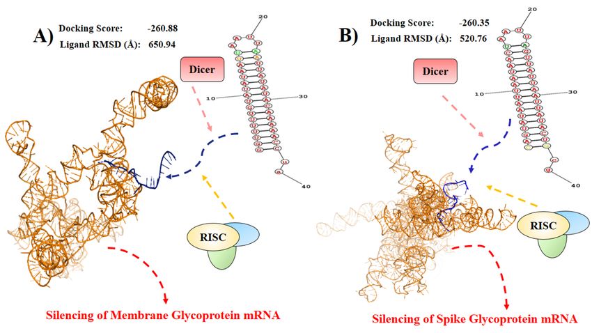
Abstract
Coronavirus disease-2019 (COVID-19) has become an immense threat to global public health. The causative
agent of this disease is a novel zoonotic pathogen called Severe Acute Respiratory Syndrome related Coronavirus-
2 (SARS-CoV-2). Since this is a newly evolved pathogen, very limited information is available to develop effective
therapeutics against this deadly virus. Although bioinformatics based analysis could be handy to unveil drugs or
vaccines against bacteria and fungus, such approaches are hardly seen for acellular viruses. However, in this
study we rationally merged several powerful in silico techniques and proposed prospective therapeutics based on
available omics data for COVID-19. Through meticulous analysis of conserved regions of 67 SARS-CoV-2 strains,
spike and membrane glycoproteins were chosen to develop and propose a chimeric vaccine against this virus.
siRNAs were also designed against these glycoprotein genes to silence them. Moreover, six drug compound
candidates were suggested to inhibit the conserved RNA-directed RNA polymerase protein. Finally, due to the close
relationship of SARS-CoV-2 and SARS-CoV, publicly available gene expression datasets of SARS-CoV were
analyzed to identify 13 immunoregulatory genes that might develop interferon based therapy. Our study will
quicken the researches among pharmaceuticals, researchers and clinicians to develop rapid therapeutics for
controlling this notorious pandemic disease.

Authors Mohammad Uzzal Hossain, Arittra Bhattacharjee, Md. Tabassum Hossain Emon, and Zeshan Mahmud
Chowdhury contributed equally to this work.

Introduction
Coronavirus Disease–2019 (COVID–19) has drastically spread throughout the globe and become a massive
threat to humanity1. Most of the healthcare system and governing bodies from developed countries failed to stop
the spreading and infection of this pandemic disease2. Regrettably, the quick prevention of COVID–19 is
challenging because the causative agent of this disease is a novel Severe Acute Respiratory Syndrome (SARS)
related Coronavirus (CoV) or SARS-CoV–2. This virus was totally unknown before the outbreak that took place in
Wuhan, China; therefore, traditional development of therapeutics will not yield a rapid solution to control this
pestilence 2,3.

CoVs, members of Coronaviridae family and Coronavirinae subfamily, were considered as insigni cant pathogens
for humans before the outbreak of SARS occurred in 2002 and 2003 from Guangdong province, China4.
Coronavirinae has been divided into four categories such as Alpha, Beta, Gamm and Deltacoronavirus4. These
pathogens were described as the “virology backwater” since very little was known about their virulence5. In recent
decades, they have become scandalous several times due to their zoonotic characteristics. Especially, the
Betacoronavirus group consists of the most perilous Human CoVs (HCoVs). For example, HCoV-OC43, HCoV-
HKU1, SARS-CoV and Middle East respiratory syndrome coronavirus (MERS-CoV) are members of
Betacoronavirus4.

Coronavirinae has vast genetic diversity among them but they mostly reside in Bat7. Therefore, this mammalian
bird always possesses a serious threat for CoV outbreaks, particularly in China8. The possible reason behind this
occurrence is that the members of Coronavirinae frequently go under RNA recombination inside bat and often
generate novel CoVs6,7. SARS-CoV–2, a member of Betacoronavirus, also originated in bats and transmitted to

                                                     Page 2/26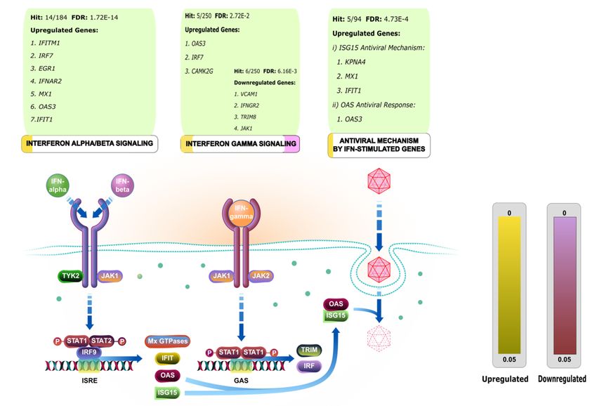
humans via unidenti ed intermediary animals in Wuhan, China. This transmission started the pandemic COVID–
19 which was instigated on December 20199.

SARS-CoV–2, a positive-stranded (+) RNA virus, spread via respiratory droplets of an infected person to a healthy
individual through sneezing and coughing3. Moreover, the presence of SARS-CoV–2 is also observed in the stool;
thereby contaminated water supply might promote transmission10. The virions enter through the upper-respiratory
tract and enter inside host by engaging Angiotensin Converting Enzyme 2 (ACE2) and Transmembrane Serine
Protease 2 (TMPRSS2) of the lung cells11. This entry triggers innate immunity as a result in ammatory cytokines
e.g., Interleukin (IL) - 2, IL–10, IL–7, Granulocyte-colony stimulating factor (GCSF), monocyte chemoattractant
protein 1 (MCP1), Macrophage In ammatory Proteins (MIP) 1A, Interferon gamma-induced protein- 10 (IP–10)
and tumor necrosis factor alpha (TNFα) get elevated9. Consequently, the patients develop indistinguishable u
like symptoms such as cough, sore throat, headache, fatigue, myalgia, breathlessness, conjunctivitis and fever. In
worst cases, patients develop pneumonia leading to respiratory failure and death12. To handle this condition,
physicians are non-speci cally applying anti-malarial Chloroquine, antiviral drug e.g. Oseltamivir, Ganciclovir,
Lopinavirritonavir etc. and plasma of the patients that recovered from COVID–199,13,14. However, this situation
demands precise therapeutics for the quick betterment of the COVID–19 patients.

A recent in vitro study revealed potential clinically proven protease inhibitor that can block TMPRSS2 and
consequently the entry of SARS-CoV–211. Similar to this study, revealing more plausible therapeutics and
druggable targets will elevate the probability to achieve successful treatments against COVID–19. In silico
approaches are the most suitable way to execute this type of research in a short time with nearly zero cost.
Powerful computational methods such as subtractive proteomics, comparative genomics or virtual screening can
disclose therapeutic targets and drugs against bacteria, fungi or non-communicable diseases15,16,17.
Unfortunately, for viral infections, this type of rational approach is hardly seen. As a result, unveiling the potential
targets, novel drugs, interferons and immunodominant vaccines are extremely di cult against COVID–19.

In this study, we merged several in silico techniques for developing treatments against COVID–19. Here, we
analyzed the genome of SARS-CoV–2 and selected several conserved regions to establish small molecules,
chimeric vaccine and Small interfering RNAs (siRNAs). Moreover, we carried out a comparative genome analysis
of SARS-CoV–2 with SARS- CoV and MERS-CoV to look for the genetic similarity. SARS-CoV showed close genetic
relation and virulence pattern with SARS-CoV–2. Thus, a transcriptomic analysis of this related virus was
performed to retrieve the interferon inducing genes for implementing interferon based therapy.

Materials And Methods
Retrieval of Whole Genomes and Conserved Region:
Public database Global Initiative on Sharing All In uenza Data (GISAID) was explored to retrieve the whole-
genomes of novel SARS CoV–218. Sixty-seven (67) whole-genomes of SARS CoV–2 were selected for this present
study (January, 2020). In quest for developing universal therapeutics, multiple sequence alignment and
construction of phylogenetic cladogram were performed using neighbor joining method in CLC Drug Discovery
Workbench (Version 3.0). For further study, consensus sequences were collected to develop a vaccine, small
molecules and SiRNAs.

                                                        Page 3/26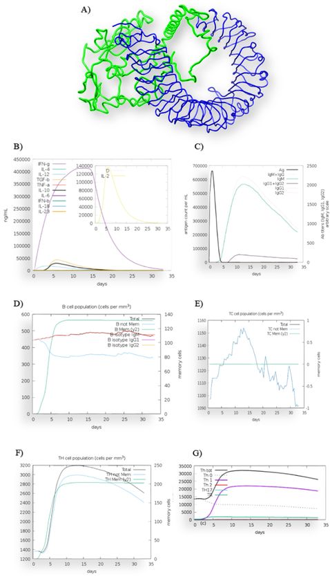
Chimeric Vaccine against SARS-CoV–2

Antigenicity analysis of the candidate proteins
The antigenicity of the selected Membrane Glycoprotein (NCBI accession:YP_009724393.1) and Spike
Glycoprotein (NCBI accession: QHR63250.2) were determined by Vaxijen 2.018 and AntigenPro19. These servers
predicted antigenicity through alignment independent methods.

Identi cation of B cell and T cell Epitopes
The outside amino acid residues of the vaccine candidates were identi ed with THMM19. The identi ed
sequences were uploaded in BepiPred–2.0 server to select the most potential B cell epitopes21. During this
selection, surface exposed amino acids were prioritized mostly. The Cytotoxic T cell (CTL), Helper T cell (HTL)
epitopes were disclosed by NetCTL 1.2 and NetMHC 4.0 respectively22,23. NetCTL 1.2 was implemented to
discover the CTL epitopes for 12 classes of Major Histocompatibility Complex 1 (MHC I) supertypes. The best CTL
epitopes were selected based on combined score. NetMHCII 2.2 was used to detect 15-mer HTL epitopes for
human HLA-DP, HLA-DQ, and HLA-DR alleles. Most potential epitopes were chosen by evaluating a nity,
percentage ranking and binding level.

Construction of Chimeric Vaccine
To construct a chimeric or multi-epitope vaccine, all the epitopes were joined with EAAK, GPGPG and AAY
linkers24. Human Beta Defensin–2 (HBD–2) (PDB ID: 1FD3) and a recombinant viral protein were added in the N
terminal and the C terminal of the vaccine respectively25,26. HBD–2 was conjugated because the protein can
activate Toll Like Receptor 4 (TLR 4) and have chemotactic activity25,27. The recombinant viral protein was added
to stimulate the antiviral responses26.

Evaluation of Immune Response and Interferon Gamma (IFNγ)
The fasta sequence of the vaccine was uploaded to an agent-based immune system simulator C-ImmSim
(http://150.146.2.1/C-IMMSIM/index.php) for measuring the immune responses28. The parameters were kept
default during this simulation. C-ImmSim showed an adequate secretion of IFNγ which was further evaluated by
IFNepitope29.

Allergenicity and Toxicity Exploration
Recombinant vaccine can initiate allergic response or lead to various types of toxicity. Therefore, evaluation of
toxicity and allergenicity is an essential step for vaccine design. Allergenicity of the vaccine was calculated by
AlgPred and AllerTop v.230,31. Toxicity was measured by ToxinPred32.

                                                      Page 4/26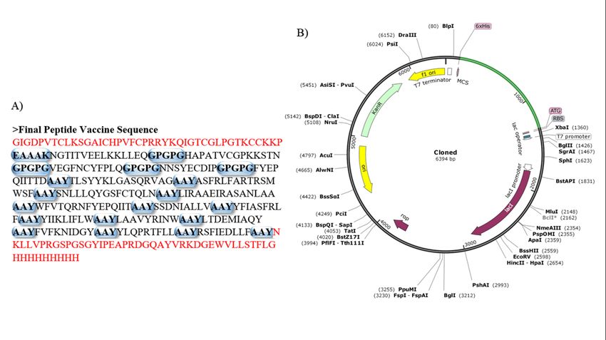
Analysis of Physicochemical Properties and Tertiary Structure
The physicochemical analysis of the protein was executed via ProtParam32. The tertiary structure was generated
through Contact-guided Iterative Threading ASSEmbly Re nement (C-I-TASSER) (17). The structure was re ned by
GalaxyRe ne35 and validated with Procheck36 and ProSAWeb37.

Molecular Docking Analysis
The crustal structure of TLR 4 was collected from Research Collaboratory for Structural Bioinformatics (RCSB)
Protein Data Bank (PDB) (www.rcsb.org). The PDB ID of the structure was 3FXI. For molecular docking analysis,
only Chain A of TLR 4 was preserved and prepared by deleting the heterogeneous atoms. This preparation was
executed by BIOVIA Discovery Studio (https://www.3dsbiovia.com/). To determine the active site pocket of Chain
A, the PDB le was uploaded in Computer Atlas of Surface Topology of Proteins (CASTp)38. Finally, the vaccine
and Chain A of TLR–4 was uploaded in ClusPro 2.0 for protein-protein docking39.

Codon Optimization and Visualization of Cloning
The vaccine sequence was reversely translated and the codons were optimized for Escherichia coli (strain K12)
through JCat40. The optimized DNA sequence was kept free from rho-independent transcription terminators and
prokaryotic ribosome binding sites. This sequence was anked by Nde I and Xho I restriction sites. Stop codons
were added at the end of 3’OH or C terminal end. After these preparations, the DNA sequence was inserted in
pET28a (+) Plasmid Vector via SnapGene tools (www.snapgene.com).

Small Molecule Therapeutics against COVID–19

Homology Modelling and Binding site analysis
The conserved RNA directed RNA polymerase (RdRp) sequence was retrieve to develop small molecules. C-I-
TASSER was employed to build the 3D model of RdRP34. Thereafter, CASTp was applied to identify the drug
binding sites that allowed to recognize the critical amino acids for drug-protein interactions38.

Screening the druggable compounds and molecular docking
simulation
DrugBank Database (www.drugbank.ca)was utilized to search for the potent drugs against RdRP. Molecular
docking of receptorand the selected compounds were performed in AutodockVina to observe the binding a nity
into the binding site of RdRp42. Here, AutoDock tools 1.5.6 was used to prepare the input pdbqt le for the
receptor. A grid box parameter were set in size with X = 70, Y = 70, Z = 36 points (center grid box:x = 23.448, y =
53.587, z = –2.012, spacing = 0.5˚A). The molecular visualization of protein-ligands were analyzed by BIOVIA
Discovery Studio and PyMol43.

                                                      Page 5/26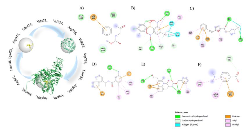
Pharmacoinformatics illustration
Osiris property explorer44, Molinspiration45, ACToR (Aggregated Computational Toxicology Resource)46,
admetSAR (absorption, distribution, metabolism, excretion, and toxicity Structure-Activity Relationship
database)47 and ACD/I-lab48 were exploited for the calculation of Absorption, distribution, metabolism, excretion
(ADME) properties and toxicity pro le. ADMET properties are necessary to establish an effective drug.

Nucleic Acid Based Therapeutics Development

Designing of potential siRNA molecules
In order to design the effective siRNA molecule, I-Score Designer was employed49. I-Score Designer computes nine
different siRNA designing scores such as Ui-Tei50, Amarzguioui51, Hsieh52‚ Takasaki53‚ s-Biopredsi54‚ i-Score55‚
Reynolds56‚ Katoh57‚ and Design of SIRna (DSIR) 58 along with other essential parameters. The server also ranks
the best siRNA molecules. From there, the best siRNA sequence was taken for further analysis. The secondary
structure of the siRNA was predicted via RNA structure webserver59. RNAfold web server was applied to calculate
the free energy of the thermodynamic ensemble for the secondary structures60. Transcription and Translation Tool
(http://biomodel.uah.es/en/lab/cybertory/analysis/trans.htm) was implemented to transcribe the viral DNA
sequences. Finally, the designated Antisense siRNAs were hybridized against viral RNA sequences using the
HNADOCK server61. HNADOCK executed RNA-RNA docking and performed molecular dynamics simulations to
re ne the best 10 predicted siRNA-mRNA complexes. The model with best docking score for Membrane
Glycoprotein mRNA and Spike Glycoprotein mRNA were visualized with PyMol.

Inteferon stimulating genes (ISGs) based Interferon Therapy

Comparative Genomics
The genome of SARS Cov–2 was compared with SARS CoV and MERS CoV. We Blasted SARS CoV–2 against
SARS CoV and MERS CoV, using Megablast and Discontiguous Megablast algorithm. The graphical
representation of side by side genome comparison is demonstrated in Artemis Comparison Tool62.

Exploring SARS-CoV expression pro le
The Gene Expression pro le (GSE5972) of SARS-CoV was collected from National center for Biotechnology
Information (NCBI). Afterwards, the normalization study was performed in between 10 Healthy samples and 54
SARS patient samples, whereas recovered cases were excluded from this study. We used limma R package to
identify the differentially expressed genes63. IDEP tools was utilized to create Hierarchical Clustering Heatmap
and Boxplot to the visualization and distribution of the data for both up and downregulated genes64.

Gene Enrichment Analysis
                                                     Page 6/26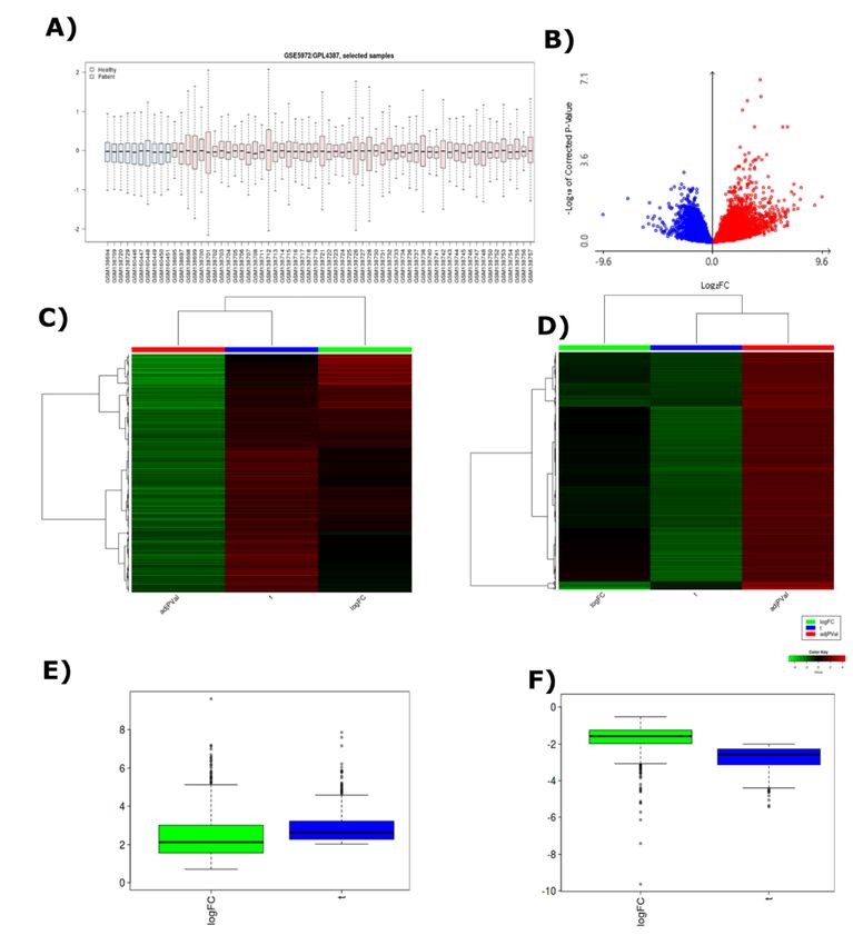
Kyoto Encyclopedia of Genes and Genomes (KEGG) and Enrichr4 were employed to identify the genes which were
involved in pathways and Gene Ontology (GO) processes such as biological process (BP), molecular function
(MF) and cellular component (CC)65–67. The genes with GO accession ID &KEGG pathways were enlisted for
further study. ShinyGO was utilized to construct a clustered tree of the top 30 GO terms for both up and
downregulated genes of BP, MF and CC68.

Identi cation of Viral Genes
We securitized the down and upregulated genes in BP, MF and CC from gene ontology dataset to nd out the viral
connected genes especially enriched viral production regulation and cytokine regulation.

Exploration of Interferon Stimulating Genes (ISGs)
INTERFEROME69 database was analyzed to recognize the Interferon Stimulating Genes (ISGs) and potential
Interferons. Further, we tried to explore the pathway of ISGs which modulate the immune system and interferon
regulation by Reactome70.

Results
Sixty seven (67) SARS-CoV-2 whole genome strains were collected from different countries (Supplementary Data
1). Multiple Sequence Alignment (MSA) and Phylogenetic Tree depicted the similarity and distant relationship of
SARS-CoV-2 among the different geographic area and populations (Supplementary Data 2 and Supplementary
Fig. 1). From these analysis, 3 consensus sequences from the whole genomes were collected (Supplementary
Data 3). The conserved sequences were translated and their functions were analyzed manually to develop
vaccine, siRNAs and drugs.

Chimeric Vaccine against SARS-CoV-2
The selected membrane glycoprotein (MG) (Vaxijen score 0.55 and AntigenPro score 0.232547) and spike
glycoprotein (SG) (Vaxijen score 0.4695 and AntigenPro score 0.717053) were predicted as “antigenic” by the
assigned programs. The MG and SG have a total of 24 and 1214 outside amino acids respectively. Among those
residues, one potential B cell epitope NGTITVEELKKLLEQ was identi ed in MG (Table 1). Four possible B-cell
epitopes (HAPATVCGPKKSTN, NNSYECDIP, FYEPQIITTD, VEGFNCYFPLQ) from the SG and only one from the MG
(NGTITVEELKKLLEQ) were found (Table 1). The selected four Cytotoxic T-cell (CTL) epitopes from MG and SG
cover HLA-A12, A24, A26, B58, B8 alleles. Moreover, Helper T-cell (HTL) epitopes can interact with HLA DRB1-0101,
DPA10103-DPB10301, DQA10101-DQB10501 and DRB1-1501 (Supplementary Table 1). The chimeric vaccine
exposed the strong binding a nity with Toll Like Receptor-4 (TLR4) (Fig. 2A). A single injection of the vaccine has
showed an adequate release of IFNγ in C-ImmSim simulator (Fig. 2B). The IFNepitope also showed 127 IFNγ
inducing epitopes by different methods. Additionally, Interleukin (IL)-2 was also released signi cantly for
lymphocyte differentiation. This differentiations are mentioned as danger signal D (Sympshon Index) (Fig. 2B).
After release of in ammatory cytokines, the immune system released anti-in ammatory IL-10 at 2.5th day and
consequently generated Immunoglobulin M (IgM) and later IgG (Fig. 2C). Generation of memory B cells was also

                                                     Page 7/26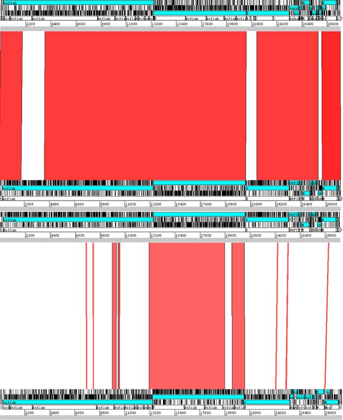
observed (Fig. 2D). The vaccine speci cally stimulated the differentiation of T helper Cell- 1 (Th 1) with certain
level of T regulatory cell (Fig. 2E). Memory Helper T Cell also released in between 2.5 days (Figs. 2F and 2G). The
vaccine does not contain any major allergenic properties according to AlgPred and AllerTop. ToxinPred identi ed
343 non-toxic peptides in different combinations and only 11 peptides with toxic properties. The vaccine is stable
according to the instability index of ProtParam (Supplementary Table 2). The estimated half-life of the protein is
30 hours in mammalian reticulocytes (in vitro), >20 hours in yeast (in vivo) and >10 hours in Escherichia coli (in
vivo) with -0.010 Grand Average of Hydropathicity (GRAVY) and 81.38 Aliphatic index (Supplementary Table 2).
The re ned three dimensional (3D) structure of the vaccine has 1.905 MolProbity score (provided by GalaxyWEB),
-2.62 Z-Score and 87.9% residues in the most favored regions of Ramachandran Plot (Supplementary Figs. 2 and
3). When this protein was docked against TLR 4 (Chain A) via ClusPro 2.0, it interacted toward the receptor via
active site pocket and HBD-2 with lowest energy score -1401.1.

Table 1: The potential B cell, Cytotoxic T cell and Helper T cell epitopes.

                      Type of Epitope   Serial No.   Epitope Sequence         Protein Name

                      B Cell epitopes   1            NGTITVEELKKLLEQ          Membrane Glycoprotein

                                        2            HAPATVCGPKKSTN           Spike Glycoprotein

                                        3            NNSYECDIP                Spike Glycoprotein

                                        4            FYEPQIITTD               Spike Glycoprotein

                                        5            VEGFNCYFPLQ              Spike Glycoprotein

                      CTL Epitopes      6            SSDNIALLV                Membrane Glycoprotein

                                        7            YFIASFRLF                Membrane Glycoprotein

                                        8            YIIKLIFLW                Membrane Glycoprotein

                                        9            LAAVYRINW                Membrane Glycoprotein

                                        10           LTDEMIAQY                Spike Glycoprotein

                                        11           FVFKNIDGY                Spike Glycoprotein

                                        12           YLQPRTFLL                Spike Glycoprotein

                                        13           RSFIEDLLF                Spike Glycoprotein

                      HTL Epitopes      14           TLSYYKLGASQRVAG          Membrane Glycoprotein

                                        15           ASFRLFARTRSMWSF          Membrane Glycoprotein

                                        16           SNLLLQYGSFCTQLN          Spike Glycoprotein

                                        17           LIRAAEIRASANLAA          Spike Glycoprotein

                                        18           WFVTQRNFYEPQIIT          Spike Glycoprotein

Codon Optimization and Cloning for chimeric vaccine
The DNA sequence of the vaccine has 1089 nucleotides that demonstrated 0.96 Codon Adaptation Index (CAI)
with 54.26% GC content. Here, the GC content of the host organism E. coli strain K12 is 50.73 (Fig. 3). This
sequence was taken under modi cations for cloning

                                                          Page 8/26
Small Molecule Drugs against COVID-19 Six experimental compounds were collected from the DrugBank. The chemical properties of the compounds showed the possibility to be suitable candidates against the RdRp (Supplementary Table 3). Further, the target protein and six compounds were selected for Molecular docking analysis to ensure the binding a nity. Strong binding a nity was observed for benzyl (2-oxopropyl)carbamate (-4.2 Kcal/mol) (Fig. 4A), 2-[(2,4-dichloro-5- methylphenyl)sulfonyl]-1,3-dinitro-5-(tri uoromethyl)benzene (-7.6 Kcal/mol) (Fig. 4B),; s-[5-(tri uoromethyl)-4h- 1,2,4-triazol-3-yl]5-(phenylethynyl)furan-2-carbothioate (-5.9 Kcal/mol) (Fig. 4C), 5-amino-2-methyl-N-[(1R)-1- naphthalen-1-ylethyl]benzamide (-6.3 Kcal/mol) (Fig. 4D), Nalpha-[(benzyloxy)carbonyl]-n-[(1r)-4-hydroxy-1-methyl- 2-oxobutyl]-l-phenylalaninamide (-7.4 Kcal/mol) (Fig. 4E), 4-(Dimethylamino)benzoic acid (-4.0 Kcal/mol) (Fig. 4F). Fifteen common amino acid residues including Val473, Glu474, Asp477, Lys478, Leu636, Val637, Arg735, Asp736, Val737, Asp304, Arg305, Arg640, His642, Arg651 and Leu648 of RdRp interacted with the druggable molecules. Later, ADMET analysis of the experimental compounds were enlisted (Supplementary Table 4). Silencing the glycoproteins of SARS-CoV-2 The Sense and Antisense RNA molecules of both MG and SG were 19mer and 21mer respectively (Supplementary Table 5 and 6). The siRNA generated hairpin like secondary structure at 310.15 K temperature. The free energy of the thermodynamic ensembles were -18.30 kcal/mol and -17.90 kcal/mol respectively for MG and SG siRNA secondary structures. Results of HNADOCK server demonstrate that the antisense siRNAs can interact with the three dimensional (3D) viral mRNA molecules with around -260 docking score (Fig. 5). Interferon Based Therapeutic for SARS-CoV-2 The whole genome of SARS-CoV and MERS-CoV were compared with SARS-CoV-2. SARS-CoV and SARS-CoV-2 share 88% query sequence coverage with 82.30% sequence identity. Besides, MERS-CoV share only 29% query coverage with 67.06% sequence identity (Supplementary Fig. 4). Genome alignment showed higher homology between SARS-CoV and SARS CoV-2 with a very few synteny break region. Whereas, MERS-CoV shares low scoring homology with SARS-CoV-2 with a larger synteny breakage area. The entry of SARS-CoV-2 and SARS-CoV are similar so differential gene expression investigation was run on SARS-CoV (Fig. 6). 64 different samples were normalized and median was centralized (Fig. 7A). 19,200 differentially expressed genes were found (Fig. 7B and Supplementary Fig. 5). Genes with o cial human gene symbol (HGNC) and have a signi cant p-value (
14), 60 MF (Supplementary Data 15) and 54 CC (Supplementary Data 16) genes with GO accession and 101
KEGG pathways (Supplementary Data 17) were enlisted as well.

The biological process of Gene Ontology (GO) reported Upregulated 91 genes and Downregulated 76 genes to be
connected with viral regulation process (Supplementary Data 18, 19, 20 and 21). The upregulated (KPNA4, IRF7,
OAS3, CAMK2G, IFITM1, IFIT1, EGR1, MX1 and IFNAR2) and the downregulated (TRIM8, VCAM1, IFNGR2 and
JAK1) genes were then identi ed to be effective to stimulate the Interferons via the pathway regulation (Fig. 8,
Supplementary Fig. 6, and Supplementary Data 21, 22, 23, 24 and 25).

Discussion
COVID-19 has become a pandemic disease and causing severe trouble in social, political and economic
stability71,72. The causative agent of this pandemic can go under frequent mutations, adaptations and
transmission that possess continuous risk of outbreaks73,74. To handle the current situation and future risks,
sustainable development of therapeutics against this deadly virus is a crying need. To address this global issue,
we have merged several computational approaches and proposed therapeutics that could be effective against all
the strains of SARS CoV-2 (Fig 1). To achieve this goal, all available whole genomes of SARS-CoV-2 from different
countries were exploited (Supplementary Data 1). Among all the strains, several consensus region was obtained
with minimal evolutionary distance (Supplementary Data 2, 3 and Supplementary Fig 1). The predicted chimeric
vaccine, drug compounds and siRNA based therapeutics were proposed on the basis of those conserved
sequences (Supplementary Data 3).

Two proteins namely Membrane and Spike Glycoprotein were identi ed through blastx of the conserved
nucleotide sequence. The vaccine candidates contain several well-conserved B and T cell epitopes (Table 1,
Supplementary Table 1). The epitopes were exploited to design a potential chimeric vaccine which is expected to
elicit TLR4 mediated Th1 antiviral response. Simulation study also showing the ability of this vaccine is producing
memory B and T cell along with the protective immune-globulins (Figs. 2 and 3). The vaccine is expected to
induce in ammatory and anti-in ammatory cytokines in su cient amounts with non-allergen activities. Finally,
since the vaccine contains very few toxic peptides; therefore, in vitro and in vivo validation are required prior to the
actual application. Again, the constructed expression vector should successfully produce adequate vaccine
molecules in E. coli strain K12 since the half-life of the protein is >10 hours inside the bacterial host with an
acceptable stability index (Fig. 3).

A universal drug is necessary with an effective vaccine to reduce the post infection consequences of COVID-19.
For coronaviruses, RNA‐directed RNA polymerase (RdRp) is an attractive therapeutic target which catalyzes the
replication of RNA from RNA template. Thus, we targeted the conserved (region 256nt-13458nt) RNA directed RNA
polymerase (RdRP) protein from SARS-CoV-2 (Supplementary Data 3). The virtual screening directed by the RdRP
protein identi ed six experimental compounds from the DrugBank server (Supplementary Table 3). Later, the
molecular docking analysis revealed a higher binding a nity between the compounds and the RdRp receptor. It is
important that all the interacting residues between drug compounds and RdRp receptor are located in the
predicted drug binding site (Fig. 4). Among the ADMET properties, all the experimental compounds exhibited the
su cient human intestinal absorption rate, a higher metabolic rate, no blood-brain barrier permeability, and
balanced amounts of drug supply to the body (Supplementay Table 4). Moreover, the toxicity levels of these
compounds appeared to be safe for the human body (Supplementay Table 4). These characteristics are

                                                       Page 10/26
signi cant for drug development because poor ADMET properties could direct a drug to be failing in phase III
clinical trials75.
RNA interference (RNAi) pathway is very e cient and speci c in silencing the viral genes just after post-
transcription. Here, siRNAs (small interfering RNAs) can play a therapeutic role76,77. The mRNA sequences have
potential sense siRNA strands CAGUAUAAUUAAUAACUAA and GGUUUUUGUAUAUAAUUAA from SG and MG
respectively (Supplementary Table 5 and 6). Therefore, 19mer and 21mer nucleotide of sense and antisense
strand were designed on the basis of siRNA universal rules. The standard thermodynamic value and strong
interaction between antisense and viral mRNA strongly supports its prospective application (Figs. 5A and 5B).
These siRNAs might be conjugated with aptamer or delivered through various delivery systems to inhibit the
intracellular growth of SARS-CoV-2.
Interferons (IFNs) instigate an array of antiviral effectors. Every virus contains a distinctive property, however,
partly overlapping antiviral Interferon Stimulating Genes or "ISG pro le" could be potential target. IFN effectors
target various phases of the virus life cycle to interfere with viral infection and proliferation. To identify the ISG
and IFN of COVID-19, the gene expression pro le of COVID-19 patients was needed. Due to a lack of the
expression pro le of COVID-19 till January 2020, three genomes of Betacoronavirus were taken including SARS-
CoV, MERS-CoV and SARS-CoV-2, which have common virulence patterns, sign and symptoms. A comparison of
their genome was performed. The whole genome comparison between SARS-CoV and SARS-CoV-2 showed 88%
similarity with each other (Fig. 6 and Supplementary Fig 4). Therefore, the SARS CoV expression pro le was
explored for the identi cation of ISG and IFNs for the IFNs based therapeutics development. On the basis of this
connection, we analyzed the genome-wide expression pro le of SARS-CoV to identify the ISG and IFNs which
might be suitable candidates for the IFNs based therapeutic development (Supplementary Data 4, 5, 6, 7, 8 and 9).
Among the up and down regulated genes, a total of 167 immunoregulatory genes (Upregulated 91 genes and
Downregulated 76 genes) were found from the biological process of Gene Ontology (GO) (Fig. 7, Supplementary
Fig. 5, Supplementary Data 9, 10, 11, 12, 13, 14, 15, 16 and 17). We then identi ed the Interferon stimulating
genes KPNA4, IRF7, OAS3, CAMK2G, IFITM1, IFIT1, EGR1, MX1 and IFNAR2 from the upregulated
immunoregulatory genes and TRIM8, VCAM1, IFNGR2, JAK1 from the downregulated immunoregulatory genes
(Fig.8). These immunoregulatory genes can prompt the interferon IFN alpha, IFN beta and IFN gamma which then
directly gets involved to induce the immunological response by regulating the pathways (Fig. 8, Supplementary
Fig. 6, Supplementary Data 17, 18, 19, 20, 21, 22, 23, 24 and 25). This analysis gives a deep insight for the IFN
based therapy during COVID-19.
The pandemic coronavirus disease 2019 (COVID-19) caused by severe acute respiratory syndrome coronavirus 2
(SARS-CoV-2) is of great global public health concern due to its spreading rate in every part of this world with
increasing death rate that ultimately has led to global socioeconomic disruption. The global urgency for the
antiviral therapy against the SARS-CoV-2 has been the superior priority of this world to survive the existence of
human as no medication is available so far. The present study proposed all possible novel therapeutics such as
chimeric vaccine, potent drug compounds, effective siRNA and interferons to the purpose of shortening the time
and reduce the cost for effective medication discovery against the global pandemic COVID-19. With the aid of
computational biology and Bioinformatics analyses, these therapeutics con rmed its effectiveness against the
COVID-19 which further offer for the wet lab validation. Lastly, we systematically proposed the possible
therapeutics in the hope of providing a reference to minimize the longer time required antiviral therapy in wet lab
and help for the prevention as well as control of the COVID-19 pandemic.
Page 11/26Conclusion
The present study has proposed four types of therapeutics including chimeric vaccine, potent drugs, siRNAs and
ISG targets against COVID–19. We have analyzed all the genomic, transcriptomic and proteomic data to
anticipate the possible therapeutics against the recent pandemic COVID–19. Therefore, these ndings might be of
interest to the researchers and pharmaceuticals company to discover the novel therapeutics against the SARS-
CoV–2. We hope this in silico pipeline will also help many researchers to combat against other viral outbreaks in
future.
Declarations
Acknowledgements
The Authors are grateful to Ministry of Science and Technology for the extensive support during this work.
Author Contributions
MS: Conceived, designed, and guided the study, analyzed the data, drafting the manuscript and performed critical
revision. CAK and KCD: Guided the study, acquisition and analyzed the data, helped in drafting the manuscript.
MM and MGM: Helped in Bioinformatics analysis and helped in drafting the manuscript. MUH, AB, MTHE, ZMC:
Helped to design the study, performed bioinformatics analysis, drafted, and developed the manuscript and
performed critical revision. All authors have approved the manuscript.
Competing Interest
The authors declare that they have competing interest on proposed therapeutics.
Funding
There is no funding source for this work
References
1. WHO. Naming the coronavirus disease (COVID-19) and the virus that causes it 2029 (2019).
2. Lai, C.C., Shih, T.P., Ko, W.C., Tang, H.J. and Hsueh, P.R., Severe acute respiratory syndrome coronavirus 2
(SARS-CoV-2) and corona virus disease-2019 (COVID-19): the epidemic and the challenges. International
journal of antimicrobial agents, p.10592 (2020).
3. Wu, Y.C., Chen, C.S. and Chan, Y.J., The outbreak of COVID-19: An overview. Journal of the Chinese Medical
Association, 83(3), pp.217-220 (2020).
4. Fung et al. Human Coronavirus: Host-Pathogen Interaction. Annual review of microbiology, 73, pp.529-557
(2019).
5. Cavanagh D. Coronaviridae: a review of coronaviruses and toroviruses. InCoronaviruses with Special
Emphasis on First Insights Concerning SARS (pp. 1-54). Birkhäuser Basel(2005).
6. Cui, J., Li, F. & Shi, Z. Origin and evolution of pathogenic coronaviruses. Nat Rev Microbiol 17, 181–192
(2019).
Page 12/267. Lau, S.K., Wong, A.C., Zhang, L., Luk, H.K., Kwok, J.S., Ahmed, S.S., Cai, J.P., Zhao, P.S., Teng, J.L., Tsui, S.K.
and Yuen, K.Y., Novel Bat Alphacoronaviruses in Southern China Support Chinese Horseshoe Bats as an
Important Reservoir for Potential Novel Coronaviruses. Viruses, 11(5), p.423 (2019).
8. Fan, Y., Zhao, K., Shi, Z.L. and Zhou, P.,. Bat Coronaviruses in China. Viruses, 11(3), p.210 (2019).
9. Wu, Z. and McGoogan, J.M., 2020. Characteristics of and important lessons from the coronavirus disease
(COVID-19) outbreak in China: summary of a report of 72 314 cases from the Chinese Center for Disease
Control and Prevention. Jama (2019).
10. Tang, A., Tong, Z.D., Wang, H.L., Dai, Y.X., Li, K.F., Liu, J.N., Wu, W.J., Yuan, C., Yu, M.L., Li, P. and Yan, J.B.,
Detection of Novel Coronavirus by RT-PCR in Stool Specimen from Asymptomatic Child, China. Emerging
infectious diseases, 26(6) (2020).
11. Hoffmann, M., Kleine-Weber, H., Schroeder, S., Krüger, N., Herrler, T., Erichsen, S., Schiergens, T.S., Herrler, G.,
Wu, N.H., Nitsche, A. and Müller, M.A., SARS-CoV-2 cell entry depends on ACE2 and TMPRSS2 and is blocked
by a clinically proven protease inhibitor. Cell (2020).
12. Chen, N., Zhou, M., Dong, X., Qu, J., Gong, F., Han, Y., Qiu, Y., Wang, J., Liu, Y., Wei, Y. and Yu, T., Epidemiological
and clinical characteristics of 99 cases of 2019 novel coronavirus pneumonia in Wuhan, China: a descriptive
study. The Lancet, 395(10223), pp.507-513 (2020).
13. Gao, J., Tian, Z. and Yang, X., Breakthrough: Chloroquine phosphate has shown apparent e cacy in
treatment of COVID-19 associated pneumonia in clinical studies. BioScience Trends (2020).
14. Cascella, M., Rajnik, M., Cuomo, A., Dulebohn, S.C. and Di Napoli, R., Features, Evaluation and Treatment
Coronavirus (COVID-19). In StatPearls [Internet]. StatPearls Publishing (2020).
15. Hossain, M.U., Omar, T.M., Alam, I., Das, K.C., Mohiuddin, A.K.M., Keya, C.A. and Salimullah, M., Pathway
based therapeutic targets identi cation and development of an interactive database CampyNIBase of
Campylobacter jejuni RM1221 through non-redundant protein dataset. PloS one, 13(6) (2018).
16. Abadio, A.K.R., Kioshima, E.S., Teixeira, M.M., Martins, N.F., Maigret, B. and Felipe, M.S.S., Comparative
genomics allowed the identi cation of drug targets against human fungal pathogens. BMC genomics, 12(1),
p.75 (2011).
17. Bhattacharjee, A., Hossain, M.U., Chowdhury, Z.M., Rahman, S.A., Bhuyan, Z.A., Salimullah, M. and Keya, C.A.,
Insight of Druggable Cannabinoids against Estrogen Receptor β in Breast Cancer. Journal of Biomolecular
Structure and Dynamics, (just-accepted), pp.1-12 (2020).
18. Doytchinova, I.A. and Flower, D.R., Identifying candidate subunit vaccines using an alignment-independent
method based on principal amino acid properties. Vaccine, 25(5), pp.856-866 (2007).
19. Magnan, C.N., Zeller, M., Kayala, M.A., Vigil, A., Randall, A., Felgner, P.L. and Baldi, P., High-throughput
prediction of protein antigenicity using protein microarray data. Bioinformatics, 26(23), pp.2936-2943 (2010).
20. Krogh, A., Larsson, B., Von Heijne, G. and Sonnhammer, E.L., Predicting transmembrane protein topology with
a hidden Markov model: application to complete genomes. Journal of molecular biology, 305(3), pp.567-580
(2001).
21. Jespersen, M.C., Peters, B., Nielsen, M. and Marcatili, P., BepiPred-2.0: improving sequence-based B-cell
epitope prediction using conformational epitopes. Nucleic acids research, 45(W1), pp.W24-W29 (2017).
22. Larsen, M.V., Lundegaard, C., Lamberth, K., Buus, S., Lund, O. and Nielsen, M., Large-scale validation of
methods for cytotoxic T-lymphocyte epitope prediction. BMC bioinformatics, 8(1), p.424 (2007).
Page 13/2623. Andreatta, M. and Nielsen, M., Gapped sequence alignment using arti cial neural networks: application to the
MHC class I system. Bioinformatics, 32(4), pp.511-517 (2016).
24. Shey, R.A., Ghogomu, S.M., Esoh, K.K., Nebangwa, N.D., Shintouo, C.M., Nongley, N.F., Asa, B.F., Ngale, F.N.,
Vanhamme, L. and Souopgui, J., In-silico design of a multi-epitope vaccine candidate against onchocerciasis
and related larial diseases. Scienti c reports, 9(1), pp.1-18 (2019).
25. Yu, L., Wang, L. and Chen, S., Endogenous toll‐like receptor ligands and their biological signi cance. Journal
of cellular and molecular medicine, 14(11), pp.2592-2603 (2010).
26. Li, J., Ulitzky, L., Silberstein, E., Taylor, D.R. and Viscidi, R., Immunogenicity and protection e cacy of
monomeric and trimeric recombinant SARS coronavirus spike protein subunit vaccine candidates. Viral
immunology, 26(2), pp.126-132 (2013).
27. Niyonsaba, F., Ogawa, H. and Nagaoka, I., Human β‐defensin‐2 functions as a chemotactic agent for tumour
necrosis factor‐α‐treated human neutrophils. Immunology, 111(3), pp.273-281 (2004).
28. Rapin, N., Lund, O., Bernaschi, M. and Castiglione, F., Computational immunology meets bioinformatics: the
use of prediction tools for molecular binding in the simulation of the immune system. PLoS One, 5(4) (2010).
29. Dhanda, S.K., Vir, P. and Raghava, G.P., Designing of interferon-gamma inducing MHC class-II binders. Biology
direct, 8(1), p.30 (2013).
30. Saha, S. and Raghava, G.P.S., AlgPred: prediction of allergenic proteins and mapping of IgE epitopes. Nucleic
acids research, 34(suppl_2), pp.W202-W209 (2006).
31. Dimitrov, I., Bangov, I., Flower, D.R. and Doytchinova, I., AllerTOP v. 2—a server for in silico prediction of
allergens. Journal of molecular modeling, 20(6), p.2278 (2014).
32. Gupta, S., Kapoor, P., Chaudhary, K., Gautam, A., Kumar, R., Raghava, G.P. and Open Source Drug Discovery
Consortium, In silico approach for predicting toxicity of peptides and proteins. PloS one, 8(9) (2013).
33. Gasteiger, E., Hoogland, C., Gattiker, A., Wilkins, M.R., Appel, R.D. and Bairoch, A., Protein identi cation and
analysis tools on the ExPASy server. In The proteomics protocols handbook (pp. 571-607). Humana press
(2005).
34. Zheng, W., Li, Y., Zhang, C., Pearce, R., Mortuza, S.M. and Zhang, Y., Deep‐learning contact‐map guided protein
structure prediction in CASP13. Proteins: Structure, Function, and Bioinformatics, 87(12), pp.1149-1164
(2019).
35. Heo, L., Lee, H. and Seok, C., GalaxyRe neComplex: Re nement of protein-protein complex model structures
driven by interface repacking. Scienti c reports, 6, p.32153 (2016).
36. Laskowski, R.A., MacArthur, M.W. and Thornton, J.M., PROCHECK: validation of protein-structure coordinates
(2006).
37. Wiederstein, M. and Sippl, M.J., ProSA-web: interactive web service for the recognition of errors in three-
dimensional structures of proteins. Nucleic acids research, 35(suppl_2), pp.W407-W410 (2007).
38. Tian, W., Chen, C., Lei, X., Zhao, J. and Liang, J., CASTp 3.0: computed atlas of surface topography of
proteins. Nucleic acids research, 46(W1), pp.W363-W367 (2018).
39. Kozakov, D., Hall, D.R., Xia, B., Porter, K.A., Padhorny, D., Yueh, C., Beglov, D. and Vajda, S., The ClusPro web
server for protein–protein docking. Nature protocols, 12(2), p.255 (2017).
40. Grote, A., Hiller, K., Scheer, M., Münch, R., Nörtemann, B., Hempel, D.C. and Jahn, D., JCat: a novel tool to adapt
codon usage of a target gene to its potential expression host. Nucleic acids research, 33(suppl_2), pp.W526-
Page 14/26W531 (2005).
41. Williams, C.J., Headd, J.J., Moriarty, N.W., Prisant, M.G., Videau, L.L., Deis, L.N., Verma, V., Keedy, D.A., Hintze,
B.J., Chen, V.B. and Jain, S., MolProbity: More and better reference data for improved all‐atom structure
validation. Protein Science, 27(1), pp.293-315 (2018).
42. Agrawal, S., Gupta, D. and Panda, S.K., The 3′ end of hepatitis E virus (HEV) genome binds speci cally to the
viral RNA-dependent RNA polymerase (RdRp). Virology, 282(1), pp.87-101 (2001).
43. Seeliger, D. and de Groot, B.L., Ligand docking and binding site analysis with PyMOL and Autodock/Vina.
Journal of computer-aided molecular design, 24(5), pp.417-422 (2010).
44. Sander, T., OSIRIS property explorer. Organic Chemistry Portal (2001).
45. Kumar Mishra, N. and PS Raghava, G., Prediction of speci city and cross-reactivity of kinase inhibitors.
Letters in Drug Design & Discovery, 8(3), pp.223-228 (2011).
46. Judson, R., Richard, A., Dix, D., Houck, K., Elloumi, F., Martin, M., Cathey, T., Transue, T.R., Spencer, R. and Wolf,
M., ACToR—aggregated computational toxicology resource. Toxicology and applied pharmacology, 233(1),
pp.7-13 (2008).
47. Cheng, F., Li, W., Zhou, Y., Shen, J., Wu, Z., Liu, G., Lee, P.W. and Tang, Y., admetSAR: a comprehensive source
and free tool for assessment of chemical ADMET properties (2012).
48. Masunov, A., ACD/I-Lab 4.5: an internet service review. Journal of chemical information and computer
sciences, 41(4), pp.1093-1095 (2001).
49. Ichihara, M., Murakumo, Y., Masuda, A., Matsuura, T., Asai, N., Jijiwa, M., Ishida, M., Shinmi, J., Yatsuya, H.,
Qiao, S. and Takahashi, M., Thermodynamic instability of siRNA duplex is a prerequisite for dependable
prediction of siRNA activities. Nucleic acids research, 35(18), p.e123 (2007).
50. Ui-Tei, K., Naito, Y., Zenno, S., Nishi, K., Yamato, K., Takahashi, F., Juni, A. and Saigo, K., Functional dissection
of siRNA sequence by systematic DNA substitution: modi ed siRNA with a DNA seed arm is a powerful tool
for mammalian gene silencing with signi cantly reduced off-target effect. Nucleic acids research, 36(7),
pp.2136-2151 (2008).
51. Amarzguioui, M. and Prydz, H., An algorithm for selection of functional siRNA sequences. Biochemical and
biophysical research communications, 316(4), pp.1050-1058 (2004).
52. Hsieh, A.C., Bo, R., Manola, J., Vazquez, F., Bare, O., Khvorova, A., Scaringe, S. and Sellers, W.R., A library of
sirna duplexes targeting the phosphoinositide 3‐kinase pathway: determinants of gene silencing for use in
cell‐based screens. Nucleic acids research, 32(3), pp.893-901 (2004).
53. Takasaki, S., Kotani, S. and Konagaya, A., An effective method for selecting siRNA target sequences in
mammalian cells. Cell cycle, 3(6), pp.788-793 (2004).
54. Huesken, D., Lange, J., Mickanin, C., Weiler, J., Asselbergs, F., Warner, J., Meloon, B., Engel, S., Rosenberg, A.,
Cohen, D. and Labow, M., Design of a genome-wide siRNA library using an arti cial neural network. Nature
biotechnology, 23(8), pp.995-1001 (2005).
55. Ichihara, M., Murakumo, Y., Masuda, A., Matsuura, T., Asai, N., Jijiwa, M., Ishida, M., Shinmi, J., Yatsuya, H.,
Qiao, S. and Takahashi, M., Thermodynamic instability of siRNA duplex is a prerequisite for dependable
prediction of siRNA activities. Nucleic acids research, 35(18), p.e123 (2007).
56. Reynolds, A., Leake, D., Boese, Q., Scaringe, S., Marshall, W.S. and Khvorova, A., Rational siRNA design for
RNA interference. Nature biotechnology, 22(3), pp.326-330 (2004).
Page 15/2657. Katoh, T. and Suzuki, T., Speci c residues at every third position of siRNA shape its e cient RNAi activity.
Nucleic acids research, 35(4), p.e27 (2007).
58. Vert, J.P., Foveau, N., Lajaunie, C. and Vandenbrouck, Y., An accurate and interpretable model for siRNA
e cacy prediction. BMC bioinformatics, 7(1), p.520 (2006).
59. Mathews, D.H. and Turner, D.H., Prediction of RNA secondary structure by free energy minimization. Current
opinion in structural biology, 16(3), pp.270-278 (2006).
60. Mathews, D.H., Disney, M.D., Childs, J.L., Schroeder, S.J., Zuker, M. and Turner, D.H., Incorporating chemical
modi cation constraints into a dynamic programming algorithm for prediction of RNA secondary structure.
Proceedings of the National Academy of Sciences, 101(19), pp.7287-7292 (2004).
61. He, J., Wang, J., Tao, H., Xiao, Y. and Huang, S.Y., HNADOCK: a nucleic acid docking server for modeling
RNA/DNA–RNA/DNA 3D complex structures. Nucleic acids research, 47(W1), pp.W35-W42 (2019).
62. Carver, T.J., Rutherford, K.M., Berriman, M., Rajandream, M.A., Barrell, B.G., Parkhill, J., ACT: the Artemis
Comparison Tool. Bioinformatics, 21(16):3422-3 (2005).
63. Ritchie, M.E., Phipson, B., Wu, D.I., Hu, Y., Law, C.W., Shi, W. and Smyth, G.K., limma powers differential
expression analyses for RNA-sequencing and microarray studies. Nucleic acids research, 43(7), pp.e47-e47
(2015).
64. Ge, S.X., Son, E.W. and Yao, R., iDEP: an integrated web application for differential expression and pathway
analysis of RNA-Seq data. BMC bioinformatics, 19(1), p.534 (2018).
65. Sherman, B.T., Tan, Q., Collins, J.R., Alvord, W.G., Roayaei, J., Stephens, R., Baseler, M.W., Lane, H.C. and
Lempicki, R.A., The DAVID Gene Functional Classi cation Tool: a novel biological module-centric algorithm to
functionally analyze large gene lists. Genome biology, 8(9), p.R183 (2007).
66. Kanehisa, M. and Goto, S., KEGG: kyoto encyclopedia of genes and genomes. Nucleic acids research, 28(1),
pp.27-30 (2000).
67. Kuleshov, M.V., Jones, M.R., Rouillard, A.D., Fernandez, N.F., Duan, Q., Wang, Z., Koplev, S., Jenkins, S.L.,
Jagodnik, K.M., Lachmann, A. and McDermott, M.G., Enrichr: a comprehensive gene set enrichment analysis
web server 2016 update. Nucleic acids research, 44(W1), pp.W90-W97 (2016).
68. Ge, S. and Jung, D., ShinyGO: a graphical enrichment tool for animals and plants. Biorxiv, p.315150 (2018).
69. Samarajiwa, S.A., Forster, S., Auchettl, K. and Hertzog, P.J., INTERFEROME: the database of interferon
regulated genes. Nucleic acids research, 37(suppl_1), pp.D852-D857 (2009).
70. Jassal, B., Matthews, L., Viteri, G., Gong, C., Lorente, P., Fabregat, A., Sidiropoulos, K., Cook, J., Gillespie, M.,
Haw, R. and Loney, F., The reactome pathway knowledgebase. Nucleic acids research, 48(D1), pp.D498-D503
(2020).
71. Ren, S. Y., Gao, R. D., & Chen, Y. L., Fear can be more harmful than the severe acute respiratory syndrome
coronavirus 2 in controlling the corona virus disease 2019 epidemic. World journal of clinical cases, 8(4),
652–657 (2020).
72. Berera, D., & Zambon, M., Antivirals in the 2009 pandemic--lessons and implications for future strategies.
In uenza and other respiratory viruses, 7 Suppl 3(Suppl 3), 72–79. (2013).
73. Andersen, K.G., Rambaut, A., Lipkin, W.I., Holmes, E.C. and Garry, R.F., The proximal origin of SARS-CoV-
2. Nature Medicine, pp.1-3 (2020).
Page 16/2674. Gurwitz, D., Angiotensin receptor blockers as tentative SARS‐CoV‐2 therapeutics. Drug Development Research
(2020).
75. Ju, S., Tardiff, D.F., Han, H., Divya, K., Zhong, Q., Maquat, L.E., Bosco, D.A., Hayward, L.J., Brown, R.H.,
Lindquist, S., Ringe, D., Petsko, G.A., A yeast model of FUS/TLS-dependent cytotoxicity. PLoS Biol,
Apr;9(4):e1001052 (2011)
76. Fire, A., Xu, S., Montgomery, M.K., et al. Potent and speci c genetics interference by double stranded RNA in
Caenorhabitis elegans. Nature, 391, 806-11 (1998).
77. Elbashir, S.M., et al., Duplexes of 21-nucleotide RNAs mediate RNA interference in cultured mammalian cells.
Nature 411, 494–498 (2001).
Figures
Page 17/26Figure 1
Overview the possible therapeutics mechanism against the pandemic-COVID-19. Here, the designed chimeric
vaccine could prompt the immune response against the SARS-CoV-2 by targeting membrane and spike
glycoprotein. Proposed drug compounds can inhibit the replication of viral RNA by targeting RNA dependent RNA
polymerase (RdRp) protein. The designed siRNAs can silence the activity of both membrane and spike
glycoproteins. Moreover, the plausible Interferon stimulating genes (ISGs) can be targeted to induce the
interferons against the viral proteins of SARS-CoV-2. Note: The designations employed and the presentation of the
material on this map do not imply the expression of any opinion whatsoever on the part of Research Square
Page 18/26concerning the legal status of any country, territory, city or area or of its authorities, or concerning the delimitation
of its frontiers or boundaries. This map has been provided by the authors.
Figure 2
Innate and adaptive immune responses from the proposed vaccine. (A) The predicted interaction of the vaccine
with Toll Like Receptor-4 (TLR4). Protein-Protein docking analysis demonstrated that the vaccine can interact with
TLR4. The constructed vaccine was injected in the immune system simulator. (B) The interaction consequently
released in ammatory and anti-in ammatory cytokines. Here, the D (Simpson index) represents the diversity of T
cell population. Moreover, (C) IgM and IgG containing B cells produced with (D) generation of Memory B cell (E)
Population of Cytotoxic T cell spiked and (F) Memory Helper T cell produced (G) The immune response stimulated
Page 19/26Th1 cells with Regulatory T cells. The immune system produced memory B cell, memory T cell, IgM and IgG.
Moreover, the induction of Th1 cells and regulatory T cells were observed.
Figure 3
Production of the vaccine can be achieved through recombinant DNA technology. (A) The primary sequence of the
chimeric vaccine. The red letters represent adjuvants and the bold capsules depict the peptide linkers. The
adjuvants were conjugated to elicit TLR 4 mediated Th1 speci c antiviral responses. The epitopes were joined
with amino acid linkers. This sequence was reversely translated and (B) The DNA sequence of was inserted in
pET-28a(+) expression vector for vaccine production in E.coli strain K12.
Page 20/26Figure 4
Drug-receptor interactions. A) benzyl (2-oxopropyl) carbamate showed the binding energy -4.2 Kcal/mol and their
interacting amino acid residues are ASP481, ASP477, GLU474, ARG640; B) 2-[(2,4-dichloro-5-
methylphenyl)sulfonyl]-1,3-dinitro-5-(tri uoromethyl)benzene showed the binding energy -7.6 Kcal/mol with
ASP304, ARG305, VAL473, ASP477, LEU636, VAL637, ARG640, HIS642 amino acid interactions ;C) s-[5-
(tri uoromethyl)-4h-1,2,4-triazol-3-yl]5-(phenylethynyl)furan-2-carbothioate docked RdRp protein with the binding
energy -5.9 Kcal/mol and their interacting residues are ASP304, ARG305, ASP477, ARG735, ASP736, ARG640,
VAL737; D) 5-amino-2-methyl-N-[(1R)-1-naphthalen-1-ylethyl]benzamide and RdRp protein had the docking energy
about -6.3 Kcal/mol with the interacting amino acid residues ASP304, ARG305, ASP477, ARG640, VAL473,
LYS478; E) Nalpha-[(benzyloxy)carbonyl]-n-[(1r)-4-hydroxy-1-methyl-2-oxobutyl]-l-phenylalaninamide had the
docking enrgy-7.4 Kcal/mol and interacting residues ARG305, ASP477, GLU474, ARG640, ARG651, LEU648; F) 4-
(Dimethylamino)benzoic acid also showed the signi cant binding energy -4.0 Kcal/mol with ARG305, ASP477,
ARG640, VAL637. The most common interacting residues are Val473, Glu474, Asp477, Lys478, Leu636, Val637,
Arg735, Asp736, Val737, Asp304, Arg305, Arg640, His642, Arg651 and Leu648 and all of these docked into the
binding site of the RdRp protein. Here, the interactions type has been marked with different color code.
Page 21/26Figure 5
Predicted pathway for siRNA based viral gene silencing. In cytosolic environment the Dicer and RNA-induced
silencing complex (RISC) might degrade the viral mRNAs. Here, (A) the guiding anti sense strand bonded with
membrane glycoprotein mRNA (B) and spike glycoprotein mRNA. The guiding anti sense strand is very speci c to
the glycoproteins to silence their activity.
Page 22/26Figure 6
The whole genome comparison among theSARS-CoV, SARS-CoV-2 and MERS CoV. MERS seems to be distantly
related as it showed only 29% similarity with the SARS-CoV-2. Besides, MERS CoV shared 88% similarity with the
newly evolved SARS-CoV-2 virus. The variation found only in ORF1ab, Spike and ORF8 region of SARS-CoV-2 with
SARS-CoV. Alternatively, orf1ab region from MERS is similar to SARS-CoV-2.
Page 23/26Figure 7
Analysis of Differential Gene Expression. A) Median centralized 10 healthy and 54 SARS patient samples for
comparison with limma normalization method. B) LogFC against adjusted normalized p-value modelled for Gene
distribution in Volcano Plot. Hierarchical Clustering Heatmap of C) Upregulated and D) Downregulated Genes.
Distance was based on Correlation and Z score in where cut off was set to 4. Distance based on Correlation & Z
score Cut off was set to 4. Frequency distribution of LogFC and T value of E) upregulated and F) downregulated
genes.
Page 24/26Figure 8
Interferon stimulating genes (ISGs) in pathway. Overrepresented pathway of interferon regulation by
overexpressed and under expressed ISGs. The gene list including KPNA4, IRF7, OAS3, CAMK2G, IFITM1, IFIT1,
EGR1, MX1 and IFNAR2 from the upregulated genes and TRIM8, VCAM1, IFNGR2 and JAK1 from the
downregulated genes might stimulate the Interferon against the SARS-CoV-2. Here, the possible pathway
mechanism has been explored to induce the immune response by the Interferon stimulating genes (ISGs).
Supplementary Files
This is a list of supplementary les associated with this preprint. Click to download.
SupplementaryData3.Consensusandconservedregionof67wholegenomesofSARSCoV2ofdifferentcountries.txt
SupplementaryData5.DifferentiallyExpressedgeneswithHGNCsymbolsofSARSCoV.csv
SupplementaryFig.4.docx
SupplementaryData8.HierarchicalClusteringHeatmapofUpregulatedgenes.csv
SupplementaryData9.HierarchicalClusteringHeatmapofDownregulatedregulatedgenes.csv
SupplementaryData1.67wholegenomesofSARSCoV2ofdifferentcountries.txt.txt
SupplementaryFig.3.docx
Page 25/26SupplementaryFig.1.docx
SupplementaryData7.DifferentiallyExpresseddownregulatedgenesofSARSCoV.csv
SupplementaryData4.DifferentiallyExpressedgenesofSARSCoV.csv
SupplementaryData6.DifferentiallyExpressedUpregulatedgenesofSARSCoV.csv
SupplementaryFig.2.docx
SupplementaryFig.5.docx
SupplementaryData2.Multiplesequencealignmentof67wholegenomesofSARSCoV2fromdifferentcountries.pdf
SupplementaryTable4.docx
SupplementaryData15.Molecularfunctionofdownregulatedgenes.csv
SupplementaryData21.InterferonstimulatinggenesISGsfromdownregulatedgenes.csv
SupplementaryData20.InterferonstimulatinggenesISGsfromupregulatedgenes.csv
SupplementaryData22.Interferonlinkedupregulatedgenes.csv
SupplementaryData23.Interferonlinkeddownregulatedgenes.csv
SupplementaryData24.InterferonlinkedISGgenesfromupregulatedgenes.csv
SupplementaryData25.InterferonlinkedISGgenesfromdownregulatedgenes.csv
SupplementaryTable5.docx
SupplementaryData14.Biologicalprocessofdownregulatedgenes.csv
SupplementaryTable6.docx
SupplementaryData16.Cellularcomponentofdownregulatedgenes.csv
SupplementaryData11.MolecularFunctionofupregulatedgenes.csv
SupplementaryData17.KEGGpathwayofdownregulatedgenes.csv
SupplementaryData18.Viralregulatoryupregulatedgenes.csv
SupplementaryData19.Viralregulatoryupregulatedgenes.csv
SupplementaryData13.KEGGpathwayofupregulatedgenes.csv
SupplementaryData10.Biologicalprocessofupregulatedgenes.csv
SupplementaryTable1.docx
SupplementaryTable2.docx
SupplementaryTable3.docx
SupplementaryFig.6.docx
SupplementaryData12.Cellularcomponentofupregulatedgenes.csv
Page 26/26You can also read