The Development of Ovine Gastric and Intestinal Organoids for Studying Ruminant Host-Pathogen Interactions
←
→
Page content transcription
If your browser does not render page correctly, please read the page content below
ORIGINAL RESEARCH
                                                                                                                                         published: 08 September 2021
                                                                                                                                        doi: 10.3389/fcimb.2021.733811

                                             The Development of Ovine Gastric
                                             and Intestinal Organoids for Studying
                                             Ruminant Host-Pathogen Interactions
                                             David Smith 1*†, Daniel R. G. Price 2†, Alison Burrells 1, Marc N. Faber 1, Katie A. Hildersley 1,3,
                                             Cosmin Chintoan-Uta 4, Ambre F. Chapuis 2,4, Mark Stevens 4, Karen Stevenson 2,
                                             Stewart T. G. Burgess 2, Elisabeth A. Innes 1, Alasdair J. Nisbet 2 and Tom N. McNeilly 1
                            Edited by:
                                             1 Department of Disease Control, Moredun Research Institute, Midlothian, United Kingdom, 2 Department of Vaccines
                   Tiago W. P. Mineo,
Federal University of Uberlandia, Brazil     and Diagnostics, Moredun Research Institute, Midlothian, United Kingdom, 3 Institute of Biodiversity, Animal Health and
                                             Comparative Medicine, University of Glasgow, Glasgow, United Kingdom, 4 The Roslin Institute, University of Edinburgh,
                         Reviewed by:        Midlothian, United Kingdom
                      Constance Finney,
         University of Calgary, Canada
                         Camila Coelho,      Gastrointestinal (GI) infections in sheep have significant implications for animal health,
La Jolla Institute for Immunology (LJI),
                                             welfare and productivity, as well as being a source of zoonotic pathogens. Interactions
                          United States
                                             between pathogens and epithelial cells at the mucosal surface play a key role in determining
                   *Correspondence:
                          David Smith        the outcome of GI infections; however, the inaccessibility of the GI tract in vivo significantly
               d.smith@moredun.ac.uk         limits the ability to study such interactions in detail. We therefore developed ovine epithelial
 †
  These authors share first authorship        organoids representing physiologically important gastric and intestinal sites of infection,
                                             specifically the abomasum (analogous to the stomach in monogastrics) and ileum. We show
                    Specialty section:
          This article was submitted to      that both abomasal and ileal organoids form self-organising three-dimensional structures
                      Parasite and Host,     with a single epithelial layer and a central lumen that are stable in culture over serial passage.
                a section of the journal
               Frontiers in Cellular and
                                             We performed RNA-seq analysis on abomasal and ileal tissue from multiple animals and on
                 Infection Microbiology      organoids across multiple passages and show the transcript profile of both abomasal and
            Received: 30 June 2021           ileal organoids cultured under identical conditions are reflective of the tissue from which they
         Accepted: 13 August 2021
                                             were derived and that the transcript profile in organoids is stable over at least five serial
      Published: 08 September 2021
                                             passages. In addition, we demonstrate that the organoids can be successfully
                                 Citation:
       Smith D, Price DRG, Burrells A,       cryopreserved and resuscitated, allowing long-term storage of organoid lines, thereby
                Faber MN, Hildersley KA,     reducing the number of animals required as a source of tissue. We also report the first
         Chintoan-Uta C, Chapuis AF,
               Stevens M, Stevenson K,
                                             published observations of a helminth infecting gastric and intestinal organoids by challenge
                 Burgess STG, Innes EA,      with the sheep parasitic nematode Teladorsagia circumcincta, demonstrating the utility of
              Nisbet AJ and McNeilly TN
                                             these organoids for pathogen co-culture experiments. Finally, the polarity in the abomasal
             (2021) The Development of
             Ovine Gastric and Intestinal    and ileal organoids can be inverted to make the apical surface directly accessible to
    Organoids for Studying Ruminant          pathogens or their products, here shown by infection of apical-out organoids with the
            Host-Pathogen Interactions.
Front. Cell. Infect. Microbiol. 11:733811.
                                             zoonotic enteric bacterial pathogen Salmonella enterica serovar Typhimurium. In summary,
       doi: 10.3389/fcimb.2021.733811        we report a simple and reliable in vitro culture system for generation and maintenance of

Frontiers in Cellular and Infection Microbiology | www.frontiersin.org              1                                    September 2021 | Volume 11 | Article 733811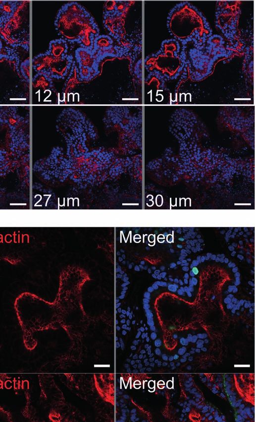
Smith et al.                                                                                                             Sheep Gastric and Intestinal Organoids

                                            small ruminant intestinal and gastric organoids. In line with 3Rs principals, use of such
                                            organoids will reduce and replace animals in host-pathogen research.
                                            Keywords: mini-guts, three-dimensional (3D) organoids, host-pathogen interactions, in vitro culture systems, stem
                                            cells, crypts, sheep, gastrointestinal

INTRODUCTION                                                                            The development of in vitro organoid culture systems has
                                                                                    transformed biomedical research as they provide a reproducible
The mammalian gastrointestinal (GI) tract is the site of digestion                  cell culture system that closely represents the physiology of the
and nutrient absorption, as well as a predilection site for many                    host. As the majority of infectious agents enter the body or reside
infectious pathogens, including bacteria, viruses and parasites.                    at mucosal surfaces, organoids derived from mucosal sites such
Understanding how pathogens attach and invade cells in the GI                       as the gastro-intestinal, respiratory and urogenital tracts promise
tract will help determine mechanisms of host infection, disease                     to transform research into host-pathogen interactions as they
pathogenesis and enable strategies to prevent and control                           allow detailed studies of early infection processes that are difficult
infectious disease. Both the gastric stomach and intestine share a                  to address using animal models.
number of common features, including a single luminal layer of                          Gastrointestinal (GI) disease in small ruminants has significant
epithelial cells sealed by tight junctions which is renewed                         implications for animal health and welfare as well as substantial
approximately every 3 – 5 days. In both organs, this huge                           economic losses because of decreased production efficiency. In
regenerative capacity is mediated by proliferation and                              sheep, gastrointestinal nematodes (GIN) have major economic and
differentiation of tissue resident adult stem cells (ASCs) (Barker                  welfare impacts worldwide, with the principal GIN of sheep
et al., 2007; Sato et al., 2009; Barker et al., 2010; Xiao and Zhou,                including: Haemonchus contortus; Nematodirus battus;
2020). In intestinal tissues, pockets of leucine-rich repeat-                       Teladorsagia circumcincta and Trichostrongylus spp. (including
containing G protein-coupled receptor 5 (LGR5)-expressing                           T. colubriformis and T. vitrinus) (Nieuwhof and Bishop, 2005;
ASCs reside in the base of the crypts of Lieberkühn and can                         Roeber et al., 2013). These parasites are transmitted by the faecal-
differentiate into all five epithelial cell types of the intestine:                  oral route where infective stage larvae develop in either the small
enterocytes, goblet cells, enteroendocrine cells, tuft cells, and                   intestine or abomasum (which is analogous to the gastric stomach)
Paneth cells (Barker et al., 2007; Sato et al., 2009). In the                       causing significant mucosal damage associated with host
stomach the epithelia is arranged into multiple gastric units,                      inflammatory immune responses (Stear et al., 2003; Roeber
which comprise of the gastric pit, isthmus, neck and base with                      et al., 2013). In addition, sheep are natural reservoirs for enteric
proliferative stem cells located in the isthmus (Barker et al., 2010;               zoonotic pathogens of worldwide significance, such as Shiga toxin
Xiao and Zhou, 2020). The ASCs of the gastric gland can                             producing Escherichia coli (STEC) and Salmonella enterica
differentiate into all five epithelial cell types of the gastric                     (Heredia and Garcı́a, 2018). The obvious challenge with studying
stomach: surface neck mucus cells, parietal cells, chief cells,                     interactions between the ovine host and GI pathogens is the lack of
enteroendocrine cells (including G cells, D cells, and                              accessibility to the site of infection, making detailed studies
enterochromaffin-like cells) and tuft cells (Barker et al., 2010;                    particularly challenging. With the current lack of physiologically
Xiao and Zhou, 2020).                                                               relevant in vitro cell culture systems to study ovine-GI pathogen
    The huge regenerative capacity of GI tract and the ability of                   interactions, research has relied heavily on use of sheep infection
ASCs to differentiate into epithelial cell types present in the GI                  models, which have led to important insights into host immune
tract has been exploited to develop GI organoids or “mini-guts”                     responses against pathogens, immune evasion by pathogens and
that reflect the cellular diversity and physiology of the organ                      pathogen transmission (Stear et al., 1995; McSorley et al., 2013;
from which they were derived (Sato et al., 2009; Barker et al.,                     Ellis et al., 2014). Despite these successes, animal experiments are
2010). Organoid models of the GI tract were first developed from                     often complex, costly and have ethical implications.
mouse stomach and intestine tissues. To achieve this, researchers                       The use of stem-cell derived GI organoids or “mini-guts” for
isolated mouse LGR5+ adult stem cells from these organs and                         farmed livestock species, including ruminants, is an exciting
cultured them in a laminin rich extracellular matrix extracted                      recent development that promises to provide a physiologically
from the Engelbreth-Holm-Swarm (EHS) mouse sarcoma, with                            relevant and host-specific in vitro cell culture system to
appropriate growth factors (including Wnt3a, epidermal growth                       interrogate host-pathogen interactions (Beaumont et al., 2021;
factor, Noggin and R-spondin 1). The resulting organoids                            Kar et al., 2021). A recent study has demonstrated the feasibility
consisted of organ-specific tissue (gastric or intestinal epithelia)                 of generating organoids from bovine ileum tissue with the derived
that self-organised into spherical three-dimensional (3D)                           organoids expressing genes associated with intestinal epithelia cell
structures with a single epithelial layer and a central lumen                       types (Hamilton et al., 2018). However, no ruminant gastric
(Sato et al., 2009; Barker et al., 2010). Since this initial discovery,             organoid model has been previously reported. In this current
organoids have been derived from a large number of different                        study, in line with 3Rs principles to reduce and replace the use of
tissue types and from numerous mammalian species using                              animals in experiments, we develop ovine ileum and abomasum
similar ASC isolation and tissue culture techniques.                                organoids as physiologically relevant in vitro culture systems to

Frontiers in Cellular and Infection Microbiology | www.frontiersin.org          2                                September 2021 | Volume 11 | Article 733811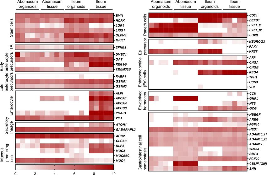
Smith et al. Sheep Gastric and Intestinal Organoids
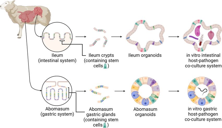
FIGURE 1 | A schematic of the development of ovine gastric and intestinal organoids for studying host-pathogen interactions. Stem cells isolated from sheep ileum
crypts and abomasum gastric glands can be cultivated into tissue-specific organoids when grown in a three-dimensional culture system. Gastric and intestinal
organoids can be co-cultured with pathogens to model host-parasite interactions in physiologically and biologically-relevant in vitro culture systems. Created with
BioRender.com.
investigate ovine GI infection and disease (Figure 1). Using RNA- placed into sterile ice-cold Hank’s buffered saline solution (HBSS)
seq of both tissue and derived organoids we demonstrate that the containing 25 µg/ml gentamicin (G1397-10ML; Sigma-Aldrich)
expression profile of abomasum and ileum organoids are and 100 U/ml penicillin/streptomycin. To expose the epithelial
representative of the tissue from which they were derived. In surfaces, the abomasum was opened along the greater curvature
addition, we demonstrate the utility of these in vitro organoid and the ileum opened longitudinally using dissection scissors. The
systems to study host-pathogen interactions by performing luminal surfaces were rinsed with tap water to remove digesta and
challenge studies with the abomasal parasite T. circumcincta then placed onto sterile Petri dishes. The majority of the mucus
and enteric bacteria Salmonella enterica serovar Typhimurium. layer was gently removed using a glass slide, after which the
surface mucosal tissue (containing the gastric glands or intestinal
crypts) was collected by firm scraping with a fresh glass slide.
MATERIALS AND METHODS Mucosal tissue was then transferred to a Falcon tube containing 50
ml of HBSS containing 25 µg/ml gentamicin and 100 U/ml
Animals penicillin/streptomycin. Samples were centrifuged at 400 x g for
All ovine abomasum and ileum tissues used in this study were 2 min, resulting in a tissue pellet with a mucus layer on top. The
derived from 7-8-month old helminth-free Texel cross male supernatant and top forming mucus layer were aspirated and
lambs (Ovis aries). The presented research was performed in discarded and the tissue was re-suspended in 50 ml of HBSS
line with 3Rs principles, particularly regarding the reduction and containing 25 µg/ml gentamicin and 100 U/ml penicillin/
replacement of animals for use in scientific research. Therefore, streptomycin. This process of centrifugation, aspiration and
the animal tissue used for developing organoids in this study was resuspension was repeated until a mucus layer was no longer
derived post-mortem from healthy control animals used in visible above the pellet. To release gastric glands and intestinal
separate research trials, thereby reducing the number of crypts from tissue, pellets were re-suspended in 25 ml of digestion
animals necessary for research. Due to the timing of the study, medium (Dulbecco’s Modified Eagle Medium [DMEM] high
we were limited to tissue derived from male lambs. glucose, (11574486; Gibco) 1% FBS, 20 µg/ml dispase
(4942086001; Roche), 75 U/ml collagenase (C2674; Sigma-
Isolation of Gastric Glands and Aldrich) 25 µg/ml gentamicin and 100 U/ml penicillin/
Intestinal Crypts streptomycin) and incubated horizontally in a shaking incubator
Tissues were removed from sheep at post-mortem. Approximately at 80 rpm for 40 minutes at 37°C. Following digestion, the tube
10 cm2 sections of fundic gastric fold were collected from the was gently shaken to loosen the cells and then left briefly at room
abomasum and approximately 10 cm sections of ileal tissue were temperate to allow large tissue debris to settle. The supernatant
collected from a region ~ 30 cm distal to the ileocecal junction. was transferred to a sterile 50 ml Falcon tube and gland/crypt
Tissues were removed using a sterile scalpel and forceps and integrity within the supernatant was assessed by light microscopy.
Frontiers in Cellular and Infection Microbiology | www.frontiersin.org 3 September 2021 | Volume 11 | Article 733811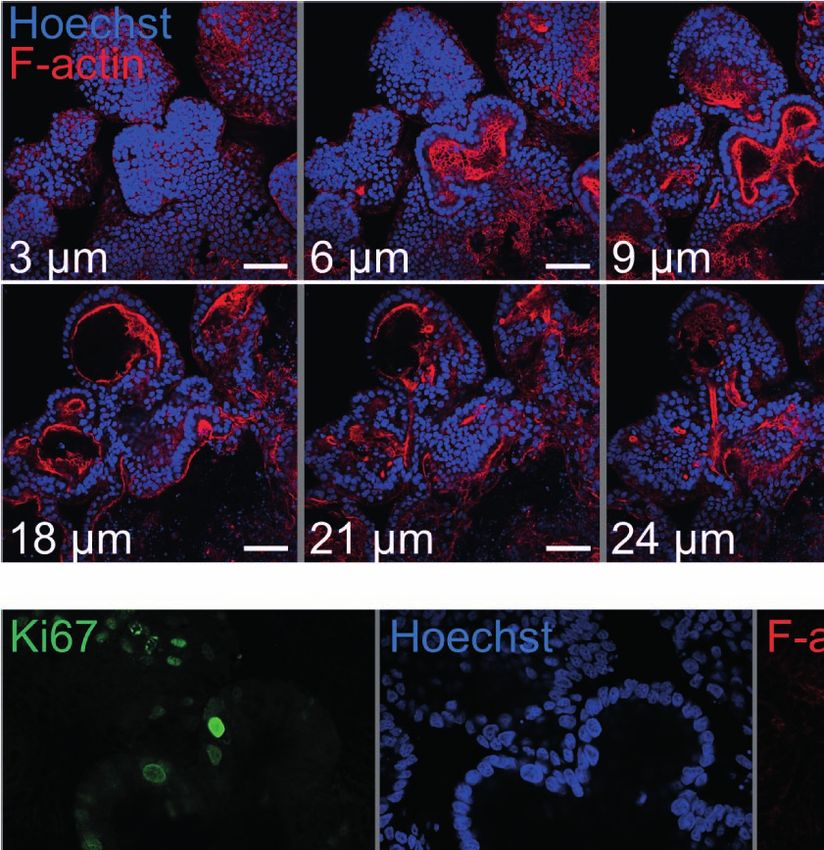
Smith et al.                                                                                                         Sheep Gastric and Intestinal Organoids

Samples were then centrifuged at 400 x g for 2 minutes, with the              cryopreservation medium (STEMCELL Technologies) at
resulting supernatant containing released glands or crypts. The               approximately 500-1000 organoids/ml before being transferred to a
gland/crypt-containing supernatant was washed by centrifugation               cryovial. Cryovials were stored in a cryogenic freezing container for
at 400 x g for 2 minutes and the glands/crypts re-suspended in 1-2            2 hours at -80°C and subsequently transferred to -196°C for long-
ml advanced DMEM/F12 (12634-010; Gibco) containing 1X B27                     term storage.
supplement minus vitamin A (12587-010; Gibco), 25 µg/ml                           Cryopreserved organoids were resuscitated by thawing
gentamicin and 100 U/ml penicillin/streptomycin.                              cryovials in a water bath at 37°C and then rapidly transferring
                                                                              the organoids into a 15 ml Falcon tube containing 8 ml of
Organoid Culture                                                              advanced DMEM/F12 medium (containing 1X B27 supplement
Two-hundred to one-thousand gastric glands or intestinal crypts               minus vitamin A, 25 µg/ml gentamicin and 100 U/ml penicillin/
were re-suspended in 100 µl advanced DMEM/F12 medium                          streptomycin). The cryovial was washed with a further 1 ml of
(containing 1X B27 supplement minus vitamin A, 25 µg/ml                       media and added to the Falcon tube. Samples were pelleted by
gentamicin and 100 U/ml penicillin/streptomycin) and were                     centrifugation at 290 x g for 5 minutes at 4°C and then re-
then added to 150 µl of BD Growth Factor Reduced Matrigel                     suspended in 200 µl of fresh advanced DMEM/F12 medium
Matrix (356230; BD Biosciences). Fifty microliter droplets were               (containing 1X B27 supplement minus vitamin A, 25 µg/ml
added to consecutive wells of a 24-well tissue culture plate (3524,           gentamicin and 100 U/ml penicillin/streptomycin). Re-
Corning). Plates were incubated at 37°C, 5% CO2 for 15-20                     suspended organoids were added to Matrigel and cultivated as
minutes to allow the Matrigel to polymerize and then 550 µl of                described in Section 2.4. Organoids were imaged by phase
pre-warmed complete IntestiCult Growth Medium (mouse)                         contrast microscopy following seven days of in vitro growth
(6005; STEMCELL Technologies) containing 500 nM Y-27632                       prior to cryopreservation and post-cryopreservation.
(10005583; Cambridge Bioscience), 10 µM LY2157299 (15312;
Cambridge Bioscience), 10 µM SB202190 (ALX-270-268-M001;                      Total RNA Extraction
Enzo Life Sciences) and gentamicin (50 µg/ml) were added to                   Total RNA was extracted from gastric and intestinal organoids
each well. Plates were incubated at 37°C, 5% CO2 to allow                     after multiple serial passages that included passage 0 (P0) through
organoids to develop, replacing complete IntestiCult medium                   to passage 4 (P4). Ovine gastric and intestinal organoids were
every 2-3 days. Organoids were typically cultured for 7-14 days               prepared as described above; organoids that were formed from
prior to passaging. Phase contrast microscopy was used to image               animal tissue-derived crypts were designated P0 and these were
organoids over the course of 14 days of in vitro growth.                      cultured by serial passage until P4. Each passage was cultured in
                                                                              triplicate wells of a 24-well tissue culture plate and allowed to
Organoid Passage                                                              mature for seven days before collecting for total RNA extraction.
IntestiCult media was removed from the cultured organoids and                 For total RNA extraction, IntestiCult media was removed from
the Matrigel matrix was dissolved by replacement with 1 ml ice-               wells and replaced with 1 ml of ice-cold advanced DMEM/F12.
cold advanced DMEM/F12. The re-suspended organoids were                       The resulting suspension containing dissolved Matrigel and
transferred to a 15 ml Falcon tube and the total volume of                    organoids was transferred to 15 ml sterile Falcon tubes and
advanced DMEM/F12 was increased to 10 ml. Samples were left                   brought up to 10 ml with ice-cold advanced DMEM/F12.
on ice for 5 minutes to allow organoids to settle and the                     Organoids were gently pelleted by centrifugation at 200 x g for
supernatant was removed. The organoids were re-suspended in                   5 min and the supernatant removed. Organoid pellets were re-
200 µl advanced DMEM/F12 medium (containing 1X B27                            suspended in 350 µl RLT buffer (Qiagen) containing b-
supplement minus vitamin A, 25 µg/ml gentamicin and                           mercaptoethanol, according to manufacturer’s guidelines and
100 U/ml penicillin/streptomycin) and then mechanically                       stored at -70°C. Total RNA was isolated from each sample
disrupted by repeatedly pipetting (approximately fifty times)                  using a RNeasy mini kit (Qiagen) with the optional on-column
using a 200 µl pipette tip bent at a 90° angle. The number of                 DNase digest and total RNA eluted in 30 µl nuclease-free water,
organoid fragments were counted by light microscopy and                       according to the manufacturers protocol. Total RNA from each
samples diluted to 200-1000 crypts per 100 µl. One-hundred                    extraction was quantified using a NanoDrop ™ One
microliters of fragments were then combined with Matrigel and                 spectrophotometer and integrity analysed using a Bioanalyzer
plated into 24-well tissue culture plates as described in section 2.4.        (Agilent) with the total RNA 6000 Nano kit. Purified total RNA
Phase contrast microscopy was used to image organoids from                    was stored at -70°C until RNA-seq analysis.
passage one to passage five, following seven days of in vitro growth               Total RNA was also extracted from ovine abomasum and
at each passage.                                                              ileum tissue harvested at post-mortem from five individual
                                                                              6-month old helminth-free Texel cross lambs and stored in
Organoid Cryopreservation                                                     RNAlater (ThermoFisher). Specifically, samples were taken
IntestiCult media was removed from the cultured organoids and the             from the same tissue regions stated above for crypt isolation.
Matrigel matrix was dissolved by replacement with 1 ml ice-cold               For total RNA isolation, approx. 30 mg of tissue was
advanced DMEM/F12. The re-suspended organoids were transferred                h o m o g e n i z e d i n 6 0 0 µ l o f R L T b u ff e r c o n t a i n i n g
to a microcentrifuge tube and pelleted by centrifugation at 290 x g for       b-mercaptoethanol using a Precellys® Tissue Homogenizer
5 minutes at 4°C. Following centrifugation, the supernatant was               with CK28 tubes using x3 10s pulses at 5500 rpm with 5 min
removed and organoid pellets were re-suspended in Cryostor CS10               on ice between each pulse (Bertin Instruments™). Total RNA

Frontiers in Cellular and Infection Microbiology | www.frontiersin.org    4                                  September 2021 | Volume 11 | Article 733811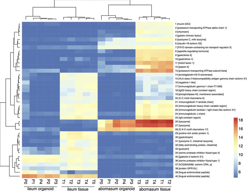
Smith et al.                                                                                                       Sheep Gastric and Intestinal Organoids

was isolated and quantified as described previously, except the               and rabbit IgG respectively. The next day, samples were washed
total RNA was eluted in 50 µl nuclease-free water. Purified total             three times with IF buffer and then secondary antibodies added
RNA was stored at -70°C until RNA-seq analysis.                              (diluted at 1:500 in blocking buffer) and incubated at room
                                                                             temperature for 1 hour. Secondary antibodies used were goat a-
RNA-Seq Analysis                                                             mouse Alexa Fluor 488 (ab150117, abcam) and goat a-rabbit Alexa
For each sample, 1 µg of total RNA was used for RNA-seq analysis.            Fluor 488 (ab150081, abcam). Phalloidin-iFluor 555 reagent
All library synthesis and sequencing were conducted at The                   (ab176756, abcam, used at a 1:1000 dilution) was also added
University of Liverpool, Centre for Genomic Research (CGR). In               during the secondary antibody step to label F-actin. Samples were
brief, dual-indexed, strand-specific RNA-seq libraries were                   washed three times with IF buffer and then Hoechst 33258 solution
constructed from submitted total RNA sample using the                        diluted 1:200 in IF buffer was added to label nuclei (94403, Sigma-
NEBNext® Poly(A) mRNA Magnetic Isolation Module (NEB                         Aldrich). Samples were incubated for a further 5 minutes at room
#E7490) and NEBNext Ultra II Directional RNA Library Prep                    temperature before three washes with IF buffer. Finally, slides were
Kit for Illumina (NEB #E7760). A total of 20 libraries were                  mounted using ProLong Gold antifade mountant (P10144,
constructed [including: ovine abomasum organoid P0-P4                        ThermoFisher Scientific) and imaged by confocal microscopy
(triplicate pooled wells for each passage); ovine ileum organoid             using a Zeiss LSM 710 Inverted Confocal Microscope and Zeiss
P0-P4 (triplicate pooled wells for each passage); ovine abomasum             Zen Black operating software.
tissues (n = 5); ovine ileum tissues (n = 5)]. The barcoded
individual libraries were pooled and sequenced on a single lane              Exsheathment of Teladorsagia
of an Illumina NovaSeq flowcell using S1 chemistry (Paired-end,               circumcincta Third Stage Larvae (L3)
2x150 bp sequencing, generating an estimated 650 million clusters            T. circumcincta L3 (Moredun isolate MTci2, CVL) were exsheathed
per lane). Following sequencing adaptors were trimmed using                  and labelled using modified protocols previously published (Dinh
Cutadapt version 1.2.1 (Martin, 2011) and reads were further                 et al., 2014; Bekelaar et al., 2019). Nine millilitres of Earle’s balanced
trimmed using Sickle version 1.200 (Joshi and Fass, 2011) with a             salts solution (EBSS) buffer in a 15 ml Falcon tube was preheated in a
minimum window quality score of 20. Reads shorter than 15 bp                 water bath to 37°C and CO2-saturated over 1 hour using an incubator
after trimming were removed. Sequence reads were checked for                 tube connected to a CO2 tank. Approximately 5x10⁴ T. circumcincta
quality using FastQC v0.11.7. Reads were pseudo-aligned to the               L3 in 1 ml of tap water were added to the CO2-saturated EBSS and
Ovis aries transcriptome (Oar_v3.1 GCA_000298735.1) using                    the sample continued to be saturated for a further 15 minutes. The
Kallisto v0.46.2 with default settings (Bray et al., 2016) and read          Falcon tube was then sealed with Parafilm® M and inverted 6 times
abundance calculated as transcripts per million (TPM). Gene                  before being placed horizontally into an incubator at 37°C, 5% CO2
expression data was analysed by principal component analysis                 for 4 hours. Following incubation, the whole sample was transferred
(PCA) using pcaExplorer version 2.12.0 R/Bioconductor package                into a 25 cm2 vented cap flask and incubated overnight at 37°C/5%
(Marini and Binder, 2019). Specific genes were also manually                  CO2, to allow L3s to continue exsheathing. Exsheathment was
retrieved from our transcriptomic dataset and their TPM values               validated the following morning by light microscopy. The larvae
log2 transformed for presenting in heat maps, which were                     were then washed 4 times by repeated centrifugation at 330 x g for 2
generated using GraphPad Prism software (v8.0).                              minutes and re-suspension in 50 ml of distilled water (pre-warmed to
                                                                             37°C). After the final wash, the L3 larvae were re-suspended in 1 ml
Immunohistochemistry                                                         distilled water and transferred to a microcentrifuge tube. Exsheathed
Abomasum and ileum organoids were cultivated in Matrigel for 7               L3 (exL3) were fluorescently labelled by the addition of 2 µl PKH26
days in 8-well chamber slides (354118; Falcon) as described in               dye (1 mM stock concentration) from the MINI26 PKH26 Red
section 2.4. To make organoids accessible to immunohistochemistry            Fluorescent Cell Linker Kit (Sigma-Aldrich) and mixed by pipetting.
reagents, the culture medium was removed and replaced with ice-              Parasites were incubated with the dye for 15 minutes at room
cold 4% paraformaldehyde. For fixation, samples were kept at 4°C              temperature, protected from light. Excess dye was removed by
for 20 minutes to also dissolve the Matrigel and prevent it from re-         washing the larvae five times with distilled water as described above
solidifying. Organoids were washed twice with IF buffer (0.1%                before finally re-suspending them in 1 ml of complete IntestiCult
Tween20 in PBS) and then permeabilised with 0.1% TritonX-100                 organoid growth medium.
in PBS for 20 minutes at room temperature. Samples were washed
three times with IF buffer and then blocked for 30 minutes with 1%           Teladorsagia circumcincta L3-Organoid
BSA in IF buffer at room temperature. Next, primary antibodies               Co-Culture
diluted in blocking solution were added to the organoids and                 Abomasum and ileum organoids were cultivated in Matrigel for
samples were left overnight at 4°C. Primary antibodies used                  7 days in 8-well chamber slides (354118; Falcon) as described in
included polyclonal rabbit a-Ki67 (ab15580, abcam, used at a                 section 2.4. Immediately prior to organoid-T. circumcincta
1:500 dilution), polyclonal rabbit a-EPCAM (orb10618, Biorbyt,               co-culture, complete IntestiCult media was removed from the
used at a 1:600 dilution), monoclonal mouse a-villin (sc-58897,              cultured organoids and replaced with 250 µl of fresh pre-warmed
Santa Cruz Biotechnology, used at a 1:200 dilution) and monoclonal           complete IntestiCult. Twenty to 50 PKH26 labelled
mouse a-pan cytokeratin (used at a 1:100 dilution). For isotype              T. circumcincta exL3 in 50 µl complete IntestiCult media were
controls, mouse or rabbit IgG were used in place of the specific              added to each well of organoids and organoid-larval cultures
primary antibodies and were diluted at 1:100 or 1:500 for mouse              incubated at 37°C, 5% CO2. Note that organoids were not

Frontiers in Cellular and Infection Microbiology | www.frontiersin.org   5                                 September 2021 | Volume 11 | Article 733811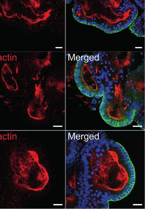
Smith et al.                                                                                                     Sheep Gastric and Intestinal Organoids

removed from their Matrigel domes prior to the addition of                    already contained organoids that had already been maintained in
T. circumcincta L3. Upon observation of multiple organoids                    conditions for generating apical-out organoids for 72 hours. The
containing T. circumcincta L3 within their lumen (after ~24-48                other half of the wells acted as negative controls, with organoids
hours of organoid-T. circumcincta co-culture) the samples were                being re-suspended in 300 µl of complete IntestiCult growth
fixed with 4% PFA for 30 min, followed by 3 washes with PBS, and               medium alone (no bacteria). After 30 minutes of incubation
stored at 4°C until fluorescence staining. Organoids were                      another 300 µl of complete IntestiCult growth medium with
permeabilized, blocked and probed with Phalloidin-488 and                     200 µg/ml gentamycin was added to kill extracellular bacteria. The
Hoechst 33258 as described for organoid immunohistochemistry                  slides were incubated at 37°C, 5% CO2 for a total of 6 hours. At the
above. Images were captured using a Zeiss LSM 710 Inverted                    end of the incubation period the entire volume of the liquid from
Confocal Microscope and Zeiss Zen Black operating software.                   each well, including the organoids, were transferred to separate
                                                                              15ml Falcon tubes (Corning, UK). All centrifugations for organoid
Generation of Apical-Out Organoids                                            collection during washing were done at 200 rpm for 5 minutes. The
Epithelial polarity was inverted in gastric and intestinal ovine              supernatant was removed and the organoids were washed twice in
organoids by following a previously published method for                      PBS, and then re-suspended in 4% PFA for 30 minutes for fixation.
reverse polarity in human intestinal organoids (Co et al.,                    The organoids were processed for immunohistochemistry as
2019). Briefly, gastric and intestinal organoids were grown in                 described in section 2.9 and stained with Phalloidin-iFluor 555
Matrigel as described above for 7 days. Matrigel domes                        reagent, prior to mounting with ProLong Diamond antifade
containing developed organoids were gently dissolved by the                   mountant (P36961, ThermoFisher Scientific). Confocal imaging
addition of 500 µl ice-cold 5 mM EDTA in PBS, taking care not                 was performed as described in section 2.9.
to rupture the organoids. The resulting suspension was
transferred to a 15 ml Falcon tube that was subsequently filled
with 14 ml of 5 mM EDTA in PBS. Samples were placed on a                      RESULTS
rocker and mixed gently for 1 hour at 4°C. Organoids were
pelleted by centrifugation at 200 x g for 3 min at 4°C and the                Growth of Ovine Gastrointestinal
supernatant was removed. Pellets were re-suspended in complete                Organoids In Vitro
IntestiCult growth media (containing 500 nM Y-27632, 10 µM                    Fragmented gastric glands and intestinal crypts isolated from the
LY2157299, 10 µM SB202190 and gentamicin (50 µg/ml), with                     abomasum fundic fold and the ileum of 7 to 8-month old Texel
the addition of 10% advanced DMEM/F12 medium (containing                      cross lambs were embedded in Matrigel and grown in complete
1X B27 supplement minus vitamin A, 25 µg/ml gentamicin and                    IntestiCult organoid growth medium. Under identical growth
100 U/ml penicillin/streptomycin). Re-suspended organoids                     conditions, epithelial stem cells from these two different organ
were transferred to the wells of 8-well glass chamber slides and              tissues were able to develop into organoids in vitro (Figure 2A).
incubated at 37°C, 5% CO2 for a period of 72 hours, prior to                  By 24 hours, sealed spherical structures containing a central
being fixed and stained with Phalloidin-iFluor 555 reagent and                 lumen had formed in both the abomasum and ileum organoids.
Hoechst 33258, as described in section 2.9. Confocal imaging was              However, while the ileum organoids became branched after 5-7
performed as described in section 2.9.                                        days of in vitro culture, the vast majority of abomasum organoids
                                                                              retained a spherical structure that persisted for the duration of a
Infection of Apical-Out Organoids With                                        culture passage (Figures 2A, B).
Salmonella enterica Serovar Typhimurium                                           Abomasum and ileum organoids could be serially passaged by
The polarity of gastric and intestinal organoids was inverted as              removal from Matrigel, fragmentation by pipetting and
described above. Salmonella Typhimurium strain ST4/74 was                     re-embedding in Matrigel. At each passage, ileum organoids
chosen for this experiment as its full genome sequence is available           continued to form into branched structures, while the abomasum
(Richardson et al., 2011) and it has been shown to efficiently invade          organoids persistently formed spherical structures. (Figure 2B).
the ovine ileal mucosa and elicit inflammatory responses in an ovine           Organoids that were cryopreserved in liquid nitrogen after
ligated ileal loop model (Uzzau et al., 2001). To aid visualization of        7 days of in vitro culture could be thawed and re-cultured,
the bacteria in organoids, the strain was electroporated with plasmid         demonstrating the potential to store organoids long-term and to
pFPV25.1 which carries gfpmut3A under the control of the rpsM                 resuscitate when required. Furthermore, we found that the
promoter resulting in the constitutive synthesis of green fluorescent          cryopreserved organoids can be resuscitated after at least 18
protein (Valdivia and Falkow, 1996). Stability of the plasmid in the          months of storage in liquid nitrogen. Abomasum and ileum
absence of antibiotic selection during Salmonella infection has been          organoids retained their spherical and branched structures,
confirmed (Vohra et al., 2019). The bacteria were grown on Luria               respectively, following resuscitation and 7 days of in vitro
Bertani (LB) agar supplemented with 100 µg/ml ampicillin at 37°C              culture (Figure 2C).
overnight. Single colonies were transferred to LB broth
supplemented with the same antibiotic and grown for 20 hours                  Epithelial Cell Markers Associated With
shaking at 180 rpm at 37 °C. The liquid cultures were diluted to 3.3 x        Ovine Gastric and Intestinal Organoids
106 CFU/ml in complete IntestiCult growth medium, described                   Immunohistochemistry was performed to identify key structural
above, and 300 µl of the dilution was added to half of the wells, which       features associated with both abomasum and ileum organoids.

Frontiers in Cellular and Infection Microbiology | www.frontiersin.org    6                               September 2021 | Volume 11 | Article 733811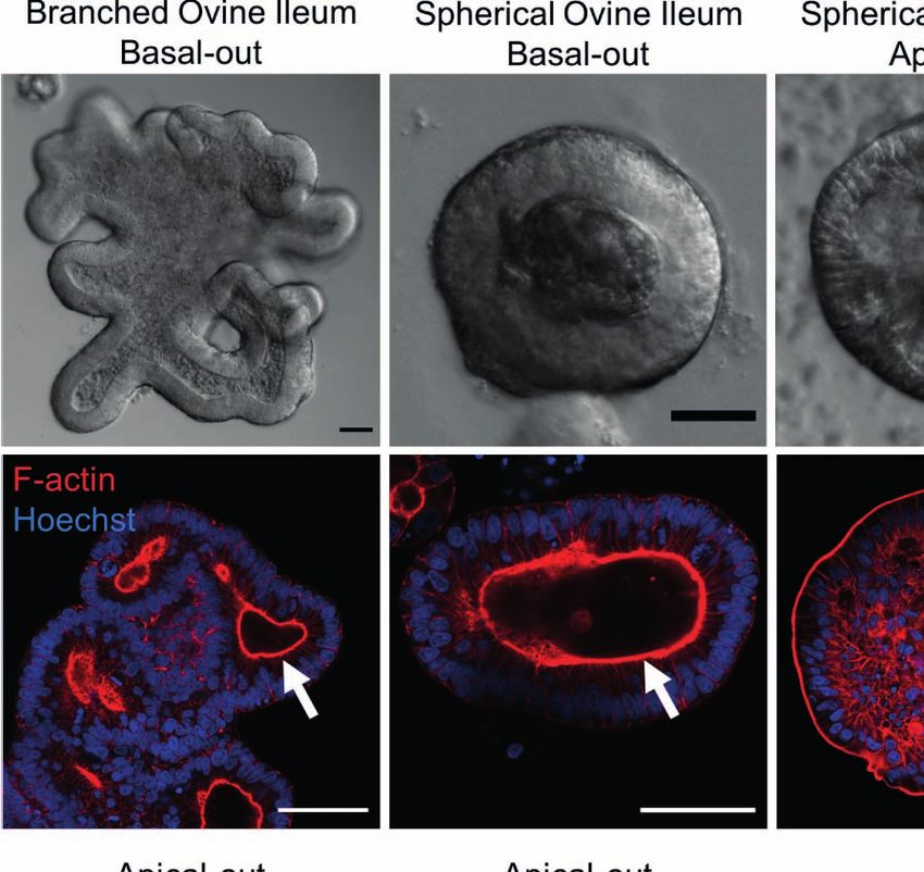
Smith et al.                                                                                                                 Sheep Gastric and Intestinal Organoids

                        A

                        B

                                     C

 FIGURE 2 | In vitro growth of ovine abomasum and ileum organoids. (A) Representative images of abomasum and ileum organoids grown over 14 days in the
 same culture conditions. (B) Representative images showing the growth and development of mature abomasum and ileum organoids across multiple consecutive
 passages (P1 - P5) at seven days of in vitro culture. (C) Representative images of abomasum and ileum organoids grown for seven days, both pre-cryopreservation
 and after resuscitation. Scale bars = 10 µm.

Individual Z-stack images of organoids stained with phalloidin to                   continued to take place at 7 days of in vitro culture
label F-actin clearly demonstrated that the apical surface of the                   (Figures 3B, 4B). The epithelial cell markers EpCAM
epithelium is present on the interior of the organoid, for both                     (epithelium cell adherence molecule), villin (epithelium-specific
abomasum and ileum organoids, indicated by the presence of a                        actin-binding protein) and cytokeratin (epithelial cell
solid F-actin-positive boundary (Figures 3A, 4A). This imaging                      cytoskeleton filament protein) were each detectable in
also confirmed the presence of a hollow lumen within the                             abomasum and ileum organoids at seven days of in vitro
organoids (Figures 3A, 4A).                                                         culture (Figures 3B, 4B), confirming the differentiation of
    The proliferation marker Ki67 was detectable in both the                        stem cells into epithelium cell-containing organoids. Control
abomasum and ileum organoids, indicating that cell division                         samples of organoids probed with mouse and rabbit serum IgG

Frontiers in Cellular and Infection Microbiology | www.frontiersin.org          7                                   September 2021 | Volume 11 | Article 733811
Smith et al.                                                                                                                 Sheep Gastric and Intestinal Organoids

                A

                B

 FIGURE 3 | Characterisation of ovine abomasum organoids by immunofluorescence confocal microscopy at seven days of in vitro culture. (A) Representative
 Z-stack images of an individual abomasum organoid with a closed luminal space and an internal F-actin-expressing brush border. Red = F-actin and blue = Hoechst
 (nuclei). (B) Representative images of abomasum organoids probed for either the cell proliferation marker Ki67, or the epithelial cell markers EpCAM, villin and
 pan-cytokeratin (all green). F-actin (red) and Hoechst (blue). Scale bars = 10 µm.

Frontiers in Cellular and Infection Microbiology | www.frontiersin.org          8                                   September 2021 | Volume 11 | Article 733811
Smith et al.                                                                                                                    Sheep Gastric and Intestinal Organoids

                 A

                 B

 FIGURE 4 | Characterisation of ovine ileum organoids by immunofluorescence confocal microscopy at seven days of in vitro culture. (A) Representative Z-stack
 images of part of an individual branched ileum organoid with a closed luminal space and an internal F-actin-expressing brush border. Red = F-actin and
 blue = Hoechst (nuclei). (B) Representative images of abomasum organoids probed for either the cell proliferation marker Ki67, or the epithelial cell markers EpCAM,
 villin and pan-cytokeratin (all green). F-actin (red) and Hoechst (blue). Scale bars = 10 µm.

Frontiers in Cellular and Infection Microbiology | www.frontiersin.org            9                                    September 2021 | Volume 11 | Article 733811
Smith et al.                                                                                                               Sheep Gastric and Intestinal Organoids

did not label positive for any of the epithelial cell markers,                           absent in all ileum samples, included genes of known gastric
confirming the specificity of the epithelial cell labelling                                function, such as: claudin-18; gastrokine; gastric lysozyme and
(Supplemental Figures 1, 2).                                                             pepsin. Similarly, ileum specific genes were detected in both
                                                                                         ileum tissue and ileum organoid samples, but absent from all
Transcriptional Analysis of Abomasum and                                                 abomasum samples included: galectin; lingual antimicrobial
Ileum Organoids and Tissue                                                               peptide; guanylin (a 15 amino acid peptide secreted from
Gene expression profiles from: ovine ileum organoids (P0 – P4);                           goblet cells). Interestingly, genes shared by ileum tissue and
ovine abomasum organoids (P0 – P4); ovine ileum tissue (n = 5)                           abomasum tissue, but largely absent from organoid cultures,
and ovine abomasum tissue (n = 5) were compared by RNA-seq                               were predominantly immune related genes (such as: C-C motif
analysis. The global gene expression profiles of the complete                             chemokine 5, regakine-1-like and various immunoglobulin
dataset, consisting of 20 individual samples, were initially                             chains) and likely reflect the presence of immune cells in ileal
compared by principal component analysis (PCA) (Figure 5).                               and abomasal mucosal tissue samples, which were not
The PCA analysis resulted in four statistically significant clusters                      represented in ASC derived ileum and abomasum organoids.
(95% confidence intervals), with each cluster representing a                              In summary, based on transcriptional profiles, abomasum and
sample type (i.e. ileum organoids; abomasum organoids; ileum                             ileum organoids are broadly representative of the tissues they
tissue; or abomasum tissue). This demonstrates that the global                           were derived from and appear to be transcriptionally stable over
transcriptome profile of ovine abomasum tissue (n = 5) and                                multiple passages.
ovine ileum tissue (n = 5) are different (Figure 5). Based on
global gene expression profiles, organoids also grouped by the
tissue type from which they derived, with ileum and abomasum                             Expression of Cell- and Tissue-Specific
organoids forming separate statistically significant clusters (95%                        Genes in Abomasum and Ileum Organoids
confidence intervals) in the PCA analysis (Figure 5).                                     and Tissues
Importantly, for both ileum and abomasum organoids, each                                 The ovine gastrointestinal transcriptomic database generated
passage (P0 – P4) is represented in each cluster, showing that                           here was manually searched for genes that are representative of
there was no global change in the transcriptome profile following                         specific cell and tissue markers. A total of 151 genes were
serial passage (Figure 5).                                                               searched in this way and their expression in abomasum and
    The expression profiles of the top 40 most variable genes (of                         ileum organoids and tissue was presented in heat maps. A
genes ranked by inter-sample variation) were compared from                               number of cell junction markers were consistently expressed in
ileum and abomasum organoids from serial passages (P0 – P4)                              both organoids and tissue, including genes encoding proteins
and ileum and abomasum tissue derived from five lambs (n = 5)                             related to tight junctions, gap junctions, adherens junctions and
(Figure 6). This analysis broadly identified three categories of                          desmosomes (Supplemental Figure 3).
genes, including genes with: i) abomasum (tissue and organoid)                               We identified genes associated with particular epithelial cell
specific expression; ii) ileum (tissue and organoid) specific                              subpopulations that were consistently expressed in abomasum
expression and iii) ileum and abomasum (tissue only) expression.                         and ileum organoids across multiple passages (P0-P4), as well as
    Based on gene expression profiles, genes that were highly                             in ileum and abomasum tissue samples from five individual
expressed in abomasum tissue and abomasum organoids, but                                 animals. These include numerous markers associated with stem
                                                                                         cells, enterocytes, secretory and mucus-producing cells and
                                                                                         Paneth cells (Figure 7). In particular, expression of the stem
                                                                                         cell marker LGR5 was higher in both abomasum and ileum
                                                                                         organoids compared to the respective tissue samples, indicating
                                                                                         the presence of a relatively higher stem cell subpopulation in the
                                                                                         organoids compared to tissues (Figure 6). Three enterocyte
                                                                                         genes associated with ileum tissue were not detected in ileum
                                                                                         organoids, namely ALPI, APOA4 and APOC3 (Figure 7). These
                                                                                         enterocyte markers were not detected in abomasum organoids or
                                                                                         abomasum tissue from any of the five individual animals.
                                                                                         Expression of several genes associated with homeostasis in
                                                                                         gastrointestinal cells was conserved between tissue samples and
                                                                                         organoids, for both abomasum and ileum samples. This included
                                                                                         HES1, ADAM10, ADAM17, FGF20 and SHH (Figure 7).
                                                                                             A number of genes associated with specific epithelial cell
 FIGURE 5 | Principal component analysis (PCA) of RNA-seq expression of
                                                                                         subpopulations were differentially expressed in ileum and
 the top 500 most variant genes (of genes ranked by inter-sample variance) in            abomasum tissue. For example, the early enterocyte precursor-
 ovine abomasum and ileum organoid and tissue samples. Sample type is                    associated gene REG3G, the Paneth cell marker DEFB1, the
 indicated in the key and includes: abomasum organoid (red); abomasum                    enteroendocrine cell marker REG4 and the enteroendocrine
 tissue (green); ileum organoid (blue); ileum tissue (purple). Ellipses indicates
                                                                                         cell-derived hormone GCG were expressed in ileum tissue and
 95% confidence intervals for each cluster.
                                                                                         not in abomasum tissue (Figure 7). These genes were also

Frontiers in Cellular and Infection Microbiology | www.frontiersin.org              10                              September 2021 | Volume 11 | Article 733811
Smith et al. Sheep Gastric and Intestinal Organoids
FIGURE 6 | Heat map showing expression level of top 40 most variant genes (of genes ranked by inter-sample variance) from ileum (ile) and abomasum (abo)
organoids from serial passages (P0 – P4) and ileum (ile) and abomasum (abo) tissue derived from five lambs (T1 – T5). Colours indicate level of expression from low
(blue) to high (red). The dendrograms indicate similarity between samples. Details of genes included in the heat map, including ENSOART sequence identifiers, are
shown in Supplemental File 1.
expressed in intestinal organoids and not abomasum organoids, was found to be highly expressed in ileum and ileal tissue, but was
indicating the conservation of tissue-specific differences in the cell not expressed in either abomasum organoids nor abomasal tissue
subpopulations of the two different types of organoids. (Figures 6 and Supplemental Figure 4).
Various genes were found to be specific for the abomasum,
being expressed in both abomasal tissue and abomasum Organoid Co-Culture With the Helminth
organoids but not in ileal tissue or ileum organoids. These Teladorsagia circumcincta
included PGA5, CCKBR and CBLIF (GIF) (Figure 8). We also In order to use gastrointestinal organoids to study host-pathogen
found that some genes specifically expressed in abomasal tissue interactions in vitro, it is important to be able to challenge
were not expressed in abomasum organoids, including SLC5A5, organoids with the pathogen-of-interest. Here, we co-cultured
DUOX2, MCT9, PGC, ATP4A, AQP4, and HDC (Figure 8). abomasum and ileum organoids with larvae of the important
The expression of immune-related genes, including toll-like ruminant helminth parasite T. circumcincta. Infective, third stage
receptors (TLRs), c-type lectin receptors (CLRs), chemokines, larvae (L3) were ex-sheathed in vitro and labelled with the
cytokines and antimicrobials were examined in abomasum and lipophilic dye PKH26. Labelled larvae were added directly to
ileum tissue and organoids. The TLRs - TLR3, TLR5 and TLR6, the well of a 24-well tissue culture plate containing abomasum or
and CLR Dectin-1 were expressed in abomasum and ileum ileum organoids embedded in Matrigel and complete IntestiCult
organoids and their respective tissues (Supplemental Figure 4). growth media. A number of T. circumcincta L3 penetrated the
A number of chemokines were expressed in abomasum organoids Matrigel, of which approximately 50% subsequently burrowed
and abomasal tissue, including CXCL16, CCL20, CCL24 and into central lumen of the organoids by 24 hours post-incubation,
ACKR3. Interestingly, the chemokine CCL17 was up-regulated in with some individual L3 invading the organoids as early as
abomasum and ileum organoids compared to the respective 2 hours. This indicated that it was possible to infect the
tissue samples (Supplemental Figure 4). The expression of organoids with the parasite in the correct orientation (i.e. with
cytokine associated genes IL18BP, IL27RA, IL4I1, IL13RA1 and the parasite residing at the luminal surface of the organoid)
IFNGR1 was detected in abomasum and ileum organoids without having to mechanically disrupt the organoids to allow
(Supplemental Figure 4). Of note, the antimicrobial gene SBD2 access to the central lumen. T. circumcincta L3 were equally
Frontiers in Cellular and Infection Microbiology | www.frontiersin.org 11 September 2021 | Volume 11 | Article 733811Smith et al. Sheep Gastric and Intestinal Organoids
FIGURE 7 | Heat map showing the expression of genes associated with gastrointestinal epithelia in abomasum and ileum tissue and organoids. RNA-seq analysis
was performed to compare gene expression in abomasal and ileal tissue derived from five lambs and abomasum and ileum organoids across multiple passages.
Squares from left to right under “abomasum tissue” and “ileum tissue” represent lambs T1-T5. Squares from left to right under abomasum organoids and ileum
organoids represent passages P0-P4. Scale = log2 transcripts per million reads. Ee: enteroendocrine. Details of genes included in the heat map, including ENSOART
sequence identifiers, are shown in Supplemental File 2.
effective at infecting both abomasum and ileum organoids and with a microvilli brush edge apparent by confocal microscopy
motile larvae were still present after 14 days of co-culture. While (Figures 10A, B).
we mainly observed abomasum organoids containing single To demonstrate the utility of apical-out ovine gastric and
larvae (Figure 9A), we found multiple larvae residing in the intestinal organoids as an in vitro model for host-pathogen
lumen of the larger ileum organoids (Figure 9B). Z-stack interactions, the apical-out organoids were exposed to the
analysis on fixed samples showed worms were present within bacterial pathogen Salmonella enterica serovar Typhimurium,
the lumen of the organoids and demonstrated L3 larvae which is known to invade the epithelium via the apical surface
burrowing directly through the epithelium of abomasum and (Finlay and Falkow, 1990). After 6 hours of organoid-bacteria co-
ileum organoids to access the central lumen (Figure 9C). culture freely suspended in complete IntestiCult growth medium,
GFP-expressing S. Typhimurium were identifiable attached
Generation of Apical-Out Organoids and to the apical surface and within epithelial cells of the
Infection With Salmonella typhimurium organoids by confocal microscopy. Although S. Typhimurium
It is necessary to expose the apical surface of the organoid is an intestinal pathogen, here we observed GFP-expressing
epithelia in order to have a working co-culture system for bacteria attached to both abomasum and ileum apical-out
some pathogens. A recently published protocol (Co et al., organoids (Figure 10C).
2019) described a method to invert the basal-out orientation of
the abomasum and intestinal organoids. When the organoids
were removed from Matrigel and incubated in 5 mM EDTA for 1 DISCUSSION
hour, the polarity of both the abomasum and intestinal
organoids was reversed following 72 hours’ incubation in Ruminants are key food-producing animals worldwide, providing
complete IntestiCult growth medium. F-actin staining of fixed a nutrient source to billions of people. Furthermore, dependency
organoid samples clearly highlighted the apical surface of the upon ruminants as a food source continues to increase in order to
epithelium, which is initially internally located in basal-out meet growing global dietary requirements. Gastrointestinal
abomasum and ileum organoids; however, after removing the disease in ruminants is a major concern and accounts for
extra cellular matrix from the organoids, the apical surface significant economic losses and reduction in production
became positioned on the exterior surface of the organoids, efficiency. It is therefore important that ruminant health and
Frontiers in Cellular and Infection Microbiology | www.frontiersin.org 12 September 2021 | Volume 11 | Article 733811Smith et al. Sheep Gastric and Intestinal Organoids
knowledge, this is the first demonstration of organoids
representing the gastric system of a ruminant.
Following the same protocol and using the same in vitro
culture conditions, we report the ability to cultivate tissue-
specific gastric and intestinal organoids from sheep. By
comparing gene expression profiles between tissue and
organoids, we found that when grown in identical conditions
in vitro, stem cells from gastric glands developed into organoids
that retained key characteristics associated with abomasum
tissue. Stem cells from ileal crypts, on the other hand,
developed into organoids which conserved important gene
expression profiles associated with the ileum. It is important to
note that there are differences in the global transcriptome
between organoids and the tissue from which they are derived.
This is largely due to the complexity associated with ex vivo tissue
which contains immune cells, fibroblasts, a microbiome, as well
as digested material at the point of collection and this will
influence overall gene expression. By contrast, the organoids
characterized here specifically represent the epithelium layer of
the organ from which they were derived, resulting in a less
complex gene expression profile than that associated with their
respective whole tissue. Despite this, the cell diversity, self-
organising properties and the conserved expression of tissue-
specific gene markers associated with organoids makes them the
FIGURE 8 | Heat map showing the expression of genes associated with most physiologically representative in vitro cell culture systems
gastric epithelia in abomasum and ileum tissue and organoids. RNA-seq developed to date.
analysis was performed to compare gene expression in abomasal and ileal
Ruminants, including cattle, sheep and goats are polygastric,
tissue derived from five lambs and abomasum and ileum organoids across
multiple passages. Squares from left to right under “abomasum tissue” and in that they have a four-chambered gastric system. The fourth
“ileum tissue” represent lambs T1-T5. Squares from left to right under chamber, the abomasum, is most closely akin to the stomach of
abomasum organoids and ileum organoids represent passages P0-P4. monogastric animals. An important differentiating characteristic
Scale = log2 transcripts per million reads. Details of genes included in the between abomasum and ileum tissue is the expression of the
heat map, including ENSOART sequence identifiers, are shown in
digestive stomach enzyme pepsinogen in the abomasum (Mostofa
Supplemental File 2.
et al., 1990). Another digestive protease associated with the
abomasum in ruminants is lysozyme, which is highly expressed
welfare is improved through prevention and control of disease in in this compartment (Stevens and Hume, 1998). Importantly, we
order to meet ethical, economic and nutrient demands found that both pepsinogen and lysozyme are expressed in
(Sargison, 2020). abomasum organoids and not in ileum organoids. We also
An obvious challenge with studying gastrointestinal host- found evidence of parietal cells specifically present in
pathogen interactions in vivo is the internal nature of infections abomasum organoids and not ileum organoids. This was
and the physical barriers associated with directly observing them. indicated by the detection of CCKBR mRNA only in
Therefore, a useful advancement for studying such infections is abomasum organoids and tissue following transcriptomic
the development of a physiologically relevant in vitro model analysis. CCKBR is a cholecystokinin receptor expressed in the
systems that allows experimental interrogation of host and gastric and central nervous systems and more specifically it is
pathogen interactions in fine detail. Stem cell-derived organoids associated with parietal cells in the stomach (Kulaksiz et al., 2000;
have become a prominent feature of modern cell and tissue Schmitz et al., 2001; Engevik et al., 2019). Conversely, we also
biology in recent years, representing in vitro cell cultures that identified genes whose expression was specific to ileum tissue that
retain structural and functional properties of the in vivo organ/ were also expressed in ileum organoids and not in abomasum
tissue they represent (Clevers, 2016). To date, organoid organoids. For example, REG4, a marker of enteroendocrine cells
cultivation has been achieved for numerous and diverse organs (specifically enterochromaffin cells) in intestinal epithelia (Gehart
and tissues from different host species. In particular, organoids et al., 2019) and SBD2, an antimicrobial sheep beta-defensin
derived from gastrointestinal tissue have been generated for associated with the mucosal surface of small intestinal crypts
numerous livestock species, including cattle (Hamilton et al., (Meyerholz et al., 2004) were found to be specifically and
2018; Beaumont et al., 2021). However, the vast majority of abundantly expressed in ileum tissue and organoids and not
these have been organoids representing the intestinal tract. abomasum. Specific expression of SBD2 in the intestinal samples
Here, we demonstrated the ability to cultivate organoids from indicates this gene plays a role in antimicrobial defence of
gastric and intestinal tissues of a small ruminant host and, to our intestinal crypts, but not in gastric glands. Collectively, these
Frontiers in Cellular and Infection Microbiology | www.frontiersin.org 13 September 2021 | Volume 11 | Article 733811Smith et al. Sheep Gastric and Intestinal Organoids
A
B
C
FIGURE 9 | Ovine gastric and intestinal organoids modelling a helminth infection. (A) Representative images of ovine abomasum and ileum organoids challenged
with the helminth parasite Teladorsagia circumcincta. Following 24 hours of co-culture, L3 stage T. circumcincta (red) are visible within the lumen of abomasum and
ileum organoids. (B) Representative images of individual ileum organoids presenting an enlarged lumen containing multiple worms (red). (C) Representative Z-stack
images showing L3 stage T. circumcincta (red) migrating through the epithelial layer in abomasum and ileum organoids. F-actin (green) and Hoescht (blue). Scale
bars = 10 µm.
key differences in gene expression indicates that the two different expression in ovine ileum organoids is similar to that previously
types of organoid are tissue-specific and representative of the reported for bovine ileum organoids (Hamilton et al., 2018), albeit
tissue from which the stem cells are derived. Intriguingly, we there was no direct comparison of gene expression in tissue for
noted that while certain mucin genes are expressed in the the bovine ileum organoids. We predict that this lower expression
organoids, the expression of particular mucins is lower than in of certain mucin genes in the organoids is due to the sterile system
their respective tissue. For example, muc2 shows lower expression in which they were cultured that does not contain a microbiome,
in ovine ileum organoids than in ileum tissue. However, muc2 pathogens or food within or passing through the lumen and it
Frontiers in Cellular and Infection Microbiology | www.frontiersin.org 14 September 2021 | Volume 11 | Article 733811You can also read