The Kr uppel-Like Factor Gene Target Dusp14 Regulates Axon Growth and Regeneration - Moore Lab
←
→
Page content transcription
If your browser does not render page correctly, please read the page content below
Glaucoma

             The Krüppel-Like Factor Gene Target Dusp14 Regulates
             Axon Growth and Regeneration
             Joana Galvao,1,2 Keiichiro Iwao,*,3 Akintomide Apara,3 Yan Wang,2,3 Masoumeh Ashouri,2 Tejas
             Nimish Shah,2 Murray Blackmore,†,3 Noelia J. Kunzevitzky,1–4 Darcie L. Moore,‡,3 and Jeffrey L.
             Goldberg1–3
             1
               Byers Eye Institute, Stanford University, Palo Alto, California, United States
             2
               Shiley Eye Center, University of California San Diego, La Jolla, California, United States
             3
               Bascom Palmer Eye Institute, University of Miami Miller School of Medicine, Miami, Florida, United States
             4
               Center for Computational Science, University of Miami, Miami, Florida, United States

             Correspondence: Jeffrey L. Gold-           PURPOSE. Adult central nervous system (CNS) neurons are unable to regenerate their axons
             berg, Byers Eye Institute, Stanford        after injury. Krüppel-like transcription factor (KLF) family members regulate intrinsic axon
             University, 1561 Page Mill Road, Palo      growth ability in vitro and in vivo, but mechanisms downstream of these transcription factors
             Alto, CA 94304, USA.                       are not known.
             JG and KI contributed equally to the
             work presented here and should
                                                        METHODS. Purified retinal ganglion cells (RGCs) were transduced to express exogenous KLF9,
             therefore be regarded as equivalent        KLF16, KLF7, or KLF11; microarray analysis was used to identify downstream genes, which
             authors.                                   were screened for effects on axon growth. Dual-specificity phosphatase 14 (Dusp14) was
                                                        further studied using genetic (siRNA, shRNA) and pharmacologic (PTP inhibitor IV)
             Current affiliation: *Kumamoto Uni-
             versity, Kumamoto, Japan.
                                                        manipulation to assess effects on neurite length in vitro and survival and regeneration in
             †Marquette University, Department of       vivo after optic nerve crush in rats and mice.
             Biomedical Sciences, Milwaukee, Wis-       RESULTS. By screening genes regulated by KLFs in RGCs, we identified Dusp14 as a critical
             consin, United States.                     gene target limiting axon growth and regeneration downstream of KLF9’s ability to suppress
             ‡University of Wisconsin, Department       axon growth in RGCs. The KLF9-Dusp14 pathway inhibited activation of mitogen-activated
             of Neurosciences, Madison, Wiscon-
                                                        protein kinases normally critical to neurotrophic signaling of RGC survival and axon
             sin, United States.
                                                        elongation. Decreasing Dusp14 expression or disrupting its function in RGCs increased axon
             Submitted: November 8, 2017                growth in vitro and promoted survival and optic nerve regeneration after optic nerve injury in
             Accepted: March 28, 2018                   vivo.
             Citation: Galvao J, Iwao K, Apara A, et
                                                        CONCLUSIONS. These results link intrinsic and extrinsic regulators of axon growth and suggest
             al. The Krüppel-like factor gene target
             Dusp14 regulates axon growth and           modulation of the KLF9-Dusp14 pathway as a potential approach to improve regeneration in
             regeneration. Invest Ophthalmol Vis        the adult CNS after injury.
             Sci. 2018;59:2736–2747. https://           Keywords: axon regeneration, Krüppel-like transcription factor, KLF, dual-specificity
             doi.org/10.1167/iovs.17-23319              phosphatase 14, Dusp14, mitogen-activated protein kinases, MAPK, retinal ganglion cells,
                                                        RGCs

                   eurons in the adult mammalian central nervous system               injury,4,12 in part by eliminating its binding and inhibiting signal
             N     (CNS) fail to regenerate their axons after injury or in
             disease, while immature neurons regenerate robustly.1,2 This
                                                                                      transducer and activator of transcription 3 (STAT3), which
                                                                                      otherwise promotes axon growth via the Janus Kinase (JAK)-
             phenomenon has been well-described in retinal ganglion cells             STAT3 signaling pathway.12 However, gene targets downstream
             (RGCs), the projection neurons of the eye that carry visual              to these transcription factors have not been elucidated in
             information to the brain via the optic nerve.3 Regenerative              neurons, and, thus, mechanisms downstream to KLFs in
             failure of mature CNS neurons is thought to be due partly to a           regulating intrinsic axon growth ability are not well under-
             developmental loss in intrinsic growth capacity. We and others           stood.
             have shown that axon growth and regeneration is strongly                     In addition to intrinsic regulators of axonal growth, extrinsic
             modulated by transcription factors, such as Krüppel-like                factors, such as neurotrophins, ephrins, myelin-associated
             transcription factors (KLFs; KLF4, KLF7, and KLF9),4–6 SRY-              inhibitors, and chondroitin sulfate proteoglycan (CSPGs),
             box containing gene (SOXs; SOX11 and SOX4),7–9 and                       influence axon growth capacity.13 A major cause of regenera-
             activating transcription factors (ATFs; ATF3 and ATF2).9–11              tive failure after injury is thought to result from an interruption
             For example, knocking down KLF9, or interfering with its                 of neurotrophic supply. Neurotrophic factors are among the
             phosphorylation or interaction with JNK3, can promote long               strongest regulators of axon growth developmentally14,15 and
             distance optic nerve axon regeneration.6 Spinal cord axon                promote growth and survival after injury.16–22 Application of
             regeneration can be induced by expression of KLF7, or more               exogenous neurotrophins, such as ciliary neurotrophic factor
             strongly by expression of a viral transcriptional activation             (CNTF)14,19,21,23 brain-derived neurotrophic factor (BDNF),14,22
             domain fused to KLF7. Knocking out KLF4 developmentally or               nerve growth factor (NGF),24,25 neurotrophin-3,13 and neuro-
             in adult RGCs promotes axon regeneration after optic nerve               trophin-4/5,22,26 promote survival and axonal growth, often via

             Copyright 2018 The Authors
             iovs.arvojournals.org j ISSN: 1552-5783                                                                                                2736

                   This work is licensed under a Creative Commons Attribution-NonCommercial-NoDerivatives 4.0 International License.

Downloaded From: https://iovs.arvojournals.org/pdfaccess.ashx?url=/data/journals/iovs/937150/ on 01/04/2019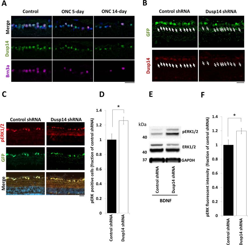
Dusp14 Regulates Axon Growth                                                                     IOVS j June 2018 j Vol. 59 j No. 7 j 2737

             mitogen activated kinase (MAPK) signaling. Similarly, directly           dent samples obtained on different days served as the starting
             manipulating MAPK family members (ERKs, JNKs, p38, and                   material for each microarray. Raw data files were normalized
             DLK) in RGCs27–32 can improve survival, axon growth, and                 using the quantile method with GeneSpring GX 11 software
             regeneration.                                                            (Agilent Technologies). Normalized data were filtered by
                 How intrinsic and extrinsic factors interact to jointly              percentile of intensity. Only the probes between the 20th
             regulate developmental growth and regeneration after injury              and 99th percentiles in at least 50% of the samples per
             is not well understood. We investigated genes downstream of              condition were considered for further analysis. Statistical
             KLFs family members, to determine the axon growth-relevant               comparison between conditions was performed using un-
             gene targets and pathways regulated by these transcription               paired Student’s t-test with Benjamini-Hochberg correction for
             factors. We discovered that KLF9 drives expression of dual               multiple testing. Final data analysis was conducted using Excel
             specific phosphatase-14 (DUSP14), and that DUSP14 dephos-                software (Microsoft Corporation, Redmond, WA, USA). All raw
             phorylates MAPK family members and its downregulation or                 data files are available at the NIH GEO Database - Series ID #
             inhibition blocks the axon growth inhibitory effects of KLF9 in          GSE92507.
             vitro and promotes axon regeneration after optic nerve crush                 For screening, we selected KLF family gene targets that met
             in vivo. This study uncovered a novel intrinsic transcription            the following criteria: (1) upregulated gene expression at least
             factor–mediated regulation of extrinsic neurotrophic signaling           3-fold by neurite growth-suppressive factor KLF9; (2) upregu-
             pathways and created a molecular link between these in the               lated at least 1.5-fold by neurite growth-suppressive KLF16; (3)
             regulation of survival, axon growth, and regeneration.                   downregulated at least 1.5-fold by neurite growth-promoting
                                                                                      factor, KLF7; and (4) no change in gene expression by KLF11.
                                                                                      Full-length clones were from an Open Biosystems (Thermo
             METHODS                                                                  Fisher Scientific, Huntsville, AL, USA) library maintained by
                                                                                      Vance Lemmon, University of Miami. Transfected RGCs were
             Constructs for Transfection and Virus                                    identified by co-transfection with pmax-GFP (Lonza), and
             Transduction                                                             FLAG-tagged mCherry was used as a control. Co-transfection
                                                                                      efficiency of pmax-GFP and FLAG-mCherry construct was 84.7
             For microarray analysis of RGCs, the open reading frames of              6 1.2% in the beta-tubulin positive RGCs.
             KLF9, -16, -7, and -11 were cloned from plasmids obtained
             from Open Biosystems (Huntsville, AL, USA). All 4 KLFs and
             FLAG-tagged mCherry33 were cloned into pLenti-MP2 expres-
                                                                                      Purification, Culture, and Transfection of Primary
             sion vector. For the screen in RGCs, plasmid constructs in               RGCs
             pCMV-SPORT6 (Open Biosystems) were co-transfected with                   RGCs (P0 through P8) were purified by immunopanning as
             pmax-GFP (Lonza, Walkserville, MD, USA) and compared to co-              described previously.14,15,35,36 RGCs were plated onto PDL-
             transfection of control FLAG-tagged mCherry33 with pmax-                 and laminin-coated tissue culture plates in growth media14
             GFP. Dusp14 and KLF9 were obtained from Open Biosystems                  including a homemade supplement similar to B27,37 forskolin
             and cDNAs were subcloned into the SpeI/XbaI site in pLenti-              (5 mM), BDNF (50 ng/mL) and CNTF (10 ng/mL). In some
             MP2 expression vector with FLAG-mCherry or GFP tag. The                  experiments, protein tyrosine phosphatase (PTP) inhibitor IV
             phosphatase-dead mutant of Dusp14 was obtained by substi-                (EMD Millipore, Billerica, MA, USA) was used as a pharmaco-
             tuting cysteine for serine at position 111 (C111S) using the             logic Dusp14 inhibitor.38 PTP inhibitor IV dissolved in dimethyl
             QuikChange II XL Site-Directed Mutagenesis Kit (Agilent                  sulfoxide (DMSO) or DMSO control solutions were added at
             Technologies, Santa Clara, CA, USA). FLAG-tagged mCherry-                various concentrations 2 hours after electroporation and
             pLenti-MP2 was used as control vector. To suppress Dusp14                plating. RGCs were cultured in 10% CO2 for 48 hours to
             expression in vivo, an inducible RNA polymerase II promoter34            analyze neurite growth.
             was subcloned upstream of four target shRNAs against the rat                 For electroporation, after purification, 500 K RGCs were
             Dusp14 gene (Gene Bank accession, BC158555) using target                 resuspended in electroporation solution containing 2 lg total
             sequences as follows: 5 0 -TCGGATGATTTCCGAGGGAGA-3 0 , 5 0 -             DNA (GFP reporter and gene of interest in a 1:9 ratio), placed
             CTGACAAGATCCACAGTGTAA-3 0 , 5 0 -CTGGAGGCAGCTGATA                        in a small cell number cuvette (Sigma-Aldrich Corp., St. Louis,
             GACTA-3 0 and 5 0 -TGGCATCATTCCAGACGTTTA-3 0 . These four                MO, USA) and electroporated using Amaxa program SCN#1.
             shRNA sequences in a SIBR cassette34 were concatenated into              Immediately following electroporation, growth media was
             a single vector and inserted into pAAV2 vector backbone,                 added and the whole solution placed into a small Eppendorf
             which also expresses eGFP as a transduction marker. Anti-                tube. RGCs were centrifuged for 16 minutes at 300g (1800 rpm
             luciferase shRNAs in the same pAAV2 system (kind gift from               in an Eppendorf 5415D centrifuge) before resuspension and
             Vance P. Lemmon, University of Miami, Miami, FL, USA) were               plating. For siRNA electroporation, 500 K postnatal rat RGCs
             used as controls. All constructs were verified by sequencing.            were electroporated immediately after purification either with
                                                                                      negative control or anti-Dusp14 siRNAs (Rn_Dusp14_pre-
             RNA Preparation, Microarray Hybridization and                            dicted_1 and Rn_Dusp14_predicted_2; Qiagen), 500 nM per
             Data Analysis                                                            electroporation reaction in 27 lL electroporation solution and
                                                                                      2 lL (2 lg) transfection DNA (KLF9 or control). All siRNAs
             RGCs from early postnatal (P4) rats were purified and KLF9,              were validated for knockdown effect by immunohistochemis-
             -16, -7, and -11 genes and control, FLAG-tagged mCherry gene             try using isolated RGCs (data not shown). RGCs were cultured
             were transduced using lentivirus described as above. Total RNA           in 10% CO2 for 48 hours to quantify cell neurite growth.
             was extracted (RNeasy; Qiagen, Valencia, CA, USA) indepen-                   For viral transduction of RGCs, 2500 P8 RGCs/well were
             dently and shipped to the National Institutes of Health (NIH,            plated on PDL/laminin-coated 48-well plates. At 16 hours after
             Bethesda, MD, USA) Neuroscience Microarray Consortium                    plating, AAV2 virus was diluted 1:1500 in media and added to
             (University of California Los Angeles, Los Angeles, CA, USA),            cultured cells. Full media changes were performed 5.5 hours
             where it was amplified and processed for hybridization onto rat          following virus exposure. For neurite growth assays, RGCs
             genome arrays (GeneChip Rat Genome 230 2.0 Array;                        were fixed in 4% paraformaldehyde 5 days after initial addition
             Affymetrix, Santa Clara, CA, USA). At least five microarrays             of AAV2 viruses. The titer of AAV2 viruses ranged from 2.0 to
             were used for each condition. RNA collected from indepen-                3.0 3 1013 genome copies/mL.

Downloaded From: https://iovs.arvojournals.org/pdfaccess.ashx?url=/data/journals/iovs/937150/ on 01/04/2019
Dusp14 Regulates Axon Growth IOVS j June 2018 j Vol. 59 j No. 7 j 2738
Immunohistochemistry template for a qPCR reaction (SsoAdvanced SYBR green; Bio-Rad
Laboratories) performed on an iCycler (Bio-Rad Laboratories)
For cultured neurons, cultures were fixed using prewarmed with Dusp14 and 18S primers. Three repeat wells (technical
378C 4% paraformaldehyde (PFA). For sections, frozen 15 lm replicates) were used for each condition. ‘‘No RT’’ control
sections were fixed with 4% PFA. Following rinses in PBS, samples also were tested. The experiment was repeated four
samples were blocked and permeabilized in 20% normal goat times with different pools of RNA (biological replicates). Primers
serum (NGS) or 20% normal donkey serum with 0.02% Triton used were as follows: Dusp14 forward: 5 0 TCGAGATCCC
X-100 in antibody buffer (150 mM NaCl, 50 mM Tris base, 1% CAACTTCAAC 3 0 ; Dusp14 reverse: 5 0 TGTCAGCCACAGTGT
BSA, 100 mM L-lysine, 0.04% Na azide, pH 7.4) for 1 hour to CAAAG 3 0 ; 18S forward 5 0 GAACTGAGGCCATGATTAAGA 3 0 ;
reduce nonspecific binding. Samples were incubated overnight and 18S reverse: 5 0 CATTCTTGGCAAATGCTTTC 3 0 .
at 48C in antibody buffer containing primary antibodies,
washed with PBS, incubated in antibody buffer containing
secondary antibodies and 4 0 ,6-diamidino-2-phenylendole (DA- Protein Extraction and Western Blot Analysis
PI) for 1 hour at room temperature, and washed with PBS. RGCs were purified by immunopanning and transduced with
Cultures were left in PBS for imaging; sections were mounted virus as above. At 48 hours after lentivirus exposure for
using ProLong Gold Antifade Reagent (Molecular Probes, Inc., Dusp14 wild-type, Dusp14 C111S, KLF9, and FLAG-mCherry as
Eugene, OR, USA). Cultures were imaged for axon growth as a control, RGC proteins were extracted with radioimmuno-
below; sections were examined using confocal laser micros- precipitation assay (RIPA) buffer containing protease and
copy (TCS SP5, Leica or Zeiss 710; Carl Zeiss Meditec, Jena, phosphatase inhibitor cocktails (Thermo Fisher Scientific).
Germany) and fluorescence microscopy (Observer.Z1; Carl For collecting protein from in vivo retina, rats were deeply
Zeiss Meditec). anesthetized and euthanized by inhalation overdose of CO2 24
Primary antibodies used for these experiments included hours after optic nerve injury, and retina proteins were
anti-b tubulin antibody from E7 hybridoma (1:500; Develop- extracted with RIPA buffer containing protease and phospha-
mental Studies Hybridoma Bank, Iowa City, IA, USA), anti-b-III tase inhibitor cocktails. Proteins in the cell extracts were
tubulin (1:500, 5568; Cell Signaling Technology, Danvers, MA, separated by SDS-PAGE and electrotransferred to polyvinyli-
USA), anti-FLAG (1:250; Sigma-Aldrich Corp., F7425 and dene fluoride (PVDF) membranes (Millipore) at 30 V for 60
F1804), anti-GFP (1:200, A11122; Invitrogen, Carlsbad, CA, minutes using the XCell SureLock Mini-Cell (Invitrogen).
USA and GFP-1020; Aves Labs, Tigard, OR, USA), anti-phospho- Membranes were blocked with chemiluminescent blocker
ERK1/2 (1:200, 4370S; Cell Signaling Technology), anti-Brn3a (Millipore) and the antibody reaction performed with the SNAP
(1:300, MAB1585; Millipore), anti-RBPMS (1:300, 1830-RBPMS; i.d. system (Millipore). Primary antibodies used for these
PhosphoSolutions, Aurora, CO, USA) and anti-Dusp14 (1:150, experiments included anti-phospho-ERK1/2 antibody (1:333;
ab110938; Abcam, Cambridge, UK). Secondary antibodies Sigma-Aldrich Corp.), anti-ERK1/2, anti-phospho-SAPK/JNK,
were Alexa Fluor 488-, 594-, or 647-conjugated, highly cross- anti-SAPK/JNK, anti-phospho-P38 MAPK, anti-P38 MAPK anti-
adsorbed antibodies (1:500; Invitrogen) and FITC-conjugated body (all 1:333; Cell Signaling Technology), and anti-GAPDH
anti-chicken Igc antibody (1:500, F-1005; Aves Labs). antibody (1:2000; Cell Signaling Technology). Secondary
Number of pERK-positive cells per 100 lm in the RGC layer antibodies were horseradish peroxidase (HRP)–conjugated
and the fluorescence intensity of pERK per area in the RGC anti-mouse IgG and anti-rabbit IgG antibody (1:500; Santa Cruz
and inner plexiform layer were compared between control Biotechnology, Dallas, TX, USA). Immunopositive bands were
retinas and retinas with Dusp14 shRNA. visualized by chemiluminescence with enhanced chemilumi-
nescence (ECL; Thermo Fisher Scientific) and imaged on an
Quantification of Neurite Length and Neurite LAS-3000 (Fujifilm, Tokyo, Japan). Densitometry was per-
Number formed with Multi Gauge version 3.1 software (Fujifilm).
For high content screening (HCS) of neuronal morphology,
Animal Surgeries
including average and maximum neurite lengths, neurite
number and branching, automated microscopy (ArrayScan Detailed protocols are available upon request. Animal exper-
VTI, Thermo Fisher Scientific) and image analysis software iments were conducted in accordance with the guidelines of
(Cellomics Neuronal Profiling BioApplication; Thermo Fisher the Institutional Animal Care and Use Committee (IACUC) and
Scientific) were used to image and trace neurons using an 35 the Institutional Biosafety Committee of the University of
or 310 objective following immunostaining. RGCs were traced Miami, University of California, San Diego, and Stanford
using b-tubulin immunoreactivity to visualize neurites. Neu- University, and complied with the ARVO Statement for the
rons with dim signal in neurites were excluded from analysis, Use of Animals in Ophthalmic and Vision Research.
due to frequent tracing errors of faint processes; the threshold Sprague-Dawley rats of varying ages and sex were obtained
for exclusion was established using a population of negative from Harlan Laboratories (Indianapolis, IN, USA). All surgeries
control (Mock-transfected) immunostained RGCs. We previ- were performed under adequate anesthesia with an intraper-
ously validated this approach’s consistency and reliability itoneal injection of ketamine hydrochloride, 60 mg/kg, and
compared to hand-tracing.4 Images and tracing were spot- xylazine hydrochloride, 8 mg/kg body weight. Postoperatively,
checked to verify that the algorithms were correctly identifying animals were allowed to recover on a heating pad and were
neurites and quantifying growth. given subcutaneous injections of buprenorphine hydrochlo-
ride, 0.1 mg/kg, twice a day for 3 consecutive days to minimize
Quantitative Reverse Transcription Polymerase discomfort.
Chain Reaction (qRT-PCR) To inhibit Dusp14 expression, adeno-associated virus (sero-
form 2: AAV2) expressing Dusp14 shRNA was injected intra-
RGCs were purified by immunopanning and transduced with vitreally (4.0 lL virus solution [titer, 7.0 3 1012]) to the left eye
virus as above. At 48 hours after virus exposure for KLF9, cells at postnatal day 21 (P21) or Dusp 14 knockout (KO) mice were
were harvested and total RNA purified with RNeasy kit (Qiagen), used. AAV2 expressing anti-luciferase shRNA and an eGFP
subjected to reverse transcription (RT, iScript; Bio-Rad Labora- reporter was injected in control eyes. One week after
tories, Hercules, CA, USA), and the resulting cDNA used as the injection, RGCs were retrogradely labeled with fluorogold
Downloaded From: https://iovs.arvojournals.org/pdfaccess.ashx?url=/data/journals/iovs/937150/ on 01/04/2019Dusp14 Regulates Axon Growth IOVS j June 2018 j Vol. 59 j No. 7 j 2739
(FG; Fluorochrome) to study RGC survival, as described phenotypes.40 Since KLFs (e.g., KLF4) require their DNA
previously.39 In brief, the animals were anesthetized and the binding domain to suppress axon growth,4 and since multiple
skull was exposed by a midline incision. Bilateral 2-mm different KLFs have opposite effects on axon growth,4,5 we
diameter craniostomies were placed 0.5 mm posterolateral to hypothesized that identifying gene targets coordinately regu-
the sagittal and transverse sutures. A small piece of Gelfoam lated by KLFs in neurons could unveil new pathways or
(Gelfoam, USP; Pfizer, New York, NY, USA) soaked in 4% FG mechanisms of axon growth regulation. We virally transduced
then was placed on the surface of the superior colliculus. Two purified postnatal day 4 (P4) RGCs15,35 to overexpress single
weeks after virus injection, the left optic nerve was exposed KLF family members, either axon growth suppressors (KLF9 or
and crushed using fine forceps (Dumont #5) at 2.0 mm behind KLF16), an axon growth promoter (KLF7), or a control KLF
the optic nerve head for 10 seconds, avoiding injury to the family member that has no effect on axon growth (KLF11), or a
ophthalmic artery. A surgeon, masked to the viral treatment, control FLAG-mCherry construct.4,5 Transcriptomes examined
performed all optic nerve crush surgeries. Two days before the by Affymetrix microarray across five to six biological replicates
end of the study, we injected 4.0 lL of 10 lg/lL cholera toxin of each KLF demonstrated low intersample variability (Supple-
subunit B (Ct B -555, Molecular Probes, Inc.) as an anterograde mentary Fig. S1). Of the targets probed, 21% changed at least
tracer to visualize axons and nerve terminals originating from 1.5-fold in at least one condition compared to FLAG-mCherry.
living RGCs. Rats with any significant postoperative complica- Remarkably, 99.6% of the 1556 targets regulated by axon
tions (e.g., retinal ischemia, cataract) were excluded from growth suppressors KLF9 and KLF16 showed expression
further analysis. Two weeks after optic nerve injury, animals changes in the same direction, and 86% of the 1139 targets
were deeply anesthetized and perfused with 4% PFA in PBS. regulated by axon growth suppressor KLF9 and axon growth
Optic nerves and retinas were dissected and fixed in 4% PFA enhancer KLF7 changed in the opposite direction (Fig. 1A),
for 1 hour and subsequently washed in PBS. Optic nerves were suggesting that KLFs’ similar (KLF9 and -16) or opposing (KLF9
cryopreserved by incubation in 20% sucrose at 48C overnight and -7) effects on axon growth are largely preserved in their
before mounting in optimal cutting temperature compound coordinated regulation of gene expression. KLF regulation of
(OCT). Longitudinal sections (10 lm) were made of the entire other KLFs did not, however, demonstrate this consistency
optic nerve and imaged with 320 magnification objective as (Supplementary Table S1), pointing to a complexity in their
above. Pictures were taken, starting with the furthest cross-regulation.
regenerating axons and working backwards toward the crush We hypothesized that genes that change similarly in
site. Lines were drawn perpendicular to the long axis of the response to KLF9 and KLF16 (axon growth suppressors), but
optic nerve 0.25, 0.5, 0.75, and 1.0 mm past the crush site (as in the opposite direction in response to KLF7 (axon growth
applicable), and Ct B-positive axons between these lines were promoter), and not in response to KLF11 (no effect on axonal
counted. Analysis of the total sum of regenerating fibers from growth), might be the best candidates to identify direct or
all sections for each animal as well as the average number of indirect KLF gene targets key in regulating intrinsic axon
axons at each measurement location/distance per number of growth ability (Fig. 1B). A total of 27 genes satisfied those
sections were performed in a masked fashion. To study RGC criteria (Supplementary Table S2). We selected the subset with
survival, retinas were dissected at days 7, 10, and 14 and full-length clones available from the National Institutes of
immunohistochemistry was performed against Brn3a (1:300, Health Mammalian Gene Collection and expressed them
Millipore) and RNA binding protein with multiple splicing individually in primary rat RGCs using GFP for a co-transfection
(RBPMS; 1:300, Phosphosolutions). Three images for each marker, FLAG-tagged mCherry as a control, and automated
quadrant of the retina were acquired using 710 Zeiss Confocal image acquisition and neurite tracing for rapid and unbiased
microscope (Carl Zeiss Meditec) and quantified by a researcher quantification of neurite growth.4,41 We found that Dusp14
blinded to the experimental condition. To evaluate phospho- strongly suppressed neurite growth, decreasing the average
ERK signaling, BDNF (5 lg/3 lL, human BDNF; PeproTech, neurite length to 72% (Fig. 1C) and decreasing the maximum
Rocky Hill, NJ, USA) was injected intravitreally immediately length of neurites to 66% (Fig. 1D) with no effect on neurite
after optic nerve crush. For sectioning, rats were deeply number (Fig. 1E). These effects were remarkably similar to
anesthetized and euthanized 3 days after optic nerve injury.
those seen in KLF9-transfected RGCs (Figs. 1C–E). For this
Rats with any significant postoperative complications (e.g.,
reason and since we had previously identified KLF9 as a major
retinal ischemia, cataract) were excluded from further analysis.
regulator of axonal growth6 further exploration of other KLFs’
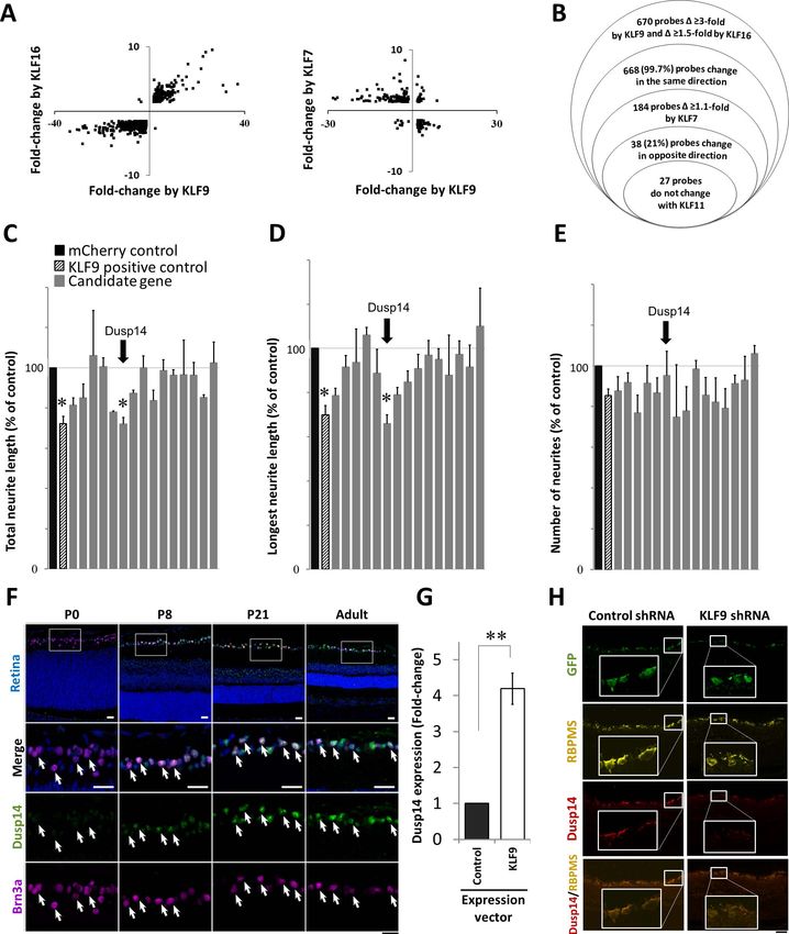
effects on Dusp14 expression was deferred.
Statistical Analysis We next investigated Dusp14 expression through retinal
Data were analyzed using the JMP version 8 statistical package development, from postnatal day 0 (P0) to adult. KLF9
program (SAS Institute, Cary, NC, USA) or Graphpad Prism 6 expression is developmentally upregulated 250-fold in RGCs
(Graphpad, San Diego, CA, USA). The Tukey-Kramer honest after birth.4 By immunofluorescence, no expression of Dusp14
significant difference (HSD) test was performed for multiple was detected in the retina at P0. By P8, Dusp14 was detectable
comparison for gene screening. For statistical comparison of in RGCs (identified by Brn3a staining) and increased through
two samples, we used a 2-tailed Student’s t-test or paired t-test. P development into adulthood (Fig. 1F). At P21 and in adult
values of less than 0.05 were regarded as statistically significant. retina, Dusp14 also was detected in cells located on the inner
side of the inner nuclear layer, but no expression of Dusp14
protein was detected in photoreceptor cells, retinal pigment
RESULTS cells or vascular endothelial cells. qRT-PCR showed Dusp14
expression increased 4-fold after KLF9 transduction compared
Microarray Analysis of KLF Family Members to control (Fig. 1G). Conversely, knocking down KLF9 in vivo
Identifies Dusp14 as Developmentally Regulated after intravitreal AAV2-anti-KLF9-shRNA (using constructs
Axon Growth Suppressor previously validated at the mRNA and protein level)6 decreased
Dusp14 protein expression compared to control (red). Taken
KLF-family Cys/His2-type zinc finger domains bind similar DNA together, KLF9 is sufficient in vitro and necessary in vivo for
consensus sequences and demonstrate redundant and com- Dusp14 expression, and the two genes increase along a similar
pensatory activities in regulating target genes and cellular time course through early postnatal development.
Downloaded From: https://iovs.arvojournals.org/pdfaccess.ashx?url=/data/journals/iovs/937150/ on 01/04/2019Dusp14 Regulates Axon Growth IOVS j June 2018 j Vol. 59 j No. 7 j 2740
FIGURE 1. Microarray analysis and screening of candidate genes regulated by KLF family members identifies Dusp14 as developmentally regulated
axon growth suppressor. (A) Scatterplots of KLF-regulated gene targets undergoing significant change in RGCs. (B) Modified Venn diagram
characterizing gene expression analysis of RGCs expressing different KLF family members. (C–E) Gene expression effects on neurite growth
screened in P0 RGCs. KLF9 (pattern column) and Dusp14 (arrow) decreased average and maximum neurite length with no effect on neurite
number (*P < 0.05, Tukey-Kramer HSD test). (F) Dusp14 expression in the retina, at different postnatal ages. (G) qRT-PCR for Dusp14 in RGCs after
KLF9 or control vector transduction (**P < 0.01, paired t-test; n ¼ 4). (H) AAV2-anti-luciferase-shRNA (control shRNA) or AAV2-anti-KLF9-shRNA
were intravitreally injected in adult rats and expressed in RGCs in vivo (GFP reporter, green). At 2 weeks, KLF9 shRNA led to a decrease in Dusp14
expression (red) in RGCs (labeled with RBPMS, yellow). Scale bar: 25 lm in (F) and 50 lm in (H). Error bars: standard error of the mean (SEM).
Downloaded From: https://iovs.arvojournals.org/pdfaccess.ashx?url=/data/journals/iovs/937150/ on 01/04/2019Dusp14 Regulates Axon Growth IOVS j June 2018 j Vol. 59 j No. 7 j 2741
Dusp14 is Required for KLF9-Mediated Neurite dephosphorylation of members of all three MAPK families,
Growth Suppression pERK1/2, pJNK, and pP38 (Figs. 3G–J). In contrast,
Dusp14C111S elicited no significant change in the phosphory-
KLF9 is the most highly developmentally regulated axon lation of MAPKs (Figs. 3G–J). Therefore, KLF9 and Dusp14 lead
growth suppressor of the KLF family, increasing over 250-fold to dephosphorylation of MAPKs, suggesting a hypothesis that
through RGC development.4 Thus, we tested whether Dusp14 this pathway limits axon growth by reducing intracellular
mediates KLF9’s suppression of axon growth. In P0 RGCs, signaling normally responsive to neurotrophic environmental
which do not express detectable levels of Dusp14 protein and cues.
express very low levels of KLF9 mRNA, two different Dusp14-
specific siRNAs had, as expected, no effect on neurite growth.
Dusp14 Regulates RGC Survival and Optic Nerve
When we overexpressed KLF9, both Dusp14-specific siRNAs
fully rescued P0 RGC neurite growth (Fig. 2A). In P8 RGCs, Axon Regeneration In Vivo
these siRNAs increased neurite growth in KLF9-overexpressing First we asked whether Dusp14 expression is dysregulated
and control-transfected neurons (Fig. 2B), consistent with the after axon injury in vivo. Using a common model of optic nerve
endogenous expression of KLF9 and Dusp14 in these older trauma, we found that surviving RGCs continued to express
neurons. Similarly, AAV2-anti-Dusp14-shRNA significantly in- Dusp14 protein after optic nerve crush without significant
creased neurite growth compared to control AAV2-anti- change detectable by immunofluorescence at days 5 and 14
luciferase shRNA (Figs. 2C, 2D). We also examined pharmaco- after injury (Fig. 4A). We evaluated molecular and cellular
logic inhibition of Dusp14 activity on neurite growth. PTP responses of RGCs to Dusp14 knockdown using AAV2-anti-
inhibitor IV, which effectively and specifically inhibits Dusp14- Dusp14-shRNA vectors containing four concatenated anti-
mediated dephosphorylation,38 similarly promoted axon Dusp14 shRNAs, which transduced 93.3% of RGCs and,
growth in a dose-dependent manner in control and KLF9- compared to control AAV2-anti-luciferase-shRNA, decreased
transfected P8 RGCs (Figs. 2E, 2F). Taken together, these expression of Dusp14 in RGCs at 2 weeks after intravitreal viral
results demonstrated that KLF9 requires expression of its target injection (Fig. 4B). Although the knockdown likely was not as
gene Dusp14 to suppress RGC axon growth ability. complete as genomic KO would be,49 we continued our
studies with viral vectors to avoid developmental changes that
Dusp14-Mediated Axon Growth Inhibition could confound a full KO mouse, to maintain a greater measure
Depends on its Catalytic Activity of RGC-specificity, and to mimic the effect of treatment in the
adult. We further found that Dusp14 shRNA significantly
To determine the relevance of Dusp14’s catalytic activity, we increased the number of BDNF-induced pERK-positive cells
examined the effect of a point mutation at cysteine-111 to (Figs. 4C, 4D) and increased BDNF-dependent pERK signaling
serine (Dusp14C111S), which leads to loss of enzymatic (Figs. 4E, 4F).
dephosphorylation activity.42 Dusp14’s cysteine-111 residue Finally, we evaluated the effect of knocking down Dusp14
catalyzes removal of phosphates initiated by a cysteine thiolate expression on RGC survival and axon regeneration after optic
anion that attacks the tyrosine phosphate to form a cysteinyl- nerve crush. Dusp14 knockdown promoted RGC survival at 7
phosphate intermediate.43 Compared to control transfected days but not at 10 or 14 days after optic nerve crush in the
neurons, neurite growth was strongly suppressed in the KLF9- adult rat (Figs. 5A, 5B). Interestingly, Dusp14 KO animal
or Dusp14-transfected P4 RGCs (Figs. 3A–D). Co-transduction robustly promoted RGC survival 14 days after optic nerve
of KLF9 and Dusp14 showed stronger suppression of axon injury (Figs. 5C, 5D). Dusp14 knockdown significantly
growth than KLF9 alone, but no significant inhibitory effect on increased the number and length of regenerating axons of
neurite growth was observed in RGCs co-transduced with RGCs in vivo (Figs. 5E, 5F). However, Dusp14 KO mice (Figs.
KLF9 and Dusp14C111S (Fig. 3D). To explore morphologic 5G, 5H) and PTP inhibitor IV (data not shown) did not promote
correlates of KLF9’s and Dusp14’s effects on inhibiting neurite any significant increase in axon regeneration after axon injury.
growth, we quantified growth cone morphologies of RGCs Thus, Dusp14 specifically suppresses axon regeneration in vivo
overexpressing all four constructs. KLF9- and Dusp14-trans- after optic nerve injury, and targeting Dusp14 expression in
fected RGCs demonstrated enlarged growth cones compared RGCs with partial knockdown by shRNA promotes axon
to control mCherry. The Dusp14C111S mutant had no effect on regeneration of RGCs.
growth cone morphology or neurite growth (Figs. 3E, 3F).
Growth cone size clearly is not related to inhibition versus
elongation as this relationship may be context dependent,44,45 DISCUSSION
but these data may help us understand the mechanism of effect
to be explored in future studies. Dusp14C111S confers a We identified Dusp14 as a critical player downstream of KLF9,
dominant negative effect, preventing KLF9 expression from mediating KLF9’s axon growth suppressive effects on axon
driving Dusp14-mediated suppression of axon growth, sup- growth in vitro and contributing to inhibition of axon
porting the conclusion that Dusp14 is critical to KLF9- regeneration in vivo after optic nerve trauma. Furthermore,
mediated axon growth suppression. these data supported the hypothesis for the interaction
How does the KLF-Dusp14 pathway regulate axon growth? between developmentally regulated, cell-intrinsic mediators
In nonneuronal cells, Dusp family members can inactivate of axon growth (e.g., KLFs), and extrinsic cues (e.g.,
mitogen-activated protein kinases (MAPKs), including ERK, neurotrophin-activated MAPKs). Enhancement of ERK phos-
JNK and P38 family members, through dephosphorylation at phorylation stimulates neurite outgrowth and specific inhibi-
threonine and tyrosine residues (TXY motif) in the kinase tors of this pathway attenuate survival and neurite growth.50
domains.42,46,47 MAPK proteins are critical for survival and p38 MAPK signaling pathway also is implicated in neurito-
axon growth of RGCs in vitro and in vivo,15 and are activated genesis and regeneration,51 and JNK signaling is involved in
by a variety of extracellular neurotrophic ligands that act on neurite initiation and elongation.52,53 Interestingly, our own
receptors at the cell body and the growth cone.48 We results showed increase in pJNK after optic nerve injury and
investigated whether Dusp14 regulates MAPK phosphorylation that JNK3 binding domain is necessary for the KLF9-induced
in P0 RGCs, as little expression of Dusp14 was detected in vivo axon growth inhibition.6 Thus, our data demonstrated that
at that age (Fig. 1F). Expression of KLF9 or of Dusp14 led to Dusp14 may decrease effects of all three contributing MAPK
Downloaded From: https://iovs.arvojournals.org/pdfaccess.ashx?url=/data/journals/iovs/937150/ on 01/04/2019Dusp14 Regulates Axon Growth IOVS j June 2018 j Vol. 59 j No. 7 j 2742
FIGURE 2. Rescue of KLF9-mediated neurite growth suppression by Dusp14 siRNA and PTP inhibitor IV. (A, B) At P0, control-transfected RGCs with
no Dusp14 protein expression showed no effect when two different siRNAs against Dusp14 were transfected, in contrast to RGCs expressing higher
levels of Dusp14 protein at P8, in which Dusp14 siRNAs significantly promoted neurite elongation. When overexpressing KLF9, siRNAs against Dusp14
rescued RGCs neurite growth at P0 (A) and P8 (B). (C, D) Similarly, AAV2 shRNA anti-luciferase (control shRNA) or AAV2 anti-Dusp14 (Dusp14 shRNA)
were expressed in P8 RGCs. Dusp14 shRNA led to a significant increase in RGCs neurite growth. Images show staining against b-III-tubulin. Positively
transduced RGCs were identified by a GFP reporter (C). Quantification showed significant increase in neurite growth after 5 days in culture (D). (E, F)
PTP inhibitor IV (Dusp14 inhibitor) rescued KLF9-induced neurite suppression at P0 (E) and P8 (F), and promoted neurite growth in control-
transfected RGCs at P8 (F), in dose-dependent fashion (*P < 0.05, **P < 0.01, paired t-test). Scale bar: 200 lm in (C). Error bars: SEM.
Downloaded From: https://iovs.arvojournals.org/pdfaccess.ashx?url=/data/journals/iovs/937150/ on 01/04/2019Dusp14 Regulates Axon Growth IOVS j June 2018 j Vol. 59 j No. 7 j 2743
FIGURE 3. Dusp14 activity is required for Dusp14- and KLF9-induced RGC axon growth inhibition and MAPK dephosphorylation. (A–D) Neurite
growth was inhibited by Dusp14 and KLF9 but not Dusp14C111S. Neurite growth of RGCs co-transfected with KLF9 and Dusp14 or Dusp14C111S (*P
< 0.05, **P < 0.01, paired t-test). (E) Morphology of the growth cones of RGCs transfected with KLF9, Dusp14, and Dusp14C111S. (F) RGCs
transfected with KLF9 and Dusp14 led to enlarged growth cones, whereas Dusp14C111S showed growth cones morphology similar with mCherry
control. (G) Western blots for total ERK1/2, phospho- (p-) ERK1/2, total JNK, p-JNK, total P38 and p-P38 were analyzed in RGCs after gene
transduction. (H–J) Densitometry of p-MAPK/total MAPK ratios: in Dusp14-transduced RGCs, the phosphorylation ratio of ERK1/2 (H), JNK (I), and
P38 (J) were decreased 31.8%, 21.9%, and 26.1%, respectively. KLF9 induced a similar reduction of phosphorylation, but Dusp14C111S showed no
effect. (*P < 0.05, **P < 0.01 vs. control, paired t-test, n ¼ 4; WT, wildtype). Scale bar: 5 lm in (A), 50 lm in (E). Error bars: SEM.
Downloaded From: https://iovs.arvojournals.org/pdfaccess.ashx?url=/data/journals/iovs/937150/ on 01/04/2019Dusp14 Regulates Axon Growth IOVS j June 2018 j Vol. 59 j No. 7 j 2744
FIGURE 4. Dusp14 regulates axon regeneration through dephosphorylation of the MAPK family in vivo. (A) No change in Dusp14 expression was
detectable at 5 and 14 days after optic nerve crush. (B) Dusp14 expression decreased after intravitreal injection of AAV2-Dusp14-shRNA in vivo
(arrows). (C–F) Dusp14 knockdown significantly increased the number of pERK1/2-positive cells (D) and pERK1/2 fluorescent intensity (E) in
retinas treated with BDNF. (*P < 0.05, Student’s t-test; n ¼ 4 [D, E]) Scale bars: 25 lm in (A) to (C), 500 lm in (D). Error bars, SEM.
signaling families (p38, ERK, and JNK), and at a minimum (DUSPs) make up a subgroup of the class-I cysteine-based
Dusp14 suppression significantly promotes BDNF-pERK sig- PTP superfamily that regulate divergent biological and patho-
naling in RGCs in vivo. These data suggested that any residual logical processes.57 They are a family of 61 phosphatases with
or exogenously applied neurotrophic factors may have their different tissue expression patterns and heterogeneous func-
pro-regenerative effects abrogated by Dusp14 expression in tions,42 including specifically dephosphorylating MAPK family
adult RGCs or other CNS neurons. Thus, the developmentally members, thereby inactivating them. Tight regulation of MAPK
regulated expression of KLFs in adult CNS neurons may act to family members is critical to control growth after develop-
suppress neurotrophic factor signaling. ment.46 While we found Dusp14 expression changed devel-
Other phosphatases have demonstrated activity in regulat- opmentally (Fig. 1F), and that Dusp14 expression is driven by
ing neurite growth and regeneration, including phosphatase KLF9 (Figs. 1G, 1H), it currently is unknown whether Dusp14
and tensin homolog (PTEN),54 protein phosphatase A2 is directly or indirectly regulated by KLF9 or other KLFs. We
(PP2A),55 and calcineurin.56 Dual specific phosphatases did not observe any change in Dusp14 expression after optic
Downloaded From: https://iovs.arvojournals.org/pdfaccess.ashx?url=/data/journals/iovs/937150/ on 01/04/2019Dusp14 Regulates Axon Growth IOVS j June 2018 j Vol. 59 j No. 7 j 2745
FIGURE 5. Dusp14 regulates optic nerve axon regeneration and RGC survival in vivo. (A) Images show RBPMS (RGC marker) staining of RGCs at days 7, 10,
and 14 after optic nerve crush, for either AAV2-shRNA Luciferase or AAV2-shRNA Dusp14 injected rats. (B) Dusp14 shRNA promotes RGC survival at 7 days
after optic nerve crush. (C) Images show RBPMS (RGC marker) staining of RGCs at day 14 after optic nerve crush, for either wild type control or Dusp14 KO
mice. (D) Dusp14 KO promotes RGC survival at 14 days after optic nerve crush. (E) Partial projections of sectioned optic nerve from Dusp14 knockdown
and control optic nerve. Regenerating axons (arrowheads) were observed in Dusp14 shRNA-treated optic nerves but not in controls. †Crush site. (F)
Significantly more fibers regenerated after shRNA-mediated knockdown of Dusp14. (G) Example partial projections of sectioned optic nerve from Dusp14
KO mice and control optic nerve. (F) No difference was detected in the number of regenerating axons in Dusp14 KO compared to controls. *Crush site (*P
< 0.05, **P < 0.01, 1-way ANOVA followed by Tukey’s test; n ¼ 4 [A, B]; n ¼ 5 [C, D]). Scale bars: 100 lm in (A), 500 lm in (C). Error bars: SEM.
Downloaded From: https://iovs.arvojournals.org/pdfaccess.ashx?url=/data/journals/iovs/937150/ on 01/04/2019Dusp14 Regulates Axon Growth IOVS j June 2018 j Vol. 59 j No. 7 j 2746
nerve injury, consistent with the premise that the develop- 3. Goldberg JL, Klassen MP, Hua Y, et al. Amacrine-signaled loss
mental regulation of KLFs is the primary, cell-autonomous of intrinsic axon growth ability by retinal ganglion cells.
regulator of Dusp14 expression. Interestingly, our bioinfor- Science. 2002;296:1860–1864.
matics analysis did not reveal a consensus KLF response 4. Moore DL, Blackmore MG, Hu Y, et al. KLF family members
element upstream in the proximal promoter region of the regulate intrinsic axon regeneration ability. Science. 2009;326:
DUSP14 locus. Other than Dusp14, no other Class-I PTPs, 298–301.
including other Dusp family members, were identified in our 5. Blackmore MG, Wang Z, Lerch JK, et al. Krüppel-like Factor 7
microarray data as potential downstream targets of KLF7, -9, engineered for transcriptional activation promotes axon
-16, or -11. Hence, Dusp14 may be one of the few Class-I PTP regeneration in the adult corticospinal tract. Proc Natl Acad
genes regulated by KLFs that specifically regulate MAPK Sci U S A. 2012;109:7517–7522.
dependent growth and regeneration in RGCs or other CNS 6. Apara A, Galvao J, Wang Y, et al. KLF9 and JNK3 interact to
neurons. suppress axon regeneration in the adult CNS. J Neurosci.
Knocking down Dusp14 in vivo yielded less regeneration 2017;37:1–13.
than manipulating KLF9 in vivo.6 This could be explain by 7. Norsworthy MW, Bei F, Kawaguchi R, et al. Sox11 expression
three reasons: first, KLF9 is a transcription factor that could be promotes regeneration of some retinal ganglion cell types but
activating other unidentified inhibitory pathways in vivo that kills others report Sox11 expression promotes regeneration of
were not present or were not activated in vitro. Second, our in some retinal ganglion cell types but kills others. Neuron.
vitro results were obtained in purified RGCs from postnatal 2017;94:1112–1120.
days 0 and 8 compared to manipulation of these factors in adult 8. Welsbie DS, Mitchell KL, Jaskula–Ranga V, et al. Enhanced
cells.15 Third, to maintain RGCs in culture, the media must be functional genomic screening identifies novel mediators of
highly supplemented with neurotrophic factors, such as BDNF dual leucine zipper kinase-dependent injury signaling in
and CNTF.6,14 Additionally, neither developmentally knocking neurons. Neuron. 2017;94:1142–1154.
out Dusp14, nor using a pharmacologic inhibitor, had any 9. Moore DL, Goldberg JL. Multiple transcription factor families
effect on axon regeneration after injury. This may reflect a regulate axon growth and regeneration. Dev Neurobiol. 2011;
requirement for Dusp14 activity at low levels in RGCs (since 71:1186–1211.
there is residual expression after shRNA knockdown), and/or 10. Tsujino H, Kondo E, Fukuoka T, et al. Activating transcription
for Dusp14 activity in other retinal or optic nerve cells, or by a factor 3 (ATF3) induction by axotomy in sensory and
compensatory effect of other Dusp family member. Indeed motoneurons: A novel neuronal marker of nerve injury. Mol
Dusp6 and Dusp1 have high homology with Dusp14. As the Cell Neurosci. 2000;15:170–182.
effect of KLF9 knockdown is greater than that of Dusp14 KO, 11. Herdegen T, Leah JD. Inducible and constitutive transcription
other targets of KLF9 should be explored in further studies on factors in the mammalian nervous system: control of gene
the differences in KLF biology within and across cells. expression by Jun, Fos and Krox, and CREB/ATF proteins.
Taken together these data strongly supported the motiva- Brain Res Rev. 1998;28:370–490.
tion to bridge biology and therapeutically target the interaction 12. Qin S, Zou Y, Zhang C-L. Cross-talk between KLF4 and STAT3
between cell-autonomous, developmentally regulated tran- regulates axon regeneration. Nat Commun. 2013;4:2633.
scriptional regulators and the signaling pathways critical for
13. Yiu G, He Z. Glial inhibition of CNS axon regeneration. Nat
regulating axon growth and regeneration. Further work linking
Rev Neurosci. 2006;7:617–627.
the developmental switch in intrinsic axon growth ability to
the extrinsic, glial-associated cues that limit survival and 14. Meyer-Franke A, Kaplan MR, Pfrieger FW, Barres BA.
growth of neurons may lead to new therapeutic approaches Characterization of the signaling interactions that promote
the survival and growth of developing retinal ganglion cells in
to promote CNS repair after injury or in degenerative disease.
culture. Neuron. 1995;15:805–819.
15. Goldberg JL, Espinosa JS, Xu Y, Davidson N, Kovacs GTA,
Acknowledgments Barres BA. Retinal ganglion cells do not extend axons by
The authors thank Vance P. Lemmon for access to a cDNA library default. Neuron. 2002;33:689–702.
and for anti-Luciferase shRNA pAAV2 plasmid; Yan Shi, Raquibul 16. Isenmann S, Klöcker N, Gravel C, Bähr M. Short communi-
Chowdhury, Pingping Jia, Eleut Hernandez, and Gabriel Gaidosh cation: protection of axotomized retinal ganglion cells by
for technical assistance from; and Evan G. Cameron and Sahil Shah adenovirally delivered BDNF in vivo. Eur J Neurosci. 1998;10:
for scientific discussion. 2751–2756.
Supported by the National Eye Institute (EY022129 to JLG; P30- 17. Harper MM, Grozdanic SD, Blits B, et al. Transplantation of
EY026877, P30-EY022589, and P30-EY014801), Department of BDNF-secreting mesenchymal stem cells provides neuropro-
Defense (W81XWH-12-1-0254), an unrestricted grant from Re- tection in chronically hypertensive rat eyes. Invest Ophthal-
search to Prevent Blindness, and the Uehara Memorial Foundation mol Vis Sci. 2011;52:4506–4515.
in Japan (to KI). 18. Mey J, Thanos S. Intravitreal injections of neurotrophic factors
Disclosure: J. Galvao, None; K. Iwao, None; A. Apara, None; Y. support the survival of axotomized retinal ganglion cells in
Wang, None; M. Ashouri, None; T.N. Shah, None; M. Black- adult rats in vivo. Brain Res. 1993;602:304–317.
more, None; N.J. Kunzevitzky, None; D.L. Moore, None; J.L. 19. Cui Q, Lu Q, So KF, Yip HK. CNTF, not other trophic factors,
Goldberg, None promotes axonal regeneration of axotomized retinal ganglion
cells in adult hamsters. Invest Ophthalmol Vis Sci. 1999;40:
References 760–766.
20. Leaver SG, Cui Q, Plant GW, et al. AAV-mediated expression of
1. Bregman BS, Kunkegbagden E, Mcatee M, Neill AO. Extension CNTF promotes long-term survival and regeneration of adult
of the critical period for developmental plasticity of the rat retinal ganglion cells. Gene Ther. 2006;13:1328–1341.
corticospinal pathway. J Comp Neurol. 1989;282:355–370. 21. Müller A, Hauk TG, Leibinger M, Marienfeld R, Fischer D.
2. Chen DF, Jhaveri S, Schneider GE. Intrinsic changes in Exogenous CNTF stimulates axon regeneration of retinal
developing retinal neurons result in regenerative failure of ganglion cells partially via endogenous CNTF. Mol Cell
their axons. Proc Natl Acad Sci U S A. 1995;92:7287–7291. Neurosci. 2009;41:233–246.
Downloaded From: https://iovs.arvojournals.org/pdfaccess.ashx?url=/data/journals/iovs/937150/ on 01/04/2019Dusp14 Regulates Axon Growth IOVS j June 2018 j Vol. 59 j No. 7 j 2747
22. Sawai H, Clarke DB, Kittlerova P, Bray GM, Aguayo AJ, 40. Moore DL, Apara A, Goldberg JL. Kruppel-like transcription
Hospital G. Brain-derived neurotrophic factor and neurotro- factors in the nervous system: Novel players in neurite
phin-4 / 5 stimulate growth of axonal branches from outgrowth and axon regeneration. Mol Cell Neurosci. 2011;
regenerating ganglion cells. J Neurosci. 1996;16:3887–3894. 47:233–243.
23. Goldberg JL, Barres BA. The relationship between neuronal 41. Buchser WJ, Pardinas JR, Shi Y, Bixby JL, Lemmon VP. 96-Well
survival and regeneration. Annu Rev Neurosci. 2000;23:579– electroporation method for transfection of mammalian
612. central neurons. Biotechniques. 2006;41:619–624.
24. Roberti G, Mantelli F, Macchi I, Massaro-Giordano M, 42. Patterson KI, Brummer T, O’Brien PM, Daly RJ. Dual-
Centofanti M. Nerve growth factor modulation of retinal specificity phosphatases: critical regulators with diverse
ganglion cell physiology. J Cell Physiol. 2014;229:1130–1133. cellular targets. Biochem J. 2009;418:475–489.
25. Maffei L, Carmignoto G, Perry V, Candeo P, Ferrari G. Schwann 43. Lountos GT, Tropea JE, Cherry S, Waugh DS. Overproduction,
cells promote the survival of rat retinal ganglion cells after optic purification and structure determination of human dual-
nerve section. Proc Natl Acad Sci U S A. 1990;87:1855–1859. specificity phosphatase 14. Acta Crystallogr Sect D Biol
26. Cohen A, Bray GM, Aguayo AJ. Neurotrophin-4 / 5 (NT-4 / 5) Crystallogr. 2009;65:1013–1020.
increases adult rat retinal ganglion cell survival and neurite 44. Kurklinsky S, Chen J, MA M. Growth cone morphology and
outgrowth in vitro. J Neurobiol. 1994;25:953–959. spreading are regulated by a dynamin-cortactin complex at
27. O’Brien GS, Sagasti A. Fragile axons forge the path to gene point contacts in hippocampal neurons. J Neurochem. 2011;
discovery: a MAP kinase pathway regulates axon regenera- 1117:48–60.
tion. Sci Signal. 2009;2:pe30. 45. Goldberg JL. How does an axon grow? Genes Dev. 2003;17:
28. Andrusiak MG, Jin Y. Context specificity of stress-activated 941–958.
MAP Kinase signaling: the story as told by C. elegans. J Biol 46. Jeffrey KL, Camps M, Rommel C, Mackay CR. Targeting dual-
Chem. 2016;291:7796:7804. specificity phosphatases: manipulating MAP kinase signalling
29. Welsbie DS, Yang Z, Ge Y, et al. Functional genomic screening and immune responses. Nat Rev Drug Discov. 2007;6:391–
identifies dual leucine zipper kinase as a key mediator of 403.
retinal ganglion cell death. Proc Natl Acad Sci U S A. 2013; 47. Huang C-Y, Tan T-H. DUSPs, to MAP kinases and beyond. Cell
110:4045–4050. Biosci. 2012;2:24.
30. Watkins TA, Wang B, Huntwork-Rodriguez S, et al. DLK 48. Steketee MB, Moysidis SN, Jin X-L, et al. Nanoparticle-
initiates a transcriptional program that couples apoptotic and
mediated signaling endosome localization regulates growth
regenerative responses to axonal injury. Proc Natl Acad Sci U
cone motility and neurite growth. Proc Natl Acad Sci U S A.
S A. 2013;110:4039–4044.
2011;108:19042–19047.
31. Hao Y, Frey E, Yoon C, et al. An evolutionarily conserved
49. Yang C-Y, Li J-P, Chiu L-L, et al. Dual-specificity phosphatase 14
mechanism for cAMP elicited axonal regeneration involves
(DUSP14/MKP6) negatively regulates TCR signaling by
direct activation of the dual leucine zipper kinase DLK. Elife.
inhibiting TAB1 activation. J Immunol. 2014;1924:1547–
2016;3:1–18.
1557.
32. Shin JE, Cho Y, Beirowski B, Milbrandt J, Cavalli V, DiAntonio
A. Dual leucine zipper kinase is required for retrograde injury 50. Washio A, Kitamura C, Jimi E, Terashita M, Nishihara T.
signaling and axonal regeneration. Neuron. 2012;74:1015– Mechanisms involved in suppression of NGF-induced neuro-
1022. nal differentiation of PC12 cells by hyaluronic acid. Exp Cell
Res. 2009;315:3036–3043.
33. Shaner NC, Campbell RE, Steinbach PA, Giepmans BNG,
Palmer AE, Tsien RY. Improved monomeric red, orange and 51. Verma P, Chierzi S, Codd A, et al. Axonal protein synthesis and
yellow fluorescent proteins derived from Discosoma sp. red degradation are necessary for efficient growth cone regener-
fluorescent protein. Nat Biotechnol. 2004;22:1567–1572. ation. J Neurosci. 2005;25:331–342.
34. Chung K, Hart CC, Al-bassam S, et al. Polycistronic RNA 52. Barnat M, Enslen H, Propst F, Davis RJ, Soares S, Nothias F.
polymerase II expression vectors for RNA interference based Distinct roles of c-Jun N-terminal kinase isoforms in neurite
on BIC / miR-155. Nucleic Acids Res. 2006;34:e53. initiation and elongation during axonal regeneration. J
Neurosci. 2010;30:7804–7816.
35. Barres B, Silverstein BE, Corey P, Chun L. Electrophysiological
variation among retinal ganglion cells purified by panning. 53. Tonges L, Planchamp V, Koch JC, Herdegen T, Bahr M, Lingor
Neuron. 1988;1:791–803. P. JNK isoforms differentially regulate neurite growth and
36. Hu Y, Cho S, Goldberg JL. Neurotrophic effect of a novel TrkB regeneration in dopaminergic neurons in vitro. J Mol Neuro-
agonist on retinal ganglion cells. Invest Ophthalmol Vis Sci. sci. 2011;45:284–293.
2010;51:1747–1754. 54. Park KK, Liu K, Hu Y, et al. Promoting axon regeneration in
37. Chen Y, Stevens B, Chang J, Milbrandt J, Barres BA, Hell JW. the adult CNS by modulation of the PTEN/mTOR pathway.
NS21: re-defined and modified supplement B27 for neuronal Science. 2008;322:963–966.
cultures. J Neurosci. 2008;171:239–247. 55. Monroe JD, Heathcote RD. Protein phosphatases regulate the
38. Park JE, Park BC, Song M, et al. PTP inhibitor IV protects JNK growth of developing neurites. Int J Dev Neurosci. 2013;31:
kinase activity by inhibiting dual-specificity phosphatase 14 250–257.
(DUSP14). Biochem Biophys Res Commun. 2009;387:795– 56. Lautermilch NJ, Spitzer NC. Regulation of calcineurin by
799. growth cone calcium waves controls neurite extension. J
39. Chiu K, Lau W, Yeung S, Chang RC, So K. Retrograde labeling Neurosci. 2000;20:315–325.
of retinal ganglion cells by application of fluoro-gold on the 57. Alonso A, Sasin J, Bottini N, et al. Protein tyrosine
surface of superior colliculus. J Vis Exp. 2008;16:819. phosphatases in the human genome. Cell. 2004;117:699–711.
Downloaded From: https://iovs.arvojournals.org/pdfaccess.ashx?url=/data/journals/iovs/937150/ on 01/04/2019You can also read