HHS Public Access Author manuscript Neurobiol Aging. Author manuscript; available in PMC 2019 November 01.
←
→
Page content transcription
If your browser does not render page correctly, please read the page content below
HHS Public Access
                                 Author manuscript
                                 Neurobiol Aging. Author manuscript; available in PMC 2019 November 01.
Author Manuscript

                    Published in final edited form as:
                     Neurobiol Aging. 2018 November ; 71: 223–233. doi:10.1016/j.neurobiolaging.2018.07.024.

                    Metabolic signature of the aging eye in mice
                    Yekai Wang1,2, Allison Grenell1,2, Fanyi Zhong3, Michelle Yam1,2, Allison Hauer1,2,
                    Elizabeth Gregor1,2, Siyan Zhu1,2, Daniel Lohner1,2, Jiangjiang Zhu3, and Jianhai Du1,2,*
                    1Department        of Ophthalmology, West Virginia University, Morgantown, WV 26506
                    2Department        of Biochemistry, West Virginia University, Morgantown, WV 26506
                    3Department        of Chemistry & Biochemistry, Miami University, Oxford, OH, 45056, USA.
Author Manuscript

                    Abstract
                         Aging is a major risk factor for age-related ocular diseases including age-related macular
                         degeneration (AMD) in the retina and retinal pigment epithelium (RPE), cataracts in the lens,
                         glaucoma in the optic nerve, and dry eye syndrome in the cornea. We used targeted-metabolomics
                         to analyze metabolites from young (6 weeks) and old (73 weeks) eyes in C57 BL6/J mice. Old
                         mice had diminished electroretinogram responses and decreased number of photoreceptors in their
                         retinas. Among the 297 detected metabolites, 45–114 metabolites are significantly altered in aged
                         eye tissues, mostly in the neuronal tissues (retina and optic nerve) and less in cornea, RPE/choroid
                         and lens. We noted that changes of metabolites in mitochondrial metabolism and glucose
                         metabolism are common features in the aged retina, RPE/choroid and optic nerve. The aging
                         retina, cornea and optic nerve also share similar changes in NAD, 1- methylnicotinamides, 3-
Author Manuscript

                         methylhistidine and other methylated metabolites. Metabolites in taurine metabolism are strikingly
                         influenced by aging in the cornea and lens. In conclusion, the aging eye has both common and
                         tissue-specific metabolic signatures. These changes may be attributed to dysregulated
                         mitochondrial metabolism, re-programed glucose metabolism and impaired methylation in the
                         aging eye. Our findings provide biochemical insights into the mechanisms of age-related ocular
                         changes.

                    Keywords
                         Metabolite; aging; retina; RPE; lens; cornea; optic nerve
Author Manuscript

                    *
                     To whom correspondence should be addressed: jianhai.du@wvumedicine.org, One Medical Center Dr, PO Box 9193, WVU Eye
                    Institute, Morgantown, WV 26505, (304)-598-6903.
                    Author Contributions: Conceptualization J.D; Investigation, Y. W., A. G., F.Z., M.Y., G.E., S.Z., D.L., J. Z., and J.D.; Writing, Y.W.,
                    and J.D.; Funding Acquisition, J.D; Supervision, J.D.
                    Disclosure statement
                    The authors have no potential conflicts of interest to disclose.
                    Publisher's Disclaimer: This is a PDF file of an unedited manuscript that has been accepted for publication. As a service to our
                    customers we are providing this early version of the manuscript. The manuscript will undergo copyediting, typesetting, and review of
                    the resulting proof before it is published in its final citable form. Please note that during the production process errors may be
                    discovered which could affect the content, and all legal disclaimers that apply to the journal pertain.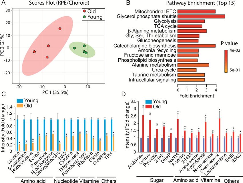
Wang et al.                                                                                                Page 2

                       1.     Introduction
Author Manuscript

                                    As we age, the eye undergoes a gradual decline of visual function. This age-related visual
                                    deterioration is a combination of structural changes in the ocular tissues including the
                                    cornea, lens, retina, retinal pigment epithelium (RPE), choroid and optic nerve. With aging,
                                    the number of corneal endothelial cells declines and the epithelium-derived glands including
                                    the lacrimal and meibomian glands decrease their production of tears to lubricate the cornea
                                    (Gambato et al., 2015; Gipson, 2013; Mustonen et al., 1998). The aging lens decreases its
                                    ability to change shape (presbyopia), which is most likely attributed to modifications in the
                                    cortical fibre cells (Duncan et al., 1997; Salvi et al., 2006). The populations of neurons in the
                                    retina decrease with loss of visual acuity and sensitivity (Lei et al., 2011; Nadal-Nicolas et
                                    al., 2018). The microcirculation in the macula, the cone photoreceptor-enriched central
                                    region of the retina also declines with age (Salvi et al., 2006). RPE loses melanin and
                                    lipofuscin deposits accumulate in the RPE (Delori et al., 2001; Sarna et al., 2003). Aged
Author Manuscript

                                    Bruch’s membrane, the innermost layer of the choroid, becomes thickened and basal laminar
                                    and basal linear deposits accumulate (Johnson et al., 2007; Li et al., 2005). There also is an
                                    age-related decrease in the number of optic nerve axons and an increase in elastic fibres
                                    (Cavallotti et al., 2003; Salvi et al., 2006).

                                    Aging is associated with many ocular diseases including Fuch’s dystrophy, dry eye
                                    syndrome, cataracts, presbyopia, age-related macular degeneration (AMD), diabetic
                                    retinopathy and glaucoma. These diseases cause visual impairment in 4–20% of adults over
                                    the age of 65 (Chader and Taylor, 2013; Delcourt et al., 2010; Lin et al., 2016; Whitcomb et
                                    al., 2013). Poor visual function significantly reduces quality of life and is a strong predictor
                                    of mortality (Lott et al., 2010). However, the biochemical basis of aging and age- related eye
                                    diseases has not been determined. Understanding how aging influences the eye should yield
Author Manuscript

                                    valuable information for both basic and clinical applications.

                                    Metabolic change is intimately entangled with aging and age-related disease at the
                                    molecular and cellular levels (Dhillon and Denu, 2017; Feng et al., 2016). Mitochondrial
                                    dysfunction including decreased energy metabolism and impaired antioxidant defenses has
                                    been reported in aging lens epithelial cells, retina, RPE and optic nerve (Ferrington et al.,
                                    2017; He et al., 2010; He and Tombran-Tink, 2010; Kubota et al., 2016; Lee et al., 2016;
                                    Lopez Sanchez et al., 2016; Maresca et al., 2013; Rohrer et al., 2016; Stahon et al., 2016).
                                    Improving mitochondrial metabolism can attenuate age-related visual decline in animal
                                    models (Mills et al., 2016; Weinrich et al., 2017). A recent study reported that
                                    supplementation with an NAD precursor could prevent glaucoma in aged mice by
                                    modulating mitochondrial dysfunction (Williams et al., 2017). Identifying metabolic
Author Manuscript

                                    changes that occur in the aging eye is critical for understanding the aging process and for
                                    identifying novel targets for disease treatment.

                                    Steady state metabolite concentrations reflect the status of metabolism. Mass spectrometry
                                    (MS) coupled with liquid chromatography (LC) or gas chromatography (GC), provides a
                                    sensitive and high- throughput platform to measure hundreds of metabolites in a single
                                    analysis (Cajka and Fiehn, 2016). In this study, we used targeted metabolomics to evaluate
                                    297 metabolites in young and old ocular tissues including cornea, lens, retina, RPE/choroid,

                                     Neurobiol Aging. Author manuscript; available in PMC 2019 November 01.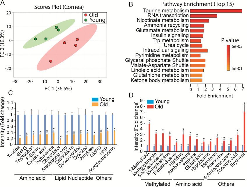
Wang et al.                                                                                                  Page 3

                                        and optic nerve from C57BL/6J mice. Mice are not an ideal model for AMD as they do not
Author Manuscript

                                        have an anatomical macula. Nevertheless, the mouse is the most widely used animal model
                                        for research in eye diseases because of the availability of genetic mutants, its capability of
                                        recapitulating key pathological processes in human eye diseases and its cost-effectiveness
                                        (Elizabeth Rakoczy et al., 2006; Pennesi et al., 2012; Zeiss, 2010). We have found that
                                        mitochondrial metabolism-related metabolites and some methylated metabolites are altered
                                        in almost all the aging tissues, suggesting a common feature for the aging eye. Additionally,
                                        we identified tissue-specific age- related metabolic changes. This study is the first
                                        comprehensive comparison of metabolites in young and old ocular tissues from mice.

                       2.     Materials and methods
                       2.1.       Animals
                                        C57 BL/6J mice of both sexes at 6 weeks were used for the young group and 73 weeks for
Author Manuscript

                                        the aged group. Both the young and aged mice were purchased from Jackson Lab. Mouse
                                        experiments were performed in accordance with the National Institutes of Health guidelines
                                        and the protocol approved by Institutional Animal Care and Use Committee of West Virginia
                                        University.

                       2.2.       Electroretinogram (ERG)
                                        ERGs were performed using the UTAS Visual Diagnostic System with BigShot Ganzfeld
                                        with UBA- 4200 amplifier and interface, and EMWIN 9.0.0 software (LKC Technologies,
                                        Gaithersburg, MD, USA). Mice were dark adapted overnight. All preparations were done
                                        under red light. Eyes were dilated using a 1:1 mixture of 2.5 % phenylephrine (Paragon) and
                                        1% tropicamide (Sandoz). Mice were sedated (1.5% isoflurane with 2.5 l/min (lpm) oxygen
                                        flow rate, 40°C) with an induction chamber (N=10 from five animals). After ten minutes of
Author Manuscript

                                        dilation, animals were placed on a heated platform (40°C) where they continued to receive a
                                        constant flow of isoflurane from a nose cone. A reference electrode was placed
                                        subcutaneously in the back of the neck (LKC Technologies, #95–016.) Electrodes made
                                        from 0.125 mm silver wire were carefully positioned closely above the cornea with contact
                                        being made by GenTeal Tears Lubricant Eye gel (Alcon). In darkness, scotopic recordings
                                        were elicited using flashes of LED white light at increasing flash intensities (−32, −24, −12,
                                        −4, 0 dB) responses were averaged at each light intensity. Immediately after scotopic
                                        recordings, animals were light adapted for 10 minutes to white background light (30 cds/
                                        m2.) To keep eyes moist, Systane Ultra Lubricant Eye Drops (Alcon) were applied
                                        periodically. With continuous background light, photopic response was elicited with
                                        increasing flash intensities (0, 3, 5, 10, 15, 25 dB) again, with LED white light. During data
                                        analysis, values were normalized to the baseline and each eye was evaluated separately to
Author Manuscript

                                        determine the A-wave and B-wave amplitudes. Amplitudes were averaged together and
                                        graphed.

                       2.3.       H&E staining
                                        Animals were euthanized by CO2 and eyes were immediately enucleated. Specimens were
                                        fixed in Excalibur’s Z-Fix and processed (Sakura VIP) to paraffin. 4–5 micron sections were
                                        cut (AO820) and placed on slides. H&E staining was performed with Gill III hematoxylin

                                            Neurobiol Aging. Author manuscript; available in PMC 2019 November 01.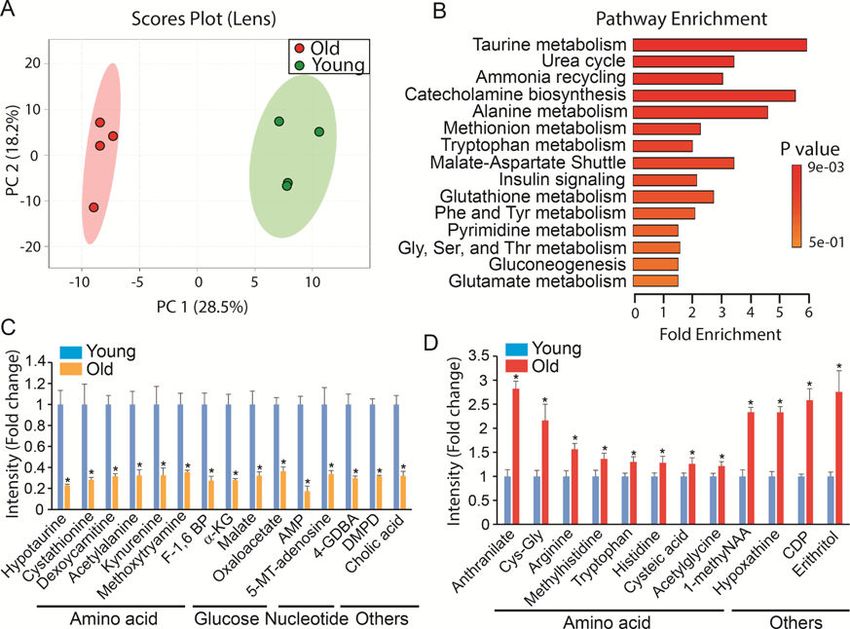
Wang et al.                                                                                                Page 4

                                         (StatLab Medical) and Alcoholic Eosin (StatLab Medical). Images were taken with a Nikon
Author Manuscript

                                         C2 confocal microscope system equipped with a Nikon Ds-Ri2 camera. Large scale images
                                         were generated using NIS elements AR 4.50 software with 5% overlap. ImageJ was used to
                                         count the nuclei of the outer nuclei layer at six different positions of the retina
                                         (Supplementary Fig 1). Nuclei of the outer nuclei layer were counted at each measurement
                                         point. The −3 and 3 positions were established by counting 20 nuclei in from the outermost
                                         edge. The −1 and 1 positions were defined 20 nuclei away from the optic nerve on the left
                                         and right side respectively. −2 and 2 positions were points in the middle of the other two
                                         measurements. Each measurement was replicated ten nuclei away from the first
                                         measurement. For each eye, ten sections were analyzed and the values averaged (20
                                         measurements per position per eye) to obtain the final nuclei count (N=30 from three eyes
                                         from different animals for each group).

                       2.4.       Isolation of retina, RPE/choroid, cornea, lens and optic nerve
Author Manuscript

                                         All mice were euthanized through quick cervical dislocation. Eyes were enucleated and
                                         submerged in 3 ml cold HBSS on ice. Upon isolation, the eye was placed under the
                                         microscope on cold HBSS-soaked filter paper. The anterior half of the eye was removed to
                                         collect the cornea and lens. The posterior half was moved and submerged in a drop of 50 μl
                                         cold HBSS solution for retina isolation. Once the retina was removed, the remaining RPE/
                                         choroid was transferred to another drop of 50 μl cold HBSS. The lingering fat and muscle
                                         tissue was cleaned before harvesting the optic nerve and RPE/choroid. Once collected in
                                         microtubes, all tissues were snap-frozen in liquid N2. The procedure from enucleation to
                                         snap-frozen took about 15–20 seconds per lens, 18–25 seconds per cornea, 40–50 seconds
                                         per retina, 75–80 seconds per optic nerve and 80–90 seconds per RPE/choroid (N=4 from
                                         four different animals).
Author Manuscript

                       2.5.       Mass spectrometry sample preparation
                                         Frozen Samples were homogenized in cold 80% methanol (methanol:water (80:20 V/V),
                                         pre-cold on dry ice) using THb Handheld Tissue Homogenizer (Omni International,
                                         #THB115). After homogenization for 10–30 seconds, the samples were stored on dry ice for
                                         30 min then centrifuged at 15000 RPM for 10 min at 4 oC. The supernatant was dried by the
                                         FreeZone 4.5 L freeze dryer (Labconco). The dried extract was either reconstituted for LC
                                         MS/MS or derivatized for GC MS. The pellets were dried and weighed for normalization.

                       2.6.       Metabolite analysis by LC-MS/MS
                                         Targeted metabolomics using LC-MS/MS was performed according to previous a validated
                                         procedure (Schelli et al., 2017; Xu et al., 2017; Zhong et al., 2017) with minor
Author Manuscript

                                         modifications. Briefly, a Thermo Fisher Scientific Dionex Ultimate 3000 HPLC system was
                                         used for chromatographic separation and analyte detection was performed using a TSQ-
                                         Quantiva triple quadrupole tandem mass spectrometer equipped with an electrospray
                                         ionization (ESI) source. Each sample was injected twice to perform detection in both
                                         negative and positive ionization modes. Regardless of ionization mode, chromatographic
                                         separations of targeted metabolites were performed on an Xbridge BEH hydrophilic
                                         interaction chromatography column (Waters Corporation, Milford, MA, 150 × 2.1 mm, 3.0
                                         μm). HPLC separation was performed (0.30 mL/min) with the autosampler thermostatted to

                                          Neurobiol Aging. Author manuscript; available in PMC 2019 November 01.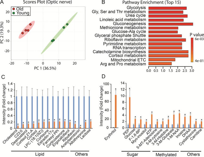
Wang et al.                                                                                                  Page 5

                                         4°C, and the column compartment to 40 °C. Mobile phase A consisted of 5 mM ammonium
Author Manuscript

                                         acetate prepared in 10% acetonitrile containing 0.2% acetic acid and mobile phase B was 5
                                         mM ammonium acetate prepared in 90% acetonitrile containing 0.2% acetic acid. Total
                                         running time for both ionization modes was 20 min with chromatographic gradient
                                         separation (0–2 min, 70% B; 5 min, 30% B; 9 min, 30% B; 11 min, 70% B; 20 min, 70% B).
                                         The LC- MS/MS was controlled by Xcalliber version 2.0 (Thermo Fisher Scientific).
                                         Authentic standards corresponding to the measured metabolites were purchased from
                                         Sigma-Aldrich (Saint Louis, MO) or IROA Technologies (Boston, MA). Stable isotope-
                                         labeled amino acids (U-13C, 97–99%; U-15N, 97–99%) were purchased from Cambridge
                                         Isotope Laboratories (Tewksbury, MA). LC-MS grade acetonitrile, ammonium acetate, and
                                         acetic acid were all purchased from Fisher Scientific (Pittsburgh, PA). The targeted
                                         metabolic profiling was performed in selected-reaction-monitoring (SRM) mode, established
                                         by running multiple authentic chemical standards and then the combination of retention time
Author Manuscript

                                         and SRM transition from standards were used to detect and identify metabolites from
                                         biological samples. The 241 metabolites were selected according to our published work
                                         (Schelli et al., 2017; Zhu et al., 2015; Zhu et al., 2014). The detection parameters for these
                                         metabolites are listed in supplementary Table 1.

                       2.7.       Metabolite analysis by GC MS
                                         The extracts were derivatized by methoxyamine (Sigma-Aldrich, # 226904) at 37 oC for 90
                                         min then derivatized by N,O-Bis(trimethylsilyl)trifluoroacetamide with
                                         trimethylchlorosilane (Sigma-Aldrich #15238) at 70 oC for 60 min. An Agilent 7890B/
                                         5977B GC/MS system with an Agilent DB-5MS column (30 m x 0.25 mm x 0.25 μm film)
                                         was used for GC separation and analysis of metabolites. Ultra-high- purity helium was the
                                         carrier gas at a constant flow rate of 1 mL/min. One microliter of sample was injected in
Author Manuscript

                                         split-less mode by the auto sampler. The temperature gradient started at 80 °C with a hold
                                         time of 2 min and increased at a rate of 10 °C/min to 180 °C, then at a rate of 5 °C/min to
                                         240 °C, finally at a rate 25 °C/min to 290 °C where it was held for 9 min. The total run time
                                         was 35 min. The temperatures were set as follows: inlet 250 °C, transfer line 280 °C, ion
                                         source 230 °C, and quadrupole 150 °C. Mass spectra were collected from 50–500 m/z under
                                         selective ion monitoring mode (SRM). All of the metabolites were tested by using authentic
                                         standards and the parameters for target ions and retention time used for SRM was listed in
                                         supplementary Table 2. Myristic acid-d27 was added into each sample as the internal
                                         standard. The data was analyzed by Agilent MassHunter Quantitative Analysis Software.

                       2.8.       Statistical analysis
                                         Principle component analysis and volcano plot (P1.2) were analyzed using MetaboAnalyst (http://www.metaboanalyst.ca/). Changed
                                         metabolites from Volcano plot were further enriched for pathways using Enrichment
                                         Analysis from MetaboAnalyst for significantly altered metabolic pathways. For univariate
                                         analysis, the significance of differences was determined by unpaired two-tailed t tests or
                                         analysis of variance with Bonferroni post hoc test. Data were mean ± SE. A p value < 0.05
                                         was considered to be significant.

                                           Neurobiol Aging. Author manuscript; available in PMC 2019 November 01.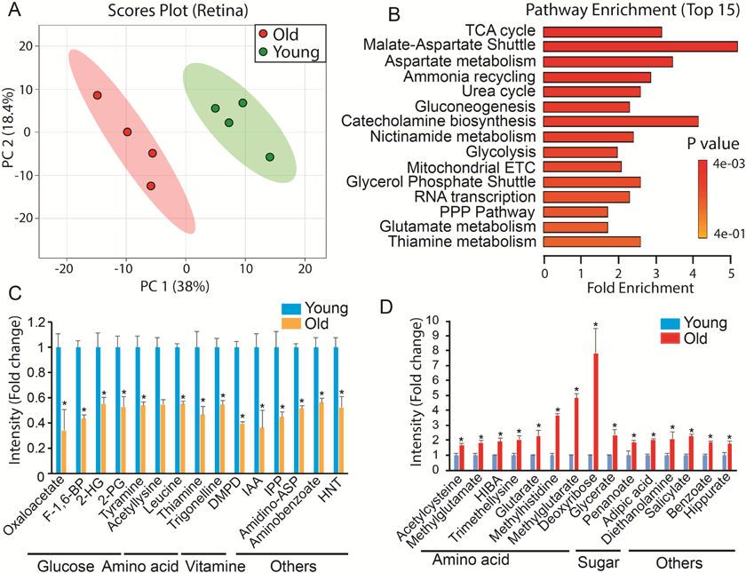
Wang et al. Page 6
3. Results
Author Manuscript
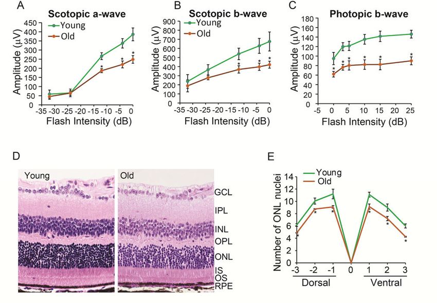
3.1. Visual function declines and photoreceptor neurons decrease in the old animals
We evaluated visual function by electroretinography (ERG) of both the young and old mice.
The animals were dark-adapted overnight and ERG a-wave and b-wave responses were
measured over a range of flash intensities. The ERG responses increased with flash intensity
(Fig 1). Old mice had significantly small dark-adapted (scotopic) a-wave and b-wave
responses starting at −10 decibel (dB). For cone responses, old mice had smaller light-
adapted (photopic) b-wave at all flash intensities (Fig 1C). To determine whether the decline
of ERG response was due to loss of photoreceptors, we stained retinal sections with H &E
and quantified the number of nuclei in the outer nuclear layer (ONL) at six different
positions (Supplementary Fig 1 and Fig 1D-E). The number of nuclei in the ONL was sparse
and significantly lower in the aging retina (Fig 1D-E), indicating loss of photoreceptors in
Author Manuscript
the old mice.
3.2. Metabolic changes in the aged retina
To study the metabolic profile of the aging eye, we combined LC MS/MS and GC MS to
target 297 metabolites from several major metabolic pathways (Supplementary Table 1-2).
The selection of 297 metabolites is based on previous literature on targeted metabolomics
(Zhu et al., 2014), our previous work on retinal metabolomics (Chao et al., 2017; Du et al.,
2016) and the availability of standards. The quantified metabolite data from young and old
retinas were evaluated by PCA analysis. Score plots separated the young and old into two
different groups, indicating that the metabolic profile is different between these retinas.
Volcano plots showed 68 significantly changed metabolites between young and old retinas
(Supplementary Fig 2, Table 3). We enriched the metabolic pathways for these changed
Author Manuscript
metabolites using MetaboAnalyst (http://www.metaboanalyst.ca/). Glucose metabolism
(TCA cycle, gluconeogenesis, glycolysis, pentose phosphate pathway) and amino acid
metabolism (malate-aspartate shuttle, aspartate and glutamate) were the major pathways that
were affected (Fig 2B). For specific metabolites, we identified metabolites that were most
decreased or increased in the aged retina (Fig 2 CD). Consistently, oxaloacetate, a critical
metabolite in the TCA cycle and Malate-Aspartate Shuttle, was the most reduced.
Metabolites in glycolysis and mitochondrial metabolism (Fructose 1,6-biphosphate (F- 1,6-
BP), 2 phosphoglyceric acid (2-PG), 2-hydroxyglutarate (2-HG)) were diminished about
half in old retinas, indicating that glucose utilization might be impaired. Carbohydrates
including deoxyribose and glycerate, increased substantially in the old retinas. Remarkably,
among the top fifteen increased metabolites in the old retina, seven were amino acids. Most
of these amino acids are acetylated or methylated such as acetylcysteine and
Author Manuscript
methylglutamate, suggesting that posttranslational modification is dysregulated in the aged
retina.
3.3. Metabolic changes in the aged RPE/choroid
Since RPE is tightly bound to the choroid, we analyzed metabolites from the RPE/choroid
complex. Multivariate analysis showed two distinctive groups for the metabolites from the
young and old RPE/choroid in the score plots (Fig 3A). We identified 45 significantly
changed metabolites by Volcano plot analysis (Supplementary Fig 3, Supplementary Table
Neurobiol Aging. Author manuscript; available in PMC 2019 November 01.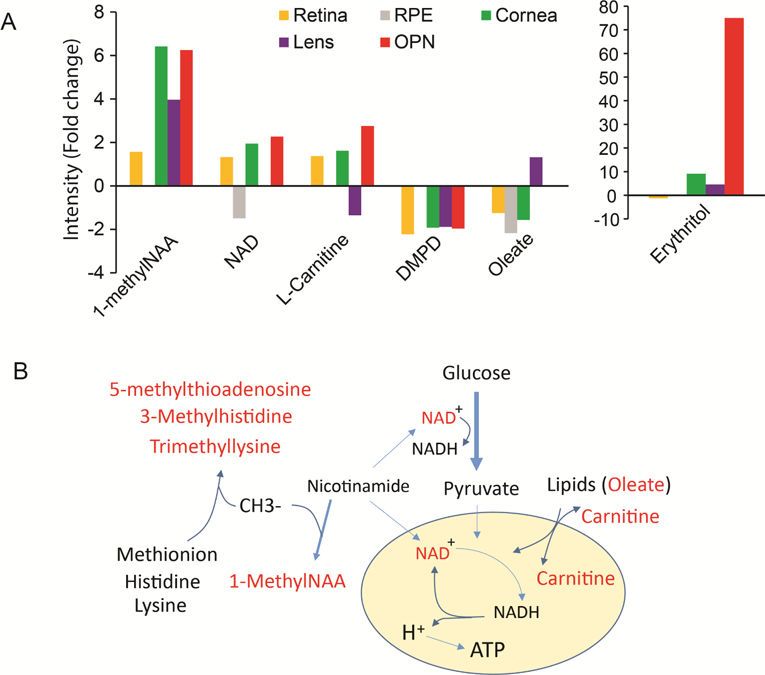
Wang et al.                                                                                                 Page 7

                                        4). Among these 45 metabolites, pathways in mitochondrial metabolism, glucose
Author Manuscript

                                        metabolism and amino acid metabolism were highly enriched (Fig 3B). Both NAD and
                                        riboflavin (a precursor for FAD) were depleted to less than half of the young levels in the
                                        aged RPE/choroid (Fig 3C). However, nicotinamide, the substrate for NAD synthesis, and
                                        the substrates for mitochondrial metabolism such as pyruvate and DHAP accumulated to
                                        higher levels in the old mice (Fig 3D), suggesting impaired mitochondrial energy
                                        metabolism. Additionally, leucine, 5- oxoproline (a metabolite in the glutathione cycle) and
                                        homocysteine were about 5 times lower in the old RPE/choroid, demonstrating that the
                                        utilization of these amino acids is severely impaired in the aged RPE/choroid (Fig 3C).

                       3.4.       Metabolic changes in the aged lens
                                        Lens metabolites in young and old mice could be divided into two distinctive groups in the
                                        score plots (Fig 4A). Volcano plots showed 54 metabolites were significantly different
Author Manuscript

                                        between these two groups (Supplementary Fig 4, Supplementary Table 5). Among the top
                                        enriched pathways, 60% were involved in amino acid metabolism such as taurine, alanine,
                                        methionine and tryptophan (Fig 4B). Taurine and hypotaurine are known to be abundant in
                                        the lens (Yanshole et al., 2014). Both hypotaurine and cystathionine, intermediates in taurine
                                        metabolism, decreased more than 70% in the aging lens (Fig 4C). Other amino acids and
                                        their intermediates such as deoxycarnitine (an intermediate in lysine and methionine
                                        metabolism), acetylalanine, kyneurenine (an intermediate for NAD synthesis from
                                        tryptophan), and methoxytryamine (an intermediate in phenylalanine and tyrosine
                                        metabolism) also were substantially decreased (Fig 4C). Additionally, the intermediates in
                                        glucose metabolism including glycolysis (F-1,6 BP) and mitochondrial TCA cycle (α-KG,
                                        malate and oxaloacetate) were diminished in the aging lens. Among the top increased
                                        metabolites, most are amino acids such as anthranilate (an intermediate for tryptophan),
Author Manuscript

                                        Cys-Gly and arginine, indicating that amino acid metabolism is severely disturbed in the
                                        aged lens.

                       3.5.       Metabolic changes in the aged cornea
                                        PCA analysis revealed significant metabolic differences between corneas from young and
                                        old mice. (Fig 5A). Among the 297 detected metabolites, we identified 45 changed
                                        metabolites in the aged cornea (Supplementary Fig 5, Supplementary Table 6). Taurine
                                        metabolism was the most enriched pathway (Fig 5B). Both taurine and cysteic acid
                                        decreased dramatically in the aged cornea while hypotaurine increased (Fig 5C-D).
                                        Additionally, the metabolism of other amino acids such as tryptophan and histidine also
                                        were highly enriched (Fig 5C-D). Different from lens, cholesterol was significantly lower
                                        and many methylated metabolites were upregulated, indicating there are some unique
Author Manuscript

                                        alterations in these pathways in the aged cornea.

                       3.6.       Metabolic changes in the aged optic nerve
                                        Similar to other aged ocular tissues, the aged optic nerve had a significantly different
                                        metabolic profile from the young optic nerve (Fig 6A). A total of 114 metabolites were
                                        changed in the volcano plot (Supplementary Fig 6, Supplementary Table 7). Besides glucose
                                        and amino acid metabolism, many of the changed metabolites were involved in lipid
                                        metabolism (Fig 6B-D). Among the top 15 downregulated metabolites, 11 were lipids

                                          Neurobiol Aging. Author manuscript; available in PMC 2019 November 01.
Wang et al. Page 8
including cholesterol, fatty acid, phospholipid and steroids (Fig 6B). Acetylserotonin, a
Author Manuscript
known neuroprotectant, decreased dramatically in the aged optic nerve (Fig 6B).
Remarkably, erythritol, a sugar alcohol, increased 75 fold over the young optic nerve (Fig
6D). Glucose and its intermediates also increased significantly in the aged optic nerve.
Similar to changes that occur in the aged cornea, many methylated metabolites such as
methylated nucleotides and amino acids accumulated in the aged optic nerve (Fig 6D).
3.7. Common metabolites in aged ocular tissue
Among the changed metabolites in five aged ocular tissues we analyzed, no metabolite was
changed in all tissues, indicating the specificity for each tissue. However, there were 31
metabolites that were changed in at least three tissues (Supplementary Table 8). Six of them
were changed in four tissues (Fig 7A). It is noteworthy that 1-methylnicotinamide (1-
methylNAA) was the only metabolite that accumulated to higher levels in all four tissues. 1-
Author Manuscript
methylNAA is produced by methylation of nicotinamide, a precursor for NAD biosynthesis.
This is important because NAD is a key cofactor that links cellular redox states with energy
metabolism. NAD accumulated to higher levels with aging in all tissues except RPE, where
it was depleted with aging (Fig 7A-B). Strikingly, other methylated metabolites such as 3-
methylhistidine, trimethyllysine and 5’-methylthioadenosine accumulated to higher levels in
the aging tissues, indicating dysregulated methyl group metabolism. Carnitine plays an
important role in transporting long-chain CoA (derived from long-chain fatty acid) into the
mitochondria for oxidation. Interestingly, the level of both carnitine and the long chain fatty
acid, oleate, changed with aging in four aged tissues (Fig 7A), indicating dysregulation of
lipid metabolism.
4. Discussion
Author Manuscript
In this report, we have described how the metabolome is altered in the aging eye in mice.
Our ERG analysis confirms that visual function and the number of photoreceptors decline in
the aging eye. Among the 297 metabolites we detected, 45–114 change significantly in
aging eye tissues. The changes are the largest in the neuronal tissues (retina and optic nerve)
and smaller in other tissues. Mitochondrial metabolism (TCA cycle, electron transport chain
and electron shuttle) and glucose metabolism (glycolysis and gluconeogenesis) are the
pathways most affected by aging in retina, RPE/choroid, optic nerve and lens. NAD and its
derivative 1-methylNAA are changed in four aging ocular tissues. Besides 1- methylNAA,
the accumulation of other methylated metabolites also is common in the aging retina,
cornea, optic nerve and lens. In the aging cornea and lens, metabolites in taurine metabolism
are strikingly influenced. Each of these substantial alterations in the metabolome may
contribute to or may be caused by age-associated neuronal cell loss and declining visual
Author Manuscript
function.
4.1. Mitochondrial metabolism in the aging eye
Metabolites in mitochondrial metabolism occur in many of the tissues in the aging eye.
These changed metabolites include intermediates in TCA cycle (oxaloacetate, a-
ketoglutarate, citrate and malate), electron transport chain (NAD and its derivatives or
precursors (nicotinamide, 1-methylNAA, trigoneline and tryptophan); FAD and its precursor
Neurobiol Aging. Author manuscript; available in PMC 2019 November 01.Wang et al. Page 9
riboflavin) and malate-aspartate shuttle (Fig 2-7, supplementary Table 3-8). Mitochondria
Author Manuscript
are abundant in the retina, RPE and optic nerve. To be transparent for light refraction, both
the cornea and lens have fewer mitochondria, which are limited to endothelial cells and
epithelial cells. Consistently, we found the mitochondrial metabolic changes occur mostly in
the aging retina, RPE and optic nerve. Mitochondrial dysfunction has been identified as an
important target for aging and age-related eye diseases (Barot et al., 2011; Lefevere et al.,
2017; Lopez Sanchez et al., 2016). The disruption of mitochondrial intermediate metabolism
and electron transport could reduce energy production and increase the generation of
reactive oxygen species, resulting in oxidative stress and damage to the aging eyes.
Additionally, several mitochondrial TCA cycle intermediates also decrease in the aging lens,
indicating that lens epithelial cells are dysfunctional in mitochondrial energy metabolism.
These results are consistent with a recent report that mitochondrial oxygen metabolism is
diminished in the older human lens epithelial cells (Kubota et al., 2016). Mitochondria-
Author Manuscript
derived oxidative stress has been regarded as an important target for cataract treatment
(Babizhayev and Yegorov, 2016).
1-methylNAA accumulates to higher levels in all of the aging tissues except RPE. 1-
methylNAA is produced from nicotinamide N-methyltransferase (NNMT) by transferring a
methyl group from S- adenosylmethionine (SAM) onto nicotinamide (Fig 7B). Since SAM
is a universal methyl donor and nicotinamide is an important precursor for NAD+ (Pissios,
2017), the enhanced production of 1- methylNAA can affect the activity of NAD+-dependent
enzymes and SAM-dependent methyltransferases. A recent study showed that both NNMT
activity and the level of 1-methylNAA are sensitive to energy deficit and their upregulation
can shift human muscle from glucose metabolism to lipid metabolism (Strom et al., 2018).
NNMT expression increases in white adipose tissue and liver of obese and diabetic mice.
Knockdown of NNMT augments cellular energy expenditure, upregulates histone
Author Manuscript
methylation and protects against diet-induced obesity (Kraus et al., 2014). In the urine
metabolome from aging mice, 1- methylNAA is the top increased metabolite (Calvani et al.,
2014). Our results suggest that 1-methylNAA might be a hallmark for the aging eye. It may
be associated with mitochondrial deficiency and stimulation of lipid metabolism that occur
with aging.
4.2. Glucose and lipid metabolism of the aging eye
Glycolysis is a predominant metabolic pathway in both the retina and lens (Ait-Ali et al.,
2015; Gillis et al., 1981; Hejtmancik et al., 2015; Hurley et al., 2015; Kanow et al., 2017).
The decrease of glycolytic intermediates in the aging retina and lens indicates a deficiency in
glucose metabolism. In contrast, in the RPE and optic nerve, glucose and glycolytic
intermediates accumulate to higher levels while lipids including fatty acids, cholesterol,
Author Manuscript
phospholipids and steroids become depleted in the aging RPE and optic nerve. Since
mitochondrial oxidative phosphorylation is very active in the RPE and optic nerve, our
findings suggest that the mitochondria of the aging RPE and optic nerve do not utilize
glucose efficiently to accumulate glycolytic intermediates. Aging mitochondria may shift to
lipid metabolism as fatty acids are depleted in the aging RPE and optic nerve. Additionally,
age-related formation of lipofuscin and perturbed cholesterol efflux (Crouch et al., 2015;
Neurobiol Aging. Author manuscript; available in PMC 2019 November 01.Wang et al. Page 10
Dolman et al., 1980; Fernandez de Castro et al., 2013; Lakkaraju et al., 2007) also may
Author Manuscript
contribute to the depletion of lipids.
4.3. Methylated metabolites in the aging eye
In addition to 1-methylNAA, many other methylated metabolites such as 3-methylglutarate,
3- methylhistidine, trimethyllysine and 5’-methylthioadenosine accumulate in the aging
retina, cornea, optic nerve and lens. 3-methylglutarate, an intermediate in leucine
degradation, can disrupt mitochondrial function, induce oxidative stress and damage DNA in
the rat brain (Colin-Gonzalez et al., 2016; da Rosa et al., 2015). The formation of 1-
methylNAA, 3-methylhistidine, trimethyllysine and 5’- methylthioadenosine relies on
methyltransferases which transfer methyl groups from SAM. Epigenetic regulation by
methylation of DNA and histone also requires activated methyl groups. Lower levels of
global DNA methylation have been reported in various aging tissues in animals and humans
Author Manuscript
(Liu et al., 2003; Unnikrishnan et al., 2018). We speculate that the consumption of methyl
groups to generate these methylated metabolites may limit the availability of SAM for DNA
methylation in the aging eye. In aged skeletal muscle, 3-methylhistidine, a marker for
muscle protein breakdown increases substantially (Sato et al., 2017). Elevated levels of 5’-
methylthioadenosine (MTA) appears in urine of older people with diabetes. Interestingly,
large amounts of trimethyllysine accumulate in intracellular deposits from patients with
Juvenile ceroid lipofuscinosis, a hereditary disorder characterized by progressive visual loss,
seizures, cognitive and psychomotor deterioration (Katz and Rodrigues, 1991). These
reports and our findings suggest that the increase of these methylated metabolites might
disrupt the balance of methyl group metabolism to impact epigenetic regulation, resulting in
aging-associated structural changes. Further studies are needed to elucidate the roles of these
methylated metabolites in the eye.
Author Manuscript
4.4. Taurine metabolism in the aging cornea and lens
Taurine metabolism is the most altered pathway in both the aged cornea and aged lens.
Taurine is one of the most abundant amino acids in the retina, cornea and lens (Ripps and
Shen, 2012). Different from all the other amino acids, taurine has a sulfonic acid group
instead of the classic carboxylic group. It is an important cytoprotective metabolite involved
in anti-oxidative stress, anti-inflammation, osmoprotection, regeneration and regulation of
Ca2+ concentration (Ripps and Shen, 2012; Rusciano et al., 2016). In the rat, the amount of
taurine level declines with age in the cornea and lens (Baskin et al., 1977; Yanshole et al.,
2014). Topical taurine application can contribute to epithelial wound healing and taurine is
typically included in contact lens cleaning solutions to protect the corneal stroma and
epithelium (Funke et al., 2012; Rusciano et al., 2016). The major route for the biosynthesis
Author Manuscript
of taurine is from methionine and cysteine, and requires the oxidation of hypotaurine or
cysteic acid to taurine. Cysteine sulfoinic acid decarboxylase (CSAD) is a key enzyme in
taurine biosynthesis from hypotaurine and it has the highest activity in the cornea among
ocular tissues (Heinamaki, 1988). In the aging cornea, the amount of taurine and cysteic acid
decreases substantially while hypotaurine increases, suggesting the aging cornea might have
impaired CSAD activity and compensate with CSAD-independent taurine synthesis from
cysteic acid. However, in the aging lens, hypotaurine and cystathionine (a precursor for
Neurobiol Aging. Author manuscript; available in PMC 2019 November 01.Wang et al. Page 11
cysteine) are depleted while cysteic acid accumulates, suggesting different mechanisms for
Author Manuscript
dysregulated taurine metabolism between the cornea and lens.
4.5. Future directions
Among different aging ocular tissues, alterations of methylated metabolites and
mitochondrial metabolites are the most striking and common features in the metabolic
profile of aging. It will be interesting to investigate whether these two common changes are
inter-connected, whether these alterations are the cause or the effect of aging in the eye, how
these metabolites are changed in age-associated ocular diseases and whether manipulation of
genes responsible for the methylations will slow down or prevent visual decline in animal
models in vivo. Furthermore, the targeted metabolomics in this study provides only a
snapshot of metabolism by measuring steady state metabolites without dynamic information,
e.g. the accumulation of metabolites can be caused by either more synthesis or less
Author Manuscript
degradation. Future studies using tracers to label the reactions in mitochondrial metabolism
and/or methylation are critical to understand the mechanisms for these metabolic alterations.
Additionally, metabolites can be exported out of cells and transported between ocular tissues
(Chao et al., 2017; Kanow et al., 2017). How the changes in the tissue metabolomes that we
report here impact metabolites in the vitreous also will be important to investigate, as it
could potentially be translated into diagnosis or prognosis in patients.
5. Conclusions
In conclusion, aging has a remarkable metabolic signature in the various tissues of the eye,
including dysregulated mitochondrial metabolism, shifted patterns in the utilization of
glucose and lipid, accumulated methylated metabolites and impaired taurine metabolism.
These findings should help to reveal the biochemical mechanisms in age-related ocular
Author Manuscript
changes and the decline of visual function, identify novel markers for the aging eye and shed
light on anti-aging research.
Supplementary Material
Refer to Web version on PubMed Central for supplementary material.
Acknowledgement:
This work was supported by NIH Grants EY026030 (to J.D., and Jennifer Chao.) and the Brightfocus Foundation
(to J.D. and Jennifer Chao). We thank Dr. James Hurley for helpful comments and suggestions.
References
Author Manuscript
Ait-Ali N, Fridlich R, Millet-Puel G, Clerin E, Delalande F, Jaillard C, Blond F, Perrocheau L,
Reichman S, Byrne LC, Olivier-Bandini A, Bellalou J, Moyse E, Bouillaud F, Nicol X, Dalkara D,
van Dorsselaer A, Sahel JA, Leveillard T, 2015 Rod-derived cone viability factor promotes cone
survival by stimulating aerobic glycolysis. Cell 161(4), 817–832. [PubMed: 25957687]
Babizhayev MA, Yegorov YE, 2016 Reactive Oxygen Species and the Aging Eye: Specific Role of
Metabolically Active Mitochondria in Maintaining Lens Function and in the Initiation of the
Oxidation- Induced Maturity Onset Cataract--A Novel Platform of Mitochondria-Targeted
Antioxidants With Broad Therapeutic Potential for Redox Regulation and Detoxification of
Oxidants in Eye Diseases. American journal of therapeutics 23(1), e98–117. [PubMed: 21048433]
Neurobiol Aging. Author manuscript; available in PMC 2019 November 01.Wang et al. Page 12
Barot M, Gokulgandhi MR, Mitra AK, 2011 Mitochondrial dysfunction in retinal diseases. Current eye
research 36(12), 1069–1077. [PubMed: 21978133]
Author Manuscript
Baskin SI, Cohn EM, Kocsis, 1977 The effect of age on taurine levels in eye tissues. Experimental eye
research 24(3), 315–319. [PubMed: 852529]
Cajka T, Fiehn O, 2016 Toward Merging Untargeted and Targeted Methods in Mass Spectrometry-
Based Metabolomics and Lipidomics. Analytical chemistry 88(1), 524–545. [PubMed: 26637011]
Calvani R, Brasili E, Pratico G, Capuani G, Tomassini A, Marini F, Sciubba F, Finamore A, Roselli M,
Marzetti E, Miccheli A, 2014 Fecal and urinary NMR-based metabolomics unveil an aging
signature in mice. Experimental gerontology 49, 5–11. [PubMed: 24184118]
Cavallotti C, Cavallotti D, Pescosolido N, Pacella E, 2003 Age-related changes in rat optic nerve:
morphological studies. Anatomia, histologia, embryologia 32(1), 12–16.
Chader GJ, Taylor A, 2013 Preface: The aging eye: normal changes, age-related diseases, and
sightsaving approaches. Investigative ophthalmology & visual science 54(14), ORSF1–4. [PubMed:
24335060]
Chao JR, Knight K, Engel AL, Jankowski C, Wang Y, Manson MA, Gu H, Djukovic D, Raftery D,
Hurley JB, Du J, 2017 Human retinal pigment epithelial cells prefer proline as a nutrient and
Author Manuscript
transport metabolic intermediates to the retinal side. The Journal of biological chemistry 292(31),
12895–12905. [PubMed: 28615447]
Colin-Gonzalez AL, Paz-Loyola AL, de Lima ME, Galvan-Arzate S, Seminotti B, Ribeiro CA,
Leipnitz G, Souza DO, Wajner M, Santamaria A, 2016 Experimental Evidence that 3-
Methylglutaric Acid Disturbs Mitochondrial Function and Induced Oxidative Stress in Rat Brain
Synaptosomes: New Converging Mechanisms. Neurochemical research 41(10), 2619–2626.
[PubMed: 27278758]
Crouch RK, Koutalos Y, Kono M, Schey K, Ablonczy Z, 2015 A2E and Lipofuscin. Progress in
molecular biology and translational science 134, 449–463. [PubMed: 26310170]
da Rosa MS, Scaini G, Damiani AP, Longaretti LM, Pereira M, Seminotti B, Zapelini HG, Schuck PF,
Streck EL, de Andrade VM, Wajner M, Leipnitz G, 2015 Evidence that 3-hydroxy-3-
methylglutaric and 3-methylglutaric acids induce DNA damage in rat striatum. Metabolic brain
disease 30(4), 1055–1062. [PubMed: 25939283]
Delcourt C, Korobelnik JF, Barberger-Gateau P, Delyfer MN, Rougier MB, Le Goff M, Malet F, Colin
J, Dartigues JF, 2010 Nutrition and age-related eye diseases: the Alienor (Antioxydants, Lipides
Author Manuscript
Essentiels, Nutrition et maladies OculaiRes) Study. The journal of nutrition, health & aging
14(10), 854–861.
Delori FC, Goger DG, Dorey CK, 2001 Age-related accumulation and spatial distribution of lipofuscin
in RPE of normal subjects. Investigative ophthalmology & visual science 42(8), 1855–1866.
[PubMed: 11431454]
Dhillon RS, Denu JM, 2017 Using comparative biology to understand how aging affects mitochondrial
metabolism. Molecular and cellular endocrinology 455, 54–61. [PubMed: 28025033]
Dolman CL, McCormick AQ, Drance SM, 1980 Aging of the optic nerve. Arch Ophthalmol 98(11),
2053–2058. [PubMed: 7436843]
Du J, Rountree A, Cleghorn WM, Contreras L, Lindsay KJ, Sadilek M, Gu H, Djukovic D, Raftery D,
Satrustegui J, Kanow M, Chan L, Tsang SH, Sweet IR, Hurley JB, 2016 Phototransduction
Influences Metabolic Flux and Nucleotide Metabolism in Mouse Retina. The Journal of biological
chemistry 291(9), 4698–4710. [PubMed: 26677218]
Duncan G, Wormstone IM, Davies PD, 1997 The aging human lens: structure, growth, and
Author Manuscript
physiological behaviour. The British journal of ophthalmology 81(10), 818–823. [PubMed:
9486018]
Elizabeth Rakoczy P, Yu MJ, Nusinowitz S, Chang B, Heckenlively JR, 2006 Mouse models of age-
related macular degeneration. Experimental eye research 82(5), 741–752. [PubMed: 16325179]
Feng Z, Hanson RW, Berger NA, Trubitsyn A, 2016 Reprogramming of energy metabolism as a driver
of aging. Oncotarget 7(13), 15410–15420. [PubMed: 26919253]
Fernandez de Castro JP, Mullins RF, Manea AM, Hernandez J, Wallen T, Kuehn MH, 2013 Lipofuscin
in human glaucomatous optic nerves. Experimental eye research 111, 61–66. [PubMed: 23567206]
Neurobiol Aging. Author manuscript; available in PMC 2019 November 01.Wang et al. Page 13
Ferrington DA, Ebeling MC, Kapphahn RJ, Terluk MR, Fisher CR, Polanco JR, Roehrich H, Leary
MM, Geng Z, Dutton JR, Montezuma SR, 2017 Altered bioenergetics and enhanced resistance to
Author Manuscript
oxidative stress in human retinal pigment epithelial cells from donors with age-related macular
degeneration. Redox biology 13, 255–265. [PubMed: 28600982]
Funke S, Azimi D, Wolters D, Grus FH, Pfeiffer N, 2012 Longitudinal analysis of taurine induced
effects on the tear proteome of contact lens wearers and dry eye patients using a RP-RP-Capillary-
HPLC- MALDI TOF/TOF MS approach. Journal of proteomics 75(11), 3177–3190. [PubMed:
22480906]
Gambato C, Longhin E, Catania AG, Lazzarini D, Parrozzani R, Midena E, 2015 Aging and corneal
layers: an in vivo corneal confocal microscopy study. Graefe’s archive for clinical and
experimental ophthalmology = Albrecht von Graefes Archiv fur klinische und experimentelle
Ophthalmologie 253(2), 267–275.
Gillis MK, Chylack LT, Jr., Cheng HM, 1981 Age and the control of glycolysis in the rat lens.
Investigative ophthalmology & visual science 20(4), 457–466. [PubMed: 6452426]
Gipson IK, 2013 Age-related changes and diseases of the ocular surface and cornea. Investigative
ophthalmology & visual science 54(14), ORSF48–53. [PubMed: 24335068]
Author Manuscript
He Y, Ge J, Burke JM, Myers RL, Dong ZZ, Tombran-Tink J, 2010 Mitochondria impairment
correlates with increased sensitivity of aging RPE cells to oxidative stress. Journal of ocular
biology, diseases, and informatics 3(3), 92–108.
He Y, Tombran-Tink J, 2010 Mitochondrial decay and impairment of antioxidant defenses in aging
RPE cells. Advances in experimental medicine and biology 664, 165–183. [PubMed: 20238015]
Heinamaki AA, 1988 Endogenous synthesis of taurine and GABA in rat ocular tissues. Acta chemica
Scandinavica. Series B: Organic chemistry and biochemistry 42(1), 39–42.
Hejtmancik JF, Riazuddin SA, McGreal R, Liu W, Cvekl A, Shiels A, 2015 Lens Biology and
Biochemistry. Progress in molecular biology and translational science 134, 169–201. [PubMed:
26310155]
Hurley JB, Lindsay KJ, Du J, 2015 Glucose, lactate, and shuttling of metabolites in vertebrate retinas.
Journal of neuroscience research 93(7), 1079–1092. [PubMed: 25801286]
Johnson M, Dabholkar A, Huang JD, Presley JB, Chimento MF, Curcio CA, 2007 Comparison of
morphology of human macular and peripheral Bruch’s membrane in older eyes. Current eye
research 32(9), 791–799. [PubMed: 17882712]
Author Manuscript
Kanow MA, Giarmarco MM, Jankowski CS, Tsantilas K, Engel AL, Du J, Linton JD, Farnsworth CC,
Sloat SR, Rountree A, Sweet IR, Lindsay KJ, Parker ED, Brockerhoff SE, Sadilek M, Chao JR,
Hurley JB, 2017 Biochemical adaptations of the retina and retinal pigment epithelium support a
metabolic ecosystem in the vertebrate eye. eLife 6.
Katz ML, Rodrigues M, 1991 Juvenile ceroid lipofuscinosis. Evidence for methylated lysine in neural
storage body protein. The American journal of pathology 138(2), 323–332. [PubMed: 1899540]
Kraus D, Yang Q, Kong D, Banks AS, Zhang L, Rodgers JT, Pirinen E, Pulinilkunnil TC, Gong F,
Wang YC, Cen Y, Sauve AA, Asara JM, Peroni OD, Monia BP, Bhanot S, Alhonen L, Puigserver
P, Kahn BB, 2014 Nicotinamide N-methyltransferase knockdown protects against diet- induced
obesity. Nature 508(7495), 258–262. [PubMed: 24717514]
Kubota M, Shui YB, Liu M, Bai F, Huang AJ, Ma N, Beebe DC, Siegfried CJ, 2016 Mitochondrial
oxygen metabolism in primary human lens epithelial cells: Association with age, diabetes and
glaucoma. Free radical biology & medicine 97, 513–519. [PubMed: 27445101]
Lakkaraju A, Finnemann SC, Rodriguez-Boulan E, 2007 The lipofuscin fluorophore A2E perturbs
Author Manuscript
cholesterol metabolism in retinal pigment epithelial cells. Proceedings of the National Academy of
Sciences of the United States of America 104(26), 11026–11031. [PubMed: 17578916]
Lee WH, Higuchi H, Ikeda S, Macke EL, Takimoto T, Pattnaik BR, Liu C, Chu LF, Siepka SM, Krentz
KJ, Rubinstein CD, Kalejta RF, Thomson JA, Mullins RF, Takahashi JS, Pinto LH, Ikeda A, 2016
Mouse Tmem135 mutation reveals a mechanism involving mitochondrial dynamics that leads to
age-dependent retinal pathologies. eLife 5.
Lefevere E, Toft-Kehler AK, Vohra R, Kolko M, Moons L, Van Hove I, 2017 Mitochondrial
dysfunction underlying outer retinal diseases. Mitochondrion 36, 66–76. [PubMed: 28365408]
Neurobiol Aging. Author manuscript; available in PMC 2019 November 01.Wang et al. Page 14
Lei Y, Garrahan N, Hermann B, Fautsch MP, Johnson DH, Hernandez MR, Boulton M, Morgan JE,
2011 Transretinal degeneration in ageing human retina: a multiphoton microscopy analysis. The
Author Manuscript
British journal of ophthalmology 95(5), 727–730. [PubMed: 21183516]
Li CM, Chung BH, Presley JB, Malek G, Zhang X, Dashti N, Li L, Chen J, Bradley K, Kruth HS,
Curcio CA, 2005 Lipoprotein-like particles and cholesteryl esters in human Bruch’s membrane:
initial characterization. Investigative ophthalmology & visual science 46(7), 2576–2586. [PubMed:
15980251]
Lin JB, Tsubota K, Apte RS, 2016 A glimpse at the aging eye. NPJ aging and mechanisms of disease
2, 16003. [PubMed: 28721262]
Liu L, Wylie RC, Andrews LG, Tollefsbol TO, 2003 Aging, cancer and nutrition: the DNA
methylation connection. Mechanisms of ageing and development 124(10–12), 989–998. [PubMed:
14659588]
Lopez Sanchez MI, Crowston JG, Mackey DA, Trounce IA, 2016 Emerging Mitochondrial
Therapeutic Targets in Optic Neuropathies. Pharmacology & therapeutics 165, 132–152. [PubMed:
27288727]
Lott LA, Schneck ME, Haegerstrom-Portnoy G, Brabyn JA, 2010 Non-standard vision measures
Author Manuscript
predict mortality in elders: the Smith-Kettlewell Institute (SKI) study. Ophthalmic epidemiology
17(4), 242–250. [PubMed: 20642347]
Maresca A, la Morgia C, Caporali L, Valentino ML, Carelli V, 2013 The optic nerve: a “mito-
window” on mitochondrial neurodegeneration. Molecular and cellular neurosciences 55, 62–76.
[PubMed: 22960139]
Mills KF, Yoshida S, Stein LR, Grozio A, Kubota S, Sasaki Y, Redpath P, Migaud ME, Apte RS,
Uchida K, Yoshino J, Imai SI, 2016 Long-Term Administration of Nicotinamide Mononucleotide
Mitigates Age-Associated Physiological Decline in Mice. Cell metabolism 24(6), 795–806.
[PubMed: 28068222]
Mustonen RK, McDonald MB, Srivannaboon S, Tan AL, Doubrava MW, Kim CK, 1998 Normal
human corneal cell populations evaluated by in vivo scanning slit confocal microscopy. Cornea
17(5), 485–492. [PubMed: 9756442]
Nadal-Nicolas FM, Vidal-Sanz M, Agudo-Barriuso M, 2018 The aging rat retina: from function to
anatomy. Neurobiology of aging 61, 146–168. [PubMed: 29080498]
Pennesi ME, Neuringer M, Courtney RJ, 2012 Animal models of age related macular degeneration.
Author Manuscript
Molecular aspects of medicine 33(4), 487–509. [PubMed: 22705444]
Pissios P, 2017 Nicotinamide N-Methyltransferase: More Than a Vitamin B3 Clearance Enzyme.
Trends in endocrinology and metabolism: TEM 28(5), 340–353. [PubMed: 28291578]
Ripps H, Shen W, 2012 Review: taurine: a “very essential” amino acid. Molecular vision 18, 2673–
2686. [PubMed: 23170060]
Rohrer B, Bandyopadhyay M, Beeson C, 2016 Reduced Metabolic Capacity in Aged Primary Retinal
Pigment Epithelium (RPE) is Correlated with Increased Susceptibility to Oxidative Stress.
Advances in experimental medicine and biology 854, 793–798. [PubMed: 26427491]
Rusciano D, Roszkowska AM, Gagliano C, Pezzino S, 2016 Free amino acids: an innovative treatment
for ocular surface disease. European journal of pharmacology 787, 9–19. [PubMed: 27090927]
Salvi SM, Akhtar S, Currie Z, 2006 Ageing changes in the eye. Postgraduate medical journal 82(971),
581–587. [PubMed: 16954455]
Sarna T, Burke JM, Korytowski W, Rozanowska M, Skumatz CM, Zareba A, Zareba M, 2003 Loss of
melanin from human RPE with aging: possible role of melanin photooxidation. Experimental eye
Author Manuscript
research 76(1), 89–98. [PubMed: 12589778]
Sato T, Ito Y, Nagasawa T, 2017 L-Lysine suppresses myofibrillar protein degradation and autophagy
in skeletal muscles of senescence-accelerated mouse prone 8. Biogerontology 18(1), 85–95.
[PubMed: 27752791]
Schelli K, Rutowski J, Roubidoux J, Zhu J, 2017 Staphylococcus aureus methicillin resistance detected
by HPLC-MS/MS targeted metabolic profiling. Journal of chromatography. B, Analytical
technologies in the biomedical and life sciences 1047, 124–130. [PubMed: 27316783]
Neurobiol Aging. Author manuscript; available in PMC 2019 November 01.Wang et al. Page 15
Stahon KE, Bastian C, Griffith S, Kidd GJ, Brunet S, Baltan S, 2016 Age-Related Changes in Axonal
and Mitochondrial Ultrastructure and Function in White Matter. The Journal of neuroscience : the
Author Manuscript
official journal of the Society for Neuroscience 36(39), 9990–10001. [PubMed: 27683897]
Strom K, Morales-Alamo D, Ottosson F, Edlund A, Hjort L, Jorgensen SW, Almgren P, Zhou Y,
Martin-Rincon M, Ekman C, Perez-Lopez A, Ekstrom O, Perez-Suarez I, Mattiasson M, de
Pablos- Velasco P, Oskolkov N, Ahlqvist E, Wierup N, Eliasson L, Vaag A, Groop L, Stenkula
KG, Fernandez C, Calbet JAL, Holmberg HC, Hansson O, 2018 N(1)-methylnicotinamide is a
signalling molecule produced in skeletal muscle coordinating energy metabolism. Scientific
reports 8(1), 3016. [PubMed: 29445118]
Unnikrishnan A, Hadad N, Masser DR, Jackson J, Freeman WM, Richardson A, 2018 Revisiting the
genomic hypomethylation hypothesis of aging. Annals of the New York Academy of Sciences.
Weinrich TW,Coyne A, Salt TE, Hogg C, Jeffery G, 2017 Improving mitochondrial function
significantly reduces metabolic, visual, motor and cognitive decline in aged Drosophila
melanogaster. Neurobiology of aging 60, 34–43. [PubMed: 28917665]
Whitcomb EA, Shang F, Taylor A, 2013 Common cell biologic and biochemical changes in aging and
age-related diseases of the eye: toward new therapeutic approaches to age-related ocular diseases.
Author Manuscript
Investigative ophthalmology & visual science 54(14), ORSF31–36. [PubMed: 24335065]
Williams PA, Harder JM, Foxworth NE, Cochran KE, Philip VM, Porciatti V, Smithies O, John SW,
2017 Vitamin B3 modulates mitochondrial vulnerability and prevents glaucoma in aged mice.
Science 355(6326), 756–760. [PubMed: 28209901]
Xu M, Zhong F, Zhu J, 2017 Evaluating metabolic response to light exposure in Lactobacillus species
via targeted metabolic profiling. Journal of microbiological methods 133, 14–19. [PubMed:
27974228]
Yanshole VV, Snytnikova OA, Kiryutin AS, Yanshole LV, Sagdeev RZ, Tsentalovich YP, 2014
Metabolomics of the rat lens: a combined LC-MS and NMR study. Experimental eye research 125,
71–78. [PubMed: 24910091]
Zeiss CJ, 2010 Animals as models of age-related macular degeneration: an imperfect measure of the
truth. Veterinary pathology 47(3), 396–413. [PubMed: 20382825]
Zhong F, Xu M, Bruno RS, Ballard KD, Zhu J, 2017 Targeted High Performance Liquid
Chromatography Tandem Mass Spectrometry-based Metabolomics differentiates metabolic
syndrome from obesity. Exp Biol Med (Maywood) 242(7), 773–780. [PubMed: 28299975]
Author Manuscript
Zhu J, Djukovic D, Deng L, Gu H, Himmati F, Abu Zaid M, Chiorean EG, Raftery D, 2015 Targeted
serum metabolite profiling and sequential metabolite ratio analysis for colorectal cancer
progression monitoring. Analytical and bioanalytical chemistry 407(26), 7857–7863. [PubMed:
26342311]
Zhu J, Djukovic D, Deng L, Gu H, Himmati F, Chiorean EG, Raftery D, 2014 Colorectal cancer
detection using targeted serum metabolic profiling. Journal of proteome research 13(9), 4120–
4130. [PubMed: 25126899]
Author Manuscript
Neurobiol Aging. Author manuscript; available in PMC 2019 November 01.Wang et al. Page 16
Highlights
Author Manuscript
45–114 metabolites are changed in the aging retina, RPE, lens, cornea and optic nerve
Altered mitochondrial metabolism and glucose metabolism in the aging ocular tissue
NAD and 1-methylnicotinamide are altered in four of aging ocular tissue Taurine
metabolism are the top influenced pathway in the aged lens and cornea
Author Manuscript
Author Manuscript
Author Manuscript
Neurobiol Aging. Author manuscript; available in PMC 2019 November 01.Wang et al. Page 17
Author Manuscript
Author Manuscript
Figure 1. Aging causes a decline in ERG response and a reduction in the number of
Author Manuscript
photoreceptors.
Dark-adapted mice were stimulated with different flash intensities. The average response of
(A) scotopic a-wave, (B) scotopic b-wave and (C) photopic b-wave decreased in older mice.
*PWang et al. Page 18
Author Manuscript
Author Manuscript
Figure 2. Differential metabolic profile between young and old retinas.
(A) Metabolites from young and old retinas were separated into two different groups in
Scores Plot by PCA. (B) Top enriched pathways from significantly changed metabolites.
Author Manuscript
ETC, electron transport chain; PPP, pentose phosphate. (C) Top decreased metabolites in old
retinas. 2-HG, 2-hydroxyglutarate; 2-PG, 2-Phosphoglyceric Acid; DMPD, N,N-
Dimethyl-1,4-Phenylenediamine; IAA, Indole-3-Acetic Acid; IPP, Isopentyl Pyrophosphate;
Amidino-ASP, N-Amidino-L-Aspartate; HNT, 1-Hydroxy-2-Naphthoate. (D) Top increased
metabolites in old retinas. Data report fold change of ion intensity of the old vs the young.
N=4 from four different animals.
Author Manuscript
Neurobiol Aging. Author manuscript; available in PMC 2019 November 01.You can also read