The Effect of Sea Buckthorn (Hippophae rhamnoides L.) Seed Oil on UV-Induced Changes in Lipid Metabolism of Human Skin Cells - MDPI
←
→
Page content transcription
If your browser does not render page correctly, please read the page content below
antioxidants
Article
The Effect of Sea Buckthorn (Hippophae rhamnoides L.)
Seed Oil on UV-Induced Changes in Lipid
Metabolism of Human Skin Cells
Agnieszka G˛egotek, Anna Jastrzab,
                                ˛ Iwona Jarocka-Karpowicz, Marta Muszyńska
and Elżbieta Skrzydlewska * ID
 Department of Inorganic and Analytical Chemistry, Medical University of Bialystok, Bialystok 15-089, Poland;
 agnieszka.gegotek@umb.edu.pl (A.G.); anna.jastrzab@umb.edu.pl (A.J.);
 iwona.jarocka-karpowicz@umb.edu.pl (I.J.-K.); marta.muszynska@umb.edu.pl (M.M.)
 * Correspondence: elzbieta.skrzydlewska@umb.edu.pl; Tel./Fax: +48-857-485-882

 Received: 9 July 2018; Accepted: 20 August 2018; Published: 23 August 2018

 Abstract: Lipids and proteins of skin cells are the most exposed to harmful ultraviolet (UV) radiation
 contained in sunlight. There is a growing need for natural compounds that will protect these
 sensitive molecules from damage, without harmful side effects. The aim of this study was to
 investigate the effect of sea buckthorn seed oil on the redox balance and lipid metabolism in UV
 irradiated cells formed different skin layers to examine whether it had a protective effect. Human
 keratinocytes and fibroblasts were subjected to UVA (ultraviolet type A; 30 J/cm2 and 20 J/cm2 ) or
 UVB (ultraviolet type B; 60 mJ/cm2 and 200 mJ/cm2 , respectively) radiation and treated with sea
 buckthorn seed oil (500 ng/mL), and the redox activity was estimated by reactive oxygen species
 (ROS) generation and enzymatic/non-enzymatic antioxidants activity/level (using electron spin
 resonance (ESR), high-performance liquid chromatography (HPLC), and spectrophotometry). Lipid
 metabolism was measured by the level of fatty acids, lipid peroxidation products, endocannabinoids
 and phospholipase A2 activity (GC/MS (gas chromatography/mass spectrometry), LC/MS (liquid
 chromatography/mass spectrometry), and spectrophotometry). Also, transcription factor Nrf2
 (nuclear erythroid 2-related factor) and its activators/inhibitors, peroxisome proliferator-activated
 receptors (PPAR) and cannabinoid receptor levels were measured (Western blot). Sea buckthorn oil
 partially prevents UV-induced ROS generation and enhances the level of non-enzymatic antioxidants
 such as glutathione (GSH), thioredoxin (Trx) and vitamins E and A. Moreover, it stimulates the activity
 of Nrf2 leading to enhanced antioxidant enzyme activity. As a result, decreases in lipid peroxidation
 products (4-hydroxynonenal, 8-isoprostaglandin) and increases in the endocannabinoid receptor
 levels were observed. Moreover, sea buckthorn oil treatment enhanced the level of phospholipid
 and free fatty acids, while simultaneously decreasing the cannabinoid receptor expression in UV
 irradiated keratinocytes and fibroblasts. The main differences in sea buckthorn oil on various skin cell
 types was observed in the case of PPARs—in keratinocytes following UV radiation PPAR expression
 was decreased by sea buckthorn oil treatment, while in fibroblasts the reverse effect was observed,
 indicating an anti-inflammatory effect. With these results, sea buckthorn seed oil exhibited prevention
 of UV-induced disturbances in redox balance as well as lipid metabolism in skin fibroblasts and
 keratinocytes, which indicates it is a promising natural compound in skin photo-protection.

 Keywords: fibroblasts; keratinocytes; sea buckthorn seeds oil; UV radiation; lipid metabolism

1. Introduction
    Skin has been recognized as an organ that performs synthesis and processing of an astounding
range of structural proteins, lipids, and signaling molecules. Moreover, skin is an integral component

Antioxidants 2018, 7, 110; doi:10.3390/antiox7090110                       www.mdpi.com/journal/antioxidants
Antioxidants 2018, 7, 110 2 of 21
of the immune, nervous and endocrine systems, and therefore is also responsible for the first line of
information and defense in the immunity process [1]. However, skin also protects the organism against
the influence of the environment and is well-known as crucial for the maintenance of temperature,
electrolyte and fluid balance [2]. As a natural biological barrier, skin is constantly exposed to numerous
external factors that can impair the functioning of this barrier, including ultraviolet (UV) radiation
contained in sunlight. High energy UVB (ultraviolet type B; 280–320 nm) and UVA (ultraviolet
type A; 320–400 nm) radiation absorption by the skin may trigger mechanisms that defend skin
integrity and also induces skin pathology, such as cancer [3]. These effects occurs as a result of the UV
electromagnetic energy transduction into chemical, hormonal, and neural signals, defined by the nature
of the chromophores and tissue compartments receiving specific UV wavelength. However, despite of
the wavelength differences, both of them are characterized by high penetration into the deep layers of
the epidermis and dermis [4]. UV radiation can upregulate local cytokines, corticotropin-releasing
hormone, urocortins, proopiomelanocortin peptides expression, however, they can be released into
circulation to exert systemic effects, including activation of the central hypothalamic-pituitary-adrenal
axis, opioidogenic effects, and immunosuppression [5]. Similar effects of UV radiation on human
organism are seen after exposure of the eyes and skin to UV, through which UV exerts very rapid
stimulatory effects on the brain and central neuroendocrine system and impairs body homeostasis [5].
Moreover, UV radiation promotes the generation of reactive oxygen species (ROS) as well as impairs
the antioxidant capacity of skin cells [6]. As a result, the redox imbalance leading to oxidative
stress is increased. UV-induced oxidative stress in skin enhances the collagen degradation and
elastin modification causes premature skin aging [7,8]. Moreover, increased oxidative phospholipid
metabolism disturbs the function of cellular membranes, and promotes the generaton of reactive
electrophiles, such as 4-hydroxyalkenals [9]. These reactive electrophiles interact with membrane
phospholipids and proteins, including receptors, causing irreparable skin damage. Additionally,
UV-induced oxidative stress affects phospholipid metabolism which influences the endocannabinoid
system [10]. The main endocannabinoids, such as anandamide and 2-arachidonoylglycerol (2-AG),
participate in cell signalling and are ligands for transmembrane receptors-CB1 and CB2 (cannabinoid
receptor type 1 and 2) [11]. Activation of CB1 is responsible for ROS generation, whereas CB2 prevents
ROS generation and inflammation, while also stimulating the MAP kinase (mitogen-activated protein
kinase) pathway [12].
In connection with the permanent exposure to harmful UV radiation, cells from different skin
layers-epidermal keratinocytes and dermal fibroblasts create a number of defense mechanisms that
protect against UV-induced oxidative stress. Examples of such mechanisms are the high activity of
repair and antioxidant enzymes, a large pool of non-enzymatic antioxidants, and redox-sensitive
transcription factors including Nrf2 that is responsible for cytoprotective protein expression [13].
Despite the above well-developed mechanisms, skin cells exposed to long-lasting UV radiation
are vulnerable to the depletion of the natural antioxidants present and therefore there is still a need
for compounds with cytoprotective properties against UV-induced changes. One potential source are
seed oils, the main source of phospholipids and triacylglycerols, which are solvents for other lipophilic
compounds, i.e., sterols, fat-soluble vitamins (vitamin A and E), carotenoids, phenolic compounds,
and free fatty acids [14]. One good source of seed oil is sea buckthorn (Hippophae rhamnoides L.) [15],
which is increasingly appearing in skin care preparations. Its antioxidant potential is based on high
content of polyphenols, vitamins (C and E), carotenoids as well as sterols [16–18]. In addition to the
antioxidant activity, sea buckthorn also shows antibacterial and antifungal activity [19]. Carotenoids
contained in the oil stimulate collagen synthesis, while phytosterols regulate inflammatory processes
and have anticancer effects [20]. Because of the high concentration of these compounds sea buckthorn
seed oil has been found as a promising therapeutic agent in the treatment of dermatitis [21]. However,
there is no research on the effect of sea buckthorn seed oil on the UV-induced oxidative lipid metabolism
of skin cells.Antioxidants 2018, 7, 110 3 of 21
The aim of this study was to estimate the effect of sea buckthorn seed oil on the correlation
between redox balance, lipid metabolism and endocannabinoid system in in vitro cultured human
keratinocytes and fibroblasts exposed to the UV-radiation.
2. Materials and Methods
2.1. Examination of Sea Buckthorn Seed Oil Composition
Sea buckthorn (Hippophae rhamnoides L.) seed oil fatty acids levels as well as vitamins A and E
were described below in the paragraphs Determination of fatty acids levels and Determination of
non-enzymatic antioxidant levels.
2.1.1. Determination of Squalene Levels
Squalene level was analyzed with separation on HP-5ms capillary column (0.25 mm; 0.25 µm,
30 m) of GC/MS system (Agilent Technologies, Palo Alto, CA, USA) with 7890A GC–7000
quadrupole MS/MS (Agilent Technologies, Palo Alto, CA, USA). Operating conditions were as follows:
Oven programming −50 ◦ C (10 min), rate 2 ◦ C/min to 310 ◦ C (30 min); ion source (EI) −230 ◦ C;
electron energy −70 eV [22]. The mass spectrometer source was run in selective ion monitoring (SIM)
mode for the following ions: 191 and 81 m/z.
2.1.2. Phytosterols Profile
The content of sterols in oils was determined by GC/MS system with 7890A GC–7000 quadrupole
MS/MS (Agilent Technologies, Palo Alto, CA, USA) [23]. Samples were separated on HP-5ms capillary
column (0.25 mm; 0.25 µm, 30 m). Operating conditions were as follows: oven programming −50 ◦ C
(2 min), rate 15 ◦ C/min to 230 ◦ C, to 310 ◦ C at the rate of 3 ◦ C/min (10 min); ion source (EI)
−230 ◦ C; electron energy −70 eV. The mass spectrometer source was run in selective ion monitoring
(SIM) mode for the following ions: 372 and 217 m/z for 5-α-cholestane (IS), 458 and 368 m/z for
cholesterol, 470 and 255 m/z for brassicasterol, 382 and 343 m/z for campesterol, 394 and 255 m/z
for stigmasterol and 396 and 357 m/z for β-sitosterol. The quantifications were carried out using the
internal standard method.
2.1.3. Determination of β-Carotene Levels
HPLC methods was used to detect the level β-carotene [24]. Samples were dissolved in
1 mL of 2-propanol and 20 µL was taken analysis. Separation was performed using C18 column
(150 nm × 4.6 mm) with UV detection at 454 nm. The concentration was determined using a calibration
curve range 0.5–50 mg/L.
2.2. Cell Culture
Human skin cell lines used in experiment were obtained from American Type Culture Collection and
cultured cultured in a humidified atmosphere of 5% CO2 at 37 ◦ C. Human immortalized keratinocytes
(CDD 1102 KERTr) were transformed with human papillomavirus 16 (HPV-16) E6/E7, while fibroblasts
(CCD 1112Sk) were isolated from normal foreskin of Caucasian newborn male and used in passage 11.
The growth media for each line were prepared as follows: for keratinocytes-keratinocyte-SFM medium
with 1% Bovine Pituitary Extract (BPE) and human recombinant Epidermal Growth Factor (hEGF);
for fibroblasts-Dulbecco’s Modified Eagle Medium (DMEM) with 10% fetal bovine serum (FBS). Media
were supplemented with 50 U/mL penicillin and 50 µg/mL streptomycin. All sterile and cell culture
reagents were obtained from Gibco (Grand Island, NY, USA).
Cells were exposed to UV radiation after reaching the 70% confluence. Radiation doses for
keratinocytes were 30 J/cm2 for UVA (365 nm, power density at 4.2 mW/cm2 ) and 60 mJ/cm2 for UVB
(312 nm, power density at 4.08 mW/cm2 ), while for fibroblasts were 20 J/cm2 for UVA and 200 mJ/cm2
for UVB (Bio-Link Crosslinker BLX 312/365, Vilber Lourmat, Germany). The distance of the cells fromAntioxidants 2018, 7, 110 4 of 21
lamps was 15 cm. Exposure doses were chosen corresponding to 70% cell viability measured by the
MTT (3-(4,5-dimethylthiazol-2-yl)-2,5-diphenyltetrazolium bromide) assay [25], what was previously
shown as a doses that lead to the activation of pro-oxidative conditions, response of cell antioxidants,
as well as activation of Nrf2 pathway and increases endocannabinoid system action [6]. Control cells
were incubated in parallel without irradiation.
To examine the effect of sea buckthorn (Hippophae rhamnoides L.) seed oil (produced by “Szarłat”
M i W Lenkiewicz Sp. J., Poland) on UV radiated skin cells, after keratinocytes and fibroblasts were
exposed to UV radiation they were incubated 24 h under standard conditions in medium containing
500 ng/mL plant oil in 0.1% DMSO. The oil concentration was chosen corresponding to 100% cell
viability compared to control cells measured by the MTT assay [25]. Parallel cells were cultured in
medium containing plant oil without irradiation.
2.3. Examination of Pro-Oxidative Activity
2.3.1. Determination of ROS Generation
The superoxide anions generation was detected using stable nitroxide CM-radicals formation
and detection with EPR spectrometer (Noxygen GmbH/Bruker Biospin GmbH, Germany) [10,26].
The results were reported in micromolar concentration of ROS per minute and normalised per
milligram of protein.
2.3.2. Determination of Pro-Oxidants Enzyme Activities
NADPH oxidase (NOX-EC 1.6.3.1) activity was analysed by luminescence measurement using
lucigenin as luminophore [27]. Enzyme specific activity was described in relative luminescence units
(RLU) per milligram protein.
Xanthine oxidase (XO-EC1.17.3.2) activity was estimated by uric acid formation from xanthine [28].
Enzyme specific activity was described in microunits per milligram of protein.
2.4. Examination of Antioxidant Defence System
2.4.1. Determination of Antioxidant Enzymes Activity
Glutathione peroxidase (GSH-Px-EC.1.11.1.6) activity was assessed spectro-photometrically [29].
Enzyme specific activity was described in microunits per milligram of protein.
Glutathione reductase (GSSG-R-EC.1.6.4.2) activity was measured by monitoring the oxidation of
NADPH at 340 nm [30]. Enzyme specific activity was described in milliunits per milligram of protein.
Superoxide dismutase (Cu/Zn–SOD-EC.1.15.1.1) activity was determined according to the method
of Misra and Fridovich [31] as modified by Sykes [32]. Enzyme specific activity was calculated in
milliunits per milligram of protein.
The thioredoxin reductase (TrxR-EC. 1.8.1.9) activity was measured using a commercially available
kit (Sigma-Aldrich, St. Louis, MO, USA) in accordance with the included instruction [33]. Enzyme
activity was measured in units of microunits per milligram of protein.
2.4.2. Determination of Non-Enzymatic Antioxidant Levels
Thioredoxin level were quantified using the ELISA (enzyme-linked immunosorbent assay) [34].
The ELISA plates coated with samples were incubated at 4 ◦ C overnight with anti-thioredoxin primary
antibody per well (diluted in 1% bovine serum albumin in phosphate buffered saline (PBS)) (Abcam,
Cambridge, MA, USA). After washing and blocking the plates were incubated with goat anti-rabbit
secondary antibody solution (Dako, Carpinteria, CA, USA). As a chromogen substrate solution
0.1 mg/mL 3,30 , 5,50 -tetramethylbenzidine was used. Spectral absorption was read at 450 nm with the
reference filter at 620 nm. Thioredoxin levels were described in micrograms per milligram of protein.Antioxidants 2018, 7, 110 5 of 21
Glutathione level was measured using the capillary electrophoresis (CE) [35] with separation on
47 cm capillary operated at 27 kV. UV detection was set at 200 ± 10 nm. Calibration curve range of
1–120 nmol/mL (r2 = 0.9985) was prepared to determined GSH concentration that was subsequently
normalized for milligrams of protein.
To detect the level of vitamins A and E HPLC method was used [36]. Vitamins were extracted
using hexane. The concentration of vitamins detected at 294 nm was determined using a calibration
curves range: 0.125–1 mg/L (r2 = 0.9998) for vitamin A, and 5–25 mg/L (r2 = 0.9999) for vitamin E.
2.4.3. Determination of Protein Expression
Protein expression level was analyzed using Western blot technique [37]. Cell lysates or nuclear
fractions (in the case of phosphorylated protein Nrf2) were separated by 10% Tris-Glycine SDS-PAGE
(sodium dodecyl sulfate polyacrylamide gel electrophoresis). Following electrophoresis, separated
proteins were transferred into a membrane and blocked with 5% skim milk. Primary antibodies against
phospho-Nrf2 (pSer40), HO-1, Keap1, p21, p62, and peroxisome proliferator-activated receptors
(PPARα, γ, and δ) (Sigma-Aldrich, St. Louis, MO, USA) were diluted 1:1000. Protein bands were
visualized calorimetrically using the BCIP/NBT (5-bromo-4-chloro-3-indolyl-phosphate/nitro blue
tetrazolium) Liquid substrate system (Sigma-Aldrich, St. Louis, MO, USA). Protein level was expressed
as a percentage of control cells.
2.5. Examination of Lipid Metabolism
2.5.1. Determination of Cellular Membrane Integrity by LDH Test
Keratinocyte and fibroblast cellular membrane integrity was monitored using the assay based
on the determination of the release of lactate dehydrogenase (LDH) into the medium. Cells were
seeded into a 96 well plates in 200 µL of growth media at 5 × 10³ for 24 h and then subjected to UV
radiation or oil treatment range 5 ng/mL–5 mg/mL. Following 24 h incubation the activity of LDH in
medium and in cell lysates was measured after 24 h. Spectrophotometric detection was carried out
at 340 nm. The final concentrations of NADH and pyruvate in the reaction mixture were 1 mM and
2 mM, respectively [25]. The rate of LDH released from the treated cells was calculated by comparing
its activity in medium to cell lysates. The results were expressed as the percentage of LDH activity
obtained for control cells.
2.5.2. Determination of Fatty Acids Levels
Fatty acids profiles were determined by gas chromatography [38]. Free fatty acids (FFA) and
phospholipids (PL) were methylated to fatty acid methyl esters (FAMEs). FAMEs were analyzed by
Clarus 500 Gas Chromatograph (Perkin Elmer, Shelton, CT, USA) following separation on capillary
column (50 m × 0.25 mm, ID 0.2 µm, Varian, Walnut Creek, USA). Identification of FAMEs was made
by comparison to their retention time with authentic standards. Quantitation was achieved using an
internal standard method.
2.5.3. Phospholipase A2 Activity
The activity of cPLA2 (cytosolic phospholipase A2 ) activity was measured using a commercially
available kit (Cayman, No. 765021). To detect cPLA2 activity specific inhibitors of sPLA2
(thioetheramide-PC) and iPLA2 (bromoenol lactone) were added to samples [39]. Enzyme specific
activity was presented in nanomol of free thiol released per minute normalized per milligram of protein.
2.5.4. Determination of Lipid Peroxidation Products
Lipid peroxidation was estimated by measuring the level of 4-hydroxynonenal (4-HNE) and
8-Isoprostaglandin F2α.Antioxidants 2018, 7, 110 6 of 21
Aldehydes were measured by GC/MS in selected ion monitoring (SIM) mode, as the O-PFB-oxime
or O-PFB-oxime-TMS derivatives using benzaldehyde-D6 as an internal standard [40]. Aldehydes
were analyzed using a 7890A GC—7000 quadrupole MS/MS (Agilent Technologies, Palo Alto, CA,
USA) equipped with a HP-5ms capillary column (30-m length, 0.25-mm internal diameter, 0.25-µm film
thickness). Target ion with a m/z 333.0 and 181.0 for 4-HNE-PFB-TMS and m/z 307.0 for IS derivatives
were selected. Obtained results were normalized for milligrams of protein.
8-Isoprostaglandin F2α (8-isoPGF2α) level was measured using LC-MS (Agilent 1290 LC with
triple quadruple mass spectrometer in negative multiple reaction mode [41]. SEP-PAK C18 column
were used to samples purification. Target ions with a m/z 353→193 were selected. Target ion with a
m/z 353→193 was selected.
2.5.5. Determination of Endocannabinoid System
Anandamide (AEA) and 2-arachidonoylglycerol (2-AG) were quantified using UPLC-MS/MS
system (Agilent 1290 UPLC system interfaced with an Agilent 6460 triple quadrupole mass
spectrometer) [42]. As internal standards AEA-d8 and 2-AG-d8 were used. The samples were analyzed
in positive-ion mode using multiple reaction monitoring (MRM) and the transition of the precursor to
the product ion were: m/z 348.3→62.1 for AEA, and m/z 379.3→287.2 for 2-AG. Endocannabinoids
concentrations were expressed as femtomols per milligram of protein.
The levels of endocannabinoid receptors CB1/2 were measured by Western blotting described
above (Determination of protein expression). Primary monoclonal antibodies against CB1 and CB2 (Santa
Cruse Biotechnology, Santa Cruz, CA, USA) were used at a concentration of 1:1000. Receptor level was
expressed as a percentage of control cells.
2.6. Statistical Analysis
Data were analyzed using standard statistical analyses, including t-test, and the results are
expressed as the mean ± standard deviation (SD) for n = 5. Experiment was performed on cell lines
and all replicates were technical repetitions and do not reflect biodiversity. p-values less than 0.05 were
considered significant.
3. Results
3.1. The Sea Buckthorn Seed Oil Composition
The composition of sea buckthorn seed oil is presented in Table 1. In addition to the large amount
of fatty acids in the free form as well as in the form of DG, TAG and PL, the seed oil of sea buckthorn
also contains phytosterols, such as cholesterol, brassicrakol, campesterol, stigmasterol and β-sitosterol,
as well as antioxidants such as vitamins A and E, squalene and β-carotene.
Table 1. The fatty acids composition, antioxidant components and phytosterols in sea buckthorn
(Hippophae rhamnoides L.) seeds oil. Mean values ± SD of five independent experiments are presented.
Fatty Acids Composition of Sea Buckthorn Needs Oil
PL TG DG FFA
mg/mL
14:0 0.060 ± 0.003 4.58 ± 0.14 0.240 ± 0.007 0.070 ± 0.003
16:0 1.14 ± 0.03 308.60 ± 9.25 3.10 ± 0.09 1.65 ± 0.05
18:0 0.51 ± 0.02 8.05 ± 0.24 0.47 ± 0.01 0.100 ± 0.003
20:0 n.d. 2.13 ± 0.06 0.130 ± 0.003 0.060 ± 0.002
22:0 n.d. 0.95 ± 0.03 0.140 ± 0.003 n.d.
24:0 n.d. 1.08 ± 0.03 0.170 ± 0.004 0.070 ± 0.002
SFA 1.71 ± 0.05 325.40 ± 9.76 4.26 ± 0.13 1.95 ± 0.06Antioxidants 2018, 7, 110 7 of 21
Table 1. Cont.
Fatty Acids Composition of Sea Buckthorn Needs Oil
PL TG DG FFA
mg/mL
16:1n7 0.48 ± 0.01 310.85 ± 9.33 4.56 ± 0.14 2.22 ± 0.07
18:1n9c 0.150 ± 0.005 45.34 ± 1.36 0.89 ± 0.03 0.34 ± 0.01
18:1n7 0.110 ± 0.003 45.25 ± 1.35 0.83 ± 0.02 0.48 ± 0.01
18:2n6 0.230 ± 0.007 87.68 ± 2.63 7.15 ± 0.21 1.53 ± 0.05
18:3γ n.d. 1.79 ± 0.05 n.d. n.d.
18:3α 0.280 ± 0.008 7.05 ± 0.21 n.d. 0.210 ± 0.006
MUFA 0.74 ± 0.02 401.44 ± 12.04 6.29 ± 0.19 3.04 ± 0.09
PUFA 0.51 ± 0.02 96.51 ± 2.90 7.15 ± 0.21 1.74 ± 0.05
USFA 1.25 ± 0.04 497.95 ± 14.94 13.44 ± 0.40 4.78 ± 0.14
Sum 2.95 ± 0.09 823.35 ± 24.70 17.70 ± 0.53 6.73 ± 0.20
Antioxidant Components of Sea Buckthorn Needs Oil [mg/100 g]
Squalene β-Carotene Vitamin E Vitamin A
240±11 15.11 ± 0.46 98.92 ± 2.89 648.0 ± 19.7
Phytosterols in Sea Buckthorn Needs Oil [mg/100 g]
Cholesterol Brassicasterol Campesterol Stigmasterol β-Sitosterol Total phytosterols
0.51 ± 0.01 4.90 ± 0.14 17.61 ± 0.48 4.95 ± 0.12 223.88 ± 6.76 251.87 ± 7.48
Abbreviations: PL: phospholipid; DG: diacylglycerol; TG: triacylglycerol; EFA: essential fatty acid; n.d.: not detected.
3.2. The Effect of Sea Buckthorn Seed Oil on Skin Cells Redox Balance
Sea buckthorn seed oil treatment was found as a factor that partially prevented the UV-induced
disturbances in redox balance occurring in experimental skin cells. It was found that fibroblast
incubation with sea buckthorn oil caused decrease in ROS generation by approximately 25% as well as
a decrease in the activity of NOX and XO by approximately 15% following UVA and 25% following
UVB radiation. Similar character of changes was observed in the case of keratinocytes only following
UVB radiation, were sea buckthorn oil caused decrease in NOX and XO activity by 10% and 15%,
respectively (Table 2).
Table 2. The reactive oxygen species (ROS) generation, xanthine oxidase and NADPH oxidase activity
in keratinocytes and fibroblasts after exposure to UVA (30 J/cm2 and 20 J/cm2 ) and UVB radiation (60
mJ/cm2 and 200 mJ/cm2 , respectively) and sea buckthorn seeds oil (500 ng/mL) treatment.
Prooxidative Keratinocytes Fibroblasts
Parameters Oil Control UVA UVB Control UVA UVB
ROS - 32.8 ± 1.6 89.1 ± 4.3 x 98.9 ± 4.8 x 50.6 ±2.4 126.5 ± 6.1 x 132.1 ± 6.4 x
[nM/min/mg protein] + 63.5 ± 3.1 x 80.8 ± 3.9 xy 96.5 ± 4.7 xy 54.7 ±2.6 94.6 ± 4.6 xya 96.5 ± 4.7 xyb
NOX - 158 ± 7.7 229 ± 11.2 x 256 ± 12.5 x 179 ± 8.7 321 ± 15.7 x 398 ± 19.5 x
[RLU/mg protein] + 173 ± 8.4 231 ± 11.3 xy 227 ± 14.0 xyb 216 ± 10.5 x 268 ± 13.1 xya 321 ± 15.7 xyb
XO - 164 ± 8.1 267 ± 13.1 x 297 ± 14.5 x 114 ± 5.5 248 ± 12.1 x 487 ± 23.8 x
[mU/mg protein] + 158 ± 7.7 248 ± 12.1 xy 254 ± 12.4 xyb 278 ± 13.6 x 215 ± 10.5 xya 348 ± 17.1 xyb
Mean values ± SD of five independent experiments are presented. x statistically significant differences vs. control
group, p < 0.05; y statistically significant differences between UVA/UVB and oil treated groups vs. oil treated control
group, p < 0.05; a statistically significant differences between UVA and oil treated group vs. UVA treated group,
p < 0.05; b statistically significant differences between UVB and oil treated group vs. UVB treated group, p < 0.05.
Abbreviations: UVA: ultraviolet type A; UVB: ultraviolet type B; ROS: reactive oxygen species; NOX: NADPH
oxidase; XO: xanthine oxidase.
Simultaneously, sea buckthorn oil altered the antioxidant system (Table 3), what was primarily
visible in the case of the non-enzymatic antioxidant thioredoxin, its level was significantly increased
by sea buckthorn oil in the keratinocytes and fibroblasts exposed to UV radiation by about 60% and
37% respectively in the case of UVA, and by 37% and 175%, respectively in the case of UVB radiation.Antioxidants 2018, 7, 110 8 of 21
Sea buckthorn oil also enhanced the level of glutathione in UV exposed cells; it was increased 2.5 fold
in the UV irradiated keratinocytes, and a 25% and 80% increase was observed in the case of UVA and
UVB irradiated fibroblasts, respectively. In the case of vitamin levels, sea buckthorn oil treatment
caused an increase in vitamin A levels in UVA and UVB irradiated keratinocytes by approximately 50%
and 60%, respectively; and in UVA and UVB irradiated fibroblasts by about 20% and 15%, respectively.
Also, the level of vitamin E following UV radiation was partially restored; by 55% in keratinocytes and
by 30% in fibroblasts following both UV types. Described changes were accompanied by enhanced
GSH-Px activity in keratinocytes (by 15% following UVA and even 2 times following UVB radiation).
Moreover, sea buckthorn oil cell treatment lead to 4- and 3-fold increase in TrxR activity in keratinocytes
following UVA and UVB radiation, and by 20% and 30% increases in the enzyme activity in fibroblasts
following UVA and UVB radiation, respectively. However, in the case of SOD activity sea buckthorn
oil additionally decreased its activity by 15% in the case of both cell lines.
Table 3. The activity of antioxidant enzymes (GSH-Px, GSSG-R, SOD, TrxR) and the level of
non-enzymatic antioxidants (GSH, Trx, vitamins A, E, and C) in keratinocytes and fibroblasts after
exposure to UVA (30 J/cm2 and 20 J/cm2 ) and UVB radiation (60 mJ/cm2 and 200 mJ/cm2 , respectively)
and sea buckthorn seeds oil (500 ng/mL) treatment.
Antioxidant Keratinocytes Fibroblasts
Parameters Oil Control UVA UVB Control UVA UVB
GSH-Px - 13.6 ± 0.6 10.8 ± 0.5 x 7.8 ± 0.4 x 11.8 ± 0.6 26.4 ± 1.2 x 29.5 ± 1.4 x
[mU/mg protein] + 15.6 ± 0.7 x 12.4 ± 0.6 ya 14.8 ± 0.7 b 16.3 ± 0.8 x 26.9 ± 1.3 xy 31.2 ± 1.5 xy
GSSG-R - 26.3 ± 1.2 23.8 ± 1.2 x 19.2 ± 0.9 x 22.3 ± 1.1 45.6 ± 2.2 x 56.2 ± 2.7 x
[mU/mg protein] + 22.5 ± 1.1 x 21.8 ± 1.1 x 18.7 ± 0.9 xy 26.9 ± 1.3 x 34.6 ± 1.7 xy 49.3 ± 2.4 xy
SOD - 28.4 ± 1.3 23.1 ± 1.1 x 21.6 ± 1.1 x 26.3 ± 1.2 21.3 ± 1.0 x 14.6 ± 0.7 x
[mU/mg protein] + 30.5 ± 1.5 25.6 ± 1.2 xy 24.7 ± 1.2 xyb 20.6 ± 1.0 x 18.5 ± 0.9 xya 12.3 ± 0.6 xyb
TrxR - 112 ± 5 93 ± 4 x 84 ± 4 x 331 ± 16 204 ± 10 x 141 ± 7 x
[µU/mg protein] + 104 ± 5 421 ± 11 xya 300 ± 9 xyb 312 ± 15 243 ± 11 xya 182 ± 8 xyb
Trx - 1.5 ± 0.07 1 ± 0.07 x 0.8 ± 0.07 x 0.9 ± 0.04 0.4 ± 0.01 x 0.4 ± 0.02 x
[µg/mg protein] + 1.6 ± 0.08 1.6 ± 0.08 xa 1.1 ± 0.07 xb 1.5 ± 0.07 x 1 ± 0.04 xya 1.1 ± 0.05 xyb
GSH - 26.2 ± 1.2 11.4 ± 0.5 x 9.6 ± 0.4 x 14.6 ± 0.7 8.2 ± 0.4 x 5.6 ± 0.3 x
[nmol/mg protein] + 37.7 ± 1.8 x 27.7 ± 1.3 ya 22.3 ± 1.1 xyb 15.6 ± 0.8 10.4 ± 0.5 xya 10.1 ± 0.5 xyb
vitamin A - 53 ± 2.5 39 ± 1.9 x 35 ± 1.7 x 41 ± 2.1 34 ± 1.6 x 33 ± 1.6 x
[nmol/mg protein] + 63 ± 3.0 x 59 ± 2.8 xa 56 ± 2.7 yb 46 ± 2.2 41 ± 2.1 a 38 ± 1.8 xyb
vitamin E - 453 ± 22.1 325 ± 15.9 x 303 ± 14.8 x 336 ± 16.5 287 ±1 4.1 x 269 ± 13.1 x
[nmol/mg protein] + 517 ± 25.3 x 491 ± 24.1 ya 478 ± 23.4 yb 401 ± 19.5 x 381 ± 18.6 xa 351 ± 17.2 yb
Mean values ± SD of five independent experiments are presented. x statistically significant differences vs. control
group, p < 0.05; y statistically significant differences between UVA/UVB and oil treated groups vs. oil treated
control group, p < 0.05; a statistically significant differences between UVA and oil treated group vs. UVA treated
group, p < 0.05; b statistically significant differences between UVB and oil treated group vs. UVB treated group,
p < 0.05. Abbreviations: UVA: ultraviolet type A; UVB: ultraviolet type B; GSH-Px: glutathione peroxidase; GSSG-R:
glutathione reductase; SOD: superoxide dismutase; TrxR: thioredoxin reductase; Trx: thioredoxin; GSH: glutathione.
3.3. The Effect of Sea Buckthorn Seed Oil on Skin Cells Transcription Factors
Sea buckthorn oil treatment also stimulated the antioxidant system, increased expression of
phosphorylated factor Nrf2 in skin cells following UV radiation was observed in both cell lines as
well as in the case of expression of Nrf2 targeted protein—HO-1. On the other hand, sea buckthorn
oil restored the decreased activity following UV radiation, Keap1 level in UVA irradiated skin cells,
and in UVB irradiated keratinocytes. Simultaneously, sea buckthorn oil lead to the decrease in Nrf2
activators—p21 and p62, in skin cells following both UVA and UVB radiation (Figure 1).
Additionally, sea buckthorn oil treatment following UV radiation induced various effects
in keratinocytes and fibroblasts and in the other transcription factors such as peroxisome
proliferator-activated receptors (PPARα, γ, and δ). In keratinocytes sea buckthorn oil lead to a decrease
by 8% in PPARα expression following UVA radiation and by 20% in PPARδ expression following UVB
radiation, while in UVA and UVB irradiated fibroblasts increases by 10% in PPARα, by 50% in PPARδ,
and 2-fold in PPARγ expression were observed following sea buckthorn oil treatment (Figure 2).Antioxidants 2018, 7, 110 9 of 21
Antioxidants
Antioxidants 2018,
2018, 7,
7, xx FOR
FOR PEER
PEER REVIEW
REVIEW 99 of
of 20
20
Figure
Figure1.1.
Figure The
1.The expression
expressionofof
Theexpression transcription
oftranscription factor
transcriptionfactor phospho-Nrf2
factorphospho-Nrf2
phospho-Nrf2(pSer40), (pSer40),
(pSer40),its its main
itsmain target
maintarget HO-1,
targetHO-1,
HO-1,and and its
andits
its
inhibitor/activators-Keap1,
inhibitor/activators-Keap1, p21, p62 in keratinocytes and fibroblasts after exposure to UVA (30 J/cm 2 22
inhibitor/activators-Keap1,p21, p21,p62
p62ininkeratinocytes
keratinocytesand andfibroblasts
fibroblastsafter afterexposure
exposuretotoUVAUVA (30(30
J/cm
J/cm
and 20 J/cm 2 2 2
and
and20 J/cm ))) and
20J/cm 2
2 andUVB
and UVBradiation
UVB radiation(60
radiation (60mJ/cm
(60 mJ/cmand
mJ/cm 2
2 and 200
and200200mJ/cm
mJ/cm
mJ/cm 2
2 ,, respectively) and
and sea
, respectively)
respectively) sea buckthorn
and sea buckthorn
buckthorn seeds
seeds
oil
oil (500
seeds (500 ng/mL)
oil (500 treatment.
ng/mL)
ng/mL) Mean
treatment.
treatment. Mean values
Mean ±± SD
values
values ± SD
SD of five
of of
five independent
five independent
independent experiments
experimentsare
experiments arepresented.
are presented.xx
presented.
xstatistically
statisticallysignificant
significant differences
differences vs.
vs. control
control group, y statistically significant
statistically significant differences group, ppAntioxidants 2018, 7, 110 10 of 21
Antioxidants 2018, 7, x FOR PEER REVIEW 10 of 20
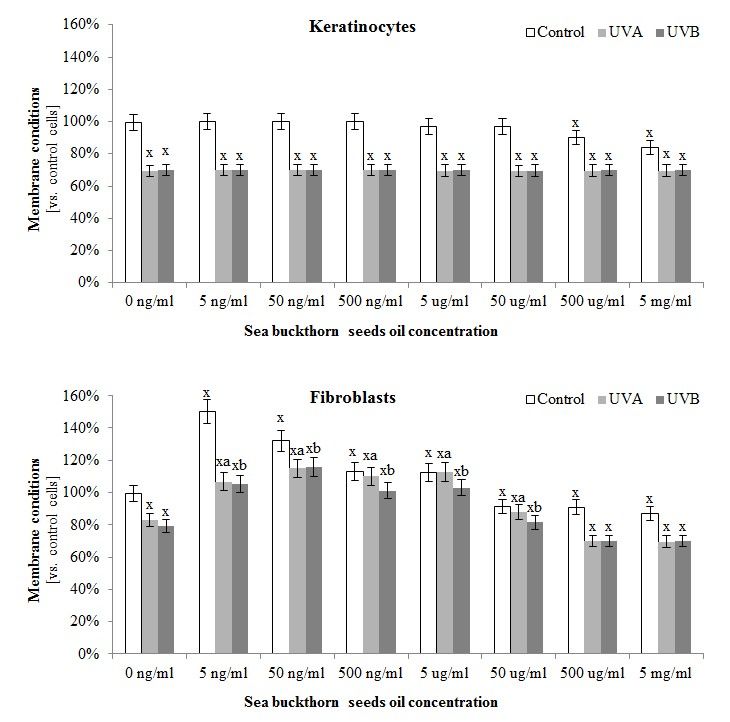
3.4. The Effect of Sea Buckthorn Seed Oil on Skin Cells Lipid Metabolism
The composition
The composition ofof sea
seabuckthorn
buckthornoiloilalso had
also hadan an
impact on skin
impact cellscells
on skin lipidlipid
metabolism. The
metabolism.
greatest protective effect of this oil on membrane conditions was observed in the case of fibroblasts,
The greatest protective effect of this oil on membrane conditions was observed in the case of fibroblasts,
where sea
where sea buckthorn
buckthorn oil
oil not
not only
only reduced
reduced the
the permeability
permeability ofof the
the membrane
membrane compared
compared to to the
the control
control
cells measured by LDH test (in the concentration rage 5 ng/mL–5 μg/mL), but
cells measured by LDH test (in the concentration rage 5 ng/mL–5 µg/mL), but also significantlyalso significantly
prevented UVA
prevented UVAandand UVB‐induced
UVB-induced membrane
membrane damage
damage (in the(in the concentration
concentration rage 5 ng/mL–50
rage 5 ng/mL–50 µg/mL).
μg/mL). These effects were not observed in the case of keratinocytes
These effects were not observed in the case of keratinocytes (Figure 3). (Figure 3).
Figure 3.3. The
The effect
effectofofdifferent
differentconcentrations
concentrations ranging
ranging50 50
ng/mL–5
ng/mL–5 µg/mL
μg/mL ofof
seasea
buckthorn
buckthorn seed oil on
seed oil
the membrane
on the membrane conditions
conditionsof control and and
of control UV irradiated keratinocytes
UV irradiated and fibroblasts
keratinocytes measured
and fibroblasts by LDH
measured by
test. 2 and 60 mJ/cm 2 for keratinocytes,
LDHTotal
test.doses
Totalofdoses
UV irradiation for each cell
of UV irradiation forline were:
each cell30line
J/cmwere: 30 J/cm2 and 60 mJ/cm2 for
and 20 J/cm2 and
keratinocytes, and200
20 mJ/cm
2
J/cm2 andfor200 fibroblasts for fibroblasts
mJ/cm2 for UVA and UVB, respectively.
for UVA and UVB, Mean values ± SD
respectively. Meanof
five independent experiments are presented. x statistically significant differences vs. control group,
values ± SD of five independent experiments are presented. statistically significant differences vs.
x
pcontrol y statistically significant
< 0.05; group, differences betweendifferences
UVA/UVBbetweenand oil treated groups vs. oil
oil treated
treated
p < 0.05; y statistically significant UVA/UVB and
a statistically significantadifferences between UVA and oil treated group vs. UVA
control vs. oil ptreated
groups group, < 0.05;control group, p < 0.05; statistically significant differences between UVA and
treated group, p < vs. b statistically significant differences between UVB and oil treated group vs. UVB
0.05;UVA
oil treated group treated group, p < 0.05; b statistically significant differences between UVB
treated group, p < 0.05.
and oil treated group vs. UVB treated group, p < 0.05.
On the other hand, sea buckthorn oil treatment on skin cells significantly increased the level of
all detected phospholipids as well as free fatty acids in both keratinocytes
keratinocytes and
and fibroblasts
fibroblasts (Table
(Table 4).
4).
Moreover, seasea buckthorn
buckthorn oil influenced the UV-decreased
UV‐decreased level
level of
of numerous
numerous fatty
fatty acids,
acids, which was
particularlyevident
particularly evidentinin thethe
casecase of keratinocytes
of keratinocytes and fibroblasts
and fibroblasts treatedtreated
with oilwith oil following
following UVB
UVB radiation.
radiation. Additionally,
Additionally, sea buckthorn sea buckthorn oil effects
oil treatment treatment effectsfatty
on short on short
acids fatty
(16:0;acids
16:1)(16:0;
in UVA 16:1) in UVA
irradiated
irradiated
cells cells was
was stronger in stronger
fibroblastsin than
fibroblasts than in keratinocytes,
in keratinocytes, while seaoilbuckthorn
while sea buckthorn treatmentoil treatment
effect on the
effect on the level of longer fatty acids (20:4) was visible
level of longer fatty acids (20:4) was visible only in the case of keratinocytes (Table 3). The Figure 3).
only in the case of keratinocytes (Table S1
The Figure
presents theS1 presents the
correlation correlation
between between
the level of freethe level
fatty of free
acids fatty
in sea acids inseeds
buckthorn sea buckthorn seeds oil
oil and changes in
and changes in the level of free fatty acids in keratinocytes
the level of free fatty acids in keratinocytes and fibroblasts. and fibroblasts.Antioxidants 2018, 7, 110 11 of 21
Table 4. The level of phospholipid and free fatty acids in keratinocytes and fibroblasts after exposure
to UVA (30 J/cm2 and 20 J/cm2 ) and UVB radiation (60 mJ/cm2 and 200 mJ/cm2 , respectively) and
sea buckthorn seeds oil (500 ng/mL) treatment.
Keratinocytes Fibroblasts
Oil Control UVA UVB Control UVA UVB
Phospholipid fatty acids [ug/mg protein]
- 5.3 ± 0.3 4.5 ± 0.2 4.5 ± 0.2 1.4 ± 0.1 1.2 ± 0.1 1.2 ± 0.1
14:0
+ 12.4 ± 0.6 x 11.3 ± 0.6 xa 11.8 ± 0.6 xb 6.3 ± 0.1 x 2.5 ± 0.1 xya 5.1 ± 0.3 xyb
- 152 ± 7.3 129 ± 6.5 x 128 ± 6.4 x 80 ± 4 68 ± 3.4 68 ± 3.4
16:0
+ 662 ± 33.1 x 145 ± 7.3 y 260 ± 13.0 xyb 190 ± 9.5 x 90 ± 4.5 ya 122 ± 6.1 xyb
- 15.6 ± 0.8 13.2 ± 0.7 13.1 ± 0.7 13.6 ± 0.7 11.6 ± 0.6 11.5 ± 0.6
16:1
+ 29.9 ± 1.5 x 27.5 ± 1.4 xa 28.5 ± 1.4 xb 23.2 ± 1.2 x 17.7 ± 0.9 xya 15.9 ± 0.8 xyb
- 123 ± 6.2 104 ± 5.2 x 10³ ± 5.2 x 69 ± 3.5 58 ± 2.9 x 58 ± 2.9 x
18:0
+ 412 ± 20.6 x 137 ± 6.9 xya
187 ± 9.4 xyb 126 ± 6.3 x 10⁹ ± 5.5 xya 110 ± 5.5 xyb
- 199 ± 10.1 169 ± 8.5 x 168 ± 8.4 x 112 ± 5.6 95 ± 4.8 x 94 ± 4.7 x
18:1nc
+ 580 ± 29.1 x 129 ± 6.5 xya
386 ± 19.3 xyb 181 ± 9.1 x 167 ± 8.4 xa 114 ± 5.7 yb
- 47 ± 2.4 39 ± 2.1 x 39 ± 2.1 x 18 ± 0.9 16 ± 0.8 15 ± 0.8 x
18:1nt
+ 170 ± 18.5 x 48 ± 2.4 ya
139 ± 7.1 xyb 58 ± 2.9 x 22 ± 1.1 xya 36 ± 1.8 xyb
- 116.2 ± 5.8 98.8 ± 4.9 x 98.1 ± 4.9 x 45.4 ± 2.3 38.6 ± 1.9 x 38.3 ± 1.9 x
18:2
+ 162.7 ± 8.1 x 111.4 ± 5.6 ya
137.2 ± 6.9 xyb 69.8 ± 3.5 x 60.4 ± 3.0 xya 64.5 ± 3.2 xb
- 24.6 ± 1.2 20.9 ± 1.1 x 20.8 ± 1.1 x 6.2 ± 0.3 5.2 ± 0.3 5.5 ± 0.3
18:3n3
+ 29.5 ± 1.5 x 30.2 ± 1.5 xa 31.2 ± 1.6 xb 12.5 ± 0.6 x 7.8 ± 0.4 xya 8.5 ± 0.4 xyb
- 38.1 ± 1.9 32.4 ± 1.6 x 32.2 ± 1.6 x 35.6 ± 1.8 30.2 ± 1.5 30.1 ± 1.5
20:4
+ 197.4 ± 9.9 x 36.4 ± 1.8 ya 104.7 ± 5.2 xyb 81.7 ± 4.1 x 30.3 ± 1.5 y 57.5 ± 2.9 xyb
- 11.1 ± 0.6 9.4 ± 0.5 9.3 ± 0.5 8.2 ± 0.4 6.9 ± 0.3 6.9 ± 0.3
22:6
+ 30.1 ± 1.5 x 18.1 ± 0.9 xya 23.1 ± 1.2 xyb 41.6 ± 2.1 x 16.2 ± 0.8 xya 33.8 ± 1.7 xyb
Free fatty acids [µg/mg protein]
- 8.8 ± 0.4 7.5 ± 0.4 7.4 ± 0.4 2.9 ± 0.1 2.4 ± 0.1 2.4 ± 0.1
16:0
+ 26 ± 1.3 x 14.1 ± 0.7 xya 19.8 ± 1 xyb 9.8 ± 0.5 x 3.9 ± 0.2 xya 7 ± 0.4 xyb
- 1.7 ± 0.1 1.4 ± 0.1 1.4 ± 0.1 0.7 ± 0.1 0.6 ± 0.1 0.6 ± 0.1
16:1
+ 5.5 ± 0.3 x 1.2 ± 0.1 y 4.4 ± 0.2 xyb 2.1 ± 0.1 x 1.3 ± 0.1 xya 1.6 ± 0.1 xyb
- 7.6 ± 0.4 6.4 ± 0.3 6.4 ± 0.3 3.4 ± 0.2 2.9 ± 0.1 2.9 ± 0.3
18:0
+ 16.8 ± 0.8 x 8.8 ± 0.4 ya 8.2 ± 0.4 yb 8.8 ± 0.4 x 3.2 ± 0.2 y 5.5 ± 0.3 xyb
- 13.3 ± 0.7 11.3 ± 0.6 11.2 ± 0.6 3.8 ± 0.2 3.3 ± 0.2 3.5 ± 0.2
18:1nc
+ 54.3 ± 2.7 x 51.1 ± 2.6 xa 45.1 ± 2.3 xyb 6.8 ± 0.3 x 4.1 ± 0.2 ya 5.5 ± 0.3 xyb
- 6.1 ± 0.3 5.2 ± 0.3 5.1 ± 0.3 1.1 ± 0.1 0.9 ± 0.1 0.9 ± 0.1
18:1nt
+ 9.8 ± 0.5 x 8.5 ± 0.4 xya 8.3 ± 0.4 xyb 4.5 ± 0.2 x 1.5 ± 0.1 xya 4.1 ± 0.2 xb
- 11.7 ± 0.6 9.9 ± 0.5 9.8 ± 0.5 3.2 ± 0.2 2.7 ± 0.1 2.7 ± 0.1
18:2
+ 15.1 ± 0.8 x 10.5 ± 0.5 y 12.7 ± 0.6 xyb 5.4 ± 0.3 x 3.3 ± 0.2 ya 3.3 ± 0.2 y
- 2.2 ± 0.1 1.8 ± 0.1 1.8 ± 0.1 0.7 ± 0.1 0.6 ± 0.1 0.6 ± 0.1
20:4
+ 9 ± 0.5 x 5.4 ± 0.3 xya 5.6 ± 0.3 xyb 3.4 ± 0.2 x 0.6 ± 0.1 y 2.7 ± 0.1 xyb
- 0.9 ± 0.1 0.8 ± 0.1 0.8 ± 0.1 0.3 ± 0.1 0.3 ± 0.1 0.3 ± 0.1
22:6
+ 11.3 ± 0.6 x 11.4 ± 0.6 xa 4.8 ± 0.2 xyb 2.3 ± 0.1 x 2.2 ± 0.1 xa 2.2 ± 0.1 xb
Mean values ± SD of five independent experiments are presented. x statistically significant differences vs. control
group, p < 0.05; y statistically significant differences between UVA/UVB and oil treated groups vs. oil treated control
group, p < 0.05; a statistically significant differences between UVA and oil treated group vs. UVA treated group,
p < 0.05; b statistically significant differences between UVB and oil treated group vs. UVB treated group, p < 0.05.
Obtained results showed that sea buckthorn oil partially prevented UV-induced increase in
phospholipase A2 activity by 50% in the case of UV irradiated keratinocytes, and by about 15% in the
case of UV irradiated fibroblasts (Figure 4). As a result of UV-induced lipid oxidative metabolism a
strong increase in the level of lipid peroxidation products was observed. Sea buckthorn oil treatment
significantly prevented these changes in the case of 4-HNE levels, which was decreased by 15% in
oil treated keratinocytes, and 30% in oil treated fibroblasts. Also, the level of isoprostaglandin in
fibroblasts exposed to UV radiation was decreased by 25% following sea buckthorn oil treatment.
However, in the case of isoprostaglandin levels in keratinocytes sea buckthorn oil lead to increases by
approximately 20% under standard conditions and did not have a significant effect on irradiated cells
(Figure 4).Antioxidants 2018, 7, 110 12 of 21
Antioxidants 2018, 7, x FOR PEER REVIEW 12 of 20
Figure
Figure 4. The
4. The activity
activity ofofphospholipase
phospholipase A2 A2 and
and level
levelofoflipid peroxidation
lipid peroxidationproducts
products(4-HNE and and
(4-HNE
8-Isoprostaglandin F2α) in keratinocytes and fibroblasts after exposure to UVA (30 J/cm 2 and 20 J/cm2 )
8-Isoprostaglandin F2α) in keratinocytes and fibroblasts after exposure to UVA (30 J/cm2 and 20
and UVB radiation (60 mJ/cm2 and 200 mJ/cm2 , respectively) and sea buckthorn seeds oil (500 ng/mL)
J/cm2) and UVB radiation (60 mJ/cm2 and 200 mJ/cm2, respectively) and sea buckthorn seeds oil (500
treatment. Mean values ± SD of five independent experiments are presented. x statistically significant
ng/mL) treatment. Mean values ± SD yof five independent experiments are presented. x statistically
differences vs. control group, p < 0.05; statistically significant differences between UVA/UVB and oil
significant differences vs. control group, p < 0.05; y statistically significant differences between
treated groups vs. oil treated control group, p < 0.05; a statistically significant differences between UVA
UVA/UVB and oilgroup
and oil treated treated groups
vs. UVA vs. group,
treated oil treated control
p < 0.05; group, psignificant
b statistically < 0.05; adifferences
statistically significant
between
differences
UVB andbetween
oil treatedUVA
groupand oil treated
vs. UVB grouppAntioxidants 2018, 7, 110 13 of 21
Antioxidants 2018, 7, x FOR PEER REVIEW 13 of 20
Figure 5. The level of endocannabinoids (AEA, 2-AG) and endocannabinoid receptors (CB1, CB2) in
keratinocytes
keratinocytes and and fibroblasts
fibroblasts after afterexposure
exposuretotoUVA UVA(30 (30J/cm
J/cm 2 and
2 and 2020J/cm 2 ) and
2) and
J/cm UVB radiation
UVB (60
radiation
mJ/cm 2
(60 mJ/cm 2
and 200
and mJ/cm 2
200 mJ/cm 2
, respectively) and sea
, respectively) and buckthorn
sea buckthornseeds seeds
oil (500
oilng/mL) treatment.
(500 ng/mL) Mean
treatment.
values ± SD of
Mean values ± five
SD ofindependent
five independent experiments
experimentsare presented.
are presented. x statistically
x statistically significant differences
significant vs.
differences
control
vs. controlgroup, pAntioxidants 2018, 7, 110 14 of 21
with UV irradiation. Results of this study partially confirm earlier observations that sea buckthorn oil
may lead to enhanced antioxidant abilities by promotion of glutathione accumulation in the whole
animal body in the case of rats treated with this oil [51]. It was also shown that supplementation of
rats with compounds of sea buckthorn extracts activate the enzymes superoxide dismutase, catalase,
glutathione peroxidase, glutathione reductase and glutathione S-transferase in animal blood [52].
However, in this study sea buckthorn oil shows the tendency to decrease fibroblast Cu, Zn-SOD
activity in control and UV-irradiated cells. This may be explained by the sea buckthorn oils ability
to capture electrons and extinguish the singlet oxygen or superoxide anion [53] that is the main
substrate of SOD in cell cytoplasm. Another important cellular redox regulator is the thioredoxin
system consisting of thioredoxin and thioredoxin reductase (TrxR) [54]. This expression is diminished
in skin cells after UV irradiation and enhanced after sea buckthorn oil treatment of keratinocytes and
fibroblasts. Previously it has been shown that this system plays a significant role in the pathogenesis of
a number of diseases, but it also participates in cellular protection against toxic compounds, which are
reduced by thioredoxin or inhibited by thioredoxin reductase [55,56]. It has been indicated that sea
buckthorn includes high levels of proteins containing thioredoxin domains and prevents the disulfide
bond formation in oxidizing environments and stabilizing the tertiary and quaternary structure
of proteins [57]. It has also been shown that Trx and TrxR synthesis is induced after exposure to
prooxidative factors [58], this explains the strong positive response of this system to UV irradiation,
particularly in fibroblasts. Therefore, enhanced effectiveness of this system after oil treatment may
explain reduction of oxidative stress in these cells.
Cellular antioxidant responses dependent on proteins is promoted by the transcription factor
Nrf2 which is responsible for cytoprotective gene transcription [59]. Under physiological conditions,
cytoplasmic Nrf2 is bound to Keap1 [60], but oxidative stress caused by UV radiation was found
to diminish Keap1 expression which prevents Nrf2–Keap1–Cul3 complex formation. Moreover, as
observed in this paper, high levels of ROS and electrophiles such as 4-HNE may lead to the oxidation of
Keap1 cysteine residues causing lack of binding and/or dissociation of Nrf2 from the complex resulting
in its translocation into the nucleus, which was reported earlier [61]. Nrf2 activation, demonstrated
also as its targeted genes-HO-1 expression, leads to cellular protection against pro-oxidative conditions.
Sea buckthorn oil treatment of UV irradiated skin cells acts in two ways—it supports the cellular
antioxidant capacity by the activation of Nrf2, and at the same time, affects the expression of proteins
strongly related with Nrf2 activation. The results showing enhanced phospho-Nrf2 translocation to
the nucleus despite of the increased Keap1 level in cytoplasm clearly suggest that sea buckthorn oil
disrupts the Nrf2–Keap1 complex formation. Moreover, sea buckthorn seed oil decreases UV-induced
levels of p21 and p62, reducing the possibility of these proteins to create adducts with Nrf2 or Keap1,
thus encouraging Nrf2 inhibition [59,62]. Keap1 might also create adducts with small antioxidant
molecules such as melatonin or GSH, what also prevents Nrf2-Keap1 binding, however, it does not
lead to Keap1 degradation [63,64]. Melatonin has been previously shown to be downregulated in
skin cells following UV radiation [65,66], while UV-induced decrease in GSH level, what is shown in
this study is significantly enhanced as a result of sea buckthorn oil treatment. These modifications
to the activators and inhibitors of the Nrf2 pathway are not able to cancel the protective effect of sea
buckthorn oil to counteract prooxidative conditions.
4.2. Sea Buckthorn Seeds Oil Effect on Lipid Metabolism
The effects of sea buckthorn seed oil on redox balance can directly protect the metabolism of
skin cells. Maintaining redox homeostasis is important for the physiological metabolism of lipids,
which is important for the proper functioning of cells. Phospholipid protection may be associated
with beneficial fatty acids present in sea buckthorn oil [67], which influences the skin cells fatty
acid profile. The strongest correlation between oil fatty acids and increase in the cells fatty acid
level is visible in the case of free fatty acids (FFA) regardless of whether that were control cells
or treated with UV radiation. This relationship between FFA composition of the applied oil andAntioxidants 2018, 7, 110 15 of 21
cells might be associated with 24 h incubation with oil when cells were able only to take up and
assimilate FFA from fatty rich oil composition. Sea buckthorn oil is rich in palmitoleic acid (16:1),
which has been reported to play a role in many metabolic processes including intracellular lipid
mediated signal transduction [68,69], which includes lipid metabolism, and is also responsible for
maintaining the fluidity of biological membranes [70–72]. Palmitoleic acid levels are enhanced in the
phospholipid profile of keratinocytes and fibroblasts after sea buckthorn oil treatment, this also causes
the reduction of mRNA expression of proinflammatory genes, i.e., TNF-α [73], therefore this oil may
affect intracellular signaling based on PI3K/Akt kinase cascades, and have anti-inflammatory activity,
which was also confirmed by experiments on rats with diabetes [74]. Moreover, sea buckthorn oil
contains a large amount of linoleic and α-linolenic acids, which can enhance the level of phospholipid
α-linolenic acid in cells [75,76]. α-Linolenic acid contained in sea buckthorn oil has been found also as a
source of eicosanoid prostaglandin E1, a signal precursor that produces antibacterial and cytoprotective
extracellular fluids [77]. Shown in this study sea buckthorn oil added to skin cells enhances the level of
these acids in phospholipid and free PUFAs fractions. Skin cells phospholipid PUFAs fraction is also
enriched in γ-linolenic acid that is a precursor of anti-inflammatory eicosanoids, such as the 1-series
prostaglandins and 15-hydroxyeicosatrienoic acid (15-HETrE) [78].
It has been previously shown that UV radiation significantly disturbs the metabolism of skin
cell membrane phospholipids [6]. In this study, active compounds contained in sea buckthorn oil
enhances the cell’s antioxidant abilities and prevents lipid peroxidation [47] as well as enzymatic
phospholipid metabolism. Sea buckthorn oil enhances, both phospholipid and free PUFA levels in
keratinocytes and fibroblasts, as well as increases the ROS level favored by ROS-dependent lipid
peroxidation manifested as oxidative fragmentation with enhanced 4-HNE levels and as oxidative
cyclisation with enhanced 8-isoprostane levels. Independently of that, one of the main sources of
peroxidation products-arachidonic acid (AA) is most enhanced in PUFAs of both skin cell lines,
what indicates for huge tributary of this acid from used oil. As a result of AA peroxidation, increases
in the 4-HNE levels can directly act as a signaling molecule or through protein adduct formation
which significantly influences their structure and activity [79]. Phospholipid AA is also metabolized
by enzymes among which the most important are phospholipases including PLA2 [80]. It has been
previously shown that UV radiation significantly increases PLA2 activity [9,81], while PLA2 inhibition
improves skin conditions [82]. Sea buckthorn oil reveals similar as other plant oils PLA2 inhibition
properties, preventing UV-induced lipid metabolism [83]. Despite the observed decrease in PLA2
activity, the levels of endocannabinoids are increased after using sea buckthorn oil. It is believed that
anandamide is a partial or full agonist of the CB1 receptor, depending on tissue and conditions, and is
suggested to have low efficacy for CB2 receptors, whereas 2-AG is a full agonist of both CB1 and CB2
receptors [84]. However, an elevated level of endocannabinoids is accompanied by a down-regulation
of cannabinoid receptors. Such response may indicate that redox and inflammatory regulation is
independent from the cannabinoid receptors. Moreover, oil-induced changes in endocannabinoids
level may influence the skin neuroendocrine capabilities regulated by UV radiation [85], what leads to
disorders in steroid hormones, neuropeptides, and neurotransmitters biosynthesis [86]. Regardless of
the above, it has been shown that a high level of AA delivered to skin cells from sea buckthorn oil may
result in increased generation of the 4-series leukotrienes, which have a strong pro-inflammatory and
hyperproliferative effect [87].
Endocannabinoid levels are enhanced by treating skin cells with sea buckthorn oil, which are
agonists of peroxisome proliferator-activated receptors (PPAR). It is known that PPARs are activated
by fatty acids and their derivatives, including lipid peroxidation products like 4-HNE, which act
as PPAR-α agonists [88,89]. PPARs act as modulators of cellular processes including lipid
metabolism, and thus create a lipid signaling network between the cell surface and the nucleus [89].
Enhanced expression of fibroblast PPARs indicates that sea buckthorn seed oil has anti-inflammatory
activity. PPAR-α controls the expression of proteins that participate in inflammatory response [89],
therefore enhanced activation of PPAR-α observed in fibroblasts indicates preventing NF-κB-dependentAntioxidants 2018, 7, 110 16 of 21
inflammation [90]. It has also been shown that anandamide as a ligand of PPAR-α can participate
in its anti-inflammatory effect through impaired production of TNF-α [91]. Similarly, enhanced
expression of PPAR-γ decreases the expression of TNF-α [92]. Moreover, sea buckthorn ethanolic
extract has been beneficial in reducing fat pad mass and preventing weight gain in mice. The extract
was effective in producing hypoglycemic effects in animals through up-regulating PPAR-γ and PPAR-α
gene expression [92].
5. Conclusions
In summary, sea buckthorn oil significantly stimulates the antioxidant system in keratinocytes
and fibroblasts. Therefore, sea-buckthorn seed oil prevents UV-induced impair in redox systems as
well as lipid metabolism disorders in skin fibroblasts and keratinocytes, which makes it a promising
natural substance in skin photo-protection. However, the influence of UV radiation on the stability
and durability of oil components, their interactions and impact on the metabolism of skin cells has not
been studied, therefore it is believed that current studies do not allow recommending the use of sea
buckthorn seed oil in direct exposure to UV radiation.
Supplementary Materials: The following are available online at http://www.mdpi.com/2076-3921/7/9/110/s1,
Figure S1: The correlation between the level of free fatty acids in sea buckthorn seeds oil and changes in the
level of free fatty acids in keratinocytes and fibroblasts after exposure to UVA (30 J/cm2 and 20 J/cm2 ) and
UVB radiation (60 mJ/cm2 and 200 mJ/cm2 , respectively) and sea buckthorn seeds oil (500 ng/mL) treatment.,
Figure S2: The electrophorogram images of Western blot analyses of phospho-Nrf2 (pSer40) in keratinocytes
and fibroblasts after exposure to UVA (30 J/cm2 and 20 J/cm2 ) and UVB radiation (60 mJ/cm2 and 200 mJ/cm2 ,
respectively) and sea buckthorn seeds oil (500 ng/mL) treatment.
Author Contributions: Conceptualization, E.S.; Formal analysis, A.G., A.J., I.J.K. and M.M.; Investigation, A.G.,
A.J., I.J.K. and M.M.; Methodology, A.G., A.J., I.J.K. and M.M.; Validation, A.G., A.J., I.J.K. and M.M.; Visualization,
A.G., A.J., I.J.K. and M.M.; Writing—Review & editing, E.S.
Funding: This research received no external funding.
Conflicts of Interest: The authors declare that they have no conflict of interests.
References
1. Chuong, C.M.; Nickoloff, B.J.; Elias, P.M.; Goldsmith, L.A.; Macher, E.; Maderson, P.A.; Sunberg, J.P.;
Tagami, H.; Plonka, P.M.; Thestrup-Pederson, K.; et al. What is the ‘ture’ function of skin? Exp. Dermatol.
2002, 11, 159–187. [PubMed]
2. Agache, P.; Lihoreau, T.; Mac-Mary, S.; Fanian, F.; Humbert, P. The human skin: An overview. In Agache’s
Measuring the Skin: Non-invasive Investigations; Springer International Publishing AG: Cham, Switzerland,
2017; pp. 1–4.
3. Moan, J.; Grigalavicius, M.; Baturaite, Z.; Dahlback, A.; Juzeniene, A. The relationship between UV exposure
and incidence of skin cancer. Photodermatol. Photoimmunol. Photomed. 2015, 31, 26–35. [CrossRef] [PubMed]
4. Natarajan, V.T.; Ganju, P.; Ramkumar, A.; Grover, R.; Gokhale, R.S. Multifaceted pathways protect human
skin from UV radiation. Nat. Chem. Biol. 2014, 10, 542–551. [CrossRef] [PubMed]
5. Slominski, A.T.; Zmijewski, M.A.; Plonka, P.M.; Szaflarski, J.P.; Paus, R. How UV Light Touches the Brain
and Endocrine System Through Skin, and Why. Endocrinology 2018, 159, 1992–2007. [CrossRef] [PubMed]
6. G˛egotek, A.; Biernacki, M.; Ambrożewicz, E.; Surażyński, A.; Wroński, A.; Skrzydlewska, E. The cross-talk
between electrophiles, antioxidant defence and the endocannabinoid system in fibroblasts and keratinocytes
after UVA and UVB irradiation. J. Dermatol. Sci. 2016, 81, 107–117. [CrossRef] [PubMed]
7. Mohamed, M.A.A.; Jung, M.; Lee, S.M.; Lee, T.H.; Kim, J. Protective effect of Disporum sessile D. Don extract
against UVB-induced photoaging via suppressing MMP-1 expression and collagen degradation in human
skin cells. J. Photochem. Photobiol. B 2014, 133, 73–79. [CrossRef] [PubMed]
8. Larroque-Cardoso, P.; Camaré, C.; Nadal-Wollbold, F.; Grazide, M.H.; Pucelle, M.; Garoby-Salom, S.;
Bogdanowicz, P.; Josse, G.; Schmitt, A.M.; Uchida, K.; et al. Elastin modification by 4-hydroxynonenal
in hairless mice exposed to UV-A. Role in photoaging and actinic elastosis. J. Investig. Dermatol. 2015, 135,
1873–1881. [CrossRef] [PubMed]You can also read