Restoration of Vision in the pde6β-deficient Dog, a Large Animal Model of Rod-cone Dystrophy
←
→
Page content transcription
If your browser does not render page correctly, please read the page content below
© The American Society of Gene & Cell Therapy
                                                                                                                  original article

Restoration of Vision in the pde6β-deficient Dog,
a Large Animal Model of Rod-cone Dystrophy
Lolita Petit1, Elsa Lhériteau1, Michel Weber2, Guylène Le Meur2, Jack-Yves Deschamps3, Nathalie Provost1,
Alexandra Mendes-Madeira1, Lyse Libeau1, Caroline Guihal1, Marie-Anne Colle4, Philippe Moullier1,5
and Fabienne Rolling1
1
 Translational Gene Therapy for Retinal and Neuromuscular Diseases, INSERM UMR 1089, Institut de Recherche Thérapeutique 1, Université de Nantes,
Nantes, France; 2CHU-Hôtel Dieu, Service d’Ophtalmologie, Nantes, France; 3Emergency and Critical Care Unit, ONIRIS, Nantes-Atlantic College of
Veterinary Medicine Food Science and Engineering, Nantes, France; 4UMR 703 PAnTher INRA/ONIRIS, Nantes-Atlantic College of Veterinary Medicine
Food Science and Engineering, Nantes, France; 5Department of Molecular Genetics and Microbiology, College of Medicine, University of Florida,
Gainesville, Florida, USA

Defects in the β subunit of rod cGMP phosphodiesterase                     of central day vision. RP may be inherited as an autosomal reces-
6 (PDE6β) are associated with autosomal recessive retini-                  sive (50–60%), autosomal dominant (30–40%), X-linked (5–15%),
tis pigmentosa (RP), a childhood blinding disease with                     or simplex/multiplex disease.2 To date, about 45 genes associated
early retinal degeneration and vision loss. To date, there                 with RP etiology have been identified; most of them are primarily
is no treatment for this pathology. The aim of this pre-                   expressed in photoreceptor cells.3
clinical study was to test recombinant adeno-associated                        Mutations in the gene encoding the β subunit of rod cGMP-
virus (AAV)-mediated gene addition therapy in the rod-                     phosphodiesterase 6 (PDE6β) are associated with one of the most
cone dysplasia type 1 (rcd1) dog, a large animal model                     prevalent forms of autosomal recessive RP, accounting for ~1–2%
of naturally occurring PDE6β deficiency that strongly                      of all human RP cases.4–12 Rod PDE6 is localized on the disc mem-
resembles the human pathology. A total of eight rcd1                       brane of rod outer segments where it plays a key role in the rod
dogs were injected subretinally with AAV2/5RK.cpde6β                       phototransduction cascade by controlling the level of cGMP and
(n = 4) or AAV2/8RK.cpde6β (n = 4). In vivo and post-                      Ca2+ in the rod outer segments. Rod PDE6 is a heterotetrameric
mortem morphological analysis showed a significant                         complex composed of two homologous catalytic subunits (PDE6α
preservation of the retinal structure in transduced areas                  and PDE6β) and two copies of an inhibitory subunit (PDE6γ).13
of both AAV2/5RK.cpde6β- and AAV2/8RK.cpde6β-                              As both PDE6α and PDE6β subunits are required for rod PDE6
treated retinas. Moreover, substantial rod-derived elec-                   activity, loss-of-function mutations in the pde6β gene result in an
troretinography (ERG) signals were recorded as soon                        improper function of PDE6 holoenzyme and an accumulation of
as 1 month postinjection (35% of normal eyes) and                          cGMP and Ca2+ in the rod outer segments. This ultimately leads to
remained stable for at least 18 months (the duration of                    rod then cone photoreceptor death through apoptosis by non-well
the study) in treated eyes. Rod-responses were undetect-                   defined mechanisms.14–16
able in untreated contralateral eyes. Most importantly,                        There is currently no cure for RP caused by PDE6β deficiency.
                                                                           Addition gene therapy is an attractive approach for PDE6β-RP.
dim-light vision was restored in all treated rcd1 dogs.
                                                                               Gene therapy approaches to delay photoreceptors degen-
These results demonstrate for the first time that gene
                                                                           eration have already been employed in three murine models
therapy effectively restores long-term retinal function
                                                                           of PDE6β-RP, the rodless 1 (rd1),17 the retinal degeneration
and vision in a large animal model of autosomal reces-
                                                                           10 (rd10)18,19 and the PDE6βH620Q.20 Various vectors encod-
sive rod-cone dystrophy, and provide great promise for
                                                                           ing murine or human pde6β gene have been tested, including
human treatment.
                                                                           adenovirus,21 adeno-associated virus (AAV),22–25 simian immu-
Received 17 March 2012; accepted 17 June 2012; advance online              nodeficiency virus,26 herpes simplex virus,27 and lentivirus.20,28
publication 24 July 2012. doi: 10.1038/mt.2012.134
                                                                           In most studies functional rescue was limited, despite significant
                                                                           improvement of retinal morphology with an increased number of
Introduction                                                               outer nuclear layer (ONL) nuclei and outer segment length. One of
Retinitis pigmentosa (RP), or rod-cone dystrophy, comprises a              the best functional results (37% preservation of rod electroretin-
wide spectrum of incurable hereditary retinal dystrophies char-            ography (ERG) responses and prolonged photoreceptor survival
acterized by sequential degeneration of rod and cone photore-              up to postnatal day (P) 35) was obtained in dark-reared rd10 mice,
ceptors.1 Early symptoms include night blindness and loss of               a model with a slower rate of degeneration than rd1 mice, treated
peripheral vision due to progressive rod photoreceptors degenera-          at P14 using an AAV2/5smCBA.mpde6β vector.23 This sug-
tion. This phase is followed by cone death and concomitant loss            gest that difficulties in achieving sustained long-term functional

Correspondence: Fabienne Rolling, Translational Gene Therapy for Retinal and Neuromuscular Diseases, INSERM UMR 1089, Institut de Recherche
Thérapeutique 1, Université de Nantes, 8 quai Moncousu, 44007 Nantes Cedex 01, France. Email: fabienne.rolling@inserm.fr

Molecular Therapy                                                                                                                              1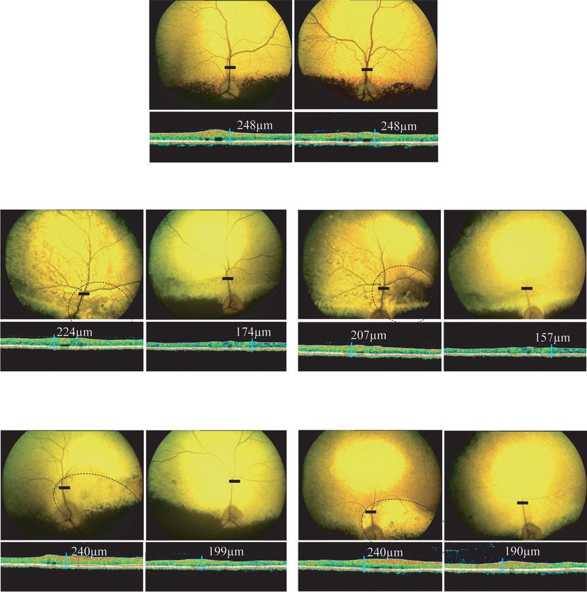
© The American Society of Gene & Cell Therapy
Restoration of Vision in pde6β-deficient Dogs

rescue in murine models of PDE6β-deficiency may not be related
to specific characteristics of the PDE6β subunit but to (i) exces-                    ITR         RK        cpde6β            BGHpA         ITR
sively rapid loss of photoreceptors in these models and mainly to
(ii) difficulties in achieving sufficient level of transgene expression                         236 bp     2,681 bp           232 bp
in mutant photoreceptors before their irreversible degeneration.                                        AAV2/5RK.cpde6β
This hypothesis was reinforced by a recent successful gene addi-                                        AAV2/8RK.cpde6β
tion therapy in the same mouse model after subretinal injection           Figure 1 Schematic structure of recombinant adeno-associated
of a fast-acting AAV2/8(Y733F)smCBA.mpde6β vector.24 In this              virus (AAV) vectors. AAV2/5RK.cpde6β and AAV2/8RK.cpde6β vectors
recent “proof-of-concept” study, the authors reported on a sub-           encode the canine pde6β cDNA under the control of a human RK pro-
sequent 58% preservation of rod ERG responses, rod-mediated               moter (–112 bp to +87 bp region of the proximal promoter36). BGHpA,
                                                                          bovine growth hormone polyadenylation signal; cpde6β, canine pde6β
vision-guided behavior and rod survival for up to 6 months.               cDNA; ITR, inverted terminal repeats from AAV2; RK, human rhodopsin
     Preclinical evaluation of recombinant AAV (rAAV)-mediated            kinase promoter.
gene transfer in large animal models (i.e., dogs, cats, pigs, and non-
human primates) is a key step for future clinical development.29          Table 1 List of dogs included in the study and ERG amplitudes
Indeed, the size and anatomy of the eye in large animals provide                                        Rod ERG (µV)                30Hz Flicker (µV)
a more relevant model than rodents in terms of pathobiology                                     1 mpi      9 mpi    18 mpi        1 mpi     9 mpi 18 mpi
and surgical approaches. Moreover, their longevity enables long-
                                                                          Dog Vector             T/U        T/U          T/U       T/U       T/U     T/U
term evaluation of the treatment, which is crucial for pathologies
                                                                          NA1                   169/166 215/182 140/128           45/46      67/65   43/42
such as PDE6β deficiency that typically progress over decades in
humans.                                                                   NA2 None              141/191      a            a
                                                                                                                                  33/39        a       a

     The Rcd1 dog is a natural large animal model of progressive          A1                      0/0      11/15         0/0      44/38      43/56    6/9
retinal degeneration. The Rcd1 dog carries a (2420G>A) non-               A2                     47/0      43/10         53/0     45/53      54/53   40/14
sense mutation in the C-terminus of the PDE6β subunit30 lead-             A3     AAV2/5RK.       34/0      34/0          42/0     39/35      17/15   25/13
ing to truncation and destabilization of the gene product and a                  cpde6β
nonfunctional PDE6 holoenzyme.31,32 Retinal development is nor-           A4     (1011 vg/ml)    40/0      52/0          nd       51/27      41/39    nd
mal in affected dogs until 13 days of age, at which point photo-
                                                                          A5                      nd         a       a
                                                                                                                                    nd         a       a
receptor development is arrested. Rod degeneration then occurs
gradually from 1 to 5 months of age, followed by cone loss within         A6                     37/0      87/0          63/0     21/46      38/15   30/11
1 to 2 years.33,34 This clinical course of generalized retinal atrophy,   A7     AAV2/8RK.       45/0      37/0          28/0     42/29      23/17   19/9
which strongly mimics retinal degeneration in humans, makes                      cpde6β
this PDE6β deficient dog a valuable large animal model for the            A8     (1012 vg/ml)    41/0      41/0          nd       36/28      27/21    nd
evaluation of gene therapy.                                               A9                    19b/64b      a            a
                                                                                                                                  37b/30b      a       a

     In the present study, we investigated the long-term efficacy of      Abbreviations: 30 Hz Flicker, photopic 30 Hz Flicker amplitude; A, affected;
early rAAV-mediated pde6β gene transfer in the rcd1 dog. Our              mpi, month(s) postinjection; NA, nonaffected; nd, not done; rod ERG, scotopic
                                                                          rod-mediated b-wave amplitude; T, treated eye; U, untreated eye; vg, vector
findings demonstrate, for the first time, that gene transfer prevents     genome.
long-term photoreceptor death and restores electrophysiological           a
                                                                            Sacrificed for RT-PCR and/or histology. bDog A9 received AAV2/8RK.cpde6β in
                                                                          both right and left eyes.
function and vision in this large animal model.

Results                                                                   cpde6β in both eyes due to the observation of significant vector
Recombinant AAV2/5 and AAV2/8 vector design                               reflux into the vitreous during the subretinal injection of the right
and subretinal administration to rcd1 dogs                                eye. Subretinal blebs were restricted to the nasal superior retina
We generated rAAV2/5 or rAAV2/8 vectors carrying the canine               and covered ~25% of the entire retinal surface, except in the right
pde6β (cpde6β) gene under the control of the photoreceptor-spe-           eye of dog A9 in which the bleb was much smaller.
cific human rhodopsin kinase (RK) promoter35,36 to drive rapid                In all treated eyes, no apparent surgically induced retinal
and strong transgene expression in photoreceptors (Figure 1).             damage was noted by fundus photography or optical coherence
     Rcd1 dogs display an early and severe rod-cone photoreceptor         tomography (OCT) at 2 months postinjection (mpi), despite the
degeneration that starts in the central retina at P25, during post-       small size of the eye and the immaturity of the retina at the time of
natal differentiation of photoreceptor cells.33,34 This rapid onset of    the subretinal injection (data not shown).
retinal degeneration in rcd1 dogs suggests that early therapeutic
intervention is critical to prevent rod photoreceptor cell death in       Assessment of transgene expression in rcd1-treated
this model. To this end, we chose to treat rcd1 dogs at P20, before       retinas
extensive rod photoreceptor loss.                                         To evaluate pde6β transgene expression after subretinal injection
     A total of eight rcd1 dogs (A2–A9) were injected subretinally        of AAV2/5RK.cpde6β and AAV2/8RK.cpde6β vectors, presence
at the age of 20 days. Dogs A2 to A5 were treated with AAV2/5RK.          of pde6β transcripts was assessed by reverse transcription (RT)-
cpde6β, whereas dogs A6 to A9 were injected with AAV2/8RK.                PCR in treated retinas of dogs A4 and A8, at 4 mpi (Figure 2).
cpde6β (Table 1). All injections were performed unilaterally in               As our preliminary RT-PCR analysis indicated that the rcd1
the right retina, except for dog A9 which received AAV2/8RK.              dogs expressed mutant (2420G>A) pde6β transcripts at the

2                                                                                                                              www.moleculartherapy.org
© The American Society of Gene & Cell Therapy
Restoration of Vision in pde6β-deficient Dogs
a AAV2/5RK.cpde6β and AAV2/8RK.cpde6β preserve
2,420
retinal structure
cDNA pde6β exon 21 exon 22 Fundus appearance and retinal thickness were evaluated in all
2056F 2561R dogs using color fundus photography and OCT. These noninva-
BtsCI
Transgene sive examinations were performed from 2 to 18 mpi for dogs A2,
385 bp 20 bp A3, A6, and A7, from 2 to 8 mpi for dogs A4 and A8, and from 2 to
Mutant (2420G>A)
405 bp 3 mpi for dogs A5 and A9. Recordings on age-matched untreated
Pde6β+/− (NA1) and Pde6β−/− (A1) dogs were performed as con-
b A5 A9 trols. All examinations were performed bilaterally to optimize the
+/+ NA2 U T T Ladder comparison between treated and untreated eyes.
Figure 3 shows the ophthalmoscopy results obtained for
no BtsCI 405 bp rAAV2/5-treated (A3), rAAV2/8-treated (A7) and age-matched
Pde6β+/− control (NA1) dogs, at 4 and 18 mpi.
Fundus photography revealed a significant preservation of
BtsCI 405 bp
385 bp the retinal vasculature in both rAAV2/5- and rAAV2/8-treated
eyes compared with an age-matched Pde6β+/− control. In contrast,
β−Actin
severe retinal degeneration, hyper-reflective fundi and attenu-
Figure 2 Detection of transgene (wild-type) and endogenous ated retinal vessels were observed in the untreated left eye of dogs
(mutant) pde6β transcripts in A5- and A9-treated retinas at 4 mpi. A3 (Figure 3b, left) and A7 (Figure 3c, left) as early as 4 mpi.
(a) Reverse transcription-PCR (RT-PCR) amplification of retinal cDNA By 18 mpi, only few central retinal vessels were still visible in the
using 2056F- and 2561R-specific primers generates a 405 bp product
encompassing a portion of exons 21 and 22 of transgene and native
untreated affected eyes and pallor of the optic nerve was observed
pde6β transcripts. Mismatched 2561R specifically creates a unique BtsCI (Figure 3b,c, right).
restriction site in PCR products arising from pde6β transgene transcripts, In all rAAV-treated eyes, this preservation of fundus appear-
dividing the 405 bp product into 385 and 20 bp fragments. This BtsCI ance was associated with a preservation of central retinal thick-
restriction site is not present in PCR products derived from mutant RNA
templates. Arrows indicate the positions of the 2056F and 2561R primers ness, as assessed by long-term OCT monitoring. At 4 and 18 mpi,
used in RT-PCR. (b) Agarose gel electrophoresis of RT-PCR products after the treated retina of dog A3 (224 and 207 µm) was 22 to 24%
complete BtsCI digestion. Lines 1 and 2 show products from Pde6β+/+ thicker than the contralateral untreated retina (174 and 157 µm)
and Pde6β+/− (NA2) untreated control retinas, respectively. Lines 3 and (Figure 3b). Meanwhile, the treated retina of dog A7 (240 µm)
4 show products from untreated and AAV2/5RK.cpde6β-treated retinas
of dog A5 at 4 mpi, respectively. Line 5 shows the AAV2/8RK.cpde6β- was 17–21% higher than the contralateral untreated retina (199
treated right retina of dog A9. +/+, Pde6β+/+ retina; AAV, adeno-associ- and 190 µm) (Figure 3c). At 18 mpi, the retinal thickness of A3
ated virus; bp, base pairs; ladder, 1 kb molecular size ladder; T, treated AAV2/5RK.cpde6β- and A7 AAV2/8RK.cpde6β-treated retinas
retina; U, untreated retina. was 207- and 240-µm thick, respectively, representing 83–97% of
that observed in age-matched noninjected Pde6β+/− eyes (248 µm)
time of injection (P20) (data not shown), we used allele-specific (Figure 3a).
RT-PCR to ensure discrimination between wild-type (trans- To more accurately define the therapeutic effect of rAAV-me-
gene) and mutant 2420G>A (endogenous) pde6β transcripts in diated gene transfer on retinal degeneration observed in vivo by
treated retinas. OCT, we serially cryosectioned the entire treated retinas of dogs
Total retinal RNA was reverse-transcribed and subjected to A5 and A9. The untreated left retinas of dogs A5 and NA2 were
PCR amplification designed to generate a 405 bp product encom- processed similarly and examined as controls.
passing a portion of exons 21 and 22 of both wild-type and mutant The expression of the PDE6β subunit in treated rcd1 eyes was
2420G>A pde6β transcripts. A mismatched reverse primer spe- not evaluated as all commercially available antibodies against the
cifically created a unique BtsCI restriction site in the PCR prod- mouse or human PDE6β subunit were tested and found to be
ucts arising from wild-type pde6β RNA templates, dividing the nonspecific in dogs due to crossreactivity with the canine PDE6α
405 bp product into 385 and 20 bp fragments (Figure 2a). This subunit (data not shown). Rod photoreceptors were thus identi-
BtsCI restriction site was not created in the PCR products derived fied by staining for the entire rod PDE6 holoenzyme or GNAT1.
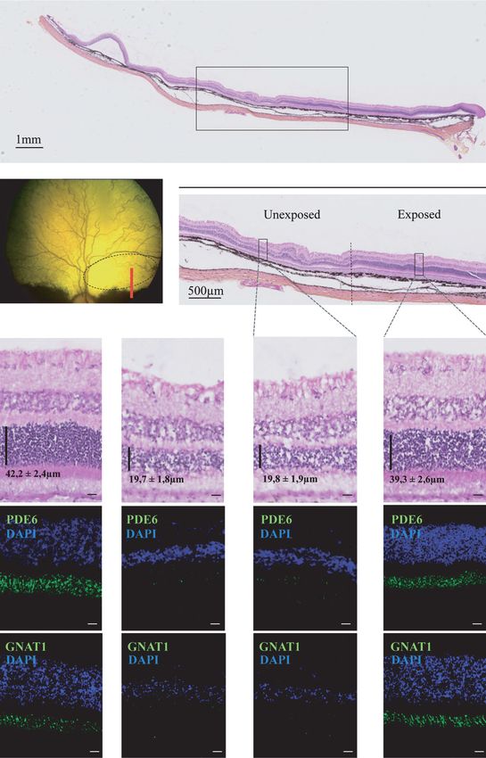
from mutant RNA templates. Thus, one could expect the complete Figure 4 shows photographs of hematoxylin and eosin stained
digestion of RT-PCR products by BtsCI endonuclease to result in a sections and images of rod PDE6 and GNAT1 immunofluores-
385-bp digestion product in RNA templates arising from Pde6β+/+ cence obtained from nasal superior retinas of dogs A5 and NA2.
retinas, a 405 bp product in RNA templates arising from untreated Low magnifications revealed a dramatic difference in retinal
Pde6β−/− retinas and both 385 and 405 bp products in RNA tem- morphology between the vector-exposed and unexposed areas of
plates arising from Pde6β+/− and rAAV-treated Pde6β−/− retinas. the treated retina of dog A5 (Figure 4a,b). The vector-exposed
The results were as expected (Figure 2b). Transgene-derived region of the treated A5 retina retained a nearly normal mor-
385 bp product was detected in both AAV2/5RK.cpde6β- and phology compared with the Pde6β+/− age-matched control retina
AAV2/8RK.cpde6β-treated retinas. On the contrary, this 385 bp (Figure 4c, top). The ONL from the treated A5 retina was 16–17
product was never detected in the untreated rcd1 retina. These rows thick (39 ± 3 µm, n = 10), accounting for ~90% of the ONL
results clearly show that vector-encoded pde6β was expressed in thickness of the control retina (18–19 rows, 42 ± 2 µm, n = 10)
both AAV2/5RK.cpde6β- and AAV2/8RK.cpde6β-treated retinas. (Figure 4c, top). Most importantly, the mean combined inner and
Molecular Therapy 3© The American Society of Gene & Cell Therapy
Restoration of Vision in pde6β-deficient Dogs
Right Left
a
NA1
18m
Treated Untreated Treated Untreated
b
A3
rAAV2/5
4 mpi 18 mpi
Treated Untreated Treated Untreated
c
A7
rAAV2/8
4 mpi 18 mpi
Figure 3 In vivo assessment of retinal morphology in dogs A3 and A7 at 4 and 18 mpi. (a) Fundus photographs and retinal cross-sectional images
obtained from control nonaffected, untreated dog NA1 at 18 months of age. (b) Fundus photographs and retinal cross-sectional images obtained
from dog A3 treated with AAV2/5RK.cpde6β at 4 and 18 mpi. (c) Fundus photographs and retinal cross-sectional images obtained from A7 treated
with AAV2/8RK.cpde6β at 4 and 18 mpi. Dark circles on fundus photographs schematically represent areas of the treated retinas exposed to recom-
binant adeno-associated virus (rAAV) vectors. Optical coherence tomography (OCT) scans were acquired on a horizontal line shown on the fundus
images (dark-line). The localization and the size of the dark-lines represent the localization and the size of the OCT scans. Retinal thicknesses at the
same location were measured using calibrated calipers and indicated on the OCT scan. mpi, months postinjection; µm, micrometers.
outer segment thickness (22 ± 3 µm, n = 10) was indistinguishable and disorganized compared with the vector-exposed region and
from that of the Pde6β+/− control retina (22 ± 1 µm, n = 10). the nonaffected control retina.
In the vector-unexposed region of the A5 treated retina, how- Strong PDE6 labeling was found in outer segments in the
ever, a massive loss of photoreceptor cell nuclei was observed vector-exposed region of the A5 treated retina (Figure 4c, mid-
(Figure 4c, top) similarly to that seen in the noninjected contral- dle), confirming the preservation of rod photoreceptor morphol-
ateral retina. The ONL was reduced to 6–7 rows (20 ± 2 µm, n ogy after rAAV-mediated gene transfer. In contrast, only mild
= 10) which represented only one-third to one-half of the num- or undetectable PDE6 immunofluorescence was detected in the
ber of rows observed in normal or in the vector-exposed retina vector-unexposed region of the A5 treated retina and in the A5
respectively, and ~50% of normal or vector-exposed ONL thick- untreated retina (Figure 4c, middle). A similar pattern of immu-
ness (Figure 4c, top). Moreover, photoreceptor inner and outer nofluorescence was obtained using an antibody directed against
segments appeared to be shorter (16 ± 2 µm, n = 10), less dense GNAT1 (Figure 4c, bottom).
4 www.moleculartherapy.org© The American Society of Gene & Cell Therapy
Restoration of Vision in pde6β-deficient Dogs
a
A5-treated
b
c NA2 A5-Untreated
GC
INL
ONL
OS
IS
RPE
ONL
OS
IS
ONL
OS
IS
Figure 4 Post-mortem assessment of retinal morphology in dog A5 at 4 months postinjection. (a–c) Nasal retinal cryosections from the treated
and untreated eyes of dog A5 at 4 months post subretinal delivery of rAAV2/5RK.cpde6β in the nasal superior retina. (a) Wide A5 retinal section dis-
playing vector exposed and unexposed-areas. Retinal layers remained intact while the choroid has been partially detached from the retina during the
embedding process. (b) Fundus photograph representing the A5 retina exposed to the rAAV2/5 vector (dark circle) and the localization of the wide reti-
nal section (red line). (c) Nasal retinal cryosections from unaffected, untreated dog NA2 at 5 months of age and from the untreated and treated eyes of
dog A5. Serial retinal cryosections were processed for hematoxylin and eosin coloration (top) and for immunohistochemistry using antibodies against
rod PDE6 (middle) or GNAT1 (bottom). Primary antibodies were detected with Alexa 488-conjugated goat anti-rabbit IgG (green). Cell nuclei were
counterstained with DAPI (blue). Bar = 10 µm. Vertical dark lines indicate ONL thickness (mean ± SEM, n = 10). GC, ganglion cells; INL, inner nuclear
layer; IS, inner segments; ONL, outer nuclear layer; OS, outer segments; rAAV, recombinant adeno-associated virus; RPE, outer retinal pigment.
Molecular Therapy 5© The American Society of Gene & Cell Therapy
Restoration of Vision in pde6β-deficient Dogs
a Rod ERG
ERG max
50 µV
NA1 25ms
Cone ERG
30 Hz Flicker 20 µV
50 ms
18 m
b
A1
1m 9m 18 m
T U T T
c U U
A2
rAAV2/5
1 mpi 9 mpi 18 mpi
T U T U
T U
d
A6
rAAV2/8
1 mpi 9 mpi 18 mpi
Figure 5 Bilateral full-field electroretinographic traces from dogs NA1, A1, A2, and A6 at 1, 9, and 18 months following subretinal injection.
(a) Electroretinographic trace from control nonaffected, untreated dog NA1 at 18 months of age. (b) Electroretinographic traces from control affected,
untreated dog A1 at 1 and 18 months of age. (c) Electroretinographic traces from dog A2 treated with AAV2/5RK.cpde6β. (d) Electroretinographic
traces from dog A6 treated with AAV2/8RK.cpde6β. The top two recordings are low- and high-intensity dark-adapted responses, whereas the bottom
two recordings show light-adapted responses (responses to single flash and 30 Hz flicker stimuli, respectively). AAV, adeno-associated virus; mpi,
month(s) postinjection; T, treated eye; U, untreated eye.
The analysis of the entire retina showed that the preserved mor- following vector delivery, starting at 1 mpi, except for dog A5 for
phology covered ~25% of the total retinal surface (data not shown) which retinal function was first assessed at 3 mpi. Follow-up was
and was mostly restricted to the region exposed to the vector. 18 mpi for dogs A2, A3, A6 and A7, 9 mpi for dogs A4 and A8, and
4 mpi for dogs A5 and A9. Recordings on age-matched untreated
AAV2/5RK.cpde6β and AAV2/8RK.cpde6β rescue Pde6β+/− (NA1) and Pde6β−/− (A1) dogs were performed as con-
retinal function trols (Table 1).
Retinal function was evaluated by the same investigator (L.L.) Representative ERG waveforms for dogs A1, A2, and A6 at 1,
in all dogs using simultaneous bilateral full-field flash ERG. 9, and 18 mpi are shown in Figure 5. The kinetics of functional
ERGs were performed on all treated dogs at different timepoints recovery in dogs A2 and A6 are presented in Figure 6.
6 www.moleculartherapy.org© The American Society of Gene & Cell Therapy
Restoration of Vision in pde6β-deficient Dogs
a Rod ERG ERG max 30 Hz Flicker
90 140
Peak-to-peak amplitude (µV)
60
80
b-wave amplitude (µV)
b-wave amplitude (µV)
120 50
70
60 100
40
A1 50 80
30
40 60
30 20
40
20
20 10
10
0 0 0
1m 3m 6m 9m 12 m 18 m 1m 3m 6m 9m 12 m 18 m 1m 3m 6m 9m 12 m 18 m
b 90 140 60
Peak-to-peak amplitude (µV)
b-wave amplitude (µV)
80
b-wave amplitude (µV)
120 50
70
100
60 40
50 80
A2 30
rAAV2/5 40 60
30 20
40
20
20 10
10
0 0 0
1 mpi 3 mpi 6 mpi 9 mpi 12 mpi 18 mpi 1 mpi 3 mpi 6 mpi 9 mpi 12 mpi 18 mpi 1 mpi 3 mpi 6 mpi 9 mpi 12 mpi 18 mpi
c 90 140 60
Peak-to-peak amplitude (µV)
80
b-wave amplitude (µV)
b-wave amplitude (µV)
120 50
70
60 100
A6 40
rAAV2/8 50 80
30
40 60
30 20
40
20
20 10
10
0 0 0
1 mpi 3 mpi 6 mpi 9 mpi 12 mpi 18 mpi 1 mpi 3 mpi 6 mpi 9 mpi 12 mpi 18 mpi 1 mpi 3 mpi 6 mpi 9 mpi 12 mpi 18 mpi
Figure 6 Kinetics of retinal function recovery in treated dogs A2 and A6. (a) Amplitudes of electroretinography (ERG) responses for control
affected, untreated dog A1 from 1 to 18 months of age. (b) Amplitudes of ERG responses for dog A2 treated with AAV2/5RK.cpde6β from 1 to 18
mpi. (c) Amplitudes of ERG responses for dog A6 treated with AAV2/8RK.cpde6β from 1 to 18 mpi. The left and middle panels show scotopic rod and
mixed cone-rod-mediated b-wave amplitudes, respectively. The right panel shows photopic 30 Hz flicker amplitude. Right eyes are shown in dark,
left eyes in white. AAV, adeno-associated virus; mpi, month(s) postinjection.
We found that both AAV2/5RK.cpde6β and AAV2/8RK. The cone-mediated ERG (30 Hz Flicker) responses in treated
cpde6β-subretinal delivery led to substantial restoration of rod and untreated eyes were similar in all AAV2/5RK.cpde6β- and
function in all treated eyes as soon as 1 mpi (Table 1 and Figures AAV2/8RK.cpde6β-injected dogs over the first 8–12 month
5c,d and 6b,c). In contrast, the rod responses were undetectable period postvector delivery (Figure 6b,c).
in the contralateral untreated eyes (Figures 5c,d and 6b,c) and in Interestingly, ERG max and 30 Hz flicker amplitudes remained
untreated control Pde6β−/− eyes (Table 1 and Figures 5b and 6a) stable in all treated eyes over the 12–18 mpi period while a pro-
at this timepoint. gressive and consistent reduction of these responses was observed
Although the general trend of rod function recovery was in all contralateral untreated eyes during this period (Table 1 and
similar in all rAAV2/5- or rAAV2/8-treated eyes, interindividual’s Figures 5c,d, and 6b,c).
variations were observed (Table 1) and might reflect small varia- At 18 mpi, the median amplitude of the 30 Hz flicker of
tions on the extent and/or localization of the vector bleb. AAV2/5RK.cpde6β- (33 ± 11 µV, n = 2) and AAV2/8RK.cpde6β-
In all rAAV2/5- and rAAV2/8-treated dogs, the amplitude of (25 ± 8 µV, n = 2) treated eyes represent 76 and 56% of that
rod function recovery increased slightly between 1 and 3 mpi and recorded on age-matched Pde6β+/− control eyes (43 ± 1µV, n = 2),
remained stable thereafter, up to 18 mpi, the longest period of obser- respectively (Supplementary Figure S1). At 18 mpi, the maximal
vation (Figure 6b,c, left). In all treated dogs, restored rod function amplitudes of the 30 Hz flicker were recorded on A2 AAV2/5RK.
exhibited typical a- and b-wave components (Figure 5c,d). cpde6β- (40 µV, 93% of the normal cone function) and A6
At 18 mpi, the median b-wave amplitude recorded on AAV2/8RK.cpde6β- (30 µV, 70% of the normal cone function)
AAV2/5RK.cpde6β- (47 ± 8 µV, n = 2) and AAV2/8RK.cpde6β- treated retinas (Table 1 and Figure 6b,c, left). They were approxi-
(46 ± 25 µV, n = 2) treated eyes represent 35% of that recorded on mately threefold higher than responses recovered for their con-
nonaffected eyes (134 ± 8 µV, n = 2) (Supplementary Figure S1). tralateral untreated eyes (14 and 11µV).
At 18 mpi, the maximal b-wave amplitudes were recorded on A2 Interestingly, the level of rod function restoration and cone
AAV2/5RK.cpde6β- (53 µV, 38% of the normal rod function) and function preservation obtained following delivery of rAAV2/5 was
A6 AAV2/8RK.cpde6β- (63 µV, 45% of the normal rod function) not significantly different from that achieved with the rAAV2/8
treated retinas (Table 1 and Figure 6b,c, left). (Supplementary Figure S1).
Molecular Therapy 7© The American Society of Gene & Cell Therapy
Restoration of Vision in pde6β-deficient Dogs
AAV2/5RK.cpde6β and AAV2/8RK.cpde6β restore Table 2 Evolution of dim- and bright-light vision in PDE6β–/– treated
vision dogs
Vision tests were performed on A2, A3, A6, and A7 treated Behavioral test
Pde6β−/− dogs at 4, 8, 12, and 18 months postvector delivery Dim-light Bright-light
(Table 2). Dim-light conditions were used to assess transmission
4 mpi 12 mpi 18 mpi 4 mpi 12 mpi 18 mpi
of rescued rod activity to higher visual pathways and improve-
Dog Vector T/U T/U T/U T/U T/U T/U
ment of visually guided behavior. Bright light conditions were
used to assess preservation of day vision in treated rcd1 dogs. A2 AAV2/5RK.cpde6β +/+ +/- +/- +/+ +/+ +/+
Under dim-light conditions at 4 and 8 mpi, none of the treated A3 +/+ +/- +/- +/+ +/+ +/+
dogs showed behavioral signs of blindness in both eyes (Table 2). A6 AAV2/8RK.cpde6β +/+ +/- +/- +/+ +/+ +/+
However, at the latest timepoints (12 and 18 mpi), all treated dogs
A7 +/+ +/- +/- +/+ +/+ +/+
avoided obstacles when their untreated eye was occluded while
Abbreviations: –, impaired vision; +, correct vision; mpi, months postinjection; T,
they showed difficulty to navigate around the obstacle panels treated eye; U, untreated eye.
when their treated eye was occluded. They moved very cautiously Vision tests were performed in dim (1.5 ± 0.8 lux) or bright (260 ± 13lux) light
at different timepoints postinjection. An opaque lens was used to alternatively
with their nose to the ground and tried to feel the panels with cover treated (right) and untreated (left) eyes.
their forelegs. Several collisions with obstacles were also noted.
These results were confirmed by a significant increase in transit
time. For example, dog A6 completed the obstacle course in 7 mouse, previous studies demonstrated that subretinal injection
seconds with the untreated eye covered and in 30 seconds with of AAV2/5smCBA.mpde6β led to the preservation of 37% of
the treated eye covered (Supplementary Video S1, part I). Dog rod ERG responses up to P35,23 and that subretinal injection of
A6 took a comparable time to complete the obstacle course with AAV2/8(Y733F)smCBA.mpde6β led to a preservation of 58% of
the untreated eye covered than a control nonaffected Pde6β+/− rod ERG responses for up to 6 months after treatment.24
dog with one eye covered (9 seconds, data not shown). This finding has obvious clinical significance because among
In bright light conditions, none of the treated dogs showed the 21 causative-mutations already identified in patients with
behavioral signs of blindness from 4 to 18 mpi regardless of which PDE6β-recessive RP,4–12 8 (38%) are nonsense mutations predicted
eye was covered (Table 2 and Supplementary Video S1, part II). to lead to a complete loss of PDE6 function.5–8,10 The most sus-
tained rescue of rod function in null animal models of progressive
Discussion photoreceptor defects reported to date was obtained in the Aipl1−/−
In this study, a total of eight rcd1 dogs were subretinally injected mouse, in which both rod and cone functions are absent at birth.
with rAAV2/5 and rAAV2/8 vectors carrying the cpde6β cDNA In this model, subretinal injection of AAV2/8RK.haipl1 at P10 led
under the control of the photoreceptor-specific RK promoter. to a 50% restoration of wild-type scotopic ERG responses for at
The results demonstrated that rAAV-mediated cpde6β expression least 4 weeks postinjection.38 When scAAV2/8(Y733F)RK.haipl1
restored rod function and consequently, prevented photorecep- was used,39 this rescued rod function was more sustained persist-
tor death and concomitant vision loss in treated dogs for at least ing until P60 (the duration of the reported study).
18 mpi (the duration of the study). In addition to this sustained restoration of rod function in
Interestingly, rod-mediated ERG responses were undetect- treated rcd1 retinas, long-term preservation of cone-mediated
able in the rcd1 canine model from the earliest age measured37 ERG responses were demonstrated up to 18 mpi (Table 1 and
(1 month of age, Table 1). At this age, the PDE6β subunit was Figures 5 and 6). This result strongly suggests that rAAV-medi-
undetectable by immunoblot and rod PDE6 function was absent, ated gene therapy inhibited or delayed the initiation of secondary
suggesting that the rcd1 dog carries a null mutation in the pde6β cone death in treated rcd1 retinas. A similar beneficial effect of
gene.30 In this matter, this canine model of PDE6β deficiency is the primary rod treatment on the cone function has been previ-
similar to the rd1 murine model in which PDE6 function is also ously obtained in rd10 mice treated with AAV2/8(Y733F)smCBA.
completely lacking,17 but different from the hypomorphic rd10 and mpde6β for at least 6 weeks postinjection24 (the duration of the
Pde6βH620Q murine models in which photoreceptors develop reported study).
and function almost normally before their degeneration.18–20 Several mechanisms have been proposed to explain the non-
Here, we show that both AAV2/5RK.cpde6β and AAV2/8RK. autonomous death of cones in rod-cone dystrophies: the loss of a
cpde6β-gene transfer restored sustained and stable rod-medi- rod-derived trophic support, the release of a toxic factor by dying
ated ERG responses in all treated rcd1 eyes as early as 1 month rods, an increase in oxidative damage to cones once the rods have
postinjection (Figure 5). From 1 to 18 mpi, median amplitudes of died or an imbalance in the metabolism of cones due to changes
dark-adapted b-waves in rAAV-treated eyes account for 28–35% in retinal architecture.14–16 In all these models, primary loss of rods
of those recorded in normal eyes (Supplementary Figure S1), is responsible for secondary cone death, indicating that protection
consistent with the estimated area of retina directly exposed to the of cones after rAAV-mediated gene transfer might be correlated
vectors (~25% of the total retinal surface). with a preservation of rod cells in treated rcd1 retinas.
This is the first demonstration that gene therapy can restore Long-term preservation of central retinal thickness in treated
a rod function in a null animal model of PDE6β deficiency as rcd1 eyes was observed by OCT from 4 to 18 mpi (Figure 3). Our
all attempts to treat the rd1 mice by gene addition therapy have histology data at 4 mpi suggested that this reflects the retention of
failed to provide functional rescue.21,26,27 In contrast, in the rd10 rod photoreceptor cells in treated retinas (Figure 4).
8 www.moleculartherapy.org© The American Society of Gene & Cell Therapy
Restoration of Vision in pde6β-deficient Dogs
Interestingly, this preservation of rod photoreceptors was not or in addition, it is possible that in both rAAV2/5- and rAAV2/8-
widespread but mainly restricted to nasal vector-exposed areas of treated eyes, the saturation threshold of pde6β expression level
treated retinas. Similar results have been recently shown in RPGR was reached.
canine models of X-linked RP following subretinal injection Interestingly, both rAAV2/5 and rAAV2/8 vectors provided
of AAV2/5IRBP.hrpgr or AAV2/5RK.hrpgr.40 This observation long-term therapy in the rcd1 dog. This result contradicts previ-
may have two notable clinical implications. First, it demonstrates ous reports in mouse models of Pde6β,24 Aipl1,38 and Rpgrip144
that degenerating nontransduced photoreceptors have no major deficiencies that have shown a strong functional advantage
negative impact on transduced photoreceptor survival as previ- of rAAV2/8 and/or rAAV2/8(Y733F) vectors compared with
ously suggested.41 Second, it surprisingly indicates that localized the rAAV2/5 vector. This significant discrepancy in long-term
preservation of rod photoreceptor cells in the nasal superior rAAV2/5 efficiency between the rcd1 dog and these murine mod-
retina (that did not comprise the cone-rich area centralis42) is els of retinal dystrophies probably reflects the slower rate of retinal
sufficient to maintain the nearly total cone function and subse- degeneration in the rcd1 dog.33,34 This result should be consid-
quently delay initiation of cone loss. We do not have yet a clear ered in the context of PDE6β-patients as they typically describe
explanation of the apparent complete retention of cone function impaired or absent night vision in childhood, followed by a reduc-
observed in the treated rcd1 retinas. It might be related to the tion of their visual field from young adulthood, which slowly pro-
physic and/or trophic interactions of preserved rods with other gresses over decades.4–6
retinal cell populations.14–16 Further monitoring of the cone func- In this study, we used rAAV vectors carrying the cpde6β under
tion for months or years in treated rcd1 dogs will be essential to the control of the human RK promoter which has been demon-
confirm its stability overtime and injection of additional dogs will strated to drive transgene expression in both rods and cones after
be required to identify the underlying molecular mechanism for subretinal delivery,35,36,40 whereas the β subunit of PDE6 is spe-
this long-term preservation of cone function. cifically expressed in rod photoreceptors.13 Recombinant AAV-
The effect of the rAAV-mediated gene therapy on the vision of mediated gene therapy in rcd1 retinas may thus led to undesirable
treated rcd1 dogs was assessed by behavioral tests under dim (1.5 ectopic expression of pde6β in transduced cones. It was unfortu-
± 0.8 lux) and bright (260 ± 13 lux) light conditions. It is impor- nately not possible to assess this pde6β expression in cones due to
tant to note that, in these two conditions, it was impossible to dis- the limitations of pde6β immunostaining. However, based on the
criminate rod- from cone-mediated vision because both types of finding that rAAV-treated eyes exhibited no appreciable reduc-
photoreceptors were stimulated. tion of cone function over the 18-mpi period (Table 1), it is likely
In dim light, all treated dogs displayed normal vision behavior that this potential RK-mediated ectopic expression of pde6β has
using either their untreated and treated eyes up to 8 mpi (Table 2), no toxic effects on the cone function, at least at this stage. These
probably due to the preservation of normal cone function in both results have high clinical relevance because they suggest the
eyes (Table 1 and Figures 5 and 6). Over the 8 to 18 mpi period, potential safety of the human RK promoter for future treatment
none of the treated dogs were still able to successfully negotiate of pde6β defects.
the obstacle course using their untreated eye (Supplementary In conclusion, this study demonstrated, for the first time, that
Video S1, part I) consistent with the progressive loss of cone func- rAAV-mediated gene transfer efficiently corrects the pde6β defect
tion observed by ERG analysis at these late timepoints (Table 1 in a large animal model of pde6β deficiency, leading to (i) robust
and Figures 5 and 6). In contrast, they maintained normal vision- and stable restoration of rod function and (ii) concomitant long-
elicited behavior using their treated eyes (Supplementary Video term preservation of cone function and vision. As mutations in
S1, part I), demonstrating that rAAV-mediated restoration of rod the pde6β gene are one of the leading causes of recessive RP4–12
function and/or preservation of cone function preserve long-term and as retinal degeneration in the rcd1 dog is highly similar to
night vision in treated rcd1 dogs. the human disease, this preclinical study represent a major step
In bright light, untreated dogs displayed vision over the towards the future development of this therapy in patients with
18-month period (Supplementary Video S1, part II) certainly due PDE6β-deficiencies. In a larger sense, these PDE6β results offer
to the residual cone function in both eyes (Table 1 and Figures 5 great promise for the treatment of many other rapid recessive rod-
and 6). It will be necessary to further monitor the vision of treated cone dystrophies due to a rod-specific defect.
rcd1 dogs over several years to determine whether the preserva-
tion of cone function observed by ERG analysis will also lead to Materials and Methods
long-term preservation of day vision over time. Plasmid construction and production of rAAV vectors. Recombinant
In this study, the efficacy of rAAV2/5 and rAAV2/8 vectors AAV2/5.RK.cpde6β and AAV2/8.RK.cpde6β vectors were produced by
was comparable with both vectors providing similar rod-me- triple transfection of 293 cells according to previously reported methods,45
diated ERG responses in treated rcd1 eyes (Table 1). This result using the SSV9RK.cpde6β vector plasmid. This construct carried the
was surprising as rAAV2/8 (1012 vg/ml) was injected at a tenfold cpde6β cDNA (2,681 bp) directly under the control of the short human RK
promoter (–112 bo to +87 bp region of the proximal promoter36) and the
higher titer than rAAV2/5 (1011 vg/ml) vector. One possible expla-
bovine growth hormone polyadenylation signal (BGHpA), flanked by two
nation might be the broader dispersion of rAAV2/8 in the canine
AAV2 inverted terminal repeat sequences.
retina after subretinal delivery.43 The higher potential of rAAV2/8 For the SSV9RK.cpde6β construction, full-length cpde6β cDNA
for widespread diffusion across synapses may reduce the global (NCBI RefSeq NM_001002934.1) was amplified by PCR from Pde6β+/+
number of rAAV-delivered vector genomes in photoreceptors canine retinal cDNA. Two primers designed to cover the entire
cells and subsequently the level of pde6β expression. Alternatively sequence of the pde6β gene were used for this purpose. The forward
Molecular Therapy 9© The American Society of Gene & Cell Therapy
Restoration of Vision in pde6β-deficient Dogs
primer encoded a HindIII end and the first 15 bp of the cpde6β gene of vector-exposed and unexposed areas of the retina were determined by
(5′-AATTAAGCTTTAGACAGCCGGACAC-3′). The reverse primer averaging ten counts, performed on ten different slices.
(5′-CTTTATTCATAGTTGAGTTT-3′) encoded a 20 bp sequence of For immunohistochemical studies, cryosections were air-dried,
the cpde6β gene located 121 bp downstream of the stop codon. An washed in PBS solution and incubated in blocking solution [20% normal
endogenous EcoRV restriction site was present in the sequence of cpde6β goat serum (Invitrogen Life Technologies, Saint Aubin, France), 0.05%
82 bp downstream of the stop codon. The PCR product (2,728 bp) was Triton X-100 in PBS] for 1 hour at room temperature. The bovine rod
purified, digested by HindIII and EcoRV and cloned into a parental PDE6 (1:500) (Cytosignal, Irvine, CA) or GNAT1 (1:250) (Santa-Cruz
plasmid SSV9RK.crpgrip1, between the RK promoter and the bovine Technology, Heidelberg, Germany) antibodies were incubated overnight
growth hormone polyadenylation site, after removal of the eGFP sequence at 4 °C in blocking solution. After three washes in 0.05% Triton X-100 in
(by HindIII and EcoRV digestion). The identity of the resulting SSV9RK. PBS solution, slides were incubated with the secondary antibody Alexa
cpde6β construct was verified by sequencing. 488 goat anti-rabbit IgG conjugate (Life Technologies, Grand Island, NY)
Viral vector titers were determined by dot-blot and by quantitative at 1:250 for 2 hours at room temperature. Slides were washed three times
real-time PCR and expressed as vector genomes (vg) per milliliter (vg/ with 0.05% Triton X-100 in PBS solution and once with PBS solution
ml). The final vector titers of AAV2/5RK.cpde6β and AAV2/8RK.cpde6β before counterstaining the nuclei with DAPI diluted at 1:500. Slides were
were 1.1011 and 1.1012 vg/ml, respectively. mounted in Prolong Gold anti-fade reagent (Life Technologies), observed
with a fluorescence microscope (Nikon) and images captured with a
Animals. A total of nine affected Pde6β−/− and 1 Pde6β+/− control Irish Setter digital camera (Nikon).
dogs were used in this study (Table 1). The first three Pde6β−/− individuals Specificity of the primary bovine rod PDE6 antibody for canine
were kindly provided D.J. Maskell (University of Cambridge, Cambridge, retina was demonstrated in the Pde6β+/+ retina by absence of co-labeling
UK). Animals were maintained at the Boisbonne Center (ONIRIS, Nantes- with peanut lectin agglutinin conjugated with fluorescein isothiocyanate
Atlantic College of Veterinary Medicine, Food Science and Engineering, (1:250; Vector Laboratories, Burlingame, CA) (data not shown).
Nantes, France) under a 12/12 hour light/dark cycle. All experiments
involving animals were conducted in accordance with the Association for
RT-PCR
Research in Vision and Ophthalmology statement for the use of animals in
RNA extraction and retro-transcription. Total RNA was isolated from indi-
ophthalmic and vision research.
vidual flash-frozen retinas or retinal cryosections of Pde6β+/+, Pde6β+/−,
Subretinal administration of rAAV vectors. Subretinal injections of or Pde6β−/− dogs using TRIzol Reagent (Invitrogen Life Technologies).
AAV2/5RK.cpde6β and AAV2/8RK.cpde6β vectors were performed on Rnase-free Dnase I (Ambion DNA-free kit; Invitrogen Life Technologies)
8 affected Pde6β−/− dogs at P20. For all dogs except A9, vector delivery was used according to the manufacturer’s instructions to remove con-
was unilateral, leaving the contralateral eyes without injection as internal taminating DNA before generation of cDNA by reverse-transcription. Five
controls (Table 1). All subretinal injections were performed under general hundred nanograms of total RNA were reverse-transcribed using oligodT
anesthesia induced by intramuscular injection of a mixture of diazepam primers and M-MLV reverse transcriptase (Invitrogen Life Technologies)
(Hoffmann-La Roche, Basel, Switzerland) and ketamine (Rhone Merieux, as per the manufacturer’s instructions. Control assays without addition of
Lyon, France) and maintained by inhalation of isofluorane gas. Pupils were reverse transcriptase were included and the products were used in the sub-
fully dilated by topical administration of 0.3% atropine (Alcon Cusi SA, sequent RT-PCR as negative controls.
Barcelona, Spain), tropicamide (Novartis, Annonay, France) and phe-
Primers. Integrity of cDNA was determined by amplification of a 650 bp
nylephrine hypochloride (Novartis).
sequence of the β-actin gene using β-actinF (5′-TGACGGGGTCACCC
Surgery was conducted using a transvitreal approach as previously
ACACTGTGCCCATCTA-3′) and β-actinR (5′-CTAGAAGCATTTGC
described,46 without vitrectomy. Under microscopic control, 80–120 µl of
GGTGGACGATGGAGGG-3′) primers.
vector solution were injected into the subretinal space. Immediately after
the injection, the localization and the extent of the retinal surface directly For pde6β amplification, 2056F (5′-GAAGATCGTGGATGAGTCTAAGA-3′)
exposed to the vector were recorded on schematic fundus drafts as it and 2561R (5′-CTACTTATCATCAGTCAAGGCCA TC-3′) primers were
was not possible to obtain clear fundus photography of the dog retina designed to specifically create a BtsCI restriction site in RT-PCR products
until 2 months of age. The precise orientation of the vector bleb was arising from wild-type RNA templates. The reverse primer was designed
further determined by fundus photography at 2 mpi, by the observation to neighbouring exon sequences to avoid amplification of genomic DNA
of brighter areas that delineate the border of the treated area (data not (Figure 2). The forward 2056F primer was designed in a pde6β-specific
shown). sequence to avoid amplification of canine pde6α RNA.
Post-surgical care included one topical administration of 0.3% To verify the specificity of the 2056F and 2561R primer set against
atropine (Alcon Cusi SA) and two topical administrations of Ocryl lotion pde6β, full-length canine pde6α cDNA (NCBI RefSeq NM_001003073.1)
(Laboratoire TVM, Lempdes, France) and gentamicin dexamethasonum was amplified by PCR from Pde6α+/+ canine retinal cDNA. The forward
(Virbac France S.A., Carros, France) daily for 10 days post-surgery. primer encoded a BamHI end and an 18 bp sequence located 72 bp upstream
of the start codon of the cpde6α gene (5′-ATTAAGGATCCTGACTC
Histology and immunohistochemistry. At 4 mpi, A5 and A9 dogs were TGTCTTGC-3′). The reverse primer encoded an EcoRV end and a 15 bp
euthanized by intravenous injection of pentobarbital sodium (Vétoquinol, sequence located 353 bp downstream of the stop codon of the cpde6α
Lure, France). Eyes were enucleated and fixed for 2 hours in 4% paraform- gene (5′- TATTGATATCTGGGTGATGAGGAGG -3′). The PCR product
aldehyde in phosphate-buffered saline (PBS) solution before removal of (3,061 bp) was purified, digested by BamHI and EcoRV and cloned into the
the anterior chamber and the lens. Eyecups were embedded in optimal plasmid SSV9RK.cpde6β previously constructed, between the RK promoter
cutting temperature compound (OCT Cryomount; Microm Microtech, and the bovine growth hormone polyadenylation site after removal of
Francheville, France), and flash-frozen in a dry ice isopentane bath. Ten to the cpde6β cDNA (by BamHI and EcoRV digestion). The identity of the
fifteen micrometer cryosections were prepared. resulting SSV9RK.cpde6α construct was verified by sequencing.
For morphological examinations, sections were stained with
hematoxylin and eosin before imaging by transmitter light microscopy Amplification and BstCI digestion. The amount of cDNA used in β-actin
(Nikon, Champigny sur Marne, France). Photoreceptor survival was and pde6β allele-specific PCR was 40 ng. Both PCR were performed using
assessed by counting the number of photoreceptor nuclei rows in one GoTaq DNA polymerase (Promega France, Charbonnicres, France) and a
ONL column. Mean combined outer and inner segment thickness values Veriti Applied Biosystems thermocycler.
10 www.moleculartherapy.org© The American Society of Gene & Cell Therapy
Restoration of Vision in pde6β-deficient Dogs
The β-actin reaction profile was as follows: an initial denaturation ACKNOWLEDGMENTS
step at 95 °C for 5 minutes, followed by 40 cycles at 94 °C for 30 seconds, We acknowledge K. Stieger (Justus-Liebig University, Giessen,
55 °C for 30 seconds, 72 °C for 30 seconds and a final incubation step at Germany) for critical reading of this manuscript. We thank D.J. Maskell
72 °C for 7 minutes. For pde6β, conditions of amplification were: an initial (University of Cambridge, Cambridge, UK) for the gift of the first rcd1
denaturation step at 95 °C for 5 minutes, followed by 10 cycles at 94 °C dogs and S. Khani (State University of New York, Buffalo, NY) for the
for 30 seconds, 60 °C for 30 seconds, 72 °C for 30 seconds, 30 cycles at gift of the human RK promoter. We also thank the Vector Core (www.
94 °C for 30 seconds, 56 °C for 30 seconds, 72 °C for 30 seconds and a final vectors.univ-nantes.fr) for production of the rAAV vectors, the staff of
incubation step at 72 °C for 7 minutes. the Boisbonne Center for animals care and Mireille Ledevin for reti-
Actin PCR products were resolved on a 1.5% agarose gel electrophoresis. nal cryosections. This work was supported by the Agence Nationale
pour la Recherche, the Association Française contre les Myopathies,
Pde6β PCR products were purified by Nucleospin Extract II (Macherey-
the INSERM, the Fondation pour la Thérapie Génique en Pays de la
Nagel, Hoerdt, France) before digestion by BtsCI (New England Biolabs,
Loire and the Ministère Français de l’Enseignement Supérieur et de la
Ipswich, MA) for 2 hours at 50 °C according to the manufacturer’s
Recherche.
instructions and analyzed by 3.5% agarose gel electrophoresis.
REFERENCES
Fundus photography and OCT. Fundus photography and OCT were per- 1. Dryja, TP (2001). Retinitis pigmentosa and stationary night blindness. The Metabolic
formed bilaterally on all treated dogs every 2 months after treatment. Before & Molecular Bases of Inherited Diseases 5903–5933.
2. Hartong, DT, Berson, EL and Dryja, TP (2006). Retinitis pigmentosa. Lancet 368:
clinical examinations, the pupils were topically dilated as described above. 1795–1809.
Dogs were anesthetized by intravenous injection of xylazine (BayerHealth 3. RETNET at . Last accessed 6 March 2012.
4. McLaughlin, ME, Sandberg, MA, Berson, EL and Dryja, TP (1993). Recessive mutations
Care, Shawnee Mission, KS) and ketamine (Rhone Merieux). in the gene encoding the beta-subunit of rod phosphodiesterase in patients with
Fundus photographs were taken with a Canon UVI retinal camera retinitis pigmentosa. Nat Genet 4: 130–134.
connected to a digital imaging system (Lhedioph Win Software; Lheritier, 5. McLaughlin, ME, Ehrhart, TL, Berson, EL and Dryja, TP (1995). Mutation spectrum of
the gene encoding the beta subunit of rod phosphodiesterase among patients with
Saint-Ouen-l’Aumône, France). autosomal recessive retinitis pigmentosa. Proc Natl Acad Sci USA 92:
Optical coherence tomography was performed by doing a 3-mm 3249–3253.
6. Danciger, M, Blaney, J, Gao, YQ, Zhao, DY, Heckenlively, JR, Jacobson, SG et al.
horizontal line scan in vector-exposed or vector unexposed areas of the (1995). Mutations in the PDE6B gene in autosomal recessive retinitis pigmentosa.
retina (Stratus 3000; Carl Zeiss S.A.S; Le Pecq, France). Genomics 30: 1–7.
7. Bayés, M, Giordano, M, Balcells, S, Grinberg, D, Vilageliu, L, Martínez, I et al. (1995).
Homozygous tandem duplication within the gene encoding the beta-subunit of rod
Electroretinography. Retinal function of treated Pde6β−/− dogs and age-
phosphodiesterase as a cause for autosomal recessive retinitis pigmentosa. Hum Mutat
matched untreated Pde6β−/− and Pde6β+/− controls was evaluated using 5: 228–234.
bilateral full-field flash ERG. Initial ERG measurements were recorded at 8. Danciger, M, Heilbron, V, Gao, YQ, Zhao, DY, Jacobson, SG and Farber, DB (1996).
A homozygous PDE6B mutation in a family with autosomal recessive retinitis
4 weeks postinjection and every subsequent 2 months up to 4 (dogs A5 pigmentosa. Mol Vis 2: 10.
and A9), 9 (dogs A4 and A8), or 18 mpi (dogs A2, A3, A6, and A7), the 9. Saga, M, Mashima, Y, Akeo, K, Kudoh, J, Oguchi, Y and Shimizu, N (1998). A
novel homozygous Ile535Asn mutation in the rod cGMP phosphodiesterase beta-
latest timepoints evaluated in this study. For all dogs, each contralateral subunit gene in two brothers of a Japanese family with autosomal recessive retinitis
eye served as intra-individual noninjected control (except for dog A9 who pigmentosa. Curr Eye Res 17: 332–335.
received injection in both eyes). 10. Hmani-Aifa, M, Benzina, Z, Zulfiqar, F, Dhouib, H, Shahzadi, A, Ghorbel, A et al.
(2009). Identification of two new mutations in the GPR98 and the PDE6B genes
Both pupils of animals were topically dilated as described above. Dogs segregating in a Tunisian family. Eur J Hum Genet 17: 474–482.
were dark-adapted for 20 minutes and anesthetized following premedication 11. Clark, GR, Crowe, P, Muszynska, D, O’Prey, D, O’Neill, J, Alexander, S et al. (2010).
Development of a diagnostic genetic test for simplex and autosomal recessive retinitis
by intravenous injection of thiopental sodium (Specia Laboratories, Paris, pigmentosa. Ophthalmology 117: 2169–77.e3.
France)followedbyisofluoranegasinhalation.Hydroxypropulmethylcellulose 12. Ali, S, Riazuddin, SA, Shahzadi, A, Nasir, IA, Khan, SN, Husnain, T et al. (2011).
(Laboratoires Théa, Clermont-Ferrand, France) was applied to each eye to Mutations in the ß-subunit of rod phosphodiesterase identified in consanguineous
Pakistani families with autosomal recessive retinitis pigmentosa. Mol Vis 17:
prevent corneal dehydration during recordings. 1373–1380.
A computer-based system MonColor (Metrovision, Pérenchies, 13. Ionita, MA and Pittler, SJ (2007). Focus on molecules: rod cGMP phosphodiesterase
type 6. Exp Eye Res 84: 1–2.
France) and contact lens electrodes (ERG-jet; Microcomponents SA, 14. Sancho-Pelluz, J, Arango-Gonzalez, B, Kustermann, S, Romero, FJ, van Veen, T,
Grenchen, Switzerland) were used. Zrenner, E et al. (2008). Photoreceptor cell death mechanisms in inherited retinal
degeneration. Mol Neurobiol 38: 253–269.
ERGs were recorded in a standardized fashion according to the 15. Wright, AF, Chakarova, CF, Abd El-Aziz, MM and Bhattacharya, SS (2010).
International Society for Clinical Electrophysiology of Vision (ISCEV) Photoreceptor degeneration: genetic and mechanistic dissection of a complex trait.
recommendations,47 using a protocol described previously.48 Nat Rev Genet 11: 273–284.
16. Punzo, C, Xiong, W and Cepko, CL (2012). Loss of daylight vision in retinal
degeneration: are oxidative stress and metabolic dysregulation to blame? J Biol Chem
Vision testing. Light and dim-light vision of selected treated Pde6β−/− dogs 287: 1642–1648.
were evaluated at regular intervals from 7 to 18 mpi by recording ambula- 17. Keeler, C (1966). Retinal degeneration in the mouse is rodless retina. J Hered 57:
47–50.
tion of dogs through an obstacle avoidance course under 260 ± 13 lux and 18. Chang, B, Hawes, NL, Pardue, MT, German, AM, Hurd, RE, Davisson, MT et al. (2007).
1,5 ± 0.8 lux ambient light intensities, respectively. Details of the apparatus Two mouse retinal degenerations caused by missense mutations in the beta-subunit of
have been described previously.48 Animals were adapted to each light con- rod cGMP phosphodiesterase gene. Vision Res 47: 624–633.
19. Hart, AW, McKie, L, Morgan, JE, Gautier, P, West, K, Jackson, IJ et al. (2005).
dition for 1 hour before beginning the test. An opaque lens was used to Genotype-phenotype correlation of mouse pde6b mutations. Invest Ophthalmol Vis Sci
alternatively cover the treated or the untreated eye. Obstacle panel combi- 46: 3443–3450.
20. Davis, RJ, Tosi, J, Janisch, KM, Kasanuki, JM, Wang, NK, Kong, J et al. (2008).
nations were randomly determined for each test to prevent the dogs mem- Functional rescue of degenerating photoreceptors in mice homozygous for a
orizing the positions of the obstacle panels. A Nightshot camera recorder hypomorphic cGMP phosphodiesterase 6 b allele (Pde6bH620Q). Invest Ophthalmol
(Sony DCR-PC110E PAL; Sony, New York, NY) was used for filming the Vis Sci 49: 5067–5076.
21. Bennett, J, Tanabe, T, Sun, D, Zeng, Y, Kjeldbye, H, Gouras, P et al. (1996).
dimmest light condition. All dim-light films were cleared before mounting Photoreceptor cell rescue in retinal degeneration (rd) mice by in vivo gene therapy.
with commercial Final Cut Pro 7 software (Apple, Cork, Ireland) using a Nat Med 2: 649–654.
22. Jomary, C, Vincent, KA, Grist, J, Neal, MJ and Jones, SE (1997). Rescue of
standardized protocol. photoreceptor function by AAV-mediated gene transfer in a mouse model of inherited
retinal degeneration. Gene Ther 4: 683–690.
23. Pang, JJ, Boye, SL, Kumar, A, Dinculescu, A, Deng, W, Li, J et al. (2008). AAV-mediated
SUPPLEMENTARY MATERIAL gene therapy for retinal degeneration in the rd10 mouse containing a recessive
Figure S1. Median ERG responses of rAAV2/5- and rAAV2/8-treated PDEbeta mutation. Invest Ophthalmol Vis Sci 49: 4278–4283.
24. Pang, JJ, Dai, X, Boye, SE, Barone, I, Boye, SL, Mao, S et al. (2011). Long-term retinal
eyes overtime. function and structure rescue using capsid mutant AAV8 vector in the rd10 mouse,
Video S1. Assessment of dim and bright light vision in dog A6 at 18 mpi. a model of recessive retinitis pigmentosa. Mol Ther 19: 234–242.
Molecular Therapy 11You can also read