Clinical analysis and nonsurgical management of 11 dogs with aural cholesteatoma
←
→
Page content transcription
If your browser does not render page correctly, please read the page content below
Vet Dermatol 2019; 30 : 42–e12                                                                                      DOI: 10.1111/vde.12707

Clinical analysis and nonsurgical management of 11
dogs with aural cholesteatoma
Akihiro Imai*, Hirotaka Kondo†, Tsunenori Suganuma‡ and Masahiko Nagata*
*Dermatology Service, Synergy Animal General Hospital, 815 Ishigami, Kawaguchi, Saitama 333-0823, Japan
†Laboratory of Veterinary Pathology, Department of Veterinary Medicine, College of Bioresource Science, Nihon University, 1866 Kameino,
Fujisawa, Kanagawa 252-0880, Japan
‡Diagnostic Image Service, Synergy Animal General Hospital, 815 Ishigami, Kawaguchi, Saitama 333-0823, Japan
Correspondence: Masahiko Nagata, Dermatology Service, Synergy Animal General Hospital, 815 Ishigami, Kawaguchi, Saitama 333-0823, Japan.
E-mail: d-nagata@syn.ne.jp

     Background – Aural cholesteatomas, also called tympanokeratomas, are destructive and expansile growths of
     keratinizing epithelium that develop in the middle ear. They have been reported sporadically in dogs, and surgery
     is usually the recommended treatment.
     Objectives – To describe the common clinical, radiological and histological findings of cholesteatoma; to report
     on the outcome of conservative management.
     Animals – Eleven dogs (13 ears) with cholesteatomas.
     Methods and materials – Medical records were reviewed for dogs diagnosed with cholesteatoma between
     2012 and 2018. All dogs had computed tomography (CT) and/or magnetic resonance imaging (MRI) followed by
     trans-canal endoscopic procedure (TEP) for removal and biopsy of middle ear lesions. Dogs were then treated
     with in-clinic flushing initially weekly tapered to monthly, as well as at-home ear cleaning and application of topical
     otic steroid medication, initially daily then tapered to once or twice weekly.
     Results – Nine dogs had a history of chronic otitis externa; head tilt or facial paralysis was present in seven and
     four cases, respectively. Otic examination identified a protruding nodule in seven ears. CT demonstrated soft tis-
     sue-like material in 12 bullae and expansion in seven bullae. MRI revealed minimally contrast-enhancing bulla
     contents in 12 ears. Post-TEP and with maintenance medical treatment, nine ears had no further signs of middle
     ear disease during a mean follow-up of 27.9 months.
     Conclusions and clinical importance – The results suggest that otitis externa may not necessarily precede
     cholesteatoma in all dogs. MRI appears to be more sensitive than CT for identifying cholesteatomas. Conserva-
     tive treatment of cholesteatomas could be useful before or as an alternative to surgery.

                                                                             The clinical signs of cholesteatoma in dogs may be non-
Introduction
                                                                          specific, including otic discharge, head shaking, rubbing
Aural cholesteatoma is an epidermoid cyst that develops                   and otic pain.1,6 Nasopharyngeal and/or neurological signs
in the middle ear and is composed of keratin debris sur-                  also may occur.1,3,7,8 In humans, early diagnosis of cho-
rounded by keratinizing stratified squamous epithelium.1–                 lesteatoma depends on its identification with otoscopic
3
   It has been colloquially described as “skin growing in                 examination.9 In veterinary medicine, otoscopy is used to
the wrong place”.4–6 The misplaced keratinizing epithe-                   diagnose the condition,2,10–12 although definitive diagno-
lium constantly sheds keratin debris, resulting in gradual                sis requires histopathological examination.1,4 Computed
enlargement of the cyst and the eventual destruction of                   tomography (CT) and magnetic resonance imaging (MRI)
the adjacent tissue due to increasing pressure and osteo-                 are considered to be reliable methods for evaluating the
clastic bone resorption activated by inflammatory chemo-                  middle ear and demonstrating middle ear-related
kines.7 In dogs, cholesteatoma is regarded as a severe                    lesions.13,14 CT is utilized to better define bony structures
complication of otitis media.4 The term tympanokeratoma                   and can yield valuable information for the detection of
has also been proposed for this condition.5                               cholesteatoma.15 MRI is used for more accurate assess-
                                                                          ment of soft tissue structures and can be a useful tool for
                                                                          the further characterization of soft tissue changes and
                                                                          recognition of potential complications;13,14 however, it is
                                                                          not routinely utilized in part because of the relatively high
Accepted 5 October 2018
                                                                          cost.
This study was presented at the World Congress of Veterinary
Dermatology, 2016, Bordeaux, France. Vet Dermatol 2016; 27
                                                                             The only curative treatment for canine cholesteatoma
(Suppl. 1): 45 (Abstract).                                                reported to date is surgery with total ear canal ablation-
Conflicts of interest: No conflicts of interest have been                 lateral bulla osteotomy (TECA-LBO) and ventral bulla
declared.                                                                 osteotomy.1,2,10,16 The prime objective of surgery for cho-
Sources of funding: This study was self-funded.                           lesteatoma is to remove all keratinous debris and
42                                                                           © 2018 ESVD and ACVD, Veterinary Dermatology, 30 , 42–e12.
Nonsurgical management of canine cholesteatoma
stratified squamous epithelium;17 recurrence postsurgery thickness = 3.5 mm, TR = 520 ms, TE = 11 ms) and dorsal T1W
is common in animals.1–3 (slice thickness = 3.0 mm, TR = 500 ms, TE = 12 ms) were
acquired. Additional transverse and dorsal T1W were obtained fol-
To the best of the authors’ knowledge, 15 reports (cov-
lowing i.v. administration of 0.2 mL/kg (0.5 mmol/mL) gadodiamide
ering a total of 70 cases) of canine cholesteatoma have hydrate (Omniscan 32% i.v. injection syringe, Daiichi-Sankyo; Tokyo,
been published.1–8,10,13,15,16,18–20 Reports in which the Japan).
diagnosis of cholesteatoma was confirmed by histopatho-
logical evaluation in combination with CT and MR imaging
TEP for cholesteatoma
are limited.1,8 No clinical studies of nonsurgical manage-
The TEP was performed in combination with CT and/or MRI examina-
ment for aural cholesteatoma have been reported. The tion. Preoperatively, the length, width and depth of the ear canal and
purpose of the present study was to report the main clini- middle ear cavity were measured, as well as the distance from the
cal and imaging findings of aural cholesteatoma; and to dorsal fold of the ear canal to the tympanic bulla on each CT/MR
report on the long-term outcome of a minimally invasive image of the cases. The dorsal fold (also called auricular projection)
therapeutic approach in 11 dogs. by the cartilaginous ridge represents a landmark that separates the
vertical and horizontal ear canals.21 In addition, the type, number,
structure, size and distribution of aural lesions were investigated to
Methods and materials clarify the anatomical position of lesions within the ear canal.
Dogs were positioned in lateral recumbency with the affected side
Case selection and clinical analysis uppermost. Cleaning and flushing the ear canal were conducted with
Medical records of dogs diagnosed with cholesteatoma, pre- NEW, and any type of obstruction in the external ear canal was
sented between January 2012 and 2018 at the Synergy Animal removed to access the middle ear. If a protruding nodule in the hori-
General Hospital, Saitama, Japan, were examined to determine zontal ear canal was present, it was grasped with curved mosquito
the following: (i) if histopathological reports stated the presence forceps or aural forceps under endoscopic visualization and then
of keratinizing epithelium and/or thick lamellar keratin debris in the removed by a traction–torsion manoeuvre. Residual portions of the
middle ear; (ii) if CT and/or MRI scanning were performed to iden- nodule were completely removed under endoscopic visualization
tify middle ear disease; (iii) if trans-canal endoscopic procedures using 5f biopsy forceps with c.2.5 mm oval cups (Karl Storz GmbH &
(TEPs) under anaesthesia were carried out to remove debris in Co. KG), or vaporized by DLV-20 diode laser (Asuka Medical Inc.).
the middle ear; and (iv) if a minimum follow-up of 12 month dura- Local irrigation with NEW was used to reduce and control the bleed-
tion was possible. Signalment, history, physical examination, ing. In addition, for cases with stenosis due to proliferative lesions at
video-otoscopy findings, diagnostic imaging, cytological evaluation, the bony part of the ear canal, the top of the lesion was vaporized
aerobic microbial culture results, histopathological features and with a diode laser in the same manner as for ablation of residual
treatment were reviewed in each case. Dogs were followed up mass fragments. For all cases, keratinaceous debris was directly
and the outcomes of dogs that did not remain in our direct care removed from the middle ear using the 5f biopsy forceps and sam-
were determined by telephone interview between the referring ples were placed in 10% formalin for histopathological evaluation
veterinarian and client. separately from the tissues removed from the external ear canal.
Impacted keratinaceous debris in the tympanic cavity was then
Video-otoscopic examination removed by either aural forceps, suction and/or 3–5f feeding tubes
Ear canal evaluations using a video-otoscope (Karl Storz GmbH & Co. under either video-otoscopic or rigid endoscopic guidance. The bulla
KG; Tuttlingen, Germany, and/or Asuka Medical Inc.; Kyoto, Japan) was lavaged to remove remaining debris with NEW. Finally, 0.1 mL
were conducted both before and during TEPs. An initial examination triamcinolone acetonide (Kenacort-A intradermal and intra-articular
was performed in conscious dogs to assess if the ear canal was ade- aqueous suspension infection, 1%, 50 mg/5 mL, Bristol-Myers
quately open to enable TEP. If stenosis of the ear canal – due to Squibb; Tokyo, Japan) was administered to the middle ear cavity.
inflammation and proliferative tissue – blocked otoscope access, For prophylactic medical management post-TEP, repeated in-clinic
topical and/or oral steroids were administered to expand the canal tube flushing by a soft feeding tube with NEW was performed in
before TEP. awake dogs once weekly for two to four weeks, followed by tapering
Imaging with CT/MRI was performed with general anaesthesia. to once every two weeks for four weeks and eventually to once
The middle ear and ear canal were evaluated using the video-oto- monthly. The indication for tapering was based on otic conditions,
scope and/or a 2.7-mm diameter, zero degree rigid endoscope such as the presence of ear discharge and behaviours including
(Asuka Medical Inc.). To facilitate visualization, the ear canal was scratching or head shaking. Additionally, routine home cleaning was
lavaged using 3–5f feeding tubes with neutral electrolyzed water performed with an ear cleaner (Zymox! ear cleanser, Pet King
(NEW) (Asahi Pretec Corp.; Kobe, Japan). Brands, Inc.; Westmont, IL, USA), and/or Tromethamine-EDTA (Triz-
EDTA Plus!, Dechra; Overland Park, KS, USA) and topical mometa-
sone furoate (Fulmeta!, Shionogi & Co., Ltd.; Osaka, Japan) was
Diagnostic imaging methods
applied to the dogs as prophylactic management to control canal
Computed tomography scanning was performed under general
inflammation. At-home ear cleaning and topical steroid administration
anaesthesia with Somatom Emotion 16 (Siemens Healthineers
was performed once daily for two to four weeks, tapering to once
Japan; Tokyo, Japan) to investigate the whole skull. Contiguous
every two days for four weeks, then once to twice weekly. If otitis
transverse 1 mm images were obtained before and after intravenous
flared despite prophylactic flushes with NEW and topical steroid
(i.v.) administration of 2.5 mL/kg (612.4 mg/mL) Iopamidol (Oypalo-
drops, dogs were treated with topical and/or systemic antimicrobials
min 300 injection syringe, Fuji Pharma; Toyama, Japan) and were
determined by clinician preference based on cytological evaluation
reconstructed using bone and soft tissue algorithms.
and culture results.
Magnetic resonance imaging was subsequently performed using
Magnetom Essenza 1.5T (Siemens Healthineers Japan). Sagittal T2-
weighted [T2W; slice thickness = 3.0 mm, repetition time Ear cytological evaluation and bacterial cultures
(TR) = 4,000 ms, echo time (TE) = 91 ms], transverse T2W (slice Under general anaesthesia, otic exudate was collected from either
thickness = 3.5 mm, TR = 4,000 ms, TE = 86 ms), dorsal T2W the middle ear or the deepest part of the ear canal with 3–5f feeding
(slice thickness = 3.0 mm, TR = 4,000 ms, TE = 86 ms), transverse tubes under video-otoscopic guidance as cytological samples and
fluid-attenuated inversion recovery (FLAIR; slice thick- sterile culture swabs. For cytological evalution, samples were placed
ness = 3.5 mm, TR = 8,500 ms, TE = 104 ms, Inversion onto sterile slides. Then, slides were stained with Microscopic Hema-
time = 2,438.7 ms), transverse T1-weighted (T1W; slice color! Rapid staining of blood smear (Merck KGaA; Darmstadt,
© 2018 ESVD and ACVD, Veterinary Dermatology, 30 , 42–e12. 43Imai et al.
Germany). Each slide was evaluated under a high-power field (940 Video-otoscopic findings
and/or 100) for the presence of cocci bacteria, rod-shaped bacteria, In 13 ears, video-otoscopy revealed otic discharge and
keratinized squamous epithelial cells and inflammatory cells. For aero-
the TM was not observed. In seven ears (cases 1, 2, 3, 4,
bic bacterial culture and antimicrobial susceptibility testing, swabs
5, 10 and 11) a smooth-surfaced, whitish to pale-pinkish
were submitted to a diagnostic laboratory (LSI Medience Corporation;
Tokyo, Japan). Swabs were inoculated onto sheep blood agar plates and rounded protruding nodule was detected, occluding
which were incubated at 35°C for 24 h. All isolates were identified the horizontal ear canal (Figure S1 in Supporting Informa-
phenotypically and biochemically. Antimicrobial susceptibility was tion; Table 1). In four ears (cases 3, 4, 8 and 9), a nar-
performed using the broth microdilution method, which determined rowed horizontal ear canal was observed (Table 1).
the minimum inhibitory concentrations of antimicrobial agents
according to Clinical and Laboratory Standards Institute guidelines.22
Diagnostic imaging findings
Histopathological diagnosis of aural cholesteatoma Computed tomography
Biopsy specimens were fixed in 10% neutral-buffered formalin. Rep- Computed tomography imaging was performed in 10
resentative trimmed tissues were routinely processed, embedded in
dogs (12 ears). All dogs had soft tissue-like material
paraffin, sectioned at 5 lm and stained with haematoxylin and eosin.
within the affected tympanic bullae. Absence of air con-
The following criteria were used for diagnosis of cholesteatoma: (i)
origin of tissue confirmed as the middle ear using the endoscopic trast within the middle ear cavity was observed in all
procedure; (ii) presence of cornified epithelium and (iii) presence of affected ears. Enlargement of the middle ear cavity was
amorphous thick lamellar keratin debris.1–3 A tentative diagnosis of identified in seven ears, osteoproliferation in seven, lysis
cholesteatoma also was made for cases which satisfied (i) and (iii) of the bulla in six, lysis of the cochlea in six (Figure 1a),
but not (ii) (i.e. they lacked cornified epithelium). sclerosis of the temporal bone in two and mineralization
of the inner lining of the bulla in one.
Four ears, in two pugs and two French bulldogs, were
Results
affected by lysis of the petrous temporal bone and bulla
Signalment and clinical findings expansion was present in three of four ears. Conversely,
Eleven dogs were included in this study; including French all dogs (five ears) without expansion of the bulla also
bulldog (n = 5); pug (n = 2), and one each of Chihuahua, were French bulldogs and only one of five had bulla lysis.
Yorkshire terrier, shiba inu and Akita inu. There were two Only one (Akita inu) of three nonbrachycephalic dogs had
intact males, four castrated males and five spayed both expansion and lysis of the tympanic bulla wall.
females. Ages ranged from 4 to 12 years, with a mean Soft tissue-like material occupying the horizontal ear
age of 8.2 years (median, 8 years). The durations of clini- canal was observed in seven ears. In five of the seven
cal signs varied from four to 104 weeks, with a mean ears, linear mineralization within the soft tissue-like mate-
duration of 28.1 weeks (median 12 weeks). Two dogs rial in the external ear canal was observed (Figure 1b) and
had bilateral disease, and nine dogs had unilateral dis- four of seven ears had bulla osteoproliferation. Enlarge-
ease, giving a total of 13 ears. ment of associated lymph nodes was observed in seven
Nine of the 11 dogs had a history of chronic otitis dogs (seven ears), specifically the submandibular lymph
externa in the affected ears. Five of the nine dogs had a nodes, retropharyngeal lymph nodes and parotid lymph
history of recurrent otitis, and four of nine had a history of node in five, six and one dogs, respectively.
continuous otitis. Suspected primary causes of otitis Postcontrast CT images were available for two ears. In
externa were atopic dermatitis (two dogs), excessive the images after administration of contrast medium, there
cerumen (two dogs), foreign body (two dogs) and idio- was no appreciable contrast enhancement of the tym-
pathic (four dogs). The duration of chronic otitis externa panic bulla content in either ear. One of the two had an
ranged from six months to over five years. Onset of otitis enhancing soft tissue mass occluding the horizontal canal.
media was suspected from one to 11 months before
their presentation. Two of the 11 dogs had no history of Magnetic resonance imaging
chronic otitis externa and were presented for nasopharyn- Magnetic resonance imaging was performed in all dogs
geal and/or neurological signs of possible middle ear dis- (13 ears). It revealed the presence of material within the
orders. bulla which was isointense to slightly hyperintense rela-
Aural signs included otorrhoea (eight dogs), ear scratch- tive to brain tissue in 12 ears (Figure 2a) and heteroge-
ing (two dogs) and head shaking (two dogs). During physi- neous hyperintense in one ear on T1W images. The mass
cal examination, neurological signs were identified in nine was heterogeneous hyperintense in nine ears, homoge-
dogs. Head tilt was present in seven dogs, facial nerve neous hyperintense in three ears and heterogeneous
paralysis in four dogs and ataxia in three dogs. Two dogs hypointense in one ear on T2W (Figure 2b) and FLAIR
were reported to have had nystagmus, and one dog was images (Figure 2c). Following contrast administration,
said to have had a seizure, but these signs were not contents of the bulla did not enhance in all but one ear.
detectable at admission. Two dogs showed an algesic However, the inner lining of the bulla showed some par-
response on palpation of the bulla and temporomandibu- tial enhancement in 11 ears. In addition, contrast
lar joint. Two dogs had increased respiratory noise and/or enhancement of cochleae was observed in nine ears.
inappropriate respiratory effort. Concurrent diseases Heterogeneous contrast enhancement of the meninges
were hypothyroidism (two dogs), spondylopathy (two also was observed with severe lysis of the petrous tem-
dogs), pyoderma, urolithiasis, cardiological disease (AV- poral bone in two dogs.
block), entropion, osteoarthritis and patella luxation (one In eight ears, soft tissue-like material in the external ear
dog each). Details are reported in Table 1. canal was enhanced (Figure 2d). Before the injection of
44 © 2018 ESVD and ACVD, Veterinary Dermatology, 30 , 42–e12.Nonsurgical management of canine cholesteatoma
Table 1. Signalment, clinical signs, duration of clinical signs, location of lesions, histopathological evaluation and outcome of the eleven dogs with
aural cholesteatoma
Duration Histopatho-
of logical
clinical Clinical Location evaluation Location of Procedure
Age signs signs Clinical signs Concurrent of of protruding for Follow-up
Case Breed Sex (years) (weeks) (Otic) (Non-otic) diseases cholesteatoma cholesteatoma nodule stenosis (months) Outcome
1 Pug SF 8 12 Otorrhoea Left head tilt Seborrhoeic Right KE, KD Right 32 No recurrence
dermatitis
2 Pug CM 7 43 Otorrhoea Right facial palsy Urolithiasis Right KE, KD Right 31 No recurrence
3 French CM 4 4 Otorrhoea Left head tilt, Bilateral KE, KD Left VOLA 28 Right: No
bulldog (left), respiratory noise, recurrence/Left:
ceruminous inappropriate First recurrence
discharge respiratory effort (recurrent
(right), Ear cholesteatoma
scratching with a nodule
confirmed) at
two months,
Second
recurrence
at one month
4 Akita inu CM 8 4 Ceruminous Right head tilt, Pyoderma, Right KE, KD Right VOLA 31 Recurrence at
discharge, ataxia hypothyroidism, one month
head shaking spondylopathy,
osteoarthritis,
cardiac
disease
(A-V-block)
5 Chihuahua SF 12 4 Otorrhoea Spondylopathy, Right KE, KD Right 28 Recurrence
seborrhoeic (recurrent
dermatitis cholesteatoma
confirmed)
at 13 months
6 Shiba inu M 8 104 Ceruminous Atopic Left KD 35 No recurrence
discharge, dermatitis,
Ear scratching Malassezia
dermatitis,
Hypothyroidism
7 French M 7 42 Otorrhoea, Left facial palsy, Osteoarthritis Left KE, KD 57 No recurrence
bulldog Ceruminous Left head tilt
discharge
8 French SF 10 8 Ceruminous Right facial palsy, Right KD VOLA 18 No recurrence
bulldog discharge right head tilt
9 French SF 11 12 Otorrhoea, Right head tilt, Osteoarthritis, Bilateral KE, KD VOLA 12 No recurrence
bulldog ceruminous ataxia patella luxation,
discharge atopic dermatitis
(right),
discharge (left)
10 French CM 7 24 Otorrhoea, Right facial palsy Right KE, KD Right 13 No recurrence
bulldog head shaking
11 Yorkshire SF 8 52 Otorrhoea Left head tilt, Left KD Left 11 Recurrence at
terrier ataxia, respiratory two months
noise
CM castrated male, F female, M male, KE keratinizing epithelium, KD keratin debris, SF spayed female, VOLA video-otoscopic laser ablation.
contrast medium, the material was isointense to brain tis- cholesteatoma (Figure 3). In the other three middle ears
sue in all ears on T1W images, whereas it was heteroge- (cases 6, 8 and 11), a tentative diagnosis of cholestea-
neous hyperintense in five ears, slightly hypointense in two toma was made because cornified epithelium was not
ears and hyperintense in one ear on T2W and FLAIR identified.
images. In seven dogs, histopathological evaluation of protrud-
ing nodules within the external ear canal (Figure 4)
Cytological evaluation and microbial culture revealed dense fibrous connective tissue surrounded by
Cytological examination of otic discharge was performed squamous epithelium. In addition, five of seven had vari-
in seven ears. Bacterial cocci, rods or a combination of able degrees of central cores of osseous metaplasia
both, were present in four, one and two of seven ears, (OM). In Case 1, a markedly abnormal bony architecture
respectively. Keratinized squamous epithelial cells and was found.
neutrophils were identified in four and six of seven ears, In one of seven ears, histopathological results for a tis-
respectively. Aerobic bacterial culture of the middle ear sue sample taken from the deeper part of the external
was performed in five ears, and all cultures were positive. acoustic meatus exhibited hyperplasia and hyperkeratosis
More than one species of organism was isolated in three of the epidermis, dermal oedema, fibrosis and lympho-
ears. Bacteria isolated were Pseudomonas aeruginosa cytic infiltration with dilation of ceruminous glands with
(four of seven ears), group G Streptococcus spp. (two of stasis of secretum.
seven ears), Staphylococcus pseudintermedius group
(one of seven ears) and Staphylococcus schleiferi (one of Outcome of TEP
seven ears). All dogs, except for Case 6, were discharged with pain
medication as a postprocedure in TEP. The duration of
Histopathological findings pain medication varied from one to five weeks. One dog
Tissue samples taken from 10 middle ears demonstrated (Case 5) was given Robenacoxib (Onsior!, Elanco Japan
keratinizing epithelium and keratin debris consistent with K.K.; Tokyo, Japan) 2.0 mg/kg orally (p.o.) once daily.
© 2018 ESVD and ACVD, Veterinary Dermatology, 30 , 42–e12. 45Imai et al.
a and in-clinic management continued until the end of the
study. In seven ears, glucocorticoid eardrops were applied
as a proactive therapy. Initially, three to five drops were
applied to each ear once daily. The frequency of adminis-
tration of topical glucocorticoids was tapered by one half
each week to a maintenance frequency of once or twice
weekly. For management of chronic aural inflammation, in
addition to topical steroid drops, oral oclacitinib was
administered to Case 11 after the second TEP and to Case
10 beginning four weeks after the first TEP. For control of
concurrent atopic dermatitis, oclacitinib (Apoquel, Zoetis;
Parsippany, NJ, USA) was prescribed to Case 9 and oral
prednisolone was intermittently administered to Case 6.
b Nine ears (seven dogs) had no recurrence of middle ear
disease in this study. Four ears (four dogs: cases 3, 4, 5
and 11) had recurrent clinical signs after the first endo-
scopic procedure. The mean time to recurrence was
4.3 months (range: one to 12 months; median: two
months). In Case 3, a protruding nodule recurred two
months after the initial procedure, despite the dog receiv-
ing prophylactic care. As the owners declined otic sur-
gery, a second TEP was immediately performed, and the
dog received systemic and topical antimicrobial medica-
tion. Recurrence of cholesteatoma was confirmed with
histopathological evaluation. No significant aggravation
was observed on either CT or MRI findings, except for
osteoproliferation in the bony part of both external ear
canals. Clinical signs waxed and waned, and then the dog
underwent a third TEP for removal of remnants of keratin
debris after which clinical signs resolved. In Case 4, ear
Figure 1. Computed tomography lesions of aural cholesteatoma of discharge recurred at one month after the initial proce-
dogs. dure. The owners declined an endoscopic or surgical
(a) At the level of the tympanic cavity, soft tissue-like material fills the
approach, and the dog was managed with only monthly
left tympanic bulla, creating enlargement, lysis and osteoproliferation
of the tympanic bulla wall, with lysis of the cochlea (Case 7).
tube flushing. Unfortunately, clinical signs persisted. In
(b) At the level of the tympanic cavity, a soft tissue mass appeared in Case 5, ear discharge recurred at 12 months after the ini-
the right external ear canal with mineralization in the centre (Case 1). tial procedure. Clinical signs waxed and waned, and even-
Soft tissue-like material also fills the right tympanic bulla, causing tually second TEP was performed due to deterioration of
mild enlargement of the tympanic bulla wall. signs 25 months after the first procedure. Recurrence of
cholesteatoma was confirmed with histopathological
Eight dogs were administered prednisolone (Predonine!, evaluation. No significant changes in CT and MRI findings
Shionogi & Co., Ltd.; Osaka, Japan) 0.5–1.0 mg/kg p.o. compared to the first procedure were observed. Clinical
once daily and one dog (Case 4) was given prednisolone signs resolved after the second procedure. In Case 11,
0.5–1.0 mg/kg p.o. once every other day; then the corti- otitis recurred two months after the initial procedure. The
costeroid treatment was tapered down. No serious post- dog received appropriate home cleaning with administra-
procedural complications were observed in any dogs, tion of oclacitinib. Subsequently, otitis resolved.
except for Case 3 which developed temporary facial None of the owners reported any adverse effects with
nerve paralysis that spontaneously resolved within eight regard to the management regimen during this study.
weeks.
Follow-up ranged from 12 to 58 months, with a median
Discussion
follow-up of 29 months (mean, 27.9 months). Post-TEP,
in-clinic tube flushing and at-home ear cleaning were per- In the veterinary literature, no significant breed predilec-
formed for all the cases once weekly and once daily, tion for cholesteatoma has been reported. In this report,
respectively. The frequency of treatment was gradually over half of the dogs were brachycephalic breeds. Previ-
tapered based on assessment of clinical signs; mainte- ous reports documented that dysfunction of the auditory
nance treatment of once monthly or twice monthly tube tube and the narrow nasopharynx could be predisposing
flushing in clinic was performed for six and five dogs, factors for developing primary acquired cholesteatoma.6,8
respectively. Routine regular home cleaning was recom- In addition, hypertrophic bulla walls and stenotic bony
mended once weekly in five dogs, every other day in one parts of the horizontal ear canals also could predispose
dog and every day in two dogs. The owner of Case 4 brachycephalic breeds to secondary acquired cholestea-
declined to continue home cleaning due to difficulties in toma.8
performing the procedure alone, but monthly in-clinic In our study, nine dogs had a history of chronic otitis
flushes continued. For all but one dog (Case 4), at-home externa, which can be considered as an important
46 © 2018 ESVD and ACVD, Veterinary Dermatology, 30 , 42–e12.Nonsurgical management of canine cholesteatoma a b c d Figure 2. Magnetic resonance images (MRIs) of aural cholesteatoma of a dog at the level of the tympanic cavity (Case 7). (a) T1-weighted, (b) T2-weighted, (c) fluid-attenuated inversion recovery and (d) postcontrast T1-weighted transverse MRIs. The expansile left bulla includes material isointense to brain tissue on T1-weighted images and heterogeneous hyperintense on T2-weighted and fluid-attenuated inver- sion recovery. On postcontrast T1-weighted images, partial enhancement is identifiable, particularly in the inner lining of the left bulla, whereas the contents of the left bulla are not significantly enhanced. predisposing factor for secondary acquired forms in any the brachycephalic breeds with expansile bulla also had breed.1,2,15 However, two of the cases were evaluated lysis of the bulla. By contrast, 80% of the brachycephalic for a history of neurological issues, not the typical chronic breeds without expansile bulla did not have bulla lysis. In history of otitis externa. The primary acquired form of humans, it is hypothesized that cholesteatoma slowly cholesteatoma may occur in dogs, although the sec- enlarges in the middle ear and gradually erodes adjacent ondary acquired form appears to be more common. bony structures with activated osteoclasts.24 Thus, the Computed tomography scanning plays an important early stages of cholesteatoma might remain unidentified role in the assessment of dogs with middle ear dis- by CT scanning. Bulla expansion in these breeds could be eases.15,23 It has been proposed that a nonenhancing but impeded due to their hypertrophic bulla wall. expansile lesion, which is hyper-attenuating to brain, is It was reported, albeit in only one case, that findings on likely to be a cholesteatoma.15 However, expansion of MR imaging included an expansile bulla containing mate- the bulla is not regularly observed.1,16 Our study identified rial isointense to brain tissue on T1W and of mixed inten- expansion of the bulla in 53.8% of the ears and 75% of sity on T2W images.13 In addition, material in the bulla © 2018 ESVD and ACVD, Veterinary Dermatology, 30 , 42–e12. 47
Imai et al.
humans, it was reported that pre- and postcontrast
conventional MRI allows the differentiation of inflamma-
tory mucosa, granulation tissue and cholesteatoma,
whereas CT was not able to differentiate between the
diseases.27 Thus, MRI might be a helpful diagnostic tool
to ensure accurate and early diagnosis of canine choles-
teatoma.
For seven of the 11 dogs in this study, a protruding nod-
ule consisting of fibrous connective tissue surrounded by
squamous epithelium was found in the external ear canal.
Histopathologically, the lesions in dogs appeared to be
different in character from typical aural polyps in cats.
Presence of ciliated columnar epithelial cells is a prerequi-
site for the histopathological definition of a polyp,28 and
as a consequence, in terms of terminology, polyp is not
an appropriate description for the canal nodules seen in
Figure 3. Photomicrograph of the histopathological findings of
this study.19 In dogs, the association between the pro-
canine aural cholesteatoma (Case 7). truding nodules and cholesteatoma remains to be fully
Section of the cholesteatoma wall constituted a multilayered squa- elucidated.1–8,10,13,15,16,18–20 It has been reported that a
mous epithelium lining a cystic lumen filled with abundant lamellar pug with cholesteatoma had an external ear canal mass
eosinophilic keratin debris. Haematoxylin and eosin stain, 920. with a thick, keratinizing epithelium.8 The authors noted
that the lesion might be related to the cholesteatoma.8 In
the present study, histopathological evaluation of external
ear canal nodules in five cases revealed a core of OM.
Likewise, it has been reported in a German shepherd dog
with a cholesteatoma in the middle ear, accompanied by
a cholesterol granuloma with a core of OM in the horizon-
tal ear canal.10 OM is a type of ectopic ossification of
fibrous connective tissue.29
Postoperative short-term complications after TEP
occurred in only one dog (9.1%) which developed facial
palsy. Procedures involving the middle ear potentially can
cause facial nerve palsy and cholesteatoma may be
attached to vulnerable underlying structures including the
facial nerve. In the veterinary literature, facial nerve palsy
and dryness of the nostrils was reported postoperatively
in two (18.2%) of 11 dogs that underwent surgery for
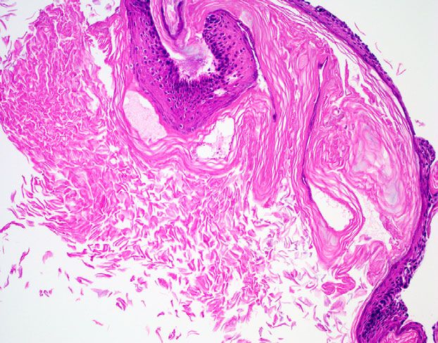
Figure 4. Gross appearance of a protruding nodule in the ear canal cholesteatoma.2 In previous retrospective studies of
of a dog (Case 1). canine TECA and LBO, 13–46% had facial nerve pare-
A whitish to pinkish, oval and pedunculated nodule was removed by sis,30–32 and 4–23% had residual deficits in facial nerve
the traction–torsion manoeuvre. function after TECA-LBO.30,32,33
It has been reported that surgical treatment is curative
in only 50% of cases of canine cholesteatoma.1,12 Recur-
was found to show minimal postcontrast enhancement, rence was seen in one of four dogs (25.0%) at 12 months
with enhancement localized to the region immediately postsurgery in one study and in 10 of 19 dogs (52.6%) at
adjacent to the bone.13 In our cases, the bulla contents a mean time of 11.3 months in another study.1,3 In addi-
commonly appeared isointense on T1W images, hetero- tion, one study reported that recurrence was suspected
geneous hyperintense on T2W images and minimally in five of 11 dogs (45.5%) at a mean time of 7.5 months.2
enhanced after contrast injection, with partial enhance- Postsurgical recurrence is likely due to the difficulty of
ment in the location of the inner epithelial lining of the complete surgical removal of abnormal keratinizing
bulla. In humans, the inner component of a cholestea- epithelium and keratin debris.6 In the present study, clini-
toma consists of keratin debris that is avascular,25 so the cal signs recurred in four of 11 dogs (four of 13 ears:
absence of contrast enhancement of the contents on MR 30.8%) at a mean time of 4.3 months post-TEP. For
imaging was not unexpected.1 Furthermore, in humans, these cases, the involved tissues could not be adequately
vascularity associated with the abnormal epithelium may removed from the middle ear with an initial TEP because
be increased due to angiogenic growth factors produced of their advanced stage. The remnants of cholesteatoma
by inflammatory cell populations in the matrix and perima- might lead to recurrence sooner. Moreover, even in cases
trix of cholesteatoma.17,26 Findings of heterogeneous without recurrence after TEP, the possibility remains that
intensity and enhancement on MRI could reflect the residues of cholesteatoma may persist in small cavities of
histopathological features of cholesteatoma, such as the bulla and keratin accumulation can slowly create clini-
increased vascularity on hyperproliferative epithelium and cal signs. Thus, the need to manage ongoing diseases
absent vascularity within keratin debris. Moreover, in suggests that owners should be prepared for monitoring
48 © 2018 ESVD and ACVD, Veterinary Dermatology, 30 , 42–e12.Nonsurgical management of canine cholesteatoma
after TEP, lifelong maintenance therapy and repeated 3. Little CJ, Lane JG, Gibbs C et al. Inflammatory middle ear dis-
TEP, if necessary. ease of the dog: the clinical and pathological features of choles-
teatoma, a complication of otitis media. Vet Rec 1991; 128: 319–
As part of maintenance therapy, oclacitinib was used in
322.
three cases in this study. In Case 9, oclacitinib was pre- 4. Banco B, Grieco V, Di Giancamillo M et al. Canine aural choles-
scribed for concurrent atopic dermatitis. The other two teatoma: a histological and immunohistochemical study. Vet J
dogs (cases 10 and 11) had unilateral chronic otitis with- 2014; 200: 440–445.
out any cutaneous lesions or identifiable primary disease. 5. Østevik L, Rudlang K, Holt Jahr T, et al. Bilateral tympanokerato-
Oclacitinib is presumed to inhibit both proallergic and mas (cholesteatomas) with bilateral otitis media, unilateral otitis
proinflammatory cytokines (IL-2, 4, 6, 13 and 31) as a interna and acoustic neuritis in a dog. Acta Vet Scand 2018; 60:
31.
specific JAK1 and -3 inhibitor. Although it is an extra-label
6. Risselada M. Diagnosis and management of cholesteatomas in
indication, we prescribed oclacitinib to the latter two dogs dogs. Vet Clin North Am Small Anim Pract 2016; 46: 623–634.
in an attempt to treat the chronic aural inflammation, 7. Davidson EB, Brodie HA, Breznock EM. Removal of a cholestea-
which seems to play a fundamental role in multiple aetio- toma in a dog, using a caudal auricular approach. J Am Vet Med
pathogenic mechanisms of acquired cholesteatoma.34 Assoc 1997; 211: 1,549–1,553.
Additionally, it is suggested in humans that the IL-6/ 8. Schuenemann RM, Oechtering G. Cholesteatoma after lateral
STAT3 signalling pathway is active and may play an bulla osteotomy in two brachycephalic dogs. J Am Anim Hosp
Assoc 2012; 48: 261–268.
important role in the epithelial hyperproliferation of
9. Chang P, Kim S. Cholesteatoma–diagnosing the unsafe ear. Aust
acquired cholesteatoma.35 During this study, the clinical Fam Physician 2008; 37: 631–638.
signs were managed with no relapse in the dogs treated 10. Newman AW, Estey CM, McDonough S et al. Cholesteatoma
with oclacitinib. More study is necessary to elucidate the and meningoencephalitis in a dog with chronic otitis externa. Vet
efficacy and risk of oclacitinib as a component of postpro- Clin Pathol 2015; 44: 157–163.
cedural management in canine cholesteatomas. 11. Radlinsky MG. Advances in otoscopy. Vet Clin North Am Small
Anim Pract 2016; 46: 171–179.
In the present study, around 70% of ears were treated
12. Angus JC, Campbell KL. Uses and indications for video-otoscopy
successfully by a single TEP, followed by maintenance in small animal practice. Vet Clin North Am Small Anim Pract
treatment involving daily to weekly ear cleaning at home 2001; 31: 809–828.
and weekly to monthly tube-flushing in the clinic. Like- 13. Harran NX, Bradley KJ, Hetzel N et al. MRI findings of a middle
wise, it was reported that the condition in a dog was con- ear cholesteatoma in a dog. J Am Anim Hosp Assoc 2012; 48:
trolled by removal of keratin debris from the bulla under 339–343.
video-otoscopic guidance, although the dog died from 14. Belmudes A, Pressanti C, Barthez PY et al. Computed tomo-
graphic findings in 205 dogs with clinical signs compatible with
unrelated causes 29 months later.1 We suggest that con-
middle ear disease: a retrospective study. Vet Dermatol 2018;
servative treatment of cholesteatoma by TEP with regular 29: 45–e20.
removal of the inflammatory stimulus in the middle ear 15. Travetti O, Giudice C, Greci V et al. Computed tomography fea-
could be useful before or in place of surgery. tures of middle ear cholesteatoma in dogs. Vet Radiol Ultra-
Limitations in the evaluation of TEP in this study include sound 2010; 51: 374–379.
its retrospective nature, the small sample size, the refer- 16. Sturges BK, Dickinson PJ, Kortz GD et al. Clinical signs, mag-
ral nature of the population and the lack of objective crite- netic resonance imaging features, and outcome after surgical
and medical treatment of otogenic intracranial infection in 11
ria to define treatment outcome. In the future, a large-
cats and 4 dogs. J Vet Intern Med 2006; 20: 648–656.
scale prospective controlled cohort study should be con- 17. Fukudome S, Wang C, Hamajima Y et al. Regulation of the
ducted to conclusively evaluate therapeutic outcomes. angiogenesis of acquired middle ear cholesteatomas by inhibitor
In conclusion, chronic otitis externa may not necessar- of DNA binding transcription factor. JAMA Otolaryngol Head
ily precede acquired cholesteatoma in dogs but the pres- Neck Surg 2013; 139: 273–278.
ence of a protruding nodule in the external canal may be a 18. Witsil AJ, Archipow W, Bettencourt AE et al. What is your diag-
nosis? Cholesteatoma in a dog J Am Vet Med Assoc 2013; 243:
significant predictor of cholesteatoma and the use of MRI
775–777.
can facilitate the early diagnosis of this disease. Nonsurgi- 19. Greci V, Mortellaro CM. Management of otic and nasopharyn-
cal therapy is not curative as surgery may be; however, geal, and nasal polyps in cats and dogs. Vet Clin North Am Small
early conservative treatment of cholesteatoma could be Anim Pract 2016; 46: 643–661.
an effective option before or as an alternative to surgery, 20. Furcas A, Gielen I, Vandenabeele S et al. Insidious progressive
even though it requires long-term at-home and in-clinic bone destruction in a dog surgically treated for otitis media: fol-
maintenance care. low-up by clinical examination and computed tomography.
Vlaams Diergeneeskundig Tijdschrift 2014; 83: 255–261.
21. Griffin C. Otitis: anatomy every practitioner should know. Com-
Acknowledgements pend Contin Educ Vet 2009; 31: 504–512.
22. CLSI. Performance standards for antimicrobial disk and dilution
The authors thank all of their colleagues who referred susceptibility tests for bacteria isolated from animals – 3rd ed.
cases and Eiji Koyama (Companion Animal Medical Imag- CLSI supplement VET01S. Wayne, PA: Clinical and Laboratory
ing Center Saitama) for diagnostic imaging. Standards Institute; 2015.
23. Garosi LS, Dennis R, Schwarz T. Review of diagnostic imaging
of ear diseases in the dog and cat. Vet Radiol Ultrasound 2003;
References 44: 137–146.
24. Hamed MA, Nakata S, Sayed RH et al. Pathogenesis and bone
1. Hardie EM, Linder KE, Pease AP. Aural cholesteatoma in twenty resorption in acquired cholesteatoma: current knowledge and
dogs. Vet Surg 2008; 37: 763–770. future prospectives. Clin Exp Otorhinolaryngol 2016; 9: 298–308.
2. Greci V, Travetti O, Di Giancamillo M et al. Middle ear cholestea- 25. Trojanowska A, Trojanowski P, Olszanski W et al. Differentiation
toma in 11 dogs. Can Vet J 2011; 52: 631–636. between cholesteatoma and inflammatory process of the middle
© 2018 ESVD and ACVD, Veterinary Dermatology, 30 , 42–e12. 49Imai et al.
ear, based on contrast-enhanced computed tomography imag- 32. Smeak DD, Kerpsack SJ. Total ear canal ablation and lateral bulla
ing. J Laryngol Otol 2007; 121: 444–448. osteotomy for management of end-stage otitis. Semin Vet Med
26. Sudhoff H, Dazert S, Gonzales AM et al. Angiogenesis and Surg (Small Anim) 1993; 8: 30–41.
angiogenic growth factors in middle ear cholesteatoma. Am J 33. Mason LK, Harvey CE, Orsher RJ. Total ear canal ablation com-
Otol 2000; 21: 793–798. bined with lateral bulla osteotomy for end-stage otitis in dogs.
27. Martin N, Sterkers O, Nahum H. Chronic inflammatory disease Results in thirty dogs. Vet Surg 1988; 17: 263–268.
of the middle ear cavities: Gd-DTPA-enhanced MR imaging. 34. Maniu A, Harabagiu O, Perde Schrepler M et al. Molecular
Radiology 1990; 176: 399–405. biology of cholesteatoma. Rom J Morphol Embryol 2014; 55:
28. Wilcock BP. Eye and ear. In: Maxie MG, ed. Jubb, Kennedy & 7–13.
Palmers Pathology of Domestic Animals, volume 1, 5th edition. 35. Xie S, Xiang Y, Wang X et al. Acquired cholesteatoma epithelial
St Louis, MO: Saunders, 2007; 460–552. hyperproliferation: roles of cell proliferation signal pathways.
29. Park JK, Lee SK, Park SJ et al. Fibroma with osseous metaplasia Laryngoscope 2016; 126: 1,923–1,930.
of external auditory canal in a dog. J Vet Diagn Invest 2010; 22:
635–638.
30. Beckman SL, Henry WB Jr, Cechner P. Total ear canal ablation Supporting Information
combining bulla osteotomy and curettage in dogs with chronic
otitis externa and media. J Am Vet Med Assoc 1990; 196: 84–90.
Additional Supporting Information may be found in the
31. Spivack RE, Elkins AD, Moore GE et al. Postoperative complica- online version of this article.
tions following TECA-LBO in the dog and cat. J Am Anim Hosp Figure S1. Video-otoscopic appearance of a protruding
Assoc 2013; 49: 160–168. nodule in the external ear canal of a dog (Case 2).
Re!sume !
Contexte – Les choleste !atomes auriculaires, aussi appele !s tympanoke !ratomes, sont une croissance
expansive et destructrice de l’e !pitelium ke!ratinise! qui se de !veloppe dans l’oreille moyenne. Ils ont e ! te
!
de!crits sporadiquement chez le chien et une chirurgie est le traitement recommande !.
Objectifs – De !crire les donne !es cliniques, radiologiques et histologiques des choleste !atomes ; de !crire
!volution d’une gestion conservatrice.
l’e
Sujets – Onze chiens (13 oreilles) avec choleste !atomes.
Mate !riel et me !thode – Les donne !es me !dicales ont e ! te
! revues pour les chiens diagnostique !s avec un cho-
!atome entre 2012 et 2018. Tous les chiens ont eu un examen tomodensitome
leste !trique (CT) et/ou une
imagerie par re !sonance magne !tique (MRT) suivi d’une endoscopie trans-canal (TEP) pour le retrait et la
biopsie des le !sions de l’oreille moyenne. Les chiens ont ensuite e ! te
! traite
!s par un nettoyage en clinique
! "
une fois par semaine puis une fois par mois, associe a des nettoyages auriculaires par les proprie !taires ainsi
que l’application d’un traitement auriculaire ste !ro€ıdien initialement une fois par jour puis une ou deux fois
par semaine.
Re!sultats – Neuf chiens pre !sentaient des comme !moratifs d’otite externe. Une te ^te penche !e ou une para-
lysie faciale e!tait pre
!sente dans sept et quatre cas respectivement. L’examen auriculaire identifiait un
nodule protrusif dans sept oreilles. Le CT mettait en e !vidence un mate !riel tissulaire dans 12 bulles avec
expansion dans sept bulles. Le MRI re !ve!lait un contenu de la bulle augmentant le contraste de fac!on
minime pour 12 oreilles. A la suite des TEP et avec le traitement de maintenance, neuf oreilles ne pressen-
taient pas de signe d’otite moyenne au cours d’une pe !riode de suivi moyenne de 27,9 mois.
Conclusions et importance clinique – Les re !sultats sugge "rent que l’otite externe peut ne pas ne !cessaire-
ment pre ! ce
!der un choleste !atome chez tous les chiens. La MRI semble e ^tre plus sensible que le CT pour
leur identification. Le traitement conservateur des choleste !atomes pourrait e ^tre utile en alternative "
a la chi-
rurgie.
Resumen
Introduccio ! n – los colesteatomas auditivos, tambie !n denominados queratomas timp! anicos, son creci-
mientos destructivos y expansivos del epitelio queratinizante que se desarrollan en el o!ıdo medio. Se han
descrito espor!adicamente en perros, y la cirug!ıa suele ser el tratamiento recomendado.
Objetivos – describir los hallazgos cl!ınicos, radiolo !gicos e histolo
!gicos comunes del colesteatoma; descri-
bir los resultados de un tratamiento conservador.
Animales – Once perros (13 orejas) con colesteatomas.
Me !todos y materiales- – se revisaron los historiales cl!ınicos de perros diagnosticados con colesteatoma
entre 2012 y 2018. Todos los perros se sometieron a tomograf!ıa computarizada (CT) y/o resonancia
magne !tica (MRI), seguidos de un procedimiento endosco !pico transcanal (TEP) para la extraccio !n y biopsia
de lesiones del o!ıdo medio. Los perros se trataron con lavado semanal en el hospital, inicialmente de forma
semanal y despue !s mensual, as!ı como limpieza del o!ıdo en su hogar y la aplicacio !n de medicamentos to !pi-
cos esteroides o !ticos, inicialmente a diario y luego se redujeron a una o dos veces por semana.
Resultados – nueve perros ten!ıan antecedentes de otitis externa cro !nica; inclinacio
!n de la cabeza o par!ali-
sis facial se observaron en siete y cuatro casos, respectivamente. El examen o !tico identifico
! un no
!dulo que
sobresal!ıa en siete orejas. La CT demostro ! material similar a tejidos blandos en 12 casos y expansio !n en
siete de las bullas. La MRI revelo ! contenidos de bulla que se realzaban m!ınimamente con contraste en 12
o!ıdos. Despue !s de la TEP y con tratamiento me !dico de mantenimiento, nueve o!ıdos no mostraron m! as
50 © 2018 ESVD and ACVD, Veterinary Dermatology, 30 , 42–e12.Nonsurgical management of canine cholesteatoma
signos de enfermedad del o!ıdo medio durante un seguimiento medio de 27,9 meses.
Conclusiones e importancia cl!ınica – los resultados sugieren que la otitis externa no necesariamente
precede al colesteatoma en todos los perros. La MRI parece ser m!as sensible que la CT para identificar
colesteatomas. El tratamiento conservador de los colesteatomas podr!ıa ser u
!til antes o como una alterna-
tiva a la cirug!ıa.
Zusammenfassung
Hintergrund – Cholesteatome im Ohr, die auch Tympanokeratome genannt werden, sind destruktive und
ausgedehnte Gew€achse, die aus keratinisierendem Epithel bestehen und im Mittelohr entstehen. Sie sind
sporadisch bei Hunden beschrieben worden und eine chirurgische Entfernung ist in der Regel die emp-
fohlene Behandlung.
Ziele – Eine Beschreibung der u €blichen klinischen, radiologischen und histologischen Befunde von
Cholestetomen; sowie ein Bericht u €ber das Ergebnis konservativer Behandlung.
Tiere – Elf Hunde (13 Ohren) mit Cholesteatomen.
Methoden und Materialien – Es wurden die Krankenakten von Hunden durchgesehen, die zwischen
2012 und 2018 mit Cholesteatomen diagnostiziert worden waren. Bei allen Hunden war eine Computerto-
mografie (CT) und/oder eine Magnetresonanztomografie (MRI) durchgefu €hrt worden. In der Folge wurde
eine Trans-Kanal Endoskopie (TEP) durchgefu €hrt, um die Ver€ anderungen im Mittelohr zu bioptieren und zu
entfernen. Die Hunde wurden danach zun€ achst wo €chentlich mit einer Ohrspu €lung in der Klinik behandelt,
was danach zu wo €chentlich und monatlich reduziert wurde sowie einer Ohrreinigung zu Hause und topis-
che Applikation von Kortisonmedikation im Ohr, anfangs t€ aglich, dann reduziert auf ein- bis zweimal
wo€chentlich.
Ergebnisse – Neun Hunde hatten eine Anamnese einer chronischen Otitis externa; Kopfschiefhaltung
bzw. Gesichtsparalyse bestanden bei sieben bzw vier F€ allen. In sieben Ohren wurde bei der Ohrunter-
suchung vorragende Knoten gefunden. Die CT zeigte Weichteil-€ ahnliches Gewebe in 12 Bullae und eine
Erweiterung bei sieben Bullae.Die MRI Untersuchung zeigte minimale Kontrast-reiche Bulla-Inhalte in 12
Ohren. Post TEP und nach Erhaltungstherapie mit Medikamenten, zeigten neun Ohren keine weiteren
Anzeichen einer Mittelohrerkrankung w€ahrend der Follow-Up Periode von 27.9 Monaten.
Schlussfolgerungen und klinische Bedeutung – Die Ergebnisse bedeuten, dass eine Otitis externa den
Cholesteatomen bei Hunden nicht unbedingt vorausgehen muss. Zur Identifizierung scheint die MRT
Untersuchung sensibler zu sein als die CT Untersuchung. Die konservative Behandlung von Cholesteato-
men ko€nnte als Alternative zur chirurgischen Behandlung sinnvoll sein.
要約
背景 – 鼓膜角化腫とも呼ばれる耳性真珠腫は、中耳に発生する角化上皮の破壊性かつ膨張性腫瘍であ
る。犬で散発的に報告されており、通常は、外科手術が推奨される治療法である。
目的 – 本研究の目的は、真珠腫の一般的な臨床的、放射線学的および組織学的所見を記述することであ
る。また、保守的管理による成果を報告することである。
被験動物 – 真珠腫を伴う11頭の犬(13 個の耳)。
方法と材料 – 2012年から2018 年にかけて真珠腫と診断された犬について、病歴をレビューした。犬全頭
において、CT検査および/または磁気共鳴画像検査(MRI)を実施し、中耳病変の除去および生検のため経
管内視鏡検査(TEP)を実施した。その後、院内における耳洗浄を、最初は週1回実施し、次第に月1回に減
少させた。同様に自宅での耳洗浄および点耳ステロイド薬の塗布を、最初は1日1回実施し、次第に週1回
または2回に減少させた。
結果 – 犬9 頭に慢性外耳炎の病歴を認めた。頭部斜頚または顔面麻痺をそれぞれ7頭および4 頭に認めた。
耳道検査では、7つの耳道に突出した結節を認めた。 CT検査では、水疱および7つの水疱の拡大を12の軟
部組織様物と認識した。 MRI検査では、12個の耳にコントラストが強調された水泡内容物を最小限に示
した。 TEP後および維持療法を実施した9 個の耳において、平均27.9 ヵ月の追跡期間中に中耳炎の兆候を
認めなかった。
結論と臨床的重要性 – 本研究結果は、外耳炎が必ずしも全ての犬において真珠腫に先行するとは限らな
いことを示唆している。 MRI検査は、真珠腫の同定に対しCT検査よりも感受性が高いようである。真珠
腫の保守的治療は、手術前治療または手術の代替療法として有用であり得る。
摘要
背景 – 耳的胆脂瘤,也称鼓膜角质瘤,是角化上皮在中耳的破坏和扩张性增生。犬的病例仅有零星报道,通常
建议外科治疗。
目的 – 描述胆脂瘤的常见临床、影像学和组织学表现; 报告保守治疗的效果。
动物 – 十一只(13只耳朵)胆脂瘤患犬。
方法和材料 – 对2012年至2018年诊断为胆脂瘤犬的医疗记录进行回顾性分析。所有犬都进行了计算机断层
扫描(CT)和/或磁共振成像(MRI),接着进行耳内镜手术(TEP),以便对中耳病变进行切除和活检。每周在医院
冲洗耳道一次,逐渐减少为每月一次;并且在家清洁耳朵和外用耳类固醇药物,最初每天,然后逐渐减至每周一
到两次。
© 2018 ESVD and ACVD, Veterinary Dermatology, 30 , 42–e12. e11Imai et al.
结果 – 9只犬有慢性外耳炎病史,7只出现头倾斜,4只出现面瘫。耳道检查发现七只耳道有突出结节;CT显示
12例犬鼓室有软组织样物质蓄积,其中7例鼓室扩张;MRI显示12只耳道至少存在鼓室内容物对比度增强。经
TEP治疗和维持治疗后,9只耳道在平均27.9个月的随访期间没有进一步的中耳疾病症状。
结论和临床价值 – 结果提示,所有犬外耳炎不一定先于胆脂瘤发生。对于识别胆脂瘤,MRI似乎比CT更敏
感。胆脂瘤的保守治疗可以作为手术前的备选方案。
Resumo
Contexto – Os colesteatomas aurais, tambe !m chamados de timpanoqueratomas, s~ ao formac!o ~es de
epite!lio queratinizado destrutivas e expans!ıveis que se desenvolvem no ouvido me !dio. Eles s~
ao relatados
esporadicamente em c~aes, e o tratamento ciru ! geralmente o mais recomendado.
!rgico e
Objetivos – Descrever os principais achados cl!ınicos, radiogr! aficos e histopatolo!gicos de colesteatomas e
relatar os resultados de tratamentos conservativos.
Animais – Onze c~aes (13 ouvidos) com colesteatomas.
Me !todos e materiais – Os histo !ricos cl!ınicos dos c~ aes diagnosticados com colesteatoma entre 2012 e
2018 foram revisados. Todos os c~aes foram submetidos " a tomografia computadorizada (TC) e/ou res-
son^ancia magne !tica (RM) seguido por procedimento endosco !pico trans-canal (TEP) para remoc!~ ao e coleta
de bio !psia das leso ~es no ouvido me !dio. Posteriormente, os c~ aes foram submetidos " a limpeza otolo !gica na
cl!ınica inicialmente uma vez por semana e depois mensalmente, bem como limpeza otolo !gica em casa e
aplicac!~ao de formulac!~ao o !tica com esteroide to !pico, inicialmente diariamente e depois reduzido para uma a
duas vezes por semana.
Resultados – Nove c~aes possu!ıam histo !rico de otite externa cro ^nica; inclinac!~
ao de cabec!a ou paralisia
facial foram observadas em sete e quatro casos, respectivamente. O exame otolo !gico revelou um no !dulo
proeminente em sete ouvidos. A TC demonstrou presenc!a de material similar a tecido mole em 12 bulas e
expans~ao em sete bulas. A RM revelou em 12 ouvidos a presenc!a de conteu !do na bula timp^ anica que
realc!ava minimamente apo !s administrac!~ao de contraste. Apo !s TEP e com o tratamento de manutenc!~ ao,
nove ouvidos n~ao apresentavam mais sinais de otopatia de ouvido me !dio por um tempo me !dio de acompa-
nhamento de 27,9 meses.
Concluso ~ es e importa ^ncia cl!ınica – Os resultados sugerem que a otite externa n~ ao necessariamente pre-
cede o colesteatoma em todos os c~aes. A RM parece ser mais sens!ıvel que a TC para a identificac!~ ao de
colesteatomas. O tratamento conservativo para colesteatomas pode ser u !til antes ou como uma alternativa
"a cirurgia.
e12 © 2018 ESVD and ACVD, Veterinary Dermatology, 30 , 42–e12.You can also read