Infectious Enteritis, Food poisoning, Protozoa and Worms - Luis S. Marsano, MD Professor of Medicine Division of Gastroenterology, Hepatology and ...
←
→
Page content transcription
If your browser does not render page correctly, please read the page content below
Infectious Enteritis, Food poisoning,
        Protozoa and Worms
                       Luis S. Marsano, MD
                      Professor of Medicine
     Division of Gastroenterology, Hepatology and Nutrition
           University of Louisville and Louisville VAMC
                               2021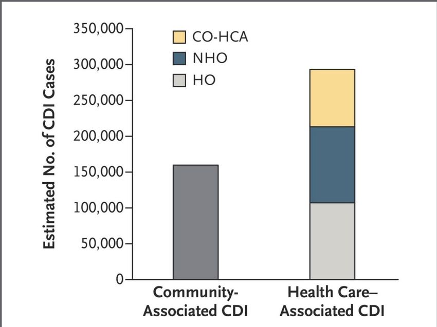
Viral Foodborne Infections
Specific Causes of Foodborne Diarrhea - Viral
                         Norovirus / Norwalk Virus
• 40-60% of acute viral gastroenteritis epidemics in older children & adults
• Pathology: Villous shortening, crypt hyperplasia, PMN & MN cells in lamina propria.
• Spread: person-to-person, contaminated food or water.
• Incubation: 12-48 hours
• Duration: 12-48 hours
• Symptoms: nausea, vomiting, diarrhea, abdominal pain, muscle aches, headache,
  tiredness and low-grade fever.
• Diagnosis: Serology, stool PCR or E/M for stool virus
• Immunity: lasts only weeks to 4 months
• Treatment:
    – ORS, supportive.
Specific Causes of Foodborne Diarrhea - Viral
                                        Rotavirus
•   60% of diarrhea in children < 2 years-old
•   Pathology: Kills mature villous-tip cells
•   Spread: fecal-oral
•   Season: late-fall, winter, early-spring
•   Duration: 3-10 days
•   Symptoms:
    – Diarrhea, nausea, vomiting, cough, rhinitis, otitis.
    – Subclinical in adults.
• Diagnosis: Stool antigen (Rotazyme for type A), PCR
• Treatment:
    – ORS, supportive.
Foodborne Bacterial Infections
    with diarrhea due to
     Mucosal Invasion
Specific Causes of Foodborne Diarrhea – Mucosal Invasion
                                   Salmonella Gastroenteritis
•   Causes 25-40% of food-borne infections in adults
•   Spread: food-borne (food, flies, fingers, feces, fomites); meat, poultry, eggs, dairy products.
•   Incubation: 8-48 hours
•   Duration: usually 3-4 days (up to 3 weeks).
•   Symptoms:
     – nausea, vomiting, abdominal cramps, low grade fever < 102 0F, watery diarrhea; sometimes severe
       dysenteria.
• Complications:
     – osteomyelitis, septic or reactive arthritis, sepsis, peritonitis, cholecystitis, pancreatitis, mycotic
       aneurism, intraabdominal abscess, Reiter S.
• Treatment: ORS & support. Antibiotics prolong the disease.
     – Treat with antibiotics only in: immunosupressed, age < 3 mo or > 50 y, hemolytic anemia, surgical
       prosthesis, valvular heart disease, severe atherosclerosis, cancer, uremia.
     – TMP-SMX DS p.o. BID x 7 days; 14 days if immunosupressed.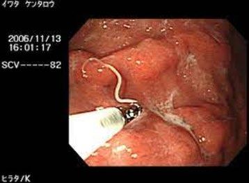
Specific Causes of Foodborne Diarrhea – Mucosal Invasion
                            Campylobacter jejuni
•   Most common cause of bacterial enteritis in many parts of the world.
•   More frequent in young children, with secondary infections in household.
•   Spread: fecal-oral, food-borne, water-borne.
•   Incubation: 24-72 hours.
•   Duration: usually 1 week
•   Symptoms:
    – prodrome of malaise, coryza, headache, and fever;
    – then colicky periumbilical pain with profuse diarrhea, that improves and then worsens,
      with WBC’s in stool.
• Complications:
    – Endocarditis, meningitis, Guillian-Barre, cholecystitis, pancreatitis, septic abortion,
      glomerulonephritis, reactive arthritis (HLA-B27), Reiter S.
• Treatment:
    – Erythromycin stearate 500 mg BID x 5 days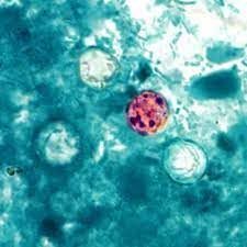
Specific Causes of Foodborne Diarrhea – Mucosal Invasion
                                            Shigella
• Spread: person to person; most common in age 6 mo-10 y; adult infected from children.
  Well water contaminated with feces.
• Incubation: 36-72 hours.
• Duration: 1-30 days (1 week) without therapy
• Symptoms:
   – biphasic illness: fever in 30-40%;
   – cramps & voluminous watery diarrhea for 2-3 days, then dysenteria, with small bloody stool and
     tenesmus.
   – Cough & meningismus in 40% of small children.
• Complications:
   – Reiter syndrome, HUS, protein-loosing enteropathy, e. nodosum, keratoconjunctivitis, pneumonia,
     seizures, and encephalopathy.
• Treatment:
   – Treat all patients.
   – Ciprofloxacin 500 mg BID x 5 days, or TMP-SMX DS po BID x 5 days.
Specific Causes of Foodborne Diarrhea – Mucosal Invasion
                               Yersinia Enterocolitica
• Spread: food-borne (undercooked meats & oysters) & contact with infected pets.
• Symptoms Children < 5y:
   – fever, abdominal cramps, diarrhea for 1 or more weeks.
• Symptoms Children > 5 y:
   – mesenteric adenitis, or ileitis; sometimes ileal perforation.
• Symptoms Adults:
   – acute diarrhea,
   – followed 2-3 weeks later by arthritis, erythema nodosum, or erythema multiformis.
• Post-infectious complications:
   – Reiter S., thyroiditis, myocarditis, pericarditis, glomerulopathy, ankylosing spondylitis, IBD, e.
     nodosum, e. multiformis, & HUS.
• Treatment: ORS & support.
   – In septicemia: gentamicin 5 mg/kg iv; 50% mortality despite treatment.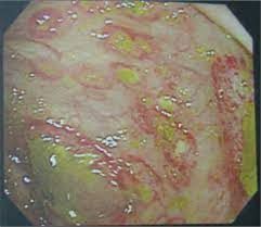
Specific Causes of Foodborne Diarrhea – Mucosal Invasion
                           Plesiomona shigelloides
• Source: contaminated water or shellfish. Common in Japan.
• Symptoms:
   – Variable; from watery diarrhea, with abdominal pain, vomiting and fever, to dysenteria and
     sepsis.
   – Usually self-limited, but 30% have diarrhea > 3 weeks.
   – Sepsis in cirrhosis and immunocompromised.
• Complications:
   – Meningitis, osteomyelitis. Endophthalmitis.
• Diagnosis: Stool culture or PCR.
• Treatment:
   – Treat only in severe (> 8 BM/d) or prolonged disease (> 7 days);
   – Ciprofloxacin 500 mg BID
Foodborne Bacterial Infections with
Toxin Mediated DiarrheaSpecific Causes of Foodborne Diarrhea – Toxin Mediated
Cholera
• Endemic in the Gulf Coast (Lousiana & Texas)
• Vibrio colonizes small bowel and produces cytotonic toxin, activating adenylate
cyclase, causing secretory diarrhea.
• Spread: Water or food contaminated with stools.
• Incubation: 18-40 hours
• Symptoms:
– vomiting and abdominal distension, followed by diarrhea of > 1 Liter/hour;
– dehydration & shock.
• Diagnosis: Stool culture neutralized by antisera. Stool PCR.
• Treatment:
– ORS; IV fluids only until ORS covers needs.
– Tetracycline 500 mg QID x 5 days.Specific Causes of Foodborne Diarrhea – Toxin Mediated
Staphylococcus aureus
• Second cause of food-borne diarrhea in USA (after salmonella).
• Spread:
– Contaminated food with preformed cytotoxic, heat-stable, enterotoxin A.
– Contamination most common in high salt & high sugar foods.
• Incubation: 1-6 hours
• Duration: 24-48 hours
• Symptoms:
– Nausea, profuse vomiting, abdominal cramps followed by diarrhea.
– No WBC in stool.
• Treatment:
– Supportive.Specific Causes of Foodborne Diarrhea – Toxin Mediated
Enterotoxigenic E. coli (ETEC)
• Major cause of Traveler’s diarrhea, and of diarrhea in infants and toddlers in
underdeveloped areas.
• Cytotonic toxins (3: one heat-labile, and two heat-stable), activate adenylate cyclase &
guanilate cyclase.
• Spread: fecal-oral.
• Symptoms:
– Profuse watery diarrhea, with abdominal cramps and nausea.
– May have low-grade fever.
• Duration: 3-5 days
• Diagnosis: stool culture and serotype; Stool PCR.
• Treatment: ORS.
– Mild: Pepto-Bismol 2 tab QID, or Loperamide.
– Severe/dysenteria: Bactrim DS 1 BID x 3d; Ciprofloxacine 500 mg BID x 3 days.Specific Causes of Foodborne Diarrhea – Toxin Mediated
Enterohemorrhagic E. coli (EHEC)
• Serotypes E. coli O157:H7 (sorbitol negative), & O26:H11,
• Has shiga-like verocytotoxin I & II; (STEC or VTEC)
– cytotoxic to endothelial cells and enterocyte.
• Sporadic and epidemic illness.
• Spread:
– Ingestion of contaminated ground beef, unpasteurized milk or apple cider. Lives in the intestine
of ruminants.
– Person-to-person.
• Symptoms:
– watery diarrhea with abdominal cramps and tenderness,
– followed by bloody stool with low-, or no fever.
• Complications:
– HUS or TTP in 7%.
• Treatment: support.
– Antibiotics increase risk of HUS or TTPSpecific Causes of Foodborne Diarrhea – Toxin Mediated
Clostridium perfringens
• Source:
– Food poisoning due to meats cooked in bulk, with inadequate internal temperature to kill spores, and later
inadequate cooling before reheating for consumption. [C. perfringes with chromosomal enterotoxin gene
(cpe)]
– C. perfringes can also cause antibiotic associated diarrhea without pseudomembranes (plasmid cpe gene).
– Heat-labile cytotoxic enterotoxin.
• Incubation: 8-24 hours.
• Duration: 24 hours.
• Symptoms:
– severe watery diarrhea, with intense abdominal cramps.
• Diagnosis: c. perfringens enterotoxin in stool, by Latex agglutination.
• Treatment:
– a) Food poisoning: support,
– b) Antibiotic associated colitis: Flagyl 500 mg po TID x 10 daysSpecific Causes of Foodborne Diarrhea – Toxin Mediated
Bacillus cereus - Diarrhea
• Source: foods cooked slowly at low temperature, permitting bacterial
proliferation.
– B. cereus colonizes the small bowel and produces heat-labile cytotonic toxin.
• Incubation: 6-14 hours
• Duration: 20-36 hours
• Symptoms:
– diarrhea and generalized abdominal cramps;
– vomit is less frequent.
• Diagnosis: clinical features
• Treatment: ORS, support.Specific Causes of Foodborne Illness – Toxin Mediated
Bacillus cereus - Vomiting
• Source: cooked food that stays unrefrigerated for long time, and has short “final
cooking”, like “fried rice”.
– Preformed heat-stable toxin
• Incubation: 2 hours
• Duration: few hours
• Symptoms:
– Vomiting and abdominal cramps.
– Diarrhea is infrequent.
• Complications:
– Acute liver failure & lactic acidosis due to mitochondrial toxicity from cereulide.
• Diagnosis: clinical features
• Treatment: support.Specific Causes of Foodborne Diarrhea – Toxin Mediated
Vibrio Parahaemolyticus
• Source: raw or poorly cooked fish or shellfish.
• Pathogenesis: variable; cytotonic and/or cytotoxic toxin, and/or
mucosal invasion
• Incubation: 12-24 hours
• Duration: hours to 10 days
• Symptoms:
– Explosive watery diarrhea, abdominal cramps, nausea, vomiting, headache; fever
in 25%.
– Infrequent dysenteria/ bloody stool
• Diagnosis: stool culture in TCBS agar medium.
• Treatment: support.
– For prolonged illness: TetracyclineSpecific Causes of Foodborne Diarrhea – Toxin Mediated
Vibrio vulnificus & V. alginolyticus
• Source: contaminated seawater or seafood; oysters; Gulf of Mexico, East & West
Coast
• Incubation: 3-7 days.
• Symptoms:
– Diarrhea, otitis media, cellulitis with myonecrosis or fasciitis.
– Cirrhotic, immunocompromised host, Fe overload patient, diabetic, & alcoholic: Sepsis, with skin
necrosis or bullae in 50-75%; 55% mortality.
• Diagnosis: culture from blood or necrotic tissue.
• Treatment:
– Doxycicline 100 mg IV BID + ceftazidime 2 g IV q 8 h, or
– Ciprofloxacin 400 mg IV BIDAntibiotic Related Diarrhea
Antibiotic Related Diarrhea (ARD)
                                   Enigmatic ARD
• Cause: antibiotic drug associated;
   – probably carbohydrate and/or bile salt malabsorption due to altered bowel flora.
• Frequency: causes 80 % of ARD
• Symptoms:
   – Watery diarrhea.
   – No pseudomembranes nor hemorrhage.
• Treatment:
   –   Discontinue antibiotics,
   –   Zn supplementation,
   –   Probiotics (Culturelle – Lactobacillus GG); hydration,
   –   Loperamide up to 16 mg/d
Antibiotic Related Diarrhea (ARD)
Clostridium difficile
• Overgrowth of C. difficile during or up to 6 weeks after antibiotics, or
MTX, cyclophosphamide, 5-FU.
– Causes 20% of ARD.
– 500,000 cases/y with 30,000 deaths/y;
– 5 billion excess cost/y.
– Cytotoxic toxin A&B
• Symptoms:
– watery diarrhea (sometimes bloody), abdominal pain, fever, leukocytosis;
– may have hypoalbuminemia (protein loosing enteropathy).
• Diagnosis:
– Toxin B(+) in stool (EIA, PCR, or cytotoxicity);
– Flex. Sigm. with typical findings +/- Bx.;
– WBC in stool may be (-); Stool lactoferrin (+) in 64-77%.Healthcare vs Community Associated CDI
Site of Onset
Community Onset
Community Onset Nursing Home Onset
Hospital OnsetInfection by Site of Exposure
Detection of C. difficile
       Toxin Assays                                                                  Bacteria Detection
      Test                  Pro                 Con                 Test                   Pro               Con
Cytotoxicity (Gold   Very sensitive (10   Expensive           GDH (common           High sensitivity   Intermediate
Standard; tests      pg Toxin B)          Takes 2 days        antigen testing for   Rapid              specificity
cytopathic effect)   Very specific                            glutamate             Cheap              Does not
                                                              dehydrogenase)                           differentiate
EIA toxin A&B        Very specific        Low sensitivity
                                                                                                       colonization from
                     (>95%)               (60-90%) (100-
                                                                                                       infection
                     Cheap                1000 pg toxin B)
                     Takes < 24 h                             Stool culture         Extremely          Turn over: 3 days
                                                              (anaerobic stool      sensitive          Does not
PCR (tests gene      Rapid (< 4h)         Expensive
                                                              culture)                                 differentiate
for toxin B)         Very sensitive       Does not
                                                                                                       colonization from
                     Very specific (80-   differentiate
                                                                                                       infection
                     99%)                 colonization from
                                          infection

     Use stool toxin test as part of a multistep algorithm rather than NAAT alone:
     - Glutamate dehydrogenase [GDH] plus toxin (EIA);
     - GDH plus toxin (EIA), arbitrated by nucleic acid amplification test [NAAT];
     - NAAT plus toxin (EIA)
Antibiotic Related Diarrhea (ARD)
Clostridium difficile
• Complications:
– protein loosing enteropathy, ascites,
– toxic megacolon requiring colectomy;
– risk high in > 64 y/o, immunosupression & hospital acquisition.
• Risk Factors for complicated nosocomial PMC:
– WBC > 15 K,
– Creat > 2 mg/dL (> 1.5 times baseline)
– (Risk: 0=10%; 1=28%; 2=60%)
• Mortality 16% over expected, due to due to “hypervirulent strain” PMC with “binary
toxin” & “deletion in tcdC”.
• Mortality due to “Fulminant” PMC: 53% (most within initial 48h)Updated Infectious Diseases Society of America guidelines for
the treatment of CDI (2018)
Clinical classification Clinical features Recommended treatment
Mild or moderate disease -Diarrhea without evidence of Severe nor -Vancomycin 125 mg four times a day x 10 days
Complicated CDI -FDX 200 mg given twice daily for 10 days
-Alternate if above agents are unavailable: metronidazole,
500 mg 3 times per day by mouth for 10 days
Severe disease or with IBD -Creatinine > 1.5 mg/dL, or -Vancomycin administered orally at a dose of 125 mg four
-Leukocytosis with a WBC count ≥15 × times daily for 10-14 days
109/l, or
-FDX 200 mg given twice daily for 10 days
-Abdominal tenderness
Complicated disease Any of the following attributable to CDI: -VAN, 500 mg 4 times per day by mouth or by nasogastric
-Admission to ICU for CDI tube.
-Hypotension +/- vasopressors
-If ileus, consider adding rectal instillation of VAN (500 mg in
-Fever ≥38.5 °C
-Ileus or significant abdominal distention 100 mL of 0.9% NaCl QID).
-Mental status changes -Intravenously administered metronidazole (500 mg every 8
-WBC ≥35,000 cells/mm3 or 2.2 mmol/l is present
-End organ failure (mechanical ventilation,
renal failure, etc.)Surgical Management for Severe CDI
• If surgical management is necessary for severely ill patients:
– Subtotal colectomy with preservation of the rectum (Strong recommendation,
moderate quality of evidence).
– Diverting loop ileostomy with colonic lavage followed by antegrade vancomycin
flushes is an alternative approach that may lead to improved outcomes (Weak
recommendation, low quality of evidence)Treatment of Recurrent CDI
Recurrence Number Regimen
First Recurrence -VAN 125 mg given 4 times daily for 10 days, if
metronidazole was used for the initial episode.
-Use a prolonged tapered and pulsed VAN regimen if a
standard regimen was used for the initial episode (eg,
125 mg 4 times per day for 10–14 days,
125 mg 2 times per day for a week,
125 mg once per day for a week, and then
125 mg every 2 or 3 days for 2–8 weeks), OR
-FDX 200 mg given twice daily for 10 days, if VAN was
used for the initial episode
Second, or -VAN in a tapered and pulsed regimen, OR
Subsequent Recurrence -VAN, 125 mg 4 times per day by mouth for 10 days
followed by rifaximin 400 mg 3 times daily for 20 days, OR
-FDX 200 mg given twice daily for 10 days, OR
-Fecal microbiota transplantationFecal Flora Reconstitution (FFR)
Rohlke, F., Surawicz, C. M. & Stollman, N. Fecal flora reconstitution for recurrent Clostridium difficile infection: results and methodology. J. Clin. Gastroenterol. 44, 567–570
(2010)
• Preparation of recipient:
– Informed consent
– The patients' prior treatment regimens (generally vancomycin) is stopped 1 to 3
days before the FFR procedure.
– Patient is prepped for the FFR with a standard 4.0 liter polyethelyne glycol purge
taken the evening before their procedureCommercial Fecal Microbiota • Vendor: OpenBiome • Cost: $ 250/each • Recommend purchase: 5 doses to decrease shipping cost • Shipping: 5 days • Storage: stored in a -20°C (-4°F) freezer, and should only be thawed immediately before treatment • http://www.openbiome.org/work-with-us/
Antibiotic Related Diarrhea (ARD)
                   Clostridium perfringes Type A
• Proliferation of C. perfringes type A with plasmid cpe gene, after antibiotics
• Causes 5-15% of cases of pseudomembranous colitis.
• Symptoms:
   – Watery diarrhea after antibiotics, abdominal pain.
   – May give fever & leukocytosis.
• Dx: culture of c. perfringes in stool (plasmid cpe (+)); have to order
  specifically.
• Treatment:
   – discontinue antibiotics.
Antibiotic Related Diarrhea (ARD)
Klebsiella Oxytoca
• Proliferation of K. oxytoca in the colon (downstream from cecum) after antibiotics
(usually penicillin derivate +/- clavulanate); toxin mediated.
• Symptoms:
– sudden onset of hemorrhagic diarrhea 3 to 7 days after antibiotics;
– abdominal cramps, leukocytosis and high CRP.
• Diagnosis:
– Culture of K. oxytoca (have to order specifically)
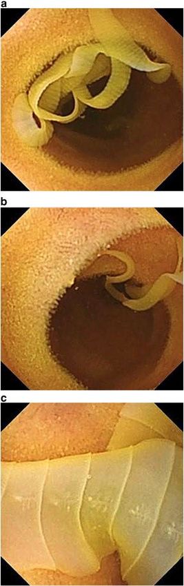
– Suggested in colonoscopy by: segmental hemorrhagic colitis (edema + petechiae +/- erosions or
linear ulcers; no pseudomembranes), more severe in right side of colon, with rectal sparing.
• Treatment:
– discontinue antibiotics and NSAIDs;
– resolution in 4 days.Antibiotic Related Diarrhea (ARD)
Others
• Staphyloccocus aureus:
– (Need to give specific order to culture for S. aureus).
– treat with Vancomycin 500 mg po QID x 10 days.
• Salmonella species:
– treat with cipro 500 mg po QID x 5-7 daysDiarrhea due to Protozoa
Giardia lamblia
• Prevalence:
   – healthy adults < 2%; homosexuals 4-18%.
• Symptoms:
   – Intermittent bloating and abdominal cramps, with watery and low grade steatorrhea;
     “sulfuric belching”.
   – Rare fever.
• Diagnosis:
   – Giardia Ag in stool; stool PCR
   – Duodenal aspirate, string-test, or Bx.
• Treatment:
   – Metronidazole 250 mg po TID x 5-7 days; Quinacrine 100 mg TID x 5 days; Nitazoxanide
     (Alinia) 500 mg TID x 3 days.
   – Patients with IgA or IgM deficiency need 6-8 weeks of therapy.
Cryptosporidium parvum
• Transmission:
– usually person-to-person; domestic animal reservoir.
– causes 4% of acute diarrhea in small children;
– frequent in AIDS.
• Symptoms:
– a) Immunocompetent host: explosive, profuse watery diarrhea, with abdominal
cramps; lasts 5-11 days.
– b) Immunocompromised host: extremely severe diarrhea (up to 17 L/day), which may
persist for months. Fever in 30%.
• Diagnosis:
– AFB stain or fluorescent Ab in stool; Stool PCR
– Small bowel Bx.
• Treatment:
– a) Immunocompetent: Nitazoxanide (Alinia) 500 mg TID x 3 days
– b) Immunosupressed: Paramomycin 500 mg with food, TID x 2-4 weeks + HAARTEntamoeba histolytica
Amebiasis
• Prevalence:
– 1-5% of US population;
– 20-30% in male homosexuals.
– Only Zymodemes II & XI are invasive.
– E. dispar and E. moshkovskii are morphologically identical but distinguished by Ag stool
Test.
• Symptoms:
– Usually asymptomatic. May have watery diarrhea of bleeding without diarrhea.
– Dysentery: Bloody diarrhea, fever > 38 oC in 10%, abdominal cramps, malaise, and
tenesmus.
– Cecal involvement more common than rectal disease.
– Infrequent: ameboma, toxic megacolon or perforation.Entamoeba histolytica
Amebiasis
• Diagnosis:
– Ag Test: (Real time PCR has similar yield in stool and pus)
• Stool in colitis >90%;
• Pus in liver abscess 40-74%.
– Serum Ag:
• In colitis 65%.
• In Abscess: 96-100%
– O&P x 4-6 samples Stool
• In colitis: 25-60%.
• In abscess: Stool 8-44%; pus exam 20%
– Colonoscopy better than Flex. Sigm (both unprep) with Bx from edge (non-specific colitis).
– Serology (Indirect Hemagglutination:
• Colitis: Acute illness = 70%; Convalescent = > 90%.
• Abscess: Acute illness = 70-80%; Convalescent = > 90%.
– Stool WBC usually (-) due to destruction.
• Treatment:
– Metronidazole 750 mg TID x 7-10 d, or Tinidazole 2 gm/d x 3-5 d, PLUS
– Diloxanide 500 mg TID x 10 d, or Iodoquinol 650 mg TID x 20 d or Paramomycin 25-35 mg/k per day,
divided TID, x 7 daysBalantidium coli
• Source:
– ingestion of contaminated short stalk vegetables
• Symptoms:
– frequently asymptomatic;
– mild to moderate, acute or chronic recurrent diarrhea.
– Dysentery in 5%;
– Appendicitis, intestinal perforation with peritonitis, lung and liver involvement
are very rare
• Treatment:
– Tetracycline 500 mg QID x 10 daysIsospora belli
• Transmission:
– fecal-oral
– more common in children and male homosexuals.
• Symptoms:
– fever, headache, abdominal cramps, diarrhea with mild malabsorption.
– In normal host lasts a few weeks;
– lasts months to years in immunocompromised host.
– In Aids may cause cholangiopathy and acalculos cholecystitis.
• Diagnosis:
– duodenal aspirate & Bx.
– Stool incubated at room temperature x 2 days; then Zn sulfate flotation & AFB
stain.
• Treatment:
– Bactrim DS: BID (QID if immunosuppressed) x 10 daysCyclospora cayetanensis • Source: – contaminated fresh berries or water • Symptoms: – Abrupt onset of watery diarrhea; fever in 30%. – Diarrhea improves in 3-4 days, and then relapses. – Anorexia, fatigue, nausea, malabsorption with 5-10% weight loss. Rare G-B and Reiter’s. – In AIDS may cause acalculous cholecystitis. • Duration: 2-12 weeks, with abrupt end. • Pathology: Acute & chronic inflammation in distal duodenum, with villous atrophy, and/or crypt hyperplasia. • Diagnosis: – spherical 9-10 micron with red stain in AFB. - Stool PCR. – Duodenal aspirate (+) in 25% • Treatment: – Bactrim DS BID x 7-10 days.
Microsporidiosis
                Enterocytozoan bienusi & Encephalitozoon intestinalis

• Symptoms:
  – Self limited diarrhea in immunocompetent.
  – In immunocompromised gives chronic diarrhea for months.
     • Can cause AIDS Cholangiopathy.
     • Rarely hepatitis, myositis, sinusitis, bronchiolitis, kidney and urogenital infection
• Treatment:
  – Enterocytozoan bienusi:
     • fumagillin 60 mg/d x 14 days.
  – Encephalitozoon intestinalis:
     • albendazole 400 mg BID x 3-4 weeks.
Foodborne Diarrhea due to Fish & Shellfish
associated ToxinsSpecific Causes of Foodborne Diarrhea – Toxin Mediated
Ciguatera
• Cause: heat-stable Ciguatoxin accumulated in large-fish muscles after eating smaller fish.
• Geography: Common in fish from Hawaii & Florida
• Associated fish:
– Barracuda, red-snapper, amberjack, grouper, and goatfish.
• Onset: minutes to 30 hours
• Duration: 1-9 days; sensory disturbance for months.
• Symptoms:
– nausea, vomiting, cramps, diarrhea, malaise, myalgia, arthralgia, blurred vision, pain in teeth, reversal
of hot-cold sensation, sharp pain in extremities, bradycardia; respiratory paralysis in severe cases.
• Treatment:
– Mannitol 20% solution; 1 g/kg IV over 45 min.
– Gastric lavage and cathartics.
– Atropine for bradycardia. May need respiratory support.
– Amitryptiline, gabapentin for chronic neuropathy.
– Amitryptiline or Fluoxetine for depression and fatigue.
– Symptoms may recur after eating fish, nuts, caffeine or alcohol.Specific Causes of Foodborne Diarrhea – Toxin Mediated
Scombroid
• Cause: histamine & saurine in flesh of fish by action of marine bacteria
– Fish tastes sharp and peppery.
• Geography: Fish from Hawaii & California.
• Associated fish: tuna, mackerel, albacore, bonito, skip jack, mahi-mahi.
• Onset: minutes to 2 hours
• Duration: 4-10 hours.
• Symptoms:
– flushing, headache, dizziness, burning in mouth, abdominal cramps, nausea, vomiting,
diarrhea & bronchospasm.
• Treatment:
– anti-histamines + H-2 blockers, bronchodilators & epinephrine for bronchospasm;
– cathartics & gastric lavage.Specific Causes of Foodborne Diarrhea – Toxin Mediated
Paralytic Shellfish Poisoning
• Cause: heat-stable saxitoxins, from dinoflagellates, concentrated in
– bivalved mollusks,
• worse in “red tide”.
• outbreaks in summer.
• Geography: New England, West Coast, Alaska.
• Onset: 30 minutes - 3 hours; may be fatal in hours.
• Duration: hours to few days.
• Symptoms:
– nausea, vomiting, diarrhea,
– paresthesias in lips, mouth, face and extremities;
– dysphonia, dysphagia, weakness, paralysis and respiratory insufficiency.
• Treatment:
– respiratory support;
– gastric lavage and cathartics.Specific Causes of Foodborne Diarrhea – Toxin Mediated
Neurotoxic Shellfish Poisoning
• Cause: heat-stable brevotoxin, from dinoflagellates, concentrated in
– Mollusks.
• Associated to "red tide".
• Geography: Gulf Coast, North Carolina, and Florida
• Onset: few hours
• Duration: hours to days.
• Symptoms:
– Nausea, vomiting, diarrhea,
– Paresthesias, reversal of hot-cold sensation, ataxia.
– Respiratory symptoms after aerolization.
• Treatment:
– Symptomatic; IV fluids, cathartics, bronchodilators.Specific Causes of Foodborne Diarrhea – Toxin Mediated
Diarrheic Shellfish Poisoning
• Cause: okadaic acid or dinophysistoxin-1 in
– mussels, scallops, or clams.
• Geography: Described in Japan & Europe;
– the organism has been found in U.S. coast.
• Onset: few hours
• Duration: hours to days.
• Symptoms:
– nausea, vomiting, abdominal pain & diarrhea.
• Treatment:
– symptomaticSpecific Causes of Foodborne Diarrhea – Toxin Mediated
Amnestic Shellfish Poisoning
• Cause: domoic acid concentrated in
– shellfish (Razor clams, Dungeness crabs), and
– anchovies.
• Geography:
– described in Canada;
– toxin-producing blooms found in Maine & Texas
• Onset: few hours
• Duration: hours to days.
• Symptoms:
– nausea, vomiting, abdominal cramps, headache, diarrhea, and loss of short-term memory.
– Anterograde memory deficits may persist for months; neuronal necrosis in hippocampus and
amygdala.
• Treatment:
– Symptomatic; cathartics; benzodiazepines for seizures.Gastro-Intestinal Disease due to Helminths Nematodes, Cestodes, Trematodes
Gastro-Intestinal Nematodes
      (Roundworms)
Ascaris Lumbricoides
• Organism: Is a nematode. Male 15-30 cm; Female 20-35 cm.
– Each female produces 200,000 eggs/d that mature in 2-3 wks.
– Life expectancy 1 y.
• Magnitude: 1.3 Billion infected worldwide; 60000 deaths/y.
• Acquisition & Life Cycle:
– Ingestion of eggs in contaminated food;
– the larva hatch in the duodenum, penetrate intestine wall into blood and lymphatic vessels, then go
through the liver, then the heart cavities, then into lung circulation.
– they then penetrate into alveoli and migrate inside bronchial tree for 20 d;
– finally enter esophagus and migrate to SB and develops into adult worm
– matures in 2-3 months an produce new eggs.
– Infestation can be up to 1000 worms/person.Ascaris lumbricoides
• Clinical Manifestations:
– Initial infection with large number of larva may cause pneumonitis with asthma attacks (Loffler syndrome),
and severe eosinophilia.
– Chronic infestation with large number of worms may cause intestinal obstruction.
– Intestinal parasitosis causes mild or no eosinophilia.
– Penetration into appendix may cause appendicitis.
– Penetration into biliary tree is more common in children and young adults. May cause severe biliary cholic
with fever, nausea, and vomiting.
• Jaundice occurs in 10-20%.
• Exquisitely tender hepatomegaly develops in 50%.
• Ascaris can cause rupture of bile duct with bile peritonitis.
• Rarely they penetrate portal or hepatic veins.
• Worm embolism to pulmonary artery may occur.
– Pancreatitis may occur when the pancreatic duct is penetrated.Ascaris lumbricoides
• Diagnosis:
– Stool or duodenal aspirate O&P.
– Contrast X-Ray may show the parasite with contrast inside its own intestine. U/S may show bile
duct worm.
– ERCP may show the parasite in the bile duct. Endoscopy may show adult worms.
• Treatment:
– Mechanical removal of biliary or hepatic worms + Albendazole 400 mg PO x 1
– In intestinal obstruction, patient are first decompressed with NGT suction, and then one dose of
Piperazine 150 mg/kg (max 3.5 gm) is given, followed by doses of 65 mg/kg given then q 12h x 6
doses.
– Pulmonary disease with Glucocorticoids + Albendazole 400 mg PO and repeat 1 month later.Strongyloides stercoralis
• Organism: Is a nematode that can be free living in the soil (male & female), or a female
hermaphrodite in the human duodenum/jejunum; adult is 2-3 mm long.
• Magnitude of the Problem: 35 million infected worldwide.
• Acquisition & Life Cycle:
– Free living adults copulate and produce rhabditiform larvae;
– they mature to filariform larvae that can penetrate the skin of feet or hands of humans (“ground itch” or “larva currens”,
advancing 5-10 cm/h), and
– then enter lymphatics and veins and are carried to the Rt heart and then to the lungs.
– then they migrate through the bronchial tree to the trachea and are finally swallowed.
– Finally they colonize the duodenum and jejunum.
– In the SB the female turns hermaphrodite and produces 40 eggs/d.
– In the bowel the eggs mature to rhabditiform larvae that are excreted, and
– sometimes the rhabditiform larvae matures in the bowel into filariform larvae and causes autoinfection, with
persistent infection for many decades.
– Infestation may persist for 75 years after leaving endemic area.Strongyloides stercoralis
• Clinical Presentation:
– Most patients are asymptomatic. Some have abdominal pain, weight loss, diarrhea, and even malabsorption.
– May develop serpiginois urticarial rash in the buttocks from stool filariform larvae re-infecting through the skin
(larva currens).
– HYPERINFECTION: Immunocompromised patients may develop massive hyper-infection syndrome that can be lethal.
– They have fever, abdominal pain, diarrhea, hematochezia, vomiting, cough, dyspnea, wheezing, hemoptysis, and shock.
– They may have bacteremia and meningitis.
– Patients may have mild jaundice, altered mental status, abdominal tenderness, peritoneal signs, and hepatomegaly but no
splenomegaly.
– Sigmoid colon may show hemorrhagic colitis.
• Laboratory:
– Eosinophilia is common, but may not be present.
– Hypoalbuminemia with protein loosing enteropathy, mild elevation of bili, variable elevation of AST, ALT and alk phosph.
– Blood cultures frequently (+) for gram (-) bacteria.Strongyloides stercoralis
• Diagnosis:
– duodenal aspirate is superior to stool study.
– May show the parasite, larvae, or less commonly eggs.
– Larvae may be found in sputum, ascites, urine, or lymph nodes.
– Serologic tests using crude larval antigens, such as the enzyme-linked immunoassay offered by the Centers for Disease
Control and Prevention, have 95% sensitivity but poor specificity to rule out Strongyloides infection when microscopic
examinations are negative or not performed.
– PCR in stool, or Serum Luciferase Immunoprecipitation systems to detect IgG antibodies to a recombinant Strongyloides
antigen (NIE) and S. stercoralis immunoreactive antigen (SsIR) has sensitivity and specificity of 100%
• Treatment:
– Uncomplicated disease: Ivermectin 0.2 mg/kg PO, then repeat in 2 weeks.
– If immunocompromised, give Ivermectin for 2 consecutive days, and repeat 2 weeks later for 2 consecutive days
– Hyperinfection: Systemic antibiotics plus Ivermectin 0.2 mg/kg PO x >/= 14 days (until stool larva is negative)Capillaria (Paracapillaria) philippinensis
• Organism: very small nematode, male 3.9 mm, and female 5.3 mm. Endemic to the
Philippines and Thailand. Reported in Egypt, Japan, Taiwan, and Iran.
• Acquisition: By eating raw fish infested with the parasite. Parasite replicates in the bowel
with ever-increasing number of intestinal worms (up to 30000 in 5 months).
• Life Cycle: The hosts are birds; in the bird small intestine, the larvae mature into adults.
Adult worms mate and female produces eggs or larvae. The bird stool contaminates ponds
and rivers and fish eat the worms. Human eats raw fish.
• Clinical Presentation:
– Vague abdominal pain and borborygmi.
– Patients begin to have diarrhea 2 to 3 weeks after infection.
– Diarrhea becomes persistent and increasingly voluminous.
– Patients rapidly waste from escalating steatorrhea and protein-losing enteropathy. Eventually they manifest
emaciation, anasarca, and hypotension; diarrhea produces severe hypokalemia.
– Patients die from cardiac failure or secondary bacterial sepsis usually 2 months after onset of symptoms.Capillaria (Paracapillaria) philippinensis
• Diagnosis:
– finding eggs and larvae in stool specimens but stool examination is not sensitive.
– Push or balloon endoscopy shows jejunal mucosal scalloping and biopsies of involved mucosa can
demonstrate the helminths.
• Treatment:
– albendazole 200 mg orally twice daily for 10 days or
– mebendazole 200 mg orally twice daily for 20 days to prevent recurrence.
– Albendazole is better tolerated.Necator americanus and
Ancylostoma duodenale
• Organism and Scope of the Problem: Worldwide, an estimated 440 million people are infested with
hookworm, usually by N. americanus, A. duodenale, or a mixture of both.
– Adult male worm measures 7 to 9 by 0.4-0.5 mm, while the female is 9 to 11 mm long by 0.4-0.5 mm wide
– N. americanus predominates in the Americas, South Pacific, Indonesia, southern India, and central Africa.
– A. duodenale is more common in North Africa, the Middle East, Europe, Pakistan, and northern India.
• Life Cycle:
– Barefoot walking in contaminated soil, infective third-stage hookworm larvae penetrate intact skin, typically between the
toes.
– Larvae migrate through the dermis to reach blood vessels, a migration that can cause a pruritic, serpiginous rash,
cutaneous larva migrans.
– Larvae migrate with venous flow through the right side of the heart to the lungs. A. duodenale larvae can arrest their
migration and become dormant for many months before proceeding to the lungs.
– Once in the lungs, larvae penetrate the alveoli and enter the air spaces, after which they migrate up the pulmonary tree,
are swallowed with saliva, and pass into the small intestine, where they mature.
– Mature worms mate and lay eggs. Each female N. americanus lays about 10,000 eggs a day, and each female A. duodenale
lays about 20,000 eggs a day.
– Eggs are deposited with feces in moist, shady soil, where they hatch to release larvae. The larvae molt twice after which
they move to the soil surface and await a suitable host.Necator americanus and
Ancylostoma duodenale
• Clinical Manifestations:
– Light infestations with N. americanus and A. duodenale cause no symptoms.
– Adult worms feed on intestinal epithelial cells and blood.
– Moderate and heavy hookworm infestation: iron deficiency.
– Intestinal blood loss is 0.01 to 0.04 mL/day per adult N. americanus and 0.05 to 0.3 mL/day per adult A.
duodenale. With a moderate number of worms, this blood loss becomes appreciable.
– The average North American diet is high in iron (more than 20 mg/day) so anemia might not develop, even
with a burden of up to 800 adult hookworms.
• Diagnosis:
– Stool exam for eggs in fixed specimen of 3 separate stool samples.
– Adult may be seen in VCE or Enteroscopy.
• Treatment: Albendazole 400 mg single dose or Mebendazole 100 mg BID x 3 days.Ancylostoma ceylanicum • A. ceylanicum have been reported in West New Guinea, Philippines, Taiwan, Thailand, India, Laos, and Malaysia. – Has multiple hosts (humans + pets like dogs and cats). – Adult worm is 8-10 mm. • Clinical manifestations: Usually asymptomatic but heavy infections can cause anemia. • Diagnosis: – Stool parasite eggs x 3 samples. – VCE and Enteroscopy may find the parasite. • Treatment: single dose Albendazole 400 mg PO
Ancylostoma Caninum
• Worldwide distribution. Is the hookworm of dogs and cats.
• A. caninum does not fully mature in the human host; eggs are not produced.
  Females are 14-16 mm; males 10-12 mm
• Clinical Presentation:
   – Cutaneous larva migrans, a distinctive serpiginous rash caused by an abortive migration of the
     parasite in an unsupportive host.
   – Eosinophilic enteritis, although not all eosinophilic enteritis is caused by this parasite.
   – Patients have colicky mid-abdominal pain and peripheral eosinophilia.
   – On endoscopy of the terminal ileum, patients may have scattered small superficial aphthous
     ulcers and mucosal hemorrhage with mucosal eosinophils; no eosinophils in gastric biopsy.
   – A. caninum also may be a cause of abdominal pain without eosinophilia or eosinophilic enteritis
• Treatment: Empirical Albendazole 400 mg x 1 dose.
Trichuris trichiura (Whipworm)
• Worldwide distribution. An estimated 800 million people harbor T. trichiura . It occurs in
temperate and tropical countries and remains prevalent in areas with suboptimal sanitation.
• Organism: The adult is about 4 cm long. The male has a curled posterior end. Looks like a
whip, with the head in the thin end and the reproductive organs in the wide section.
• Life Cycle:
– Eggs, passed in the feces, contain a zygote and are not infective until embryonation, which takes
place in the soil over 2–4 weeks, producing the L1 larva inside the egg.
– Following human ingestion, the larva is released in the stomach and passes into the intestine. It
penetrates the epithelium in the mucosal crypts of the cecum.
– The larva develops by molting, and the adult develops from the L4 stage, migrating with the
epithelial cells up the sides of the crypts in the cecum.
– Each female produces 3,000–20,000 eggs per day; the life expectancy of a worm within the host
has been estimated at 1–3 yearsTrichuris trichiura (Whipworm)
• Clinical Presentation:
– Most infections are asymptomatic.
– In heavy infections, stools become loose and frequent and there is tenesmus.
– Frequency can exceed 12 stools per 24 hours and nocturnal stooling is especially characteristic.
– Stools consist largely of mucus but may also be watery. Frank blood is common.
– Trichuriasis can causes of recurrent rectal prolapse and the worms may be seen on the prolapsed
mucosa. Children may have severe anemia and growth-retardation.
• Diagnosis: Stool for parasite eggs x 3 (Kato-Katz technique). Proctoscopy showing
worms on the rectal mucosa is more reliable evidence of Trichuris colitis.
• Treatment: mebendazole 200 mg/day on 3 successive days is recommended, or
albendazole 400 mg/day for 3 days. Heavily infested patients might require 7 days of
treatment.Enterobius vermicularis (Pinworm)
• Worldwide distribution. Small nematode with female measuring 9–12 mm in length
and 0.5 mm in width and male 2.5 mm. The eggs that are deposited on the perianal
skin are immediately infective.
• Life Cycle:
– Adult worms inhabit the lumen of the cecum and appendix.
– The life span of the adult female is 4–10 weeks; the adult male, only about 2 weeks.
– Following fertilization, the gravid adult female migrates from the large intestine onto the perianal
skin, where she deposits up to 11,000 eggs by uterine contraction and rupture.
– The sticky eggs adhere to the anal skin and embryonate rapidly over about 6 hours to reach the
infective L3 larval stage, still inside the eggshell.
– The intense pruritus induced by the adult female and the eggs facilitates fecal–oral transfer of
eggs.
– The minimum interval between egg ingestion and the next egg deposition is between 3 and 4
weeks.Enterobius vermicularis
• Clinical Manifestations:
– The most common is pruritus ani. When very heavy infection is present, an eczematous reaction
with bacterial superinfection may occur.
– Other manifestations include teeth grinding, enuresis, insomnia, nausea, vomiting, abdominal
pain, conjunctivitis, and less likely appendicitis.
– Infection of the female genital tract: vulvar and cervical granulomas, salpingitis, oophoritis,
tuboovarian abscess, and peritonitis.
– Eosinophilic colitis has also been reported and appears to occur early in infection, since only
larvae rather than eggs have been identified in such cases.
• Treatment:
– Single 100-mg oral dose of mebendazole or 400-mg oral dose of albendazole.
– Re-infestation is common, and patients should receive a second treatment after 15 days.
– All members of the family should be treated and clothes and bed linens should be washed.Trichostrongyliasis
• The Organism: Female is 3-10 mm long, and males 2-8 mm. Normal host are herbivorous.
Distribution: worldwide, but prevalences are highest in the Middle East, the former southern Soviet
republics, India, North Africa, Southeast Asia, Japan, Siberia, Central Africa, and central and southern
China. Reported sporadic cases are in Australia, Hawaii, the United States, France, and South
America. The parasite lives embedded in the intestinal mucosa of herbivorous animals worldwide.
– Trichostrongylus produce eggs that, after passage in feces reach the soil, mature rapidly and hatch into
larvae that become infective.
– Infection is acquired by the ingestion of larvae with soil or more commonly with infected vegetation (grass)
• Clinical Manifestations: Male and female worms live embedded in the small intestinal mucosa where,
if in sufficient numbers, they are capable of producing trauma, desquamation of the mucosa, and
hemorrhages. Eosinophilia can be very elevated
• Treatment: Pyrantel pamoate is recommended. Alternative agents include mebendazole and
albendazole.Anisakis and Pseudoterranova
(Anisakiasis and Pseudoterranoviasis)
• Organism: Anisakiasis (“herring worm disease”) and pseudoterranoviasis (“cod worm
disease”). Adults are 2-3 cm long.
• Acquisition: by eating raw marine fish in food such as sushi, sashimi, oka, poisson cru, and
ceviche.
• Mechanism of Injury:
– Anisakiasis is mostly a GI tract infection, with rare ectopic locations in the peritoneal cavity, other
abdominal organs, and once in the lungs.
– Usually one larva, uncommonly two and rarely several.
– The parasite burrows the cephalic end into the GI mucosa. Because humans are abnormal hosts,
the parasite dies, producing an abscess characterized by marked inflammation and tissue
eosinophilia.
– This abscess occurs anywhere in the GI tract, often of sufficient size to allow clinical palpation or
radiographic imaging. Sometimes the larva reaches the peritoneal cavity, producing an abscess in
the omentum, occasionally on the surface of other viscera.Anisakis and Pseudoterranova
(Anisakiasis and Pseudoterranoviasis)
• Clinical Presentation:
– The main symptoms are epigastric pain accompanied by nausea and vomiting, sometimes with expulsion of
the worm.
– Clinical and radiographic signs and symptoms of intestinal obstruction may be evident, accompanied by
diarrhea, followed by normal stools or constipation, and blood and mucus in the stools.
– Palpation may demonstrate a discrete mass, which in contrast radiographs appears as an intestinal wall
defect. The mass and symptoms resolve spontaneously in some instances.
– Mild fever, leukocytosis, and eosinophilia are usually present, as is a certain amount of peritoneal fluid,
which is rich in leukocytes and eosinophils.
– Endoscopic examination of the stomach and duodenum may reveal a red, bleeding, often-ulcerated lesion,
sometimes with the worm at the center attached by its cephalic end to the mucosa, the rest of the body
extending into the lumen.
• Treatment: Extracting the worm cures the condition.Oesophagostomum bifurcum, O. Stephanostomum
(Nodule Worm)
• Organism: small nematode; adult measures 1.5-3 cm.
– Prevalent in Togo and northern Ghana were 250,000 people infected and at least 1 million at risk.
– Also found in Uganda, Kenya, Côte d’Ivoire, Ethiopia, Sudan, Guinea, Nigeria, Brunei, Malaysia, Indonesia,
and Brazil.
• Infection Cycle:
– Adult worms live attached to the mucosa of the colon, mate, and produce 5000 eggs per day.
– Eggs passed in feces mature in soil and produce larvae that enter a new host orally and reach the wall of the
large intestine to develop in abscess-like cavity or nodule of 1-2 cm diameter.
– The larvae will mature into adulthood in the nodule and then leave the abscess and attach to the colonic
mucosa and produce eggs.
– The nodules may affect peritoneal surface, omentum, kidneys, spleen, other viscera, and sometimes lesions
push into the abdominal wall, where they can be seen and felt as well-demarcated painful tumors.
– Lesions encountered in the periumbilical area are known as “Dapaong tumor”.Oesophagostomum bifurcum, O. Stephanostomum
• Clinical manifestations:
– Abdominal mass, sometimes large or painful;
– Acute intestinal symptoms, often of obstruction, due mostly to peritoneal adhesions.
– Asymptomatic persons, mostly children, may present because of a painless or disfiguring abdominal mass.
• Diagnosis:
– Eggs are identical to hookworm eggs.
– A PCR assay has detected O. bifurcum in human feces, and multiplex real-time PCR studies of fecal samples
differentiate Ancylostoma , Necator , and Oesophagostomum infections.
• Treatment:
– In acute cases, treatment is often surgical, and diagnosis is confirmed by gross and microscopic examination
of the removed specimen.
– Albendazole 400 mg po once, is effective against hookworms and Oesophagostomum.
– Pyrantel pamoate is effective against Oesophagostomum , but not against hookwormsIntestinal Cestodes (Taenia or Tapeworms)
Diphyllobothrium Species
• The Organism: Fish tapeworm ( Diphyllobothrium species) is the largest parasite of
  humans, reaching lengths of up to 40 feet (12 m).
• Prevalence: Trout, salmon, pike, perch, and whitefish all can harbor D. latum .
    – About 20 million people worldwide are infected with Diphyllobothrium species and prevalence seems to be
      increasing in Russia, South Korea, Japan, and Brazil.
    – Endemic in northern Europe, Russia, and Alaska, but fish tapeworm has been reported in Africa, Japan,
      Taiwan, Australia, South America, North America, and Canada.
• Life Cycle: Has 2 intermediate hosts.
    – Parasite eggs that reach water embryonate and then release free-swimming larvae called coracidia.
    – Coracidia are ingested by water fleas ( Cyclops and Diaptomus ) and develop into procercoid larvae.
    – Fish eat these procercoid larvae, and the parasite changes into the infective plerocercoid form.
    – The plerocercoid larva migrates to and embeds in fish muscle and various organs, growing to 2 cm in length.
    – If an infected fish is consumed by another fish, the plerocercoid larva simply migrates into the flesh of the
      second fish.
    – People acquire the parasite by eating raw or undercooked freshwater fish.
    – Larvae matures to adult in the SB lumen, then develop proglottids with eggs.
Diphyllobothrium Species
• Clinical manifestations:
– Fish tapeworm is not invasive and usually causes no direct symptoms. The worm obtains nutrients such as vitamin B12 by
absorbing luminal contents through its surface. D. latum also produces a substance that splits B 12 from intrinsic factor in
the intestine, thereby further preventing host absorption of the vitamin. Rarely, B 12 deficiency is severe enough to result
in megaloblastic anemia and neurologic symptoms.
– Some patients complain of vague abdominal pain and others describe the sensation that “something is moving inside.”
– Others describe bloating, sore tongue, sore gums, allergic symptoms, headache, hunger pains, loss of appetite, or
increased appetite.
– Rarely, mechanical intestinal obstruction may occur as a result of several worms becoming entangled. Diarrhea may also
occur.
• Diagnosis:
– Identification of D. latum eggs in stool specimens.
– Occasionally, diagnosis is made because the patient passes proglottids and brings them in for identification or
– Worm is seen on endoscopy.
• Treatment:
– Praziquantel is effective in a single oral dose of 10 mg/kg. Patients should be warned that they might pass a rather long
worm 2 to 5 hours after taking the medication.
– Albendazole 400 mg each day for 3 days also kills the tapeworms.Taenia saginata, Taenia asiatica, and Taenia solium
• Organism:
– Pork tapeworms: Taenia solium is 2-8 m with 1000 proglotides, and T. asiatica 4-10 m with 1-2000
proglotides.
– Beef tapeworm: T. saginata 4-10 m with 1-2000 proglotides.
• Global distribution with > 50 million infected. Human is the definitive host.
• Acquisition:
– Via ingestion of poorly cooked pork or beef that carries metacestodes (cysticerci);
– from there the scolex evaginates and attaches to the upper jejunum by its well-developed
holdfast organs.
– After 12 weeks of maturation the scolex neck generates Proglottids that mature and fill with
eggs and are replaced by new proglottids.
– Mature proglotides are eliminated in the stool.Taenia saginata, Taenia asiatica, and
Taenia solium
• Clinical intestinal manifestations:
– Most are asymptomatic. T. saginata, carriers eliminate motile proglottids through migration to the perianal area and
appear in their underwear. T. solium proglottids are not motile, and fewer are passed in the stool.
– Symptomatic patients complain of mild abdominal discomfort, cramps, colicky pain, nausea, vomiting, fatigue, anorexia,
and weight loss.
– Intestinal perforation, intestinal obstruction, pancreatitis, cholangitis, and cholecystitis as a result of aberrant proglottid
migration have rarely been reported.
• T. solium can cause cysticercosis.
– Human cysticercosis follows ingestion of eggs from a human tapeworm carrier (fecal-oral route).
– Eggs hatch in the upper intestines, releasing oncospheres (invasive larvae) that penetrate the intestinal mucosa using
their hooklets and excretory proteases, enter the bloodstream, and migrate to the tissues, where they mature into
cysticerci.
– Cysticerci may lodge in skeletal and cardiac muscle, subcutaneous tissue, and even lung tissue, but, in most of these
locations, cysticerci cause few symptoms and spontaneously degenerate, which may lead to formation of calcified
granulomas.
– Neurocysticercosis results from the minority of parasites that invade the central nervous system (CNS), including the
brain, cerebral ventricles, or eye.Taenia saginata, Taenia asiatica, and Taenia solium
• Diagnosis:
– Microscopic exam of proglottids and eggs.
– Stool CoAg ELISA for worm somatic or excretory-secretory products is at least twofold more
sensitive at identifying human carriers compared with traditional stool examination.
– Copro-PCR, can make species-specific diagnoses before treatment, using a single fecal sample to
detect a T. solium oncosphere-specific protein (100% sensitive and specific).
• Treatment:
– All three forms of human taeniasis can be eliminated (85%–98% efficacy) with a single oral dose
of Niclosamide (2 g PO) as preferred agent, or praziquantel (5–10 mg/kg PO) x 1 code.
– Praziquantel presents a small risk that asymptomatic viable brain cysts of T. solium will be
activated during treatment, resulting in neurologic sequelae of seizures and headache.Hymenolepis nana and Hymenolepis diminuta
• They are cosmopolitan in distribution. H. nana is common in warmer climates whereas H. diminuta
only occasionally infects humans.
• Organism: H. nana is approximately 5 mm long and H. diminuta is 3-5 mm.
• Life Cycle:
– H. nana is spread from hand to mouth.
– H. nana eggs are passed in the feces and ingested by a new human or the same host (autoinfection).
– The embryo hatches in the small intestine and penetrates a villus, where it becomes a cysticercoid larva.
– Upon maturation, in 3 or 4 days, it emerges from the tissue and attaches to the intestinal mucosa by its scolex.
– In 2 or 3 weeks, the new worm is producing eggs.
– Hyperinfection can occur when eggs liberated in the small intestine hatch and immediately penetrate a villus to undergo a
new cycle. As a result of hyperinfection, children may harbor many hundreds or even thousands of adult worms.
– The entire life history from ingestion of the egg to adulthood requires approximately 10–14 days. Eggs are first seen in the
stools in approximately 25–30 days.Hymenolepis nana and Hymenolepis diminuta
• Clinical Manifestations:
– Most cases are asymptomatic. Young children may develop symptoms when many worms are present.
– Diffuse, persistent abdominal pain is the most common complaint.
– Patients may have loose bowel movements or occasionally frank diarrhea with mucus but no blood.
– Pruritus ani and nasi are occasionally encountered.
– Many children have sleep and behavioral disturbances that resolve after successful therapy.
– Serious neurologic disturbances such as seizures have been reported.
– Many patients with hymenolepiasis have a moderate eosinophilia of 5–10% and skin eruptions.
• Diagnosis: by finding ova in stool.
• Treatment:
– Praziquantel is the drug of choice and is highly effective in a single dose of 25 mg/kg. It not only eliminates adult worms
but also, unlike other anthelminthics, is efficiently absorbed and kills the larval stages (cysticercoids) in the submucosa.
– Stools of the entire family must be checked before therapy is initiated because other members of a household are
commonly infected, and they must also be treated for therapy to be successful.
– Post-treatment stool examinations should be done after 5 weeks and again after 3 months.You can also read