Arthramid Vet Whitepaper 2019 - Ihr ESS Ansprechpartner: Frau Benita Müller - Service & Vertrieb Mail
←
→
Page content transcription
If your browser does not render page correctly, please read the page content below
Arthramid® Vet
Whitepaper 2019

Ihr ESS Ansprechpartner:

Frau Benita Müller – Service & Vertrieb
Mail: sales@ess-supplements.de

Stand: 01/2021

                             www.ess-supplements.de
Arthramid® Vet
2.5% polyacrylamide hydrogel for the treatment of lameness

   Contents

   Chapter 1                                                                 Chapter 5

   Treatment of joint Lameness &                                     3       Mechanism of Action                                      13
   Osteoarthritis in horses
                                                                             Introduction                                             13
   Introduction                                                       3
                                                                             2.5% PAAG - A Novel Treatment                            13
   Conventional Therapies for Osteoarthritis (OA)                     3
                                                                             Mechanism of Action 2.5%PAAG                             14

                                                                             Conclusion                                               15
   Chapter 2

    Pathophysiology of Osteoarthritis                                5
                                                                             Chapter 6
   Introduction                                                       5      Clinical Safety                                          16
   Histological Anatomy                                               5      Safety Studies                                           16

   Inflammation and Synovitis                                         6      In Vitro                                                 16

   Pain and Synovitis                                                 7      In Vivo                                                  17

                                                                             Synoviocentesis Studies                                  17
   Chapter 3
                                                                             Conclusion                                               17
   Chemistry and Manufacturing                                       9
                                                                             Putting it into Practice                                 18
   Chemistry                                                          9

   Manufacturing                                                      9
                                                                             Chapter 7

                                                                             Putting it into Practice                                 18
   Chapter 4                                                                 Case Selection                                           18
   History of its use in clinical studies                           10
                                                                             Case Management                                          18
   Clinical Efficacy                                                 10
                                                                             Dosages                                                  19
   Prospective Double-Blinded Positive Control Study                  11
                                                                             Key Points                                               19
   Histological Studies                                              12

   Arthramid Vet Prices and Information: sales@ess-supplements.de
   ESS - Equine Supplement Service GmbH - Essener Straße 39a - 49456 Bakum                                                   Page 2 | 23
                                                                                                        E   contact@arthramid.co.nz
Chapter 1

  Arthramid®
Arthramid®
           Vet                                  Vet 2.5%PAAG
   Treatment of joint Lameness & Osteoarthritis in horses
2.5% polyacrylamide hydrogel for the treatment of lameness                                                                           Chapter 1

   Introduction                                                              Conventional Therapies for
   Osteoarthritis (OA) is cited as the most important
                                                                             Osteoarthritis (OA)
   musculoskeletal disorder in both humans and horses.1, 2
                                                                             Osteoarthritis (OA) is treated symptomatically in the
   Clinically, it is associated with lameness and dysfunction
                                                                             horse.12 Current conventional therapies include non-ste-
   of the affected joint, and approximately 60% of all equine
                                                                             roidal anti-inflammatory drugs (NSAIDs), polysulphated
   lameness is due to OA.3, 4 Significant economic loss
                                                                             glycosaminoglycans (PSGAGs), corticosteroids, glucos-
   to the equine industry occurs as a result and coupled
                                                                             amine, hyaluronic acid and a combination of the above,
   with welfare concerns, motivates ongoing research
                                                                             along with biotechnological substances such as gene
   into innovative treatments.5, 6, 7 The rapid resolution
                                                                             therapy, recombinant or autologous growth factors
   of lameness by a reduction in pain is paramount, but
                                                                             (platelet-rich plasma and interleukin-1 receptor antago-
   treatments ideally, should also serve to arrest or slow the
                                                                             nist) and stem cells (allogeneic and autologous chondro-
   progression of the disease.8
                                                                             cyte transplantation).13
   The use of an intra-articular 2.5% cross-linked
                                                                             The challenge is to develop therapeutic options that
   polyacrylamide (2.5%PAAG- Arthramid® Vet, Contura Vet,
                                                                             both reduce pain and are disease modifying. This would
   Denmark) to treat OA is novel. 2.5%PAAG is integrated
                                                                             reduce the progression of the deleterious effects OA
   into the synovial membrane through a combination
                                                                             has on the articular cartilage and surrounding joint
   of vessel in-growth and molecular water exchange
                                                                             structures.
   and persists long term in the joint.9 This significantly
   improves joint lameness caused by early and late stages
   of OA, with trials showing over 75% of cases becoming
                                                                             A recent study also showed a positive
   lame free10 and for up to 24 months. Its use in racing
   Thoroughbreds is also confirmed where it is shown to                      association between musculoskeletal
   be both superior to and longer lasting than conventional                  injury (MSI) rates in Thoroughbred
   therapies.11
                                                                             racehorses and local corticosteroid

   The rapid resolution of lameness by                                       injection.15 The International

   a reduction in pain is paramount, but                                     Federation of Horse Racing

   treatments ideally, should also serve                                     Authorities acknowledged this study

   to arrest or slow the progression of                                      and through its welfare committee

   the disease.8                                                             recommended a 14-day stand-down
                                                                             period as a result.

                                                                             Intra-articular (IA) corticosteroid administration is one
                                                                             of the most commonly used treatments.14 Concerns for
                                                                             its ongoing use include steroid-induced deterioration
                                                                             of articular tissues, known as “steroid arthropathy.” The
                                                                             overuse of a treated joint can also result in accelerated
                                                                             cartilage degeneration.2 These primary concerns are
                                                                             compounded by additional reports of the adverse effects
                                                                             corticosteroids have on chondrocyte metabolism.8 A
                                                                             recent study also showed a positive association between
                                                                             musculoskeletal injury (MSI) rates in Thoroughbred

   Arthramid Vet Prices and Information: sales@ess-supplements.de
   ESS - Equine Supplement Service GmbH - Essener Straße 39a - |49456
                                     www.imsvet.com                   Bakum
                                                                  sales@imsvet.com   | +64 (09) 8013 253                        Page
                                                                                                                             Page 3 | 233
racehorses and local corticosteroid injection.15 The                      treatment of OA.
International Federation of Horse Racing Authorities                      Notwithstanding, whilst the use of MSCs, BMAC or
acknowledged this study and through its welfare                           ciMSCs may have been shown in some studies to aid in
committee recommended a 14-day stand-down period                          the treatment of OA, 17, 18 their exact mechanism of action
as a result.                                                              within the OA environment is still poorly understood.
IA corticosteroids are commonly combined with                             MSCs have been shown to produce IL-1Ra post
hyaluronic acid (HA). There is the perception that the                    administration into a joint and inhibit inflammation,20
HA might be protective against any deleterious effects                    and this mechanism possibly supports findings in
of corticosteroids.16 Studies, however, reveal little or no               those studies where positive outcomes following MSCs
improvement of OA scores and show little effect against                   treatment in the OA model or OA patient are seen. But
the induced cartilage matrix proteoglycan catabolism                      whilst inhibiting inflammatory mediators attenuates the
in cartilage explants (caused by Methylprednisolone                       disease, further work is required to determine if the use
acetate (MPA) in particular).16                                           of MSCs are disease modifying in the long-term.
                                                                          The ideal therapeutic agent for a horse suffering from
                                                                          OA is an agent that alleviates the symptoms of lameness
The ideal therapeutic agent for
                                                                          while also providing a positive effect on the articular
a horse suffering from OA is an                                           components resulting in stabilisation or even repair of
                                                                          the pathologic processes occurring in the affected joint.
agent that alleviates the symptoms
                                                                          The era of OA management is shifting towards new
of lameness while also providing                                          therapeutic concepts.12

a positive effect on the articular
components resulting in stabilisation
or even repair of the pathologic
processes occurring in the affected
joint.

Mesenchymal Stem Cells (MSCs) have been used for the
treatment of tendonitis, arthritis and intra-articular soft
tissue injury in horses, as well as for cartilaginous disease.
Historically however, several factors have hindered the
use of MSCs such as cell source, propagation techniques,
and the effects of transportation on cell viability.
Furthermore, it is unknown whether MSCs applied
directly to an environment full of pro-inflammatory and
catabolic substances function successfully or die.12
Even so, the use of autologous bone marrow derived
concentrate, produced patient-side for the treatment of
early partial cranial cruciate ligament and meniscal tears,
has been found to provide potential for clinical use.17
More recently, chondrogenic induced mesenchymal stem
cells (ciMSCs) have been shown to offer an alternative
to autologous therapy18, with a study showing that after
ciMSCs application in an OA model, treated horses were
less lame, and with reduced joint effusion and improved
synovial fluid quality compared to saline controls.
Nevertheless, another study of 24 horses with induced
carpal OA treated with MSCs showed an increase in
concentration of PGE2 and TNF-alpha in the synovial
fluid.19 MSCs were not, therefore, recommended for the

Arthramid Vet Prices and Information: sales@ess-supplements.de
ESS - Equine Supplement Service GmbH - Essener Straße 39a - |49456
                                  www.imsvet.com                   Bakum
                                                               sales@imsvet.com   | +64 (09) 8013 253                        Page
                                                                                                                          Page 4 | 234
Chapter 2

  Arthramid®
Arthramid®
           Vet                                           Vet 2.5%PAAG
   Pathophysiology of Osteoarthritis
2.5% polyacrylamide hydrogel for the treatment of lameness                                                                                      Chapter 2

   Introduction                                                                     bone and soft tissues of the joint.26 Although OA can
                                                                                    be classified as a non-inflammatory disease, there are
   The two primary functions of a synovial joint are to                             multiple studies indicating that synovitis is an essential
   enable efficient movement and to transfer load.5, 21 All of                      component in its pathogenesis,2, 5, 27, 28 with it almost
   the tissues comprising the synovial joint can be affected                        always being present in OA cases.2
   by injury, including the subchondral bone, articular                             Indeed, it is now well recognised that the OA disease
   cartilage, synovium, and joint capsule as well as intra-                         process starts with disease in the synovial membrane,
   articular and extra-articular ligaments and, if present,                         fibrous joint capsule, subchondral bone, or ligaments, as
   meniscus.22, 23, 24, 25                                                          well as articular cartilage or any combination of all of the
                                                                                    above.29

   It is now well recognised that the
   OA disease process starts with
   disease in the synovial membrane,                                                Histological Anatomy
   fibrous joint capsule, subchondral                                               Synovial joints are considered complex organs in which
                                                                                    all constituent tissues (articular cartilage, subchondral
   bone,                                                                            bone, and synovial membrane) interact with each other,
   or ligaments, as well as articular                                               both directly and via the synovial fluid, in health and
                                                                                    disease.30
   cartilage or any combination of all of
                                                                                    The synovial intima is lined by a diverse population of
   the above.29
                                                                                    synoviocytes, classified according to their ultrastructure.31
                                                                                    Type A cells are macrophages, implicated in
                                                                                    phagocytosis of fluid, foreign material, and microbes.
                                                                                    Type B cells are fibroblasts; locally derived cells that
                                                                                    produce structural components including collagen. Type
   Trabecular Bone
                                                                                    C cells appear to be an intermediate between type A and
   Joint Capsule
                                                                                    B forms. Beneath this synoviocyte cellular layer is the
   Synovial Membrane                                                                subintima, comprised of fibrous and adipose tissue, with
   Subchondral Bone                                                                 blood vessels and small numbers of inflammatory cells.
   Plate
                                                                                    The deepest layer is loose connective tissue that allows
   Articular Cartilage
                                                                                    the membrane to move freely. Ligaments, tendons, or
   Joint Cavity                                                                     capsular fibrous tissue are located outside of that.
   Containing Synovial
   Fluid

                                                                                    The synovial membrane becomes
                                                                                    a source of proinflammatory and
   Figure 1. Schematic representation of a synovial (diarthrodial) joint.
                                                                                    catabolic products, which contribute
   Adapted from: van Weeren P.R (2016). General Anatomy and Physiology of
   Joints. In: Joint Disease in the horse. 2nd Edition. Elselvier. St Louis; 1: 3   to articular matrix degradation

   Equine (OA) is a group of disorders characterised by                             Two important molecules produced by synovial
   a common end stage: the progressive deterioration of                             lining cells are lubricin and hyaluronic acid which
   the articular cartilage accompanied by changes in the                            help to protect and maintain the integrity of articular

   Arthramid Vet Prices and Information: sales@ess-supplements.de
   ESS - Equine Supplement Service GmbH - Essener Straße 39a - |49456
                                     www.imsvet.com                   Bakum
                                                                  sales@imsvet.com        | +64 (09) 8013 253                             Page
                                                                                                                                       Page 5 | 235
cartilage surfaces in synovial joints. 32 Together, these
                                                                          Inflammation and Synovitis
two molecules reduce friction by providing boundary
lubrication at the articular surface. As part of the                      Multiple pathologic states develop after either single
OA complex, elastoviscosity of the synovial fluid is                      or repetitive traumas that are the initial “take-off” for
abnormally low.33                                                         progression towards OA. These can include traumatic
                                                                          injury, synovitis, capsulitis, sprain, intra-articular fractures,
                                                                          and meniscal tears.       4, 5
                                                                                                           All of which lead to a common
The clinical syndrome of OA is                                            final end-stage of joint failure.5, 23, 35, 36, 37 Inflammation
                                                                          is most intense in acute synovitis and is one of the
quite variable, with differences in                                       initial changes to occur in the development of OA24,
affected joint patterns, risk factors,                                    25, 38
                                                                                   Furthermore, the presence of synovitis in OA is
                                                                          associated with more severe pain, and joint dysfunction.39
rates of progression, and severity of                                     This is shown to correlate with symptom severity, the
symptoms.32                                                               rate of cartilage degeneration, and osteophytosis.

Previous studies have shown that alteration of the
                                                                          In athletic and young horses, synovitis
friction-lowering function of synovial fluid may
contribute to the deterioration of articular cartilage                    and capsulitis are changes that occur
in joint disease and, after joint injury in the horse.34
                                                                          early on and are assumed to be
Furthermore, the synovial membrane becomes a source
of proinflammatory and catabolic products, which                          associated with repetitive trauma.4, 5
contribute to articular matrix degradation.                               This aetiology originates most often
Although structural joint damage in OA is a constant
                                                                          from overuse and conformational
feature, the clinical syndrome of OA is quite variable,
with differences in affected joint patterns, risk factors,                problems predisposing the horse to
rates of progression, and severity of symptoms.32 It is                   inappropriate biomechanical forces
recognised that early structural changes seen associated
with OA may remain asymptomatic for many years.30                         on the articular cartilage.5
This again highlights the opportunity for a novel
therapeutic agent that assists in disease modification
                                                                          There are three hypothetically pathogenic pathways
and allows for preventative and therapeutic strategies.
                                                                          for OA.

                                                                          1.       The most commonly accepted theory is that cartilage
                                                                                   gets damaged due to different mechanical forces,
                                                                                   which generates injury to cells and matrix, and to
                                                                                   metabolic alterations of chondrocytes, which will
                                                                                   start a cascade of a release of proteolytic enzymes
               Intima                           Subintima                          resulting in cartilage fibrillation and breakdown of
       Layers of synoviocytes          Fibrous tissue, blood vessels
                                                                                   the proteoglycan network. Cartilage is relatively
                                                                                   susceptible to repetitive trauma compared to its
                                                                                   ability to resist shear forces.40 Therefore, repeated
                                                                                   trauma is assumed to be one of the most common
            Subintima                                                              factors of OA in horses.31

                                                                          2. A second pathway describes the cartilage as being
                                                                                   principally defective, with abnormal biomechanical
                                  Tissue here varies – usually                     properties that will fail under normal loading.5, 31
                Capsule          a layer of dense collagenous
                                  tissue, can be fat here too.
                                                                          3. The third pathway involves physical changes in
                                                                                   the subchondral bone.31 The thin articular cartilage
Figure 2. Histological anatomy of the synovial membrane using electron             cannot work as an effective shock absorber.
microscopy. Images captured at 2x, 20x and 40x magnification.
                                                                                   Therefore, the subchondral bone protects the
                                                                                   cartilage from damage by providing a flexible surface
                                                                                   to absorb forces placed on the joint.23

Arthramid Vet Prices and Information: sales@ess-supplements.de
ESS - Equine Supplement Service GmbH - Essener Straße 39a - |49456
                                  www.imsvet.com                   Bakum
                                                               sales@imsvet.com      | +64 (09) 8013 253                            Page
                                                                                                                                 Page 6 | 236
Pain and Synovitis
                                                                                  Two general types of pain stimuli in synovial joints can
                                                                                  be distinguished. Mechanical stimuli, generated by
                                                                                  (severe) mechanical changes in the environment of
                                                                                  the joint (e.g., by direct trauma), and chemical stimuli
                                                                                  resulting from tissue inflammation.2 The stimuli are
                                                                                  detected and forwarded by different types of receptors;
                                                                                  mechanoreceptors and nociceptors, to peripheral nerves,
                                                                                  spinal cord and ultimately to the brain to be processed,
                                                                                  modulated and perceived.

Figure 3. Joint overview: normal vs osteoarthritic lesions

                                                                                  Synovitis, however, is a single
                                                                                  important factor that contributes to
In athletic and young horses, synovitis and capsulitis
are changes that occur early on and are assumed to                                the pain of OA through joint effusion,
be associated with repetitive trauma.             4, 5
                                                         This aetiology           swelling and capsulitis that in turn
originates most often from overuse and conformational
problems predisposing the horse to inappropriate
                                                                                  will activate mechanoreceptors in
biomechanical forces on the articular cartilage.5                                 the joint capsule and result in direct
Inflammatory mediators (cytokines, prostaglandin E2,                              chemical stimulation of nociceptors.2
and matrix metalloproteinases) are released by reactive
synovial cells in response to cartilage wear products
within synovial fluid and occurs in both naturally and                            The rapid resolution of lameness attributable to pain is
experimentally induced OA.31, 41, 42, 43, 44 Clinically, synovitis                noticeably the principal concern of owners and trainers
is seen as a palpable joint swelling due to either synovial                       and the reason why horses present for veterinary care.8
effusion or thickening of the synovium.              38, 45
                                                              If this persists,   There is a weak correlation between the magnitude of
fibrosis and increased friction in the joint capsule will                         pain and the severity of articular damage observed.31
develop, resulting in thickening of the joint capsule and                         Synovitis, however, is a single important factor that
loss of the normal range of joint motion.                46
                                                              Therefore           contributes to the pain of OA through joint effusion,
rapid resolution of synovitis and capsulitis is critical                          swelling and capsulitis that in turn will activate
in the management of OA because synovitis induces                                 mechanoreceptors in the joint capsule and result in
cartilage matrix degradation.16                                                   direct chemical stimulation of nociceptors.2

                                                                                  In articular tissues, four types of afferent receptors are
                                                                                  found; Type 1 (low-threshold mechanoreceptors with
                                                                                  proprioceptive function, located in the joint capsule),
                                                                                  Type 2 (low-threshold mechanoreceptors activated
                                                                                  during motion with dynamic proprioceptive function
                                                                                  and located at the joint capsule/sub-synovial tissue
                                                                                  junction), Type 3 (high threshold mechanoreceptors
                                                                                  and nociceptors, activated during physiologic limits and
                                                                                  located near bony insertions of IA and peri-articular
                                                                                  ligaments) and Type 4 (polymodal high threshold
                                                                                  nociceptors found as free nerve endings responding to
                                                                                  thermal, chemical and mechanical stimuli, located in the
                                                                                  synovial membrane).2

                                                                                  Nociception will in most cases be stimulated
                                                                                  or enhanced by inflammation. In addition to
                                                                                  mechanoreceptors stimulated by mechanical influences,

Arthramid Vet Prices and Information: sales@ess-supplements.de
ESS - Equine Supplement Service GmbH - Essener Straße 39a - |49456
                                  www.imsvet.com                   Bakum
                                                               sales@imsvet.com        | +64 (09) 8013 253                            Page
                                                                                                                                   Page 7 | 237
they can become hypersensitized by chemical stimuli
released during the inflammatory process.2 Furthermore,
mechanical stimulation itself may, through tissue
damage, elicit an inflammatory response with the release
of pro-nociceptive mediators. Such as the OA cascade
ensues within the joint tissues, pain results attributable
to synovitis and capsulitis as well as subchondral
bone exposure, remodeling or bone marrow oedema
and marginal periosteal activation associated with
osteophytosis.47

                               Trauma/Insult

       Tissue catabolism                             Direct Tissue
          > anabolism                                  Damage

    Nociceptor                     Joint                 Mechanoreceptor
    activation                     Pain                     activation

         Joint swelling,                            Inflammatory
            effusion                                   response

                                Release of
                            cytokines mediators

Figure 4. Simplified schematic diagram of the vicious cycle of osteochondral
damage and cartilage degeneration in OA, showing key processes that may
contribute to joint pain associated with the disease.

Adapted from: Van Weeren P.R., de Grauw J.C. Pain in osteoarthritis. Vet
Clin North Am Equine Pract, 26(3), 619-642.

Arthramid Vet Prices and Information: sales@ess-supplements.de
ESS - Equine Supplement Service GmbH - Essener Straße 39a - |49456
                                  www.imsvet.com                   Bakum
                                                               sales@imsvet.com   | +64 (09) 8013 253      Page
                                                                                                        Page 8 | 238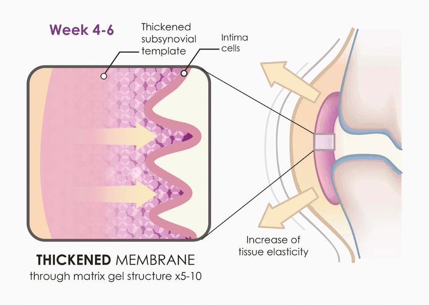
Chapter 3

  Arthramid®
Arthramid®
           Vet                                        Vet 2.5%PAAG
   Chemistry and Manufacturing
2.5% polyacrylamide hydrogel for the treatment of lameness                                                                                           Chapter 3

                                                                             for the augmentation of connective tissues such as skin
   Chemistry                                                                 and bladder neck.51 The current direction of hydrogel
   Arthramid® Vet 2.5% Polyacrylamide hydrogel                               research is focusing on their use as bioactive materials
   (2.5%PAAG) is an inert, non-pyrogenic and neuro-                          to regulate stem cell fate, drug delivery, and now for the
   innocuous polymer gel consisting of 97.5% sterile                         management of OA in both animals and humans.52, 53
   water and 2.5% cross-linked polyacrylamide with water
   exchanging capabilities. (Contura International A/S,
   Soborg, Denmark). Its biocompatibility in soft tissues
   has been demonstrated and histopathological studies
   of subcutaneous tissues from mice, rats, rabbits, pigs,
   horses and humans have shown it supports cell growth
   and tissue integration and possesses a permanent, stable
   augmentation effect due to constant molecular water
   exchange with its host tissue.9, 48, 49, 50

   Shown it supports cell growth and
   tissue integration and possesses a
   permanent, stable augmentation
   effect in host tissue                                                     Figure 6. Representative image of 2.5%PAAG using scanning electron
                                                                             microscopy. Scale Bar = 1.0 µm showing porosity of the gel structure.

                                                                             Manufacturing
                                                                             Arthramid® Vet (AV) is produced by a patented
                                                                             technology called In-line Cross-Linking Technology (ILX
                                                                             Technology), forcing water molecules between the cross-
                                                                             linked polymers of polyacrylamide (CAS No. 9003-05-8),
                                                                             that provides the gel with exceptional molecular stability
                                                                             and the ability to retain its viscoelastic properties in situ.
                                                                             AV is hydrophilic, due to its chemical structure, and has
                                                                             an irreversible and steady-state backbone, with lightly
                                                                             bound water molecules that can interchange with water
                                                                             molecules of surrounding tissue.

   Figure 5. 3D molecular representation of 2.5%PAAG showing cross linking   Patented technology called In-
   between two molecules of polyacrylamide.
                                                                             line Cross-Linking Technology (ILX
   2.5%PAAG is a gel similar to hyaluronic acid in overall                   Technology) giving exceptional mole-
   structure and tissue compatibility,        33
                                                   but with a longer
                                                                             cular stability and, the ability to retain
   lasting viscous effect, as it is non-degradable. 2.5% PAAG
   has been used in human medicine for more than 16 years
                                                                             its viscoelastic properties in situ

   Arthramid Vet Prices and Information: sales@ess-supplements.de
   ESS - Equine Supplement Service GmbH - Essener Straße 39a - |49456
                                     www.imsvet.com                   Bakum
                                                                  sales@imsvet.com   | +64 (09) 8013 253                                     Page
                                                                                                                                          Page 9 | 239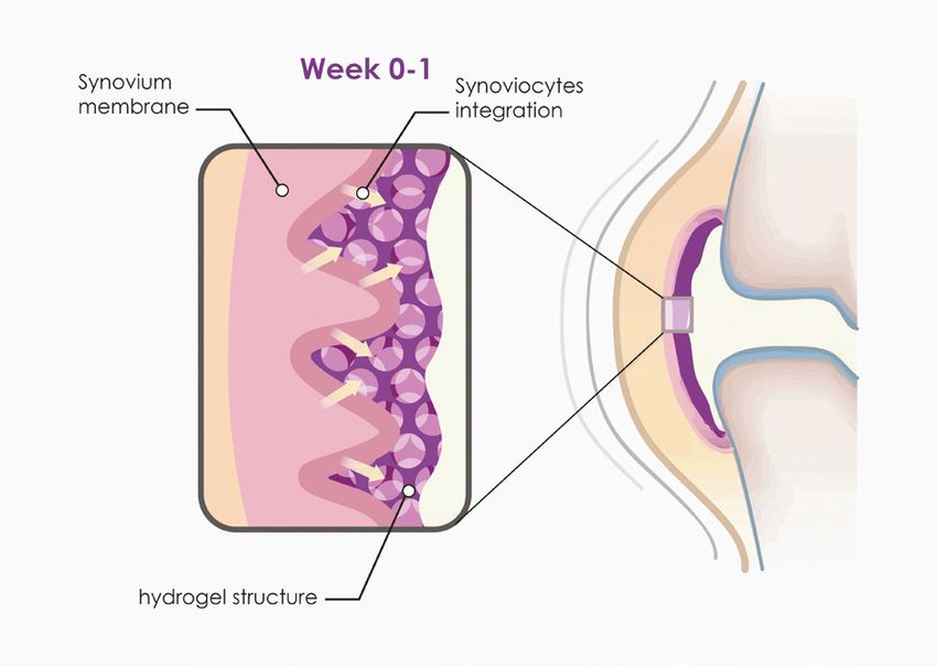
Chapter 4

  Arthramid®
Arthramid®
           Vet                                  Vet 2.5%PAAG
   History of its use in clinical studies
2.5% polyacrylamide hydrogel for the treatment of lameness                                                                            Chapter 4

                                                                             with both goats in the control group showing marked
   Clinical Efficacy                                                         deterioration of OA lesions.
   Results in animals and humans show long-lasting positive                  Tnibar et al. (2014) also compared the efficacy of 2mls of
   effects on the symptoms of OA after treatment with                        2.5% PAAG with 12mg of triamcinolone acetonide (TA)
   2.5%PAAG.9 Two papers published in 2012, were the                         and 20mg of hyaluronic acid (HA), when injected into a
   first to report prospective clinical trials on the efficacy               metacarpal/metatarsophalangeal (fetlock) joint, in 40
   of 2.5% PAAG in horses. Tnibar et al. (2012) evaluated                    Warmblood horses used for dressage, showjumping or
   33 horses older than two years with confirmed OA in                       eventing.10, 20 Horses were assigned into 1 of 2 treatment
   only one joint based on clinical evaluation, intra-articular              groups after clinical examination, intra-articular
   anaesthesia and imaging (radiography, magnetic                            anaesthesia, radiological and MRI assessment, and were
   resonance imaging (MRI) or arthroscopy) and followed                      clinically evaluated at 1, 3 and 6-months post-treatment.
   up at 1, 3 and 6 months after treatment with 2mls of 2.5%                 The proportion of lame-free horses were 55%, 65%,
   PAAG.54 At six months 70% (23/33) of the horses were                      and 75% respectively in the 2.5% PAAG treated group
   lame-free concluding 2.5% PAAG is an effective and safe                   and 15%, 40%, and 35% in the control (TA-HA) group.
   treatment for symptomatic OA in horses and warranted                      The study concluded that horses treated with 2.5%
   further studies.                                                          PAAG were significantly less lame (p
intra-articular analgesia, and radiological assessment. The percentage of horses lame-free following a single intra-articular injection of 2ml of 2.5% PAAG was 65% (p
3 y.o. TB filly; 14 days; LFC Prox
Histological Studies
Christensen et al. (2016) present a thorough histological                                                                             Blue Haze

review of the synovial incorporation of 2.5% PAAG after
injection into normal and OA animal joints.9 Vessel in-
growth begins immediately after gel injection with host
macrophages entering the gel, which is unable to engulf
                                                                                          Subintima
the polymer. These are gradually replaced by fibroblasts
and endothelial cells, which in time develop into a thin
vessel-bearing fibrous network inside the gel. Integration
and invasion of synovial cells in the gel is seen by day
14 with a sub-synovial layer traversed by thin strands of
connective tissue and vessels covered by a synovial lining
facing the joint cavity formed by day 30 and at up to 2
years post injection.9
                                                                          5 y.o. TB gelding; 42 days; LFC Prox
De Clifford and Lowe (unpublished) have demonstrated
similar histological findings. The 2.5% PAAG appears
                                                                                  Blue Haze
as a blue granular material gel in the subintima,
when injected into normal and OA equine synovial
joints. The local response comprised of macrophages
and surface synoviocytes (type A cells) that were
attempting to phagocytose the hydrogel at day 14 and
                                                                                  Subintima
42 post-treatment. There is evidence of binucleate
and multinucleate macrophages indicating a response
to a persistent stimulus. Also noted was an increase
in fibroblasts and collagenous tissue surrounding
blood vessels, together with visible hyperplasia and
hypertrophy of the synoviocyte layers of the intima. This
noticeable rejuvenation of the synovium may improve
the nature of the synovial fluid. There were no significant
                                                                          Figure 8. Histological sections of the subintima layer of the synovial
amounts of gel material overlying the synovial surface.                   membrane. 2.5%PAAG can be seen as a ‘blue haze’ integrated into the
This study also demonstrated no evidence of neutrophilic                  subintima, 14 days and 42 days after treatment. Images captured at 4x
                                                                          magnification.
inflammation, fibrin deposition, mineralisation, or cell
death, or other inflammatory cell types. This implies a
low level of irritation and antigenicity. Indeed, studies
                                                                          5 y.o. TB gelding; 42 days; RFC Prox
on 2.5% PAAG have not found the polymer to act as
a nidus or foreign body with any potential to harbour
bacterial infection,11, 33 and is unlikely to be detrimental
to managing joint sepsis or surgical intervention in the
future.

                                                                               Synoviocytes

                                                                          Figure 9. Histological section of the intima layer of the synovial membrane.
                                                                          Synoviocytes are larger than normal (hypertrophy) and present in greater
                                                                          numbers (hyperplasia) in response to 2.5%PAAG 42 days after treatment.
                                                                          Images captured at 20x magnification.

Arthramid Vet Prices and Information: sales@ess-supplements.de
ESS - Equine Supplement Service GmbH - Essener Straße 39a - |49456
                                  www.imsvet.com                   Bakum
                                                               sales@imsvet.com   | +64 (09) 8013 253                                      Page
                                                                                                                                         Page 12 | 12
                                                                                                                                                   23
Chapter 5
Arthramid®
Arthramid®
Vet Vet 2.5%PAAG
Mechanism of Action
2.5% polyacrylamide hydrogel for the treatment of lameness Chapter 5
Introduction 2.5% PAAG - A Novel
Arthritis describes inflammation of a joint and occurs
Treatment
after single or repetitive episodes of trauma.34 As
2.5% PAAG is a novel treatment for OA, and its clinical
previously described, the term incorporates synovitis,
efficacy recorded in horses,10, 11, 33, 55, goat and human
capsulitis, sprain, intra-articular fractures, meniscal tears,
models.32, 51, 57 2.5% PAAG is different from other
and OA. These pathological conditions are ‘a group of
described hydrogels which have different concentrations
overlapping distinct diseases which may have different
of polyacrylamide and may contain other components as
aetiologies, but with similar biologic, morphologic, and
well e.g. silver ions. Furthermore, PAAG products,
clinical outcomes. Although conventional concepts of OA
although often considered equal, have clear differences
emphasize the direct and predominant involvement of
in composition, manufacturing and injection techniques
cartilage and bone in OA development, it is increasingly
as well as their ability to interact with surrounding
recognised that the synovium has a significant effect on
tissues. Characteristics that ultimately determine the
the central pathophysiological event of cartilage matrix
safety and effectiveness of each hydrogel.58
depletion.51
PAAG products, although often
considered equal, have clear
differences in composition,
manufacturing and injection
techniques as well as their ability
to interact with surrounding
tissues. Characteristics that
ultimately determine the safety and
effectiveness of each hydrogel.58
Hydrogels are 3-dimensional, hydrophilic, polymeric
networks capable of absorbing large amounts of water
or biological fluids.59 Due to their high-water content,
porosity and soft consistency, they closely simulate
natural living tissue. In addition to biocompatibility,
2.5%PAAG is shown to be long-lasting and non-
degradable.11, 49, 51 Histological examinations of treated OA
joints, while showing a large number of infiltrating
macrophages with some evidence of phagocytosis,
illustrate the 2.5% PAAG is fully integrated into the
synovial membrane by between 14 to 42 days post-
treatment,11 and still present at 2 years.9 By comparison,
clinical trials investigating the use of a 4% PAAG,
highlight those differences in composition, metabolism,
and mechanism of action. i.e. 4% PAAG is degradable,
mechanically removed by phagocytosing synoviocytes
Arthramid Vet Prices and Information: sales@ess-supplements.de
ESS - Equine Supplement Service GmbH - Essener Straße 39a - |49456
www.imsvet.com Bakum
sales@imsvet.com | +64 (09) 8013 253 Page
Page 13 | 13
23over time and positive outcomes are believed to result showing that the stiffness coefficient was higher in
from direct lubrication and protection, acting as a individuals with painful OA.61 By augmenting with the
boundary lubricant. 60
synovial membrane, which subsequently decreases
joint capsule and joint stiffness, the 2.5% PAAG may
relieve the pain associated with the OA joint.51 Indeed
experimental studies and clinical trials demonstrate
Mechanism of Action a significant reduction in pain on joint flexion tests in
2.5%PAAG horses diagnosed with OA and treated with 2.5% PAAG.10,
11, 33
Tnibar et al. (2015) investigated possible biomechanical
mechanisms of action of 2.5% PAAG in OA joints,
based on MRI, pathology and joint capsule elasticity An increase in joint capsule elasticity,
investigations.33 Stabilization of OA lesions was seen on
MRI indicating a possible protective effect of the 2.5%
caused by augmentation of the soft
PAAG attributable to the high visco-supplementation. tissues of the joint and in particular,
Additionally, an increase in joint capsule elasticity, caused
the synovial membrane, may reduce
by augmentation of the soft tissues of the joint and
in particular, the synovial membrane, may reduce the the overall joint capsule stiffness
overall joint capsule stiffness. 2.5% PAAG significantly
alleviated lameness during the first month after
treatment and lasted and increased progressively until
six months, with stabilization between 6 and 24 months.
Success was attributed to improved load transfer
capacity of the joint capsule, which in turn reduces
mechanoreceptor activation and disrupting the catabolic
pathways characteristic of OA.
2.5% PAAG has a disease-modifying
effect from its incorporation and
augmentation of the synovial
membrane and high visco-
supplementation properties 32
These findings are supported in clinical trials in horses,
where OA joints that respond to treatment of 2.5%
PAAG show a greater chance of resolution of lameness,
joint effusion, and reaction to flexion at four and six
weeks.11 The dramatic and significant reduction in joint
effusion supports the suggestion that 2.5% PAAG has
a disease-modifying effect from its incorporation and
augmentation of the synovial membrane and high
visco-supplementation properties.32 The 24 month follow
up trial also demonstrated that joint effusion scores
decreased significantly over time.33
Fig. 10: Mechanism of action of 2.5%PAAG. A. Integration of AV into the
Joints effected by OA typically show joint stiffness, connective tissue of the synovial joint capsule at Week 0-1 after treatment
with 2.5%PAAG. B. By Week 4-6, 2.5%PAAG increases thickness of synovial
which is a significant source of pain.51 A recent study membrane, improving tissue elasticity.
on knee joint stiffness in humans supports this concept,
Arthramid Vet Prices and Information: sales@ess-supplements.de
ESS - Equine Supplement Service GmbH - Essener Straße 39a - |49456
www.imsvet.com Bakum
sales@imsvet.com | +64 (09) 8013 253 Page
Page 14 | 14
23Conclusion
Upon injection into the joints the 2.5% PAAG adheres to
the synovial lining through its ability to exchange water
molecules. This will reduce exposure of synoviocytes to
pro-inflammatory cytokines in the inflamed or diseased
joint. The infiltration of mononuclear cells observed in
synoviocentesis may further lead to the release of anti-
inflammatory cytokines (such as IL-1 receptor antagonist
protein, transforming growth factor - beta 1, and insulin-
like growth factor) among others. Throughout 14 up to
42 days the gel then becomes fully integrated into the
synovial lining and its immediate surrounding tissue of
the inner capsule by a combination of cell migration and
vessel ingrowth forming a thick, cushion-like membrane
consisting of vessel integrated gel covered by a new
and hypercellular synovial cell lining. (Refer to previous
figures 8 and 9)
The gel then becomes fully
integrated into the synovial lining and
its immediate surrounding tissue of
the inner capsule by a combination of
cell migration and vessel ingrowth
forming a thick, cushion-like
membrane consisting of vessel
integrated gel covered by a new and
hypercellular synovial cell lining. Fig. 11. Scanning electron microscopy images from treated joints 49 days
post injection with 2.5%PAAG. After integration of 2.5%PAAG into the
subintima, thin strands of collagen deposition can be visualised. Scale bar =
1.0 um.
As a result, 2.5% PAAG has a long-lasting augmentation
effect on both the joint capsule and synovium. It
increases the elasticity and tensile strength of the
capsule improving its capacity to transfer load (Fig
12). It is understood that this augmentation and
cushioning causes a reduction in mechanoreceptor and
nociceptor activation in the capsule itself. The formation
of a new and hypercellular synovial cell lining further
improves the nature of synovial fluid within the joint
itself and, combined these properties reduce the pain
and inflammation of synovitis and restore the joint to
healthier function.
Fig. 12. Comparison of joint capsule elasticity of an osteoarthritic knee
(purple) vs. non osteoarthritic knee (yellow) in one of the 2.5%PAAG treated
goats.32 Tnibar 2014
Arthramid Vet Prices and Information: sales@ess-supplements.de
ESS - Equine Supplement Service GmbH - Essener Straße 39a - |49456
www.imsvet.com Bakum
sales@imsvet.com | +64 (09) 8013 253 Page
Page 15 | 15
23Chapter 6
Arthramid®
Arthramid®
Vet Vet 2.5%PAAG
Clinical Safety
2.5% polyacrylamide hydrogel for the treatment of lameness Chapter 6
The active substance used to manufacture 2.5% PAAG
Safety Studies is the same as the finished product i.e. cross-linked
Both in vitro and in vivo studies have taken place, polyacrylamide hydrogel (CAS No. 9003-05-8). The
designed to generate data on the safety of AV and to product is known to be exceptionally stable and
support regulatory submissions for market authorisation. extensive washing occurs during the manufacturing
Current OA treatments are focused on reducing process to remove any potential contaminants.
symptoms and there are few effective treatments.9 In
Monomers of acrylamides are known to be neurotoxic
addition, some treatments have been associated with
to animals and humans whereas polyacrylamides
significant toxicities and contra-indications, and their
are non-toxic.58 Any possible toxic effects of residual
use restricted across populations.9, 62 Multiple studies
monomers from manufacturing have been calculated
have shown 2.5% PAAG is safe for use in animals and
using recommendations from the United States
humans.9, 10, 11, 32, 33, 51, 55, 57
Environmental Protection Agency (2007).67 Levels do
not raise biological safety concerns, either in data-
derived or worst-case scenarios. The European Medical
Multiple studies have shown 2.5% Agency (EMA) likewise recently ruled that 2.5%PAAG is
PAAG is safe for use in animals and considered as not falling within the scope of regulation
with regard to residues for veterinary medicinal
humans. products.68
Acrylamide in toxic levels would also be capable of
producing an axonopathy by transection of neurons –
that portion of the axon which is separated anatomically
In Vitro from the nerve cell body and the myelin surrounding
the axon degenerates.69 Tnibar. et al. (2017) assessed
Any potential cytotoxic effects of Arthramid® Vet have the presence of nerves in the synovial membrane in
been independently analysed using cell growth analysis response to 2.5% PAAG treatment.51 In treatment groups,
via BCA-Staining. The methodology employed in this nerves were seen in similar patterns as those in control
manner of cytotoxicity testing represents one of the groups; nerves were intact with normal morphology and
easiest methods for the analysis of detrimental effects in normal numbers, further highlighting no neurotoxic
of substances, and cell culture techniques allow rapid effects.
yet sensitive diagnosis of the biological reactivity of
diffusible components of materials.63, 64 The BCA-Staining
test predicts cytotoxic or necrotic effects of medical
devices or materials with good correlation to animal
experiments and high sensitivity.65, 66 Under this testing
model no cytotoxic substances are released from 2.5%
PAAG.
Monomers of acrylamides are known
to be neurotoxic to animals and
humans whereas polyacrylamides are
non-toxic.
Arthramid Vet Prices and Information: sales@ess-supplements.de
ESS - Equine Supplement Service GmbH - Essener Straße 39a - |49456
www.imsvet.com Bakum
sales@imsvet.com | +64 (09) 8013 253 Page
Page 16 | 16
23healthy horses at up to 90 days after treatment. In this
In Vivo case, pre-treatment samples from the same joint were
In vivo, studies have investigated the safety of used as controls. In general although horses showed
Arthramid® Vet at 1x, 2x, and 5x the standard mild elevations in total nucleated cell counts around
recommended dose, against controls (de Clifford, Lowe Day 14, mostly mononuclear cells (macrophages) and
and Sommerville, pending publication). Follow up lymphocytes, these differences were less apparent
examinations were performed at Days 1, 3, 7, and 14 after by Day 42. Again, all levels remained within normal
treatment. The safety of the product was evaluated using laboratory limits at all times.
physical examination, including joint health and mobility,
and evaluation of complete blood hematology, serum
biochemistry and acute phase proteins (SAA). While Independent analysis of the
results showed some mild variations between individuals
and groups, they were unrelated to timing of treatment
synoviocentesis samples showed all
and consistent with normal variations due to breed, results stayed within normal limits at
exercise, diet, climate, and history, and not the treatment
itself. The study was conducted following VICH GL43
all times and matched controls at 42
guidelines and demonstrates that the intraarticular days. There were no elevations in any
treatment of equines with 2.5% PAAG is safe and with no
of the pro-inflammatory cytokines in
adverse reactions or detrimental clinical effects in any
treatment group, even at up to 5x the recommended any treatment group at any time.
dose. These findings align with numerous published
clinical studies where no adverse reactions using 2.5%
PAAG have been recorded.9, 10, 32, 33, 51, 55 Thirdly, ten matched synovial fluid samples were
collected from mature horses with clinical evidence of
OA. In this case the horses had been treated with one
of either 2.5% PAAG, Triamcinolone acetate (TA), or
Studies demonstrate that the
Hyaluronic Acid (HA) at Day 0. Synovial samples were
intraarticular treatment of equines collected pre-treatment and at 30 days post treatment.
A 23-plex equine assay kit at an independent laboratory,
with 2.5% PAAG is safe and with no
blinded to the treatment groups, measured levels of
adverse reactions or detrimental pro-inflammatory cytokines. There were no elevations in
any of the pro-inflammatory cytokines in any treatment
clinical effects in any treatment
group at any time. It was concluded that 2.5% PAAG
group, even at up to 5x the does not cause any pro-inflammatory reaction in
recommended dose the joint and was non-inferior to currently registered
treatments (TA and HA).
Synoviocentesis Studies
Further reports (pending publication) have serially
analysed synoviocentesis samples to investigate synovial
Conclusion
fluid composition and biomarkers in both healthy and
2.5% PAAG has had widespread use in human medicine
OA horses, pre and post injection, with 2.5% PAAG. The
for many years and, together these studies are consistent
first study performed serial synovial fluid analysis at Day
in their findings, that 2.5% PAAG is safe, non-pyrogenic
0 (baseline), and Days 7, 14, 28 and 42 post injection
and neuro-innocuous.
with 2mls 2.5% PAAG injected into the intercarpal joint
of 3 healthy horses. The contralateral carpus was used
as a control. Independent analysis of the synoviocentesis
samples showed all results stayed within normal limits at
all times and matched controls at 42 days.
A second study similarly analysed pre and post-
treatment synoviocentesis samples from a total of ten
Arthramid Vet Prices and Information: sales@ess-supplements.de
ESS - Equine Supplement Service GmbH - Essener Straße 39a - |49456
www.imsvet.com Bakum
sales@imsvet.com | +64 (09) 8013 253 Page
Page 17 | 17
23Chapter 7
Arthramid®
Arthramid®
Vet Vet 2.5%PAAG
Putting it into Practice
2.5% polyacrylamide hydrogel for the treatment of lameness Chapter 7
Case Selection
Understanding the complexity of disease processes
associated with joint pain remains a constant dilemma
in clinical practice and, as with any disease process
an accurate diagnosis is essential. Arthritis describes
inflammation of a joint and can occur after single or
repetitive episodes of trauma. The term incorporates
synovitis, capsulitis, sprain, intra-articular fractures,
meniscal tears and osteoarthritis(OA). Sub-chondral
bone injury also plays a role.
Cases suitable for treatment with Arthramid® Vet
are those in which lameness is localised to the joint
by clinical examination, intra-articular analgesia, +/-
radiography, ultrasound, MRI, CT and/or Scintigraphy.
Conditions that respond to treatment with Arthramid®
Vet include acute and chronic synovitis, capsulitis,
meniscal tears, OA and subchondral bone cysts. It is
essential that anamnesis of data of ongoing infection,
concomitant medication, surgery or potential fracture is
reviewed prior to injection to prevent possible infections
or use of the product for conditions other than for which
it is indicated.
Initially, Arthramid® Vet was used only in chronic OA
cases, but it is now recommended for use as early
as possible in the disease process, e.g. persistent
lameness-causing synovitis and capsular stiffness. There
is even further work being done to investigate its use
prophylactically to reduce joint lameness and lost days in
training.
Case Management
Following treatment animals should be rested for 48
hours. After this time the animal can return to low
impact exercise and until a response to treatment
is seen- typically 2-4 weeks after treatment. Clinical
studies show that tissue integration and subsequent
augmentation of the joint capsule takes between 2 and
4 weeks to occur, although a response to treatment
can be seen earlier than that in some cases; it is
understood that mononuclear cells producing a myriad
of anti-inflammatory compounds in response to the
Arthramid Vet Prices and Information: sales@ess-supplements.de
ESS - Equine Supplement Service GmbH - Essener Straße 39a - |49456
www.imsvet.com Bakum
sales@imsvet.com | +64 (09) 8013 253 Page
Page 18 | 18
23initial exposure to the 2.5% PAAG temporarily reduce severe case of OA in a metacarpophalangeal joint may
inflammation within the medicated joint, whilst the be treated with 1, 2, or 3mls of 2.5% PAAG, respectively).
secondary change in joint capsule elastance occurs. It is necessary to reassess the response to treatment at
Animals typically show a gradual reduction in lameness 4-6 weeks and re-medicate ‘partial responders’ at this
during the first week after treatment and a concurrent time. Repeated doses can be given at 6 to 12 month
reduction in reaction to passive flexion. This continues intervals if clinically indicated.
to improve over the ensuing weeks. By 4 to 6 weeks no
further improvement is expected. Re-examination at 4
to 6 weeks is therefore indicated to either administer
a second dose - in those that have only partially
responded (around 15% of cases) or to reassess accuracy
Key Points
of the diagnosis.
• For use in joint lameness that responds to IA
It is important for owners to understand this time lag anaesthesia.
for a treatment effect to be seen as this contrasts with • Cases that have joint effusion and react to flexion
conventional therapies. In this respect and, due to its appear to respond the best.
long lasting effect, it may also be prudent to consider • There is no necessity for radiographic changes of OA
treating the animal during periods of reduced exercise to justify early treatment
demands or earlier in the animals training programme • Dose can be varied depending on the severity of the
that normally considered. disease.
Arthramid® Vet can be used concurrently with other • Repeat injections can take place in ‘partial-responders’
medications that assist with subchondral bone pain. 4-6 weeks after initial treatment.
It will not directly treat subchondral bone pain, but it
may aid joint function by reducing shear forces on the
subchondral bone plate. Veterinarian’s should also still
consider using conventional IA medications when a more
immediate reduction in joint inflammation is required,
and treatment of Arthramid® Vet taking place 2-4 weeks
later (depending on the IA medication used) to assist
in longer term management of the affected joint(s).
Concurrent use of NSAID’s with Arthramid® Vet may also
be useful and carries no contra-indications.
Dosages
• The following doses are recommended based on
clinical efficacy;
• Distal and proximal interphalangeal (coffin) joints:
1-2ml
• Navicular bursa: 1ml
• Metacarpophalangeal/metatarsophalangeal (fetlock)
joints: 2ml
• Carpal joints: 2ml
• Tarsocrural joint: 2ml
• Tarsometatarsal and distal intertarsal joints: 1ml
• Stifle joints: 1-4ml per joint compartment.
There is evidence that there is a dose-dependent
response. These doses may therefore be altered
depending on disease severity. (e.g. a mild, moderate or
Arthramid Vet Prices and Information: sales@ess-supplements.de
ESS - Equine Supplement Service GmbH - Essener Straße 39a - |49456
www.imsvet.com Bakum
sales@imsvet.com | +64 (09) 8013 253 Page
Page 19 | 19
23References
Arthramid®
Vet
2.5% polyacrylamide hydrogel for the treatment of lameness
1. Goodrich, L.R. and Nixon, A.J., Medical treatment of M., David, F., A single site, double-blinded,
osteoarthritis in the horse – A review. Vet J. 2006; prospective study on the comparative efficacy of a
171: 51-69. 2.5% polyacrylamide hydrogel in horses with inter-
carpal joint lameness. Journal Equine Vet Science;
2. van Weeren, P.R. and de Grauw, J.C., Pain in
[Online] 2019. https://www.sciencedirect.com/
osteoarthritis. Vet Clin Equine. 2010; 26: 619-642.
science/article/pii/S0737080618307615?dgcid=rss_
3. National Animal Health Monitoring Systems, sd_all
Lameness and laminitis in US horses. Fort Collins,
12. Sandoval, J.A., Lopez, C., Carmona, J.U., Therapies
CO. USDA, APHIS, Veterinary Services-Centres for
intended for joint regenerations in the horse. Arch
Epidemiology in Animal Health. 2000.
Med Vet. 2013; 45: 229-236.
4. McIlwraith, C.W., Principles and practices of joint
13. Bogers, S.H., Cell-based therapies for joint disease
disease treatment. In: Ross, M.W., Dyson, S.J., editors.
in veterinary medicine: What we have learned and
Diagnosis and management of lameness in the
what we need to know. Front Vet Sci. 2018; 5: 70.
horse. 2nd edition. Saunders. Missouri, 2011; 840-852.
14. Kamm, J.L., Nixon, A.J., Witte, T.H., Cytokine and
5. McIlwraith, C.W., General pathobiology of the joint
catabolic enzyme expression in synovium, synovial
and response to injury. In: McIlwraith, C.W., Trotter,
fluid and articular cartilage of naturally osteoarthritic
G.W., editors. Joint disease in the horse. Saunders.
equine carpi. Equine Vet J. 2010; 42(8): 693-699.
Philadelphia, 1996; 40-70.
15. Whitton, R.C., Jackson, M.A., Campbell, A.J.,
6. Frisbie, D.D., Kawcak, C.E., Werpy, N.M., McIlwraith,
Anderson, G.A., Parkin, T.D., Morton, J.M., Boden,
C.W., Evaluation of polysulfated glycosaminoglycan
L.A., Musculoskeletal injury rates in Thoroughbred
or sodium hyaluron administered intra-articularly
racehorses following local steroid injection. Vet J.
for treatment of horses with experimentally induced
2013; 200(1): 71-76.
osteoarthritis. AM J Vet Res. 2009; 70: 203-209.
16. McIlwraith, C.W., Principles and practices of joint
7. Frisbie, D.D., McIlwraith, C.W., Kawcak, C.E., Werpy,
disease treatment. In: Ross, M., Dyson, S., editors.
N.M., Evaluation of intraarticular hyaluron, sodium
Diagnosis and management of lameness in the
chondroitin sulfate and N-acetyl-D-glucosamine
horse. 2nd edition. Saunders. Missouri, 2011b; 840-
combination versus saline (0.9% NaCl) for
852.
osteoarthritis using an equine model. Vet J. 2013; 197:
824-829. 17. Canapp Jr, S.O., Leasure, C.S., Cox, C., Ibrahim, V.,
Carr, B.J., Partial cranial cruciate ligament tears
8. Caron, J.P., Intra-articular injections for joint diseases
treated with stem cell and platelet-rich plasma
in horses. Vet Clin North Am Equine Pract. 2005; 21:
combination therapy in 36 dogs: A retrospective
559-573.
study. Front Vet Sci. 2016. 3(112).
9. Christensen, L., Camitz, L., Illigen, K.E., Hansen, M.,
18. Broeck, S.Y., Seys, B., Suls, M., Vandenberghe, A.,
Sarvaa, R., Conaghan, P.G., Synovial incorporation of
Marien, T., Adriaensen, E., Declercq, J., Van Hecke, L.,
polyacrylamide hydrogel after injection into normal
Braun, G., Hellman, K., Spaas, J.H., Equine allogenic
and osteoarthritic animal joints. Osteoarthritis
chondrogenic induced mesenchymal stem cells are
Cartilage. 2016; 24: 1999-2002.
an effective treatment for degenerative joint disease
10. Tnibar, A., Schougaard, H., Koene, M., Christensen, in horses. Stem Cells Dev. 2019; 28(6): 410-422.
L.H., Markussen, B., A controlled clinical trial on the
19. Frisbie, D.D., Kisiday, J.D., Kawcak, C.E., Werpy, M.,
efficacy of an intra-articular polyacrylamide hydrogel
McIlwraith, C.W., Evaluation of adipose-derived
in horses with osteoarthritis. 23rd Annual Scientific
stromal vascular fraction or bone marrow-
Meeting of the European College of Veterinary
derived mesenchymal stem cells for treatment of
Surgeons (ECVS), Copenhagen, July 2014.
11. De Clifford, L.T., Lowe, J.N., McKellar, C.D., Chambers,
Arthramid Vet Prices and Information: sales@ess-supplements.de
ESS - Equine Supplement Service GmbH - Essener Straße 39a - |49456
www.imsvet.com Bakum
sales@imsvet.com | +64 (09) 8013 253 Page
Page 20 | 20
23osteoarthritis. J Orthop Res. 2009; 27: 1675-1680. fluid and serum biomarkers. In: Joint disease in the
horse. 2nd edition. Elselvier. St Loius, 2016; 10:179-191.
20. Lee, K., Park, N., Jung, H., Rim, Y.A., Nam, Y., Lee,
J., Park, S.H., Ju, J.H., Mesenchymal stem cells 31. Caron, J.P., Osteoarthritis. In: Ross, M.W., Dyson, S.J.,
ameliorate experimental arthritis via expression of editors. Diagnosis and management of lameness
interleukin-1 receptor antagonist. PLos ONE. 2018; in the horse. 2nd edition. Saunders. Missouri, 2011;
13(2). https://doi.org/10.1371/journal.pone.0193086 655-668.
21. Todhunter. R.J., Anatomy and physiology of synovial 32. Tnibar, A., Persson, A., Jensen, H.E., Svalastoga,
joints. In: McIlwraith, C.W., Trotter, G.W., editors. Joint E., Westrup, U., McEvoy, F., Evaluation of a
disease in the horse. Saunders. Philadelphia, 1996; polyacrylamide hydrogel in the treatment of induced
1-28. osteoarthritis in a goat model: A pilot randomized
controlled Study [abstract]. Osteoarthritis Cartilage.
22. McIlwraith, C.W., Diseases of joints, tendons and
2014; 22: 477.
related structures. In: Stashak, T.S., editor. Adams’
lameness in horses. 5th edition. Lippincott Williams 33. Tnibar, A., Schougaard, H., Camitz, L., Rasmussen, J.,
and Wilkins. Philadelphia, 2002; 457-644. Koene, M., Jahn, W., Markussen, B., An international
multi-centre prospective study on the efficacy of an
23. Brandt, K.D., Dieppe, P., Radin, E., Ethiopthogenesis
intrarticular polyacrylamide hydrogel in horses with
of osteoarthritis. Med Clin North Am. 2009; 93: 1-24.
osteoarthritis: a 24 month follow up. Acta Vet Scand.
24. Sutton, S., Clutterbuck, A., Harris, P., Gent, T., 2015; 57: 20-27.
Freeman, S., Foster, N., Barrett-Jolley, R., Mobasheri,
34. McIlwraith, C.W., Traumatic arthritis and
A., The contribution of the synovium, synovial
posttraumatic arthritis in the horse. In: McIlwraith,
derived inflammatory cytokines and neuropeptides
C.W., Kawcak, C.E., van Weeren, P., editors. Joint
to the pathogenesis of osteoarthritis. Vet J. 2009;
disease in the horse. 2nd edition. Elsevier. St Louis,
179: 10-24.
2016; 2: 33-48.
25. McIlwraith, C.W., Joint injuries and disease and
35. Radin, E.L., Burr, D.B., Caterson, B., Mechanical
osteoarthritis. In: Baxter, G.M., editor. Adams &
determinants of osteoarthritis. Semin Arthritis
Stashak’s Lameness in horses. 6th edition. Wiley-
Rheum. 1991; 21: 12-21.
Blackwell. Chichester, 2011a; 1267-1291.
36. Kapoor, M., Martel-Pelletier, J., Lajeunesse, D.,
26. McIlwraith, C.W., Frisbie, D.D., Kawcak, C.E., The horse
Pelletier, J.P., Fahmi, H., Role of proinflammatory
as a model of naturally occurring osteoarthritis. Bone
cytokines in the pathophysiology of osteoarthritis.
Joint Res. 2012; 1(11): 297-309.
Nat Rev Rheumatol. 2011; 7: 33-42.
27. Bondeson, J., Wainwright, S.D., Lauder, S., Amos,
37. Loeser, R.F., Goldring, S.R., Scanzello, C.R., Goldring,
N., Hughes, C.E., The role of synovial macrophages
M.B., Osteoarthritis: a disease of the joint as an
and macrophage-produced cytokines in driving
organ. Arthritis Rheum. 2012; 64: 1697-1707.
aggrecanases, matrix metalloproteinases, and
other destructive and inflammatory responses in 38. Sellam, J., Berenbaum, F., The role of synovitis
osteoarthritis. Arthritis Res Ther. 2006; 8:R187: 1-12. in pathophysiology and clinical symptoms of
osteoarthritis. Nat Rev Rheumatol. 2010; 6: 625-635.
28. Loeser, R.F., Molecular mechanisms of cartilage
destruction: mechanics, inflammatory mediators and 39. Scanzello, C.R. and Goldring, S.R., The role of
aging collide. Arthritis Rheum. 2006; 54: 1357-1360. synovitis in osteoarthritis pathogenesis. Bone. 2012;
51(2): 249-257.
29. McIlwraith, C.W., From arthroscopy to gene
therapy—30 years of looking in joints. 51th Annual 40. Radin, E.L. and Paul, I.L., Does cartilage compliance
Convention of the American Association of reduce skeletal impact loads? The relative force-
Equine Practitioners: Proceedings of the American attenuating properties of articular cartilage, synovial
Association of Equine Practitioners, Lexington. 2005; fluid, periarticular soft tissue and bone. Arthritis
51: 65-113. Rheum. 1970; 13: 139-144.
30. Frisbie, D.D., McIlwraith, C.W., de Grauw, J.C., Synovial 41. Mow, V.C., Ratcliffe, A., Poole, A.R., Cartilage and
diarthrodial joints as paradigms for hierarchical
materials and structures. Bio-materials. 1992; 13: 67-
97.
Arthramid Vet Prices and Information: sales@ess-supplements.de
ESS - Equine Supplement Service GmbH - Essener Straße 39a - |49456
www.imsvet.com Bakum
sales@imsvet.com | +64 (09) 8013 253 Page
Page 21 | 21
23You can also read