Persistence of myelofibrosis treated with ruxolitinib: biology and clinical implications
←
→
Page content transcription
If your browser does not render page correctly, please read the page content below
Persistence of myelofibrosis treated with ruxolitinib:
biology and clinical implications

by David M. Ross, Jeffrey J. Babon, Denis Tvorogov and Daniel Thomas

Haematologica 2021 [Epub ahead of print]

Citation: David M. Ross, Jeffrey J. Babon, Denis Tvorogov and Daniel Thomas.
Persistence of myelofibrosis treated with ruxolitinib: biology and clinical implications.
Haematologica. 2021; 106.:xxx
doi:10.3324/haematol.2020.262691

Publisher's Disclaimer.
E-publishing ahead of print is increasingly important for the rapid dissemination of science.
Haematologica is, therefore, E-publishing PDF files of an early version of manuscripts that
have completed a regular peer review and have been accepted for publication. E-publishing
of this PDF file has been approved by the authors. After having E-published Ahead of Print,
manuscripts will then undergo technical and English editing, typesetting, proof correction and
be presented for the authors' final approval; the final version of the manuscript will then
appear in print on a regular issue of the journal. All legal disclaimers that apply to the
journal also pertain to this production process.
REVIEW ARTICLE

Persistence of myelofibrosis treated with                                                         Ferrata Storti Foundation
ruxolitinib: biology and clinical implications
David M. Ross,1,2,3 Jeffrey J. Babon,4 Denis Tvorogov2 and Daniel Thomas3
1
 Department of Hematology and Bone Marrow Transplantation, Royal Adelaide Hospital,
Adelaide; 2Centre for Cancer Biology, University of South Australia and SA Pathology,
Adelaide; 3Precision Medicine Theme, South Australian Health & Medical Research
Institute, and Adelaide Medical School, University of Adelaide, Adelaide and 4The Walter
and Eliza Hall Institute of Medical Research and Department of Medical Biology,
University of Melbourne, Parkville, Australia.

                                        ABSTRACT                                           Haematologica 2021
                                                                                           Volume 106(5):1-10

A
        ctivation of JAK-STAT signaling is one of the hallmarks of
        myelofibrosis, a myeloproliferative neoplasm that leads to
        inflammation, progressive bone marrow failure, and a risk of
leukemic transformation. Around 90% of patients with myelofibrosis
have a mutation in JAK2, MPL, or CALR: so-called ‘driver’ mutations
that lead to activation of JAK2. Ruxolitinib, and other JAK2 inhibitors in
clinical use, provide clinical benefit but do not have a major impact on
the abnormal hematopoietic clone. This phenomenon is termed ‘persis-
tence’, in contrast to usual patterns of resistance. Multiple groups have
shown that type 1 inhibitors of JAK2, which bind the active conforma-
tion of the enzyme, lead to JAK2 becoming resistant to degradation with
consequent accumulation of phospho-JAK2. In turn, this can lead to
exacerbation of inflammatory manifestations when the JAK inhibitor is
discontinued, and it may also contribute to disease persistence. The
ways in which JAK2 V617F and CALR mutations lead to activation of
JAK-STAT signaling are incompletely understood. We summarize what
is known about pathological JAK-STAT activation in myelofibrosis and
how this might lead to future novel therapies for myelofibrosis with
greater disease-modifying potential.

Introduction                                                                               Correspondence:
                                                                                           DAVID M ROSS
   Primary myelofibrosis is a peculiar illness that has features of both a slowly pro-     david.ross@sa.gov.au
gressive cancer and a chronic inflammatory disorder. It is a clonal neoplasm driven
by a handful of somatic mutations that activate cell signaling, presumably residing        Received: September 15, 2020.
in the long-term stem cell compartment,1-3 but it also has a constellation of
cytokine-mediated symptoms that are disproportionately severe. Myelofibrosis               Accepted: December 11, 2020.
can also arise from antecedent polycythemia vera or essential thrombocythemia,             Pre-published: January 21, 2021.
leading to substantially overlapping clinical features. Progression of myelofibrosis
leads to bone marrow failure with extensive marrow fibrosis or transformation to
secondary acute myeloid leukemia. After the discovery of activating JAK2 muta-             https://doi.org/10.3324/haematol.2020.262691
tions in 50-60% of patients with primary myelofibrosis,4,5 clinicians were hopeful
that myelofibrosis would respond to tyrosine kinase inhibitor (TKI) therapy in a
similar fashion to chronic myeloid leukemia, another myeloproliferative neoplasm           ©2021 Ferrata Storti Foundation
(MPN). Chronic myeloid leukemia is driven by the BCR-ABL1 fusion which
                                                                                           Material published in Haematologica is covered by copyright.
responds to ABL1 TKI therapy with rapid log-fold reductions in the number of               All rights are reserved to the Ferrata Storti Foundation. Use of
BCR-ABL1-mutated cells in the majority of patients.6-8 Ruxolitinib was the first dual      published material is allowed under the following terms and
JAK1/JAK2 TKI approved for the treatment of myelofibrosis and was demonstrated             conditions:
to reduce splenomegaly and improve many symptoms related to myelofibrosis but,             https://creativecommons.org/licenses/by-nc/4.0/legalcode.
unlike TKI treatment in chronic myeloid leukemia, it does not result in elimination        Copies of published material are allowed for personal or inter-
of the mutant clonal population of cells (a phenomenon termed “disease persist-            nal use. Sharing published material for non-commercial pur-
                                                                                           poses is subject to the following conditions:
ence” in contrast to the conventional understanding of TKI resistance) nor does it         https://creativecommons.org/licenses/by-nc/4.0/legalcode,
cause widespread regression of fibrosis. Understanding the molecular basis of clin-        sect. 3. Reproducing and sharing published material for com-
ical responses to ruxolitinib is of great relevance to cancer biology and has implica-     mercial purposes is not allowed without permission in writing
tions for prescribing (and stopping) therapy, as well as the future design of kinase       from the publisher.
inhibitors for other cancers. Here we review what is known to date about the
mechanism of ruxolitinib persistence, its relationship to various recurrent somatic

    haematologica | 2021; 106(5)                                                                                                                       1
D.M. Ross et al.
mutations in myelofibrosis, and potential ways to circum- sesses no definitive kinase activity, even though it is
vent persistence to improve outcomes. required for cytokine receptor activation. The JH2 domain
has, however, been shown to phosphorylate two negative
regulatory residues of JAK2 (Ser523 and Tyr570), which
Physiological JAK-STAT signaling may contribute to the increased kinase activity.19 A bio-
chemical study showed that the orthologous mutation in
JAK family kinases are non-receptor tyrosine kinases JAK1 (JAK1 V658F, which has been found in patients with
that are crucial for signal transduction of many cytokines acute lymphoblastic leukemia and confers cytokine-inde-
and growth factors. The family comprises four members: pendence in transduced Ba/F3 cells20,21) does not enhance
JAK1, JAK2, JAK3, and TYK2.9 JAK family kinases are pre- the catalytic activity of the isolated kinase or indeed any
associated with the cytoplasmic portion of their cognate other measurable enzymatic parameter.10 These findings
receptors via their FERM and SH2 domains. Cytokine- are all consistent with the hypothesis that the V617F
induced receptor dimerization facilitates JAK kinase trans- mutation does not render JAK2 more “active” when
activation and phosphorylation of tyrosine residues in the switched on but rather leads to its being switched on in
activation loop, as well as local phosphorylation of recep- inappropriate circumstances, such as in the absence of
tor cytoplasmic-tail tyrosine residues and tyrosine cytokine. Consistent with this, it was shown recently that
residues on associated signaling molecules. JAK2 contains JAK2 V617F promotes cytokine-independent receptor
a carboxy-terminal JAK homology domain, JH1, which dimerization, thereby activating the kinase in the absence
has tyrosine kinase activity and transfers ATP to a protein of an appropriate signal.22 Understanding the exact molec-
substrate (such as STAT3 or STAT5), together with a JH2 ular effects of V617F will be important in the design of
pseudokinase domain, which is believed to regulate the V617F-specific therapies that have lower activity against
activity of JH1. wild-type JAK2. Emerging in vitro and clinical data suggest
Uncontrolled signaling by JAK2 is prevented by at least that various JAK-STAT pathway-activating mutations
three major negative regulatory mechanisms. Activation (JAK2, MPL, SH2B3, NRAS, KRAS, PTPN11), as well as
loop phosphorylation of Tyr1007/1008 can be removed by mutations in the protein scaffold CALR, all activate JAK-
tyrosine phosphatases including PTP1B, TC-PTP, SHP1, STAT signaling by subtly distinct mechanisms. This may
SHP2, CD45, and PTP-RT. Secondly, SOCS proteins have implications for treatment outcomes and suggests
(SOCS1-7 and CIS) are transcriptionally upregulated fol- the existence of mutation-specific differences in ruxoli-
lowing receptor activation and provide negative feedback tinib sensitivity and mechanisms of persistence.
loops that restrict the duration of active signaling by either More than 90% of cases of myelofibrosis show muta-
directly inhibiting JAK or by promoting the degradation of tional evidence of JAK-STAT activation,16 suggesting that
the associated cytokine receptor.10-13 Thirdly, phosphory- this pathway is a critical “necessary” driver of the patholo-
lated JAK2 (p-JAK2) is ubiquitinated by CBL-family E3 gy. However, the converse is not true: having a mutation in
ubiquitin ligases,14 as well as SOCS1, leading to proteaso- the JAK-STAT pathway does not inevitably lead to myelofi-
mal degradation, normally within minutes of receptor brosis, i.e., JAK2 V617F or mutant MPL is not sufficient for
activation. Ubiquitination is itself a reversible process the disease phenotype. This is perhaps best exemplified by
mediated by de-ubiquitinases, such as USP9X.15 polycythemia vera, in which the JAK2 V617F variant allele
frequency is commonly in the range 50-100%,23 yet pro-
gression to myelofibrosis occurs in only 20-30% of individ-
Molecular aspects of JAK2 and ruxolitinib uals. This risk is time-dependent and was 15% with a
median follow-up of 8 years in one study.24 Emerging stud-
The commonest somatic mutation in myelofibrosis is ies comprehensively detailing the genetic landscape high-
JAK2 V617F, which is present in 50-60% of patients with light that myelofibrosis is a multi-mutation disease in the
primary myelofibrosis and post-essential thrombo- majority of patients.16 Importantly, mutations in epigenetic
cythemia myelofibrosis, and in 95% of patients with genes such as EZH2, ASXL1 or splicing factors as well the
myelofibrosis following polycythemia vera.16 The V617F presence of specific inflammatory cytokines may link to
point mutation has been shown to disrupt the normal fibrotic aspects of the pathology.25 In a large cohort of MPN
auto-inhibitory function of the JH2 domain leading to dys- patients, Grinfeld and colleagues performed an analysis to
regulated activation of JAK-STAT signaling which, both in identify mutations that were associated with myelofibrosis
animal models and in patients, contributes to many of the versus chronic phase MPN. Five of the six genes with the
cardinal manifestations of the disease. Other mutations highest odds ratio for myelofibrosis were epigenetic regula-
that occur in MPN may activate a cytokine receptor (e.g., tors (ZRSR2, U2AF1, SRSF2, EZH2, and ASXL1) and all had
thrombopoietin receptor) or downstream signaling pro- an odds ratio higher than that for JAK2 at an allele burden
teins, including NRAS, KRAS, and PTPN11. The homod- >50%.16 Although some of these mutations are likely
imeric type 1 receptor for thrombopoietin regulates acquired during disease evolution, there is also evidence
platelet formation and is encoded by MPL. that epigenetic changes early in MPN development may
The exact molecular mechanism of JAK2 V617F, in com- affect the phenotype. In a few patients with MPN who had
parison with the action of wild-type JAK2 and other mutations in both TET2 (which regulates DNA methyla-
kinases such as JAK1 or TYK2, is still being elucidated. tion) and JAK2, the progenitor cells that appeared to have
The molecular signature of this mutation is that it induces acquired TET2 mutations first were shown to be less sensi-
JAK autophosphorylation (activation) in the absence of tive to ruxolitinib than progenitors from other patients in
cytokine. In vitro the presence of a homodimeric cytokine whom JAK2 was acquired first.26 Determining the epigenet-
receptor is necessary for this to occur.17,18 Interestingly, the ic mechanisms that contribute to ruxolitinib persistence/
JH2 domain of mutated JAK2 in myelofibrosis lacks an resistance is an exciting area of ongoing research that is
Asp in the His/Arg/Asp motif of its catalytic loop and pos- beyond the scope of this review.
2 haematologica | 2021; 106(5)Persistence of myelofibrosis treated with ruxolitinib
Molecular aspects of CALR and ruxolitinib inhibitors, in contrast to “type II” ATP-competitive
inhibitors which bind and stabilize the protein in its inac-
Somatic mutations in exon 9 of the gene coding for the tive conformation. Exactly how these varying binding
endoplasmic chaperone protein calreticulin (CALR) are mechanisms play out in the clinic and their association
found in 70-80% of patients with JAK2-negative myelofi- with susceptibility to disease persistence are exciting
brosis and account for ~30% of myelofibrosis cases over- areas of ongoing research.
all.2,27 Virtually all CALR mutations in MPN are small JAK2 V617F mutations are remarkable among recurrent
insertions or deletions clustered in exon 9, resulting in a oncogenic single nucleotide variants reported in the
+1 frameshift and loss of the last four amino acids (KDEL) Catalogue of Somatic Mutations In Cancer (COSMIC) for
that form the endoplasmic reticulum retention signal, having a consistent inflexible substitution, namely
leading to altered distribution of CALR. The two com- replacement of a small hydrophobic by a large hydropho-
monest mutations are a 52 bp deletion (type 1, which is bic residue outside the catalytic domain, generally imply-
present in 45-53% of patients) and a 5 bp insertion (type ing a “change in function” rather than simply a loss or
2, present in 32-41% of patients). Although more than 50 non-specific gain in function. Notably, the mutated JH2
CALR mutations have been described, the majority can be pseudokinase domain does not bind ruxolitinib directly.
classified as type 1-like or type 2-like based on bioinfor- This is in contrast to activating point mutations in FLT3
matic predictions of protein structure.28 This functional (such as D835Y, D835H, F691L) or gate-keeper mutations
classification is clinically relevant as it influences progno- found in ABL1, which involve a range of substitutions and
sis in myelofibrosis.29,30 The reason for differing prognosis directly alter binding of the drug with the enzyme.
is unclear, but there are biological differences that might
be relevant: type 1 mutations eliminate all the negative
charge of the C-terminal domain eliminating its calcium
binding, thus potentially activating proteases and protein
misfolding in the endoplasmic reticulum,31 and potentially
altering the chaperone function of CALR.
Data from independent laboratories have shown that
mutant CALR protein requires MPL for signaling and fac-
tor-independent cell growth, and that the normal lectin
domain of CALR is essential to bind glycosylated sites on
MPL.32-34 More recently, it was reported that both mutant
and wild-type CALR proteins are present at higher levels
in the plasma of myelofibrosis patients (compared to nor-
mal individuals),35,36 and may function in a paracrine fash-
ion by binding the extracellular domains of MPL to facil-
itate receptor dimerization.34,36 However, the relative con-
tribution of autocrine versus paracrine versus endosomal
signaling of mutant CALR protein to aberrant activation
of the JAK-STAT pathway in MPN has not been fully elu-
cidated (Figure 1).
As stated, activating mutations in the juxtamembrane
domain of MPL are found in 5-10% of patients with
myelofibrosis. Both CALR and MPL mutations presum-
ably signal through JAK2, and there is evidence of thera-
peutic benefit from ruxolitinib in experimental models
and in patients with these mutations. In these cases, the
JAK inhibitor is binding wild-type JAK2, so there is limit-
ed selectivity of the TKI for MPN cells. Consistent with
this, patients with JAK2-negative myelofibrosis may have
slightly inferior clinical responses to ruxolitinib (discussed
more fully below) but, because of the relatively small
number of such patients in the COMFORT studies, addi-
tional studies are needed to confirm this.37
Ruxolitinib is a “type I” ATP-competitive
inhibitor
Historically, pan-JAK inhibitors (such as AG-490) were
first developed as molecules that were substrate-compet-
itive for tyrosine residues, and either mixed competitive
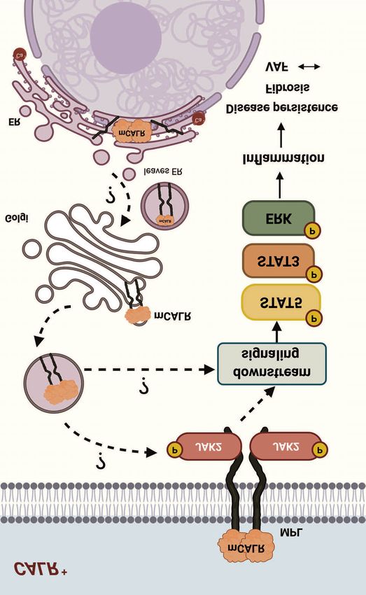
or non-competitive for ATP. In recent years, drug devel- Figure 1. Activation of JAK-STAT signaling in CALR-mutant cells is inhibited by
opment has focused on ATP-competitive inhibitors, such ruxolitinib. Schematic representation of mutant CALR (mCALR) trafficking in
MPN cells showing inhibition by ruxolitinib. The exact mechanism(s) by which
mCALR activates JAK-STAT signaling are still being elucidated. Mutant CALR
as ruxolitinib. All clinically approved JAK inhibitors (rux-
olitinib, fedratinib) bind and stabilize the kinase-active leaves the endoplasmic reticulum and is associated with MPL to promote
conformation of JAK2, and are known as “type I” homodimerization and activation of JAK2.
haematologica | 2021; 106(5) 3D.M. Ross et al.
What are the long-term outcomes for patients colony assays. New models of myelofibrosis will help to
treated with ruxolitinib? characterize the disease-propagating stem-like cells and
identify critical cell surface markers for monitoring future
The COMFORT-1 study compared ruxolitinib with disease-modifying therapies.
placebo for the treatment of myelofibrosis,38 and the
COMFORT-2 study compared ruxolitinib with best avail-
able therapy, including supportive care, hydroxyurea, and What are the possible mechanisms of
a range of other medical therapies.39 The primary endpoint ruxolitinib persistence?
in both studies was the proportion of patients achieving a
35% or greater reduction in spleen volume. The proportion A number of models have been proposed to explain rux-
of patients who achieved this response after 24 weeks of olitinib persistence, i.e., the ongoing outgrowth of mutated
ruxolitinib treatment was 42% in COMFORT-1 and 32% cells despite JAK-STAT pathway sensitivity to ruxolitinib,
in COMFORT-2.38,39 The response rates in the respective with varying levels of evidence. These include activation of
comparator arms of the two studies were 0.7% and 0%. alternative kinases not inhibited by ruxolitinib,46 epigenetic
In a subsequent analysis of a subset of patients from the mutations leading to growth advantage,26,47 phosphatase
COMFORT-2 study the response rate after 48 weeks was negative feedback inhibition (as noted with the failure of
20% (confidence interval: 5.7-43.7%) in 20 patients with a RAFK inhibitors in melanoma),48 lack of specificity for
CALR mutation versus 34% (confidence interval: 24.8- JAK2 V617F versus wild-type JAK2,10 and accumulation of
44.1%) in 100 CALR-negative patients, most of whom had p-JAK2 during exposure to ruxolitinib. The last of these
a JAK2 V617F mutation.37 Overall, there was no statistically phenomena is the most studied, with multiple lines of evi-
significant difference in splenic response rate or survival dence across several independent studies suggesting that it
between patients with and without JAK2 mutations, but is a contributory mechanism in clinically observed ruxoli-
the JAK2-positive patients had a numerically greater reduc- tinib persistence.49-51 Various therapeutic strategies have
tion in spleen size.40 JAK2 V617F is almost universally pres- been proposed in addition to targeting JAK2 (reviewed by
ent in post-polycythemia vera myelofibrosis, which is Bankar and Gupta52). Here we focus on the evidence
characterized by loss of heterozygosity and high allelic regarding persistent activation of JAK-STAT signaling and
burden,41 and can perhaps be considered to epitomize its downstream proteins, and how these phenomena may
JAK2-driven myelofibrosis. In this subgroup the hazard be targeted by current or emerging therapies.
ratio for overall survival was 0.25, compared with 0.65 in
primary myelofibrosis.40 Whether this reflects the higher
frequency of high-risk genomic lesions in primary myelofi- Why does phosphorylation of JAK2 increase
brosis or ‘on-target’ efficacy through inhibition of JAK2 during ruxolitinib treatment?
V617F remains to be established. Overall, these clinical
data emphasize that ruxolitinib is not a JAK2 mutant-spe- Initial laboratory studies studying ruxolitinib activity in
cific therapy, although patients with CALR mutations vitro noted that, paradoxically, phosphorylation of JAK2
might have mildly inferior responses. on Tyr1007/1008 located in the activation loop was
observed at increased levels in V617F-positive cells fol-
lowing prolonged exposure to ruxolitinib.49 Both the total
Does ruxolitinib eradicate cells containing level of phosphorylation and the total amount of JAK2
disease-causing driver mutations? protein were increased.49 Similar observations were also
made for the type I inhibitor “JAK inhibitor 1” and
Although ruxolitinib is now established as the standard Go6976, a protein kinase C inhibitor with potent activity
therapy for symptomatic myelofibrosis, evidence of a against JAK2.53,54 Accumulation of p-JAK2 was noted to
long-term effect on disease biology is limited. The average occur in the presence of total blockade of kinase function
reduction in JAK2 allelic burden was 7-22% after 48 weeks and inhibition of STAT and ERK phosphorylation down-
of treatment in evaluable patients;38,42 regression of fibrosis stream (Figure 2). It was noted that ruxolitinib-induced
in the marrow was seen in around 16% of patients at last phosphorylation of JAK2: (i) was staurosporine-sensitive
follow-up (median 2.2 years),43 and there was no change in and ATP-dependent; (ii) required cytokine receptor inter-
the risk of leukemic transformation. Cases of complete action and intact JH1, FERM and JH2 domains; and (iii)
hematologic or molecular remission have been reported,44 could occur in V617F+ SET2 cells in the absence of JAK1
but are very uncommon. Typically there is a gradual loss of and TYK2.50 In contrast, JAK2 wild-type cells (such as TF-
response over time, with approximately 27% of patients 1 cells) showed little or no type I inhibitor-induced loop
remaining on first-line ruxolitinib treatment after 5 years in phosphorylation after growth factor starvation. A car-
the COMFORT studies. In the subgroup of patients who boxyterminal-directed antibody could not immunopre-
achieved a 35% or greater reduction in spleen volume, the cipitate JAK2 after exposure to type I inhibitor. All these
median duration of the response was 3.2 years.43,45 findings are consistent with a conformational change in
The immunophenotype of myelofibrosis-initiating cells the kinase domain generated by ruxolitinib. Because of
has not been well studied, principally because of a lack of presumed structural flexibility, high resolution crystallo-
robust engraftment models, but in a few patients analyzed graphy data are not available for the activation loop dur-
carefully, the stem cell population appeared to reside with- ing type I inhibitor binding but, critically, activation of
in the CD34+CD38–CD90+ compartment, as determined downstream STAT phosphorylation could be repro-
using a humanized bone marrow niche.3 Flow cytometry ducibly detected following removal of the drug. This
studies suggest a high level of circulating CD34+CD38– implies that the accumulated p-JAK could act as a patho-
hematopoietic stem-like cells in patients with mutant logical signaling node as the drug level falls in patients if
CALR, but all these cells were resistant to ruxolitinib in drug is abruptly stopped or a dose missed.
4 haematologica | 2021; 106(5)Persistence of myelofibrosis treated with ruxolitinib
More recently, Tvorogov and co-authors extended these ceals Tyr1007/1008 from phosphatase access, and ruxoli-
findings to show that accumulation of p-JAK2 is due to tinib-bound p-JAK2 is no longer susceptible to dephospho-
resistance of p-JAK2 to ubiquitination and degradation rylation or degradation.
while bound to ruxolitinib.51 Specifically, immunoprecipi- Interestingly, primary CALR-mutant cells did not exhibit
tation with an anti-p-JAK2 antibody produced a band for either JAK2 phosphorylation in the presence of ruxolitinib
p-JAK2 in untreated cells, but not in ruxolitinib-treated or striking withdrawal signaling to the same degree as the
cells, consistent with an altered conformation induced by JAK2 V617F samples. CALR-mutant myelofibrosis cells
the drug. Accordingly, it was noted that cells exhibiting showed undetectable levels of activated JAK2
delayed phosphorylation kinetics (i.e., those with wild- Tyr1007/1008 phosphorylation in the presence of ruxoli-
type JAK2) were significantly more sensitive to ruxolitinib tinib and in some CALR+ samples and in a CALR-mutated
cytotoxicity and growth inhibition than cells with rapid cell line (MARIMO) total JAK2 protein was difficult to
phosphorylation kinetics (cells with JAK2 V617F). This detect, if not completely absent. This is consistent with a
was an important observation that raised the possibility number of emerging reports using CALR-mutated models
that ruxolitinib allows drug-bound JAK protein to escape of myelofibrosis55-57 suggesting fundamental differences in
the negative feedback loops of dephosphorylation and the nature of JAK activation in myelofibrosis patients with
degradation (Figure 2B). mutated CALR (Figure 1).
To test this, recombinant JAK2 kinase was treated with
a phosphatase in the presence of ruxolitinib. Ruxolitinib
blocked the dephosphorylation by PTP1B for up to 20 h Lessons from the clinic
whereas reduction of Tyr1007/1008 phosphorylation nor-
mally occurred within 2 h.51 In keeping with this, ruxoli- Ruxolitinib discontinuation syndrome
tinib prevented any detectable ubiquitination of JAK2 after Interruption of ruxolitinib treatment typically leads to
cytokine stimulation. These results suggested that binding recrudescence of cytokine-mediated symptoms within a
of ruxolitinib induces a conformational change that con- week,38 often accompanied by increasing spleen size,
A B C
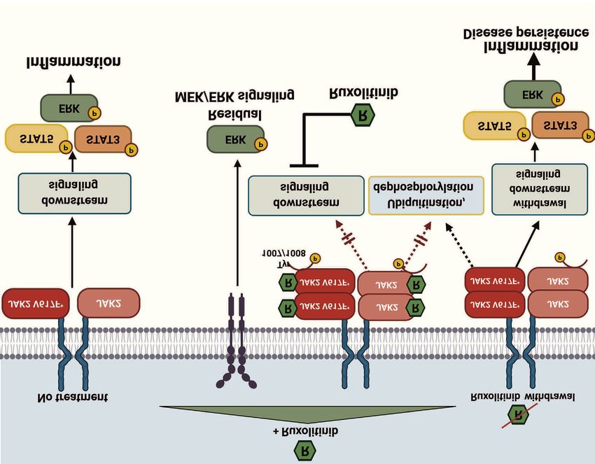
Figure 2. Signaling in JAK2 V617F cells before, during, and after discontinuation of ruxolitinib. Schematic representation of JAK-STAT activation in JAK2 V617F myelo-
proliferative neoplastic cells. (A) JAK2 V617F leads to increased signaling through STAT5/STAT3/ERK leading to proliferation and inflammation. (B) In the presence
of a type 1 JAK2 inhibitor, such as ruxolitinib, signaling through the JAK-STAT pathway is abrogated, but so too is ubiquitination and degradation of JAK2, leading to
accumulation of p-JAK2. Signaling through MEK/ERK remains activated and contributes to disease persistence. (C) Ruxolitinib discontinuation leads to transiently
increased signaling through the accumulated pool of p-JAK2 with inflammatory symptoms.
haematologica | 2021; 106(5) 5D.M. Ross et al.
highlighting the persistence of the underlying disease. The Ruxolitinib wash-out and rechallenge
accumulation of p-JAK2 likely accounts for the prompt A number of cases have been reported in which a ‘drug
onset of withdrawal symptoms when ruxolitinib is holiday’ led to restoration of ruxolitinib response upon re-
stopped, since the increased p-JAK2 will rapidly lead to challenge,70,71 as predicted by the experimental models of
downstream signaling in the absence of the drug. ruxolitinib persistence and wash-out. When ruxolitinib is
Ruxolitinib has a short half-life of approximately 3 h,58 so withdrawn the accumulated p-JAK2 becomes susceptible
the drug is washed out rapidly if dosing is interrupted. again to ubiquitination and degradation, so that after a
Although the accumulation of p-JAK2 is likely to be a class wash-out period the untreated cell biology is restored for
effect of type 1 JAK inhibitors, we are not aware of any a variable period of time. Understanding this phenome-
reports of discontinuation syndrome with fedratinib, non has important implications for the design of clinical
momelotinib, or pacritinib. Both fedratinib and pacritinib trials of second-line agents after ruxolitinib. It is often in
have very long half-lives of more than 24 h.59,60 Delayed the patient’s interest to minimize the interval between
clearance of these drugs may reduce the risk of withdraw- stopping ruxolitinib and starting a second-line agent,
al phenomena. Momelotinib has a half-life of 4-6 h,61 sim- because of the increase in symptom burden that common-
ilar to that of ruxolitinib, so withdrawal phenomena ly occurs. However, if baseline response assessments for
might be predicted to occur with abrupt discontinuation the second-line agent are undertaken too early there may
of momelotinib. be further deterioration after the ‘baseline’ assessment. In
In most patients who discontinue ruxolitinib there is a the randomized FREEDOM-2 study, wash-out and
return to near baseline severity of symptoms, but in rare resumption of ruxolitinib will be compared to wash-out of
cases there are life-threatening manifestations thought to ruxolitinib followed by institution of fedratinib
be due to an exaggerated inflammatory response. (NCT03952039). This study will provide important new
Ruxolitinib discontinuation syndrome is a diagnosis of data on the effects of ruxolitinib rechallenge.
exclusion based on a temporal relationship between drug
withdrawal and onset of clinical manifestations, which Inhibition of JAK1 versus JAK2 versus both
can appear from less than 24 h to up to 3 weeks after dis- Many of the symptoms of myelofibrosis are related to
continuation. In the original phase I/II trial, three of 47 increased levels of pro-inflammatory cytokines, including
patients who discontinued ruxolitinib developed acute interleukin-6 and tumor necrosis factor-α. Symptomatic
respiratory distress syndrome.62 In the phase III COM- improvement in response to ruxolitinib is correlated with
FORT-I study, one patient developed acute respiratory dis- a reduction in cytokine levels.72 Importantly, many
tress, pyrexia and splenic infarction following ruxolitinib cytokines signal through JAK1 and suppression of JAK1
discontinuation.38 Coltro and co-workers described a case signaling by dual inhibitors, such as ruxolitinib and
of JAK2 V617F essential thrombocythemia evolving to momelotinib, may contribute to the symptomatic benefit
acute myeloid leukemia with acute respiratory distress observed with these treatments. A selective JAK1
syndrome developing after ruxolitinib was discontinued, inhibitor, itacitinib (INCB039110), has been tested in a
and improving within 48 h of re-introduction of the drug.63 phase II clinical trial involving 87 patients with myelofi-
Other reports include a patient who developed tumor brosis.73 Symptomatic improvement was comparable to
lysis-like syndrome,64 a case of acute respiratory failure,65 that seen with ruxolitinib (total symptom score reduced
and a case of acute respiratory distress syndrome that by 50% at 24 weeks in 49% of patients versus 46% in the
twice resolved after ruxolitinib re-introduction.66 COMFORT-I study) whereas the splenic responses were
Of note, of the nine published cases of life-threatening less (≥35% reduction in spleen volume in 17% of patients
ruxolitinib discontinuation syndrome reported in the liter- at 24 weeks versus 32-42% in the COMFORT studies).
ature, all were reported to have a JAK2 mutation. This is a Conversely, a 50% reduction in total symptom score was
disproportionate enrichment compared to the prevalence seen in 36% of patients treated with the JAK2-selective
of the CALR mutation (~35% of cases of myelofibrosis). inhibitor, fedratinib.74
Further research into mutation-specific side effects of It may be an oversimplification that JAK1 inhibition
kinase inhibitors is warranted. mediates improvement in inflammatory symptoms and
Some experts recommend tapering the ruxolitinib dose JAK2 inhibition mediates cytoreductive effects. There
rather than abrupt discontinuation, especially when the may be some crosstalk between JAK1 and JAK2, or splenic
reason for the therapy interruption is an adverse event responses could perhaps be due to inhibition of cytokine-
other than cytopenia.67,68 One of the commonest causes of mediated recruitment of cells to sites of extramedullary
ruxolitinib discontinuation is infection.69 By stabilizing the hematopoiesis.
active conformation of JAK2 in a manner that prevents
dephosphorylation and degradation, type I inhibitors may
rarely induce a signaling state that heuristically resembles Secondary resistance to ruxolitinib: comparison
a cytokine storm (Figure 2C).51 Experimental co-culture of with chronic myeloid leukemia
JAK2 V617F cells with increased levels of cytokines in the
medium promoted the accumulation of p-JAK2, since sig- Secondary resistance to TKI therapy is perhaps best
naling through cytokine receptors is still capable of induc- understood in chronic myeloid leukemia. Resistance often
ing p-JAK2 in the presence of ruxolitinib. Intriguingly, acti- occurs relatively abruptly and leads to near-complete loss
vation of JAK1 by interleukin-3 also led to phosphoryla- of therapeutic benefit, typically over a number of months.
tion of JAK2, suggesting that both JAK1 and JAK2 may be In contrast, loss of response to ruxolitinib in myelofibrosis
involved in withdrawal signaling.51 These findings provide is characterized by a gradual waning of the initial thera-
a biological rationale for why inflammation and discontin- peutic benefit, sometimes associated with late-emerging
uation of ruxolitinib could interact to cause more severe cytopenia that is likely to represent progression of the
withdrawal phenomena. underlying disease rather than direct toxicity of the TKI.
6 haematologica | 2021; 106(5)Persistence of myelofibrosis treated with ruxolitinib
In chronic myeloid leukemia, around half of cases of kinases, such as BCR-ABL1 and PCM1-JAK2) it does not
secondary resistance are associated with the emergence of strongly favor the active conformation. In contrast, the
single amino acid changes in the BCR-ABL1 kinase KIT D816V mutation that is commonly found in systemic
domain which impair binding of the TKI or stabilize the mastocytosis leads to a bias in favor of the active confor-
active conformation of the protein.75 Ruxolitinib-resistant mation of the stem cell factor receptor (which KIT
kinase domain mutations have been isolated using in vitro encodes). Midostaurin and avapritinib (both type 1
mutagenesis screening, and such mutations have also been inhibitors) have significant clinical activity against KIT
observed clinically in patients with acute lymphoblastic D816V: they show greater potency against KIT D816V
leukemia treated with ruxolitinib.76 However, fewer than than against wild-type KIT, whereas the converse is true
50% of resistant clones after saturation mutagenesis in the for type 2 inhibitors, such as imatinib and sunitinib.78 An
presence of ruxolitinib harbor additional mutations in experimental type 2 JAK inhibitor, CHZ868, did not result
JAK2.49 Fedratinib, another ATP-competitive JAK2 in accumulation of p-JAK2 and led to more potent sup-
inhibitor in clinical use, appears to be less susceptible to pression of myelofibrosis cells.49,51 This compound is not
this pattern of resistance because it inhibits not only ATP being developed for clinical use, but these pre-clinical data
binding, but also the binding of peptide substrates to suggest that the development of type 2 JAK inhibitors for
JAK2.77 However, JAK2 kinase domain mutations have not clinical use might offer advantages over the available TKI
yet been observed in clinical samples from myelofibrosis for myelofibrosis. A potential risk of this approach is
patients treated with JAK inhibitors, which could indicate greater suppression of normal hematopoiesis.
either incomplete inhibition of JAK2 or that myelofibrosis The JH2 pseudokinase domain of JAK2 binds ATP, and
is not critically dependent on the kinase activity of JAK2 mutation of certain residues abrogates ATP binding, as
in the way that chronic myeloid leukemia is addicted to a well as leading to loss of the activated JH1 tyrosine kinase
fusion oncogene. activity in V617F.79 In a mouse model of JAK2 V617F MPN,
co-mutation with K581A prevented the development of
polycythemia. Notably, mutation of the same residue had
Strategies to improve inhibition of JAK-STAT little or no effect on cytokine signaling through wild-type
signaling JAK2, potentially opening the opportunity for the devel-
opment of mutation-specific allosteric inhibitors that tar-
Here we discuss various approaches that might be get this site.
employed to achieve more complete inhibition of JAK- JAK2 is a client protein of Hsp90, and inhibition of
STAT signaling (summarized in Table 1) with the aim of Hsp90 leads to degradation of JAK2 (including p-JAK2).
improving treatment response, and ultimately finding Combining ruxolitinib with an Hsp90 inhibitor was more
treatments with greater disease-modifying potential. Type effective than ruxolitinib monotherapy in a mouse model
2 kinase inhibitors, such as the BCR-ABL1 inhibitor, ima- of myelofibrosis.80 A clinical trial using the Hsp90 inhibitor
tinib, are ATP-competitive inhibitors that bind the inactive AUY922 was terminated prematurely because of safety
conformation of the kinase. All of the JAK2 inhibitors that concerns, despite evidence of splenic responses.81 Other
have been tested clinically to date for myelofibrosis are studies using the same drug documented a high frequency
type 1 inhibitors. Since JAK2 V617F is a weak activating of visual alterations, including night blindness.82 An alter-
mutation (relative to constitutive activation of fusion native approach to target the accumulation of p-JAK2 is to
Table 1. Possible strategies to inhibit JAK2 and downstream signaling in myeloproliferative neoplasms.
Mechanism of action Examples Notes
Current standard of care
Type 1 inhibitors ATP-competitive inhibitors that bind the Ruxolitinib Lead to accumulation of p-JAK2
active conformation of JAK2 Fedratinib Limited disease-modifying potential
Momelotinib
Pacritinib
Alternative JAK inhibitors
Type 2 inhibitors ATP-competitive inhibitors that bind CHZ868 Do not lead to accumulation of p-JAK2
the active conformation of JAK2 BBT594
Allosteric inhibitors Non-ATP-competitive inhibitors of LS104 Could confer greater specificity for JAK2
the pseudokinase domain or substrate binding V617F over wild-type JAK2
Drugs to degrade JAK2
Hsp90 inhibitors Inhibit chaperone function to expose AUY922 Clinical development discontinued
JAK2 to degradation because of ocular toxicity
De-ubiquitinase inhibitors Small molecules that promote WP1130 Could reduce accumulation of p-JAK2
ubiquitination without specificity for JAK2 in combination with a type 1 JAK2 inhibitor
Proteolysis-activating chimeras Designed to target specific proteins - Could reduce accumulation of p-JAK2
(PROTAC) to degrade in combination with a type 1 JAK2 inhibitor
Inhibitors of downstream signaling
MEK/ERK inhibitors Inhibitors of bypass signaling in Trametinib Synergistic with type 1 JAK2 inhibitor
the presence of JAK inhibitor Binimetinib in experimental models
Ulixertinib Not tested clinically in MPN
Approaches that have been used clinically (in any disease) are shown in bold.
haematologica | 2021; 106(5) 7D.M. Ross et al.
use a proteolysis-targeting chimera (PROTAC). These inhibitors may contribute to disease persistence by pre-
small-molecule drugs contain two functional domains: venting JAK2 de-phosphorylation and proteasomal degra-
one binds the target protein and the second engages an E3 dation, allowing heterodimerization of JAK2 with JAK1 or
ubiquitin ligase, thereby triggering proteasomal degrada- TYK2. Type 2 inhibitors of JAK2 do not cause this phe-
tion of the target protein. JAK family PROTAC have been nomenon and have greater impact on disease biology in a
developed that recruit inhibitor of apoptosis protein lead- mouse model. The V617F mutation leads to a highly
ing to proteasomal degradation.83 An inhibitor of the de- inflexible substitution, which may be indicative of a neo-
ubiquitinase, USP9X, has been shown to accelerate the morphic change in function, rather than non-specific over-
degradation of JAK2 in vitro.84 Whether any of these activation. Several lines of evidence suggest that it may be
approaches could selectively target pathological accumu- possible to exploit the unique properties of the mutant
lation of p-JAK2 in patients with myelofibrosis remains to pseudokinase to develop mutation-specific JAK2
be tested. inhibitors that spare normal hematopoiesis. Whereas cur-
One of the pathways activated downstream of JAK2 in rent clinical trials are mostly targeting cooperating path-
MPN is RAS/RAF/MEK/ERK. Ruxolitinib treatment of a ways or pathways downstream of JAK2, these observa-
cell line expressing JAK2 V617F inhibited downstream tions suggest that novel approaches to the targeting of
phosphorylated ERK1 and ERK2 (p-ERK1/2), but ruxoli- JAK2 could lead to substantial benefits for patients with
tinib treatment of a V617F mouse MPN model did not, myelofibrosis.
despite effective suppression of other proteins down-
stream of JAK2.46 Persistence of MEK/ERK signaling in vivo Disclosures
was found to be due to activation of PDGFRA by PDGF- DMR has received research funding and honoraria from
BB signaling as a bypass pathway that enables persistence Novartis and BMS/Celgene unrelated to the current work. JJB,
of MPN cells despite effective inhibition of JAK2 kinase DTv and DTh declare that they have no conflicts of interest.
activity. Combined inhibition of JAK2 and MEK with rux-
olitinib and binimetinib (an approved treatment for RAF- Contributions
mutant melanoma) was more efficient than either TKI DMR and DTh reviewed the literature and wrote the paper.
alone in achieving regression of splenomegaly, fibrosis and JJB and DTv critically reviewed the content and wrote the paper.
JAK2 V617F allele burden.46
Acknowledgments
We thank Suraiya Onnesha for assistance with the figures.
Conclusion
Funding
Despite the symptomatic benefits of currently available Research support for DMR was provided through the Medical
JAK2 inhibitors, there is a need for agents that have a Research Future Fund and The Hospital Research Foundation.
greater effect on the disease clone and its natural history. Research support for JJB was provided by the National Health
JAK2 remains a crucial target in myelofibrosis, and dele- and Medical Research Council (APP1113577; APP1121755;
tion of JAK2 in a mouse model of myelofibrosis substan- APP1122999). Research support for DTh was provided through
tially abrogates disease manifestations. Data from several the National Health and Medical Research Council
groups have shown that a conformational change in JAK2 (APP1182564), Medical Research Future Fund, The Hospital
induced by the binding of ruxolitinib and other type 1 Research Foundation and a CSL Centenary Fellowship.
References imatinib or interferon alfa plus cytarabine in 12. Kershaw NJ, Murphy JM, Lucet IS, Nicola
newly diagnosed chronic myeloid leukemia. NA, Babon JJ. Regulation of Janus kinases by
N Engl J Med. 2003;349(15):1423-1432. SOCS proteins. Biochem Soc Trans.
1. Wernig G, Mercher T, Okabe R, Levine RL, 7. Kantarjian HM, Hochhaus A, Saglio G, et al. 2013;41(4):1042-1047.
Lee BH, Gilliland DG. Expression of Nilotinib versus imatinib for the treatment 13. Liau NPD, Laktyushin A, Lucet IS, et al. The
Jak2V617F causes a polycythemia vera-like of patients with newly diagnosed chronic molecular basis of JAK/STAT inhibition by
disease with associated myelofibrosis in a phase, Philadelphia chromosome-positive, SOCS1. Nat Commun. 2018;9(1):1558.
murine bone marrow transplant model. chronic myeloid leukaemia: 24-month mini- 14. Lv K, Jiang J, Donaghy R, et al. CBL family
Blood. 2006;107(11):4274-4281. mum follow-up of the phase 3 randomised E3 ubiquitin ligases control JAK2 ubiquitina-
2. Nangalia J, Massie CE, Baxter EJ, et al. ENESTnd trial. Lancet Oncol. 2011;12(9): tion and stability in hematopoietic stem cells
Somatic CALR mutations in myeloprolifera- 841-851. and myeloid malignancies. Genes Dev.
tive neoplasms with nonmutated JAK2. N 8. Cortes JE, Saglio G, Kantarjian HM, et al. 2017;31(10):1007-1023.
Engl J Med. 2013;369(25):2391-2405. Final 5-year study results of DASISION: the 15. Chou DH, Vetere A, Choudhary A, et al.
3. Reinisch A, Thomas D, Corces MR, et al. A Dasatinib Versus Imatinib Study in Kinase-independent small-molecule inhibi-
humanized bone marrow ossicle xenotrans- Treatment-Naive Chronic Myeloid tion of JAK-STAT signaling. J Am Chem Soc.
plantation model enables improved engraft- Leukemia Patients trial. J Clin Oncol. 2015;137(24):7929-7934.
ment of healthy and leukemic human 2016;34(20):2333-2340. 16. Grinfeld J, Nangalia J, Baxter EJ, et al.
hematopoietic cells. Nat Med. 2016;22(7): 9. Stark GR, Darnell JE Jr. The JAK-STAT path- Classification and personalized prognosis in
812-821. way at twenty. Immunity. 2012;36(4):503- myeloproliferative neoplasms. N Engl J
4. Baxter EJ, Scott LM, Campbell PJ, et al. 514. Med. 2018;379(15):1416-1430.
Acquired mutation of the tyrosine kinase 10. Liau NPD, Laktyushin A, Morris R, et al. 17. Lu X, Huang LJ, Lodish HF. Dimerization by
JAK2 in human myeloproliferative disor- Enzymatic characterization of wild-type a cytokine receptor is necessary for constitu-
ders. Lancet. 2005;365(9464):1054-1061. and mutant Janus kinase 1. Cancers (Basel). tive activation of JAK2V617F. J Biol Chem.
5. Kralovics R, Passamonti F, Buser AS, et al. A 2019;11(11):1701. 2008;283(9):5258-5266.
gain-of-function mutation of JAK2 in myelo- 11. Kershaw NJ, Murphy JM, Liau NP, et al. 18. Lu X, Levine R, Tong W, et al. Expression of
proliferative disorders. N Engl J Med. SOCS3 binds specific receptor-JAK com- a homodimeric type I cytokine receptor is
2005;352(17):1779-1790. plexes to control cytokine signaling by direct required for JAK2V617F-mediated transfor-
6. Hughes TP, Kaeda J, Branford S, et al. kinase inhibition. Nat Struct Mol Biol. mation. Proc Natl Acad Sci U S A. 2005;102
Frequency of major molecular responses to 2013;20(4):469-476. (52):18962-18967.
8 haematologica | 2021; 106(5)Persistence of myelofibrosis treated with ruxolitinib
19. Ungureanu D, Wu J, Pekkala T, et al. The activation specifically in CALR mutated (V617F, T875N and K539L) counteracts
pseudokinase domain of JAK2 is a dual- cells: perspectives for MPN therapy. Blood. cytokine-independent signaling. Oncogene.
specificity protein kinase that negatively 2018;132(Suppl 1):4. 2009;28(34):3069-3080.
regulates cytokine signaling. Nat Struct Mol 37. Guglielmelli P, Rotunno G, Bogani C, et al. 54. Grandage VL, Everington T, Linch DC,
Biol. 2011;18(9):971-976. Ruxolitinib is an effective treatment for Khwaja A. Go6976 is a potent inhibitor of
20. Jeong EG, Kim MS, Nam HK, et al. Somatic CALR-positive patients with myelofibrosis. the JAK 2 and FLT3 tyrosine kinases with
mutations of JAK1 and JAK3 in acute Br J Haematol. 2016;173(6):938-940. significant activity in primary acute myeloid
leukemias and solid cancers. Clin Cancer 38. Verstovsek S, Mesa RA, Gotlib J, et al. A leukaemia cells. Br J Haematol. 2006;135
Res. 2008;14(12):3716-3721. double-blind, placebo-controlled trial of rux- (3):303-316.
21. Staerk J, Kallin A, Demoulin JB, Vainchenker olitinib for myelofibrosis. N Engl J Med. 55. Marty C, Pecquet C, Nivarthi H, et al.
W, Constantinescu SN. JAK1 and Tyk2 acti- 2012;366(9):799-807. Calreticulin mutants in mice induce an MPL-
vation by the homologous polycythemia 39. Harrison C, Kiladjian JJ, Al-Ali HK, et al. JAK dependent thrombocytosis with frequent
vera JAK2 V617F mutation: cross-talk with inhibition with ruxolitinib versus best avail- progression to myelofibrosis. Blood.
IGF1 receptor. J Biol Chem. 2005;280(51): able therapy for myelofibrosis. N Engl J 2016;127(10):1317-1324.
41893-41899. Med. 2012;366(9):787-798. 56. Balligand T, Achouri Y, Pecquet C, et al.
22. Wilmes S, Hafer M, Vuorio J, et al. 40. Verstovsek S, Mesa RA, Gotlib J, et al. The Pathologic activation of thrombopoietin
Mechanism of homodimeric cytokine recep- clinical benefit of ruxolitinib across patient receptor and JAK2-STAT5 pathway by
tor activation and dysregulation by onco- subgroups: analysis of a placebo-controlled, frameshift mutants of mouse calreticulin.
genic mutations. Science. 2020;367(6478): phase III study in patients with myelofibro- Leukemia. 2016;30(8):1775-1778.
643-652. sis. Br J Haematol. 2013;161(4):508-516. 57. Nivarthi H, Chen D, Cleary C, et al.
23. Moliterno AR, Williams DM, Rogers O, 41. Godfrey AL, Chen E, Pagano F, et al. Thrombopoietin receptor is required for the
Isaacs MA, Spivak JL. Phenotypic variability JAK2V617F homozygosity arises commonly oncogenic function of CALR mutants.
within the JAK2 V617F-positive MPD: roles and recurrently in PV and ET, but PV is char- Leukemia. 2016;30(8):1759-1763.
of progenitor cell and neutrophil allele bur- acterized by expansion of a dominant 58. Shi JG, Chen X, McGee RF, et al. The phar-
dens. Exp Hematol. 2008;36(11):1480-1486. homozygous subclone. Blood. 2012;120(13): macokinetics, pharmacodynamics, and safe-
24. Stein BL, Saraf S, Sobol U, et al. Age-related 2704-2707. ty of orally dosed INCB018424 phosphate in
differences in disease characteristics and 42. Cervantes F, Vannucchi AM, Kiladjian JJ, et healthy volunteers. J Clin Pharmacol.
clinical outcomes in polycythemia vera. al. Three-year efficacy, safety, and survival 2011;51(12):1644-1654.
Leuk Lymphoma. 2013;54(9):1989-1995. findings from COMFORT-II, a phase 3 59. Zhang M, Xu C, Ma L, et al. Effect of food
25. Gangat N, Tefferi A. Myelofibrosis biology study comparing ruxolitinib with best avail- on the bioavailability and tolerability of the
and contemporary management. Br J able therapy for myelofibrosis. Blood. JAK2-selective inhibitor fedratinib
Haematol. 2020;191(2):152-170. 2013;122(25):4047-4053. (SAR302503): results from two phase I stud-
26. Ortmann CA, Kent DG, Nangalia J, et al. 43. Harrison CN, Vannucchi AM, Kiladjian JJ, et ies in healthy volunteers. Clin Pharmacol
Effect of mutation order on myeloprolifera- al. Long-term findings from COMFORT-II, a Drug Dev. 2015;4(4):315-321.
tive neoplasms. N Engl J Med. 2015;372(7): phase 3 study of ruxolitinib vs best available 60. Younes A, Romaguera J, Fanale M, et al.
601-612. therapy for myelofibrosis. Leukemia. Phase I study of a novel oral Janus kinase 2
27. Klampfl T, Gisslinger H, Harutyunyan AS, et 2016;30(8):1701-1707. inhibitor, SB1518, in patients with relapsed
al. Somatic mutations of calreticulin in 44. Masarova L, Wang W, Newberry KJ, lymphoma: evidence of clinical and biologic
myeloproliferative neoplasms. N Engl J Kantarjian H, Verstovsek S. Complete remis- activity in multiple lymphoma subtypes. J
Med. 2013;369(25):2379-2390. sion in a patient with JAK2- and IDH2-posi- Clin Oncol. 2012;30(33):4161-4167.
28. Eder-Azanza L, Navarro D, Aranaz P, Novo tive myelofibrosis. Blood. 2016;128(6):877- 61. Pardanani A, Laborde RR, Lasho TL, et al.
FJ, Cross NC, Vizmanos JL. Bioinformatic 880. Safety and efficacy of CYT387, a JAK1 and
analyses of CALR mutations in myeloprolif- 45. Verstovsek S, Gotlib J, Mesa RA, et al. Long- JAK2 inhibitor, in myelofibrosis. Leukemia.
erative neoplasms support a role in signal- term survival in patients treated with ruxoli- 2013;27(6):1322-1327.
ing. Leukemia. 2014;28(10):2106-2109. tinib for myelofibrosis: COMFORT-I and -II 62. Tefferi A, Pardanani A. Serious adverse
29. Tefferi A, Lasho TL, Finke C, et al. Type 1 vs pooled analyses. J Hematol Oncol. events during ruxolitinib treatment discon-
type 2 calreticulin mutations in primary 2017;10(1):156. tinuation in patients with myelofibrosis.
myelofibrosis: differences in phenotype and 46. Stivala S, Codilupi T, Brkic S, et al. Targeting Mayo Clin Proc. 2011;86(12):1188-1191.
prognostic impact. Leukemia. 2014;28(7): compensatory MEK/ERK activation increas- 63. Coltro G, Mannelli F, Guglielmelli P, Pacilli
1568-1570. es JAK inhibitor efficacy in myeloprolifera- A, Bosi A, Vannucchi AM. A life-threatening
30. Guglielmelli P, Lasho TL, Rotunno G, et al. tive neoplasms. J Clin Invest. 2019;129(4): ruxolitinib discontinuation syndrome. Am J
MIPSS70: Mutation-enhanced International 1596-1611. Hematol. 2017;92(8):833-838.
Prognostic Score System for transplantation- 47. Nangalia J, Nice FL, Wedge DC, et al. 64. Dai T, Friedman EW, Barta SK. Ruxolitinib
age patients with primary myelofibrosis. J DNMT3A mutations occur early or late in withdrawal syndrome leading to tumor
Clin Oncol. 2018;36(4):310-318. patients with myeloproliferative neoplasms lysis. J Clin Oncol. 2013;31(29):e430-432.
31. Clinton A, McMullin MF. The calreticulin and mutation order influences phenotype. 65. Beauverd Y, Samii K. Acute respiratory dis-
gene and myeloproliferative neoplasms. J Haematologica. 2015;100(11):e438-442. tress syndrome in a patient with primary
Clin Pathol. 2016;69(10):841-845. 48. Hatzivassiliou G, Song K, Yen I, et al. RAF myelofibrosis after ruxolitinib treatment dis-
32. Elf S, Abdelfattah NS, Baral AJ, et al. Defining inhibitors prime wild-type RAF to activate continuation. Int J Hematol. 2014;100(5):
the requirements for the pathogenic interac- the MAPK pathway and enhance growth. 498-501.
tion between mutant calreticulin and MPL in Nature. 2010;464(7287):431-435. 66. Herman DD, Kempe CB, Thomson CC,
MPN. Blood. 2018;131(7):782-786. 49. Koppikar P, Bhagwat N, Kilpivaara O, et al. McCallister JW. Recurrent hypoxemic respi-
33. Elf S, Abdelfattah NS, Chen E, et al. Mutant Heterodimeric JAK-STAT activation as a ratory failure. Beyond the usual suspects.
calreticulin requires both its mutant C-ter- mechanism of persistence to JAK2 inhibitor Ann Am Thorac Soc. 2014;11(7):1145-1148.
minus and the thrombopoietin receptor for therapy. Nature. 2012;489(7414):155-159. 67. Harrison C, Mesa R, Ross D, et al. Practical
oncogenic transformation. Cancer Discov. 50. Andraos R, Qian Z, Bonenfant D, et al. management of patients with myelofibrosis
2016;6(4):368-381. Modulation of activation-loop phosphoryla- receiving ruxolitinib. Expert Rev Hematol.
34. Pecquet C, Chachoua I, Roy A, et al. tion by JAK inhibitors is binding mode 2013;6(5):511-523.
Calreticulin mutants as oncogenic rogue dependent. Cancer Discov. 2012;2(6):512- 68. National Comprehensive Cancer Network.
chaperones for TpoR and traffic-defective 523. NCCN Clinical Practice Guidelines in
pathogenic TpoR mutants. Blood. 2019;133 51. Tvorogov D, Thomas D, Liau NPD, et al. Oncology: Myeloproliferative Neoplasms.
(25):2669-2681. Accumulation of JAK activation loop phos- 2020 [accessed August 5, 2020].
35. Sollazzo D, Forte D, Polverelli N, et al. phorylation is linked to type I JAK inhibitor 69. Palandri F, Breccia M, Bonifacio M, et al. Life
Circulating calreticulin is increased in withdrawal syndrome in myelofibrosis. Sci after ruxolitinib: reasons for discontinuation,
myelofibrosis: correlation with interleukin-6 Adv. 2018;4(11):eaat3834. impact of disease phase, and outcomes in
plasma levels, bone marrow fibrosis, and 52. Bankar A, Gupta V. Investigational non-JAK 218 patients with myelofibrosis. Cancer.
splenomegaly. Mediators Inflamm. 2016; inhibitors for chronic phase myelofibrosis. 2020;126(6):1243-1252.
2016:5860657. Expert Opin Investig Drugs. 2020;29(5):461- 70. Gisslinger H, Schalling M, Gisslinger B,
36. Pecquet C, Balligand T, Chachoua I, et al. 474. Skrabs C, Mullauer L, Kralovics R.
Secreted mutant calreticulins as rogue 53. Haan S, Wuller S, Kaczor J, et al. SOCS- Restoration of response to ruxolitinib upon
cytokines trigger thrombopoietin receptor mediated downregulation of mutant Jak2 brief withdrawal in two patients with
haematologica | 2021; 106(5) 9D.M. Ross et al.
myelofibrosis. Am J Hematol. 2014;89(3): GIMEMA Working Party on Chronic 2075-2083.
344-346. Myeloid Leukemia. Clin Cancer Res. 81. Hobbs GS, Hanasoge Somasundara AV,
71. Gerds A, Su D, Martynova A, et al. 2006;12(24):7374-7379. Kleppe M, et al. Hsp90 inhibition disrupts
Ruxolitinib rechallenge can improve consti- 76. Sadras T, Heatley SL, Kok CH, et al. A novel JAK-STAT signaling and leads to reductions
tutional symptoms and splenomegaly in somatic JAK2 kinase-domain mutation in in splenomegaly in patients with myelopro-
patients with myelofibrosis: a case series. pediatric acute lymphoblastic leukemia with liferative neoplasms. Haematologica.
Clin Lymphoma Myeloma Leuk. 2018;18 rapid on-treatment development of LOH. 2018;103(1):e5-e9.
(11):e463-e468. Cancer Genet. 2017;216-217:86-90. 82. Johnson ML, Yu HA, Hart EM, et al. Phase
72. Verstovsek S, Kantarjian H, Mesa RA, et al. 77. Kesarwani M, Huber E, Kincaid Z, et al. I/II study of HSP90 inhibitor AUY922 and
Safety and efficacy of INCB018424, a JAK1 Targeting substrate-site in Jak2 kinase pre- erlotinib for EGFR-mutant lung cancer with
and JAK2 inhibitor, in myelofibrosis. N Engl vents emergence of genetic resistance. Sci acquired resistance to epidermal growth fac-
J Med. 2010;363(12):1117-1127. Rep. 2015;5:14538. tor receptor tyrosine kinase inhibitors. J Clin
73. Mascarenhas JO, Talpaz M, Gupta V, et al. 78. Evans EK, Gardino AK, Kim JL, et al. A pre- Oncol. 2015;33(15):1666-1673.
Primary analysis of a phase II open-label cision therapy against cancers driven by 83. Shah RR, Redmond JM, Mihut A, et al. Hi-
trial of INCB039110, a selective JAK1 KIT/PDGFRA mutations. Sci Transl Med. JAK-ing the ubiquitin system: the design and
inhibitor, in patients with myelofibrosis. 2017;9(414):eaao1690. physicochemical optimisation of JAK
Haematologica. 2017;102(2):327-335. 79. Hammaren HM, Ungureanu D, Grisouard J, PROTACs. Bioorg Med Chem. 2020;28(5):
74. Pardanani A, Harrison C, Cortes JE, et al. Skoda RC, Hubbard SR, Silvennoinen O. 115326.
Safety and efficacy of fedratinib in patients ATP binding to the pseudokinase domain of 84. Akiyama H, Umezawa Y, Watanabe D, et
with primary or secondary myelofibrosis: a JAK2 is critical for pathogenic activation. al. Inhibition of USP9X downregulates
randomized clinical trial. JAMA Oncol. Proc Natl Acad Sci U S A. 2015;112(15): JAK2-V617F and induces apoptosis syner-
2015;1(5):643-651. 4642-4647. gistically with BH3 mimetics preferentially
75. Soverini S, Colarossi S, Gnani A, et al. 80. Bhagwat N, Koppikar P, Keller M, et al. in ruxolitinib-persistent JAK2-V617F-posi-
Contribution of ABL kinase domain muta- Improved targeting of JAK2 leads to tive leukemic cells. Cancers (Basel). 2020;
tions to imatinib resistance in different sub- increased therapeutic efficacy in myelopro- 12(2):406.
sets of Philadelphia-positive patients: by the liferative neoplasms. Blood. 2014;123(13):
10 haematologica | 2021; 106(5)You can also read