Dual Role of Lysophosphatidic Acid Receptor 2 (LPA2) in Amyotrophic Lateral Sclerosis - Frontiers
←
→
Page content transcription
If your browser does not render page correctly, please read the page content below
ORIGINAL RESEARCH
                                                                                                                                             published: 25 March 2021
                                                                                                                                       doi: 10.3389/fncel.2021.600872

                                           Dual Role of Lysophosphatidic Acid
                                           Receptor 2 (LPA2) in Amyotrophic
                                           Lateral Sclerosis
                                           Maria Puigdomenech-Poch 1,2 , Anna Martínez-Muriana 1 , Pol Andrés-Benito 2,3 ,
                                           Isidre Ferrer 2,3 , Jerold Chun 4 and Rubèn López-Vales 1,2 *
                                           1
                                            Departament de Biologia Cellular, Fisiologia i Immunologia, Institut de Neurociències, Centro de Investigación Biomédica en
                                           Red sobre Enfermedades Neurodegenerativas (CIBERNED), Universitat Autònoma de Barcelona, Bellaterra, Spain, 2 Centro
                                           de Investigación Biomédica en Red de Enfermedades Neurodegenerativas (CIBERNED), Madrid, Spain, 3 Departament de
                                           Patologia i Terapèutica Experimental, Hospital Universitari de Bellvitge, IDIBELL, L’Hospitalet de Llobregat, Universitat de
                                           Barcelona, Barcelona, Spain, 4 Sanford Burnham Prebys Medical Discovery Institute, La Jolla, CA, United States

                                           Lysophosphatidic acid (LPA) is a pleiotropic extracellular lipid mediator with many
                            Edited by:
                        Ertugrul Kilic,    physiological functions that signal through six known G protein-coupled receptors
   Istanbul Medipol University, Turkey     (LPA1–6 ). In the central nervous system (CNS), LPA mediates a wide range of effects
                       Reviewed by:        including neural progenitor cell physiology, neuronal cell death, axonal retraction, and
                     Savina Apolloni,
 University of Rome Tor Vergata, Italy
                                           inflammation. Since inflammation is a hallmark of most neurological conditions, we
                Mustafa Caglar Beker,      hypothesized that LPA could be involved in the physiopathology of amyotrophic lateral
  Istanbul Medipol University, Turkey      sclerosis (ALS). We found that LPA2 RNA was upregulated in post-mortem spinal cord
                  *Correspondence:         samples of ALS patients and in the sciatic nerve and skeletal muscle of SOD1G93A
                   Rubèn López-Vales
                 ruben.lopez@uab.cat       mouse, the most widely used ALS mouse model. To assess the contribution of LPA2
                                           to ALS, we generated a SOD1G93A mouse that was deficient in Lpar2. This animal
                   Specialty section:
        This article was submitted to      revealed that LPA2 signaling accelerates disease onset and neurological decline but,
             Cellular Neuropathology,      unexpectedly, extended the lifespan. To gain insights into the early harmful actions of
                a section of the journal
    Frontiers in Cellular Neuroscience
                                           LPA2 in ALS, we studied the effects of this receptor in the spinal cord, peripheral nerve,
                                           and skeletal muscle of ALS mice. We found that LPA2 gene deletion increased microglial
      Received: 09 September 2020          activation but did not contribute to motoneuron death, astrogliosis, degeneration, and
         Accepted: 01 March 2021
         Published: 25 March 2021
                                           demyelination of motor axons. However, we observed that Lpar2 deficiency protected
                                           against muscle atrophy. Moreover, we also found the deletion of Lpar2 reduced the
                             Citation:
              Puigdomenech-Poch M,         invasion of macrophages into the skeletal muscle of SOD1G93A mice, linking LPA2
Martínez-Muriana A, Andrés-Benito P,       signaling with muscle inflammation and atrophy in ALS. Overall, these results suggest for
  Ferrer I, Chun J and López-Vales R
(2021) Dual Role of Lysophosphatidic
                                           the first time that LPA2 contributes to ALS, and its genetic deletion results in protective
             Acid Receptor 2 (LPA2 ) in    actions at the early stages of the disease but shortens survival thereafter.
       Amyotrophic Lateral Sclerosis.
    Front. Cell. Neurosci. 15:600872.      Keywords: amyotrophic lateral sclerosis, LPA2 , lysophosphatidic acid, lysophosphatidic acid receptor,
     doi: 10.3389/fncel.2021.600872        inflammation, motoneuron, muscle atrophy

Frontiers in Cellular Neuroscience | www.frontiersin.org                           1                                         March 2021 | Volume 15 | Article 600872
Puigdomenech-Poch et al.                                                                                                             LPA2 in ALS

INTRODUCTION                                                               physiopathology of spinal cord injury by modulating microglial
                                                                           activation (Santos-Nogueira et al., 2015; López-Serrano et al.,
Amyotrophic lateral sclerosis (ALS) is a fatal neurodegenerative           2019). However, whether LPA exerts harmful or beneficial
disease caused by the progressive degeneration of motoneurons              actions in ALS is currently unknown.
in the spinal cord and the brain. ALS leads to muscle weakness,               In the present work, we provide evidence for the first time that
spasticity, paralysis, and eventually the death of the patients            LPA contributes to ALS pathology. In particular, we found that
within 2–5 years of clinical onset in the majority of the cases.           the lack of LPA2 signaling reduces macrophage infiltration into
At present, therapy is mainly symptomatic and fails to halt                the muscle and muscle atrophy in ALS animals, and slows disease
disease progression (Philips and Robberecht, 2011; Robberecht              onset and neurological deficits. However, the lack of this receptor
and Philips, 2013; Hardiman et al., 2017) ALS is inherited in              paradoxically shortens the survival of SOD1G93A mice.
5–10% of cases but approximately 90–95% of ALS patients have
no apparent family history and are often termed ‘‘sporadic ALS’’
                                                                           MATERIALS AND METHODS
(sALS; Philips and Robberecht, 2011; Robberecht and Philips,
2013). Nevertheless, they all share similar pathological features          Human Samples
and are clinically indistinguishable.                                      Human samples obtained from ALS patients and controls were
    A common feature of ALS and other neurological disorders               gently provided by the Institute of Neuropathology HUB-ICO-
is the occurrence of an inflammatory response in the central               IDIBELL Biobank. The post-mortem interval between death and
nervous system (CNS), peripheral nervous system (PNS), and                 tissue processing was between 2 and 17 h. Necropsy samples
muscles (Nagai et al., 2007; Yamanaka et al., 2008; Frakes                 of the spinal cord from human ALS patients (n = 22) and
et al., 2014; De Virgilio et al., 2016; Newcombe et al., 2018;             age-matched controls (n = 17) were removed and kept at −80◦ C
Bright et al., 2019). Although immune cells play a crucial                 or fixed by immersion in 4% buffered formalin. The lumbar
role in protecting host tissues from infection and help at                 anterior spinal cord was dissected frozen on a plate over dry
maintaining homeostasis, immune cells also secrete various                 ice using a binocular microscope at a magnification ×4. Cases
cytotoxic mediators that promote damage in healthy neighboring             with frontotemporal dementia were not included in the present
cells and cause cell death (López-Vales and David, 2012; Gilhus            series. Patients with associated pathology including Parkinson’s
and Deuschl, 2019) This double-edged sword of inflammation                 disease, Alzheimer’s disease (excepting neurofibrillary 103 tangle
likely depends on regulatory mediators that are present in the             pathology stages I–II of Braak and Braak), tauopathies, vascular
extracellular milieu. In neurological conditions, including ALS,           diseases, neoplastic diseases affecting the nervous system,
an inflammatory response is believed to trigger more hazardous             metabolic syndrome, hypoxia, and prolonged axonal states such
than protective actions, and thus, attenuating inflammation                as those occurring in intensive care units were excluded. Cases
could reduce axonal damage and motor neuron degeneration                   with infectious, inflammatory, and autoimmune diseases, either
in ALS (Appel et al., 2010; Philips and Robberecht, 2011;                  systemic or limited to the nervous system were not included.
Robberecht and Philips, 2013).                                             Age-matched control cases had not suffered from neurologic
    Several studies have revealed the importance of                        or psychiatric diseases and did not have abnormalities in the
lysophosphatidic acid (LPA) in modulating inflammation,                    neuropathologic examination, excepting sporadic neurofibrillary
including in the central nervous system (CNS; Choi et al.,                 tangle pathology stages I–II of Braak and Braak. No C9ORF72,
2010; Choi and Chun, 2013; Yung et al., 2014, 2015). LPA is                SOD1, TARDBP, and FUS mutations occurred in any case.
a lysophospholipid that is a potent lipid-signaling molecule
with many different actions in the CNS including neural                    Animals
development (Hecht et al., 1996), axonal retraction (Bräuer                SOD1G93A mice [B6-Tg (SOD1-G93A)1Gur] were purchased
et al., 2003; Broggini et al., 2010), neuronal death (Kingsbury            from Jackson Laboratory (Bar Harbor, ME, USA). SOD1G93A
et al., 2003; Steiner et al., 2006; Ramesh et al., 2018; Zhang             mice were heterozygous for the mutant SOD1 gene (Bright et al.,
et al., 2020), and inflammation (Kwon et al., 2018; Plastira et al.,       2019). This experimental model replicates the most relevant
2020). Moreover, LPA signaling has been related to various                 phenotypical and histopathological features of the human disease
neurological disorders including psychiatric diseases (Mirendil            (Turner and Talbot, 2008). Lpar2 deficient mice (mixed BL6/SVJ
et al., 2015; Yamada et al., 2015; Schneider et al., 2018; Tabbai          background) were provided by Dr. Jerold Chun (Contos et al.,
et al., 2019), Alzheimer’s disease (Hwang et al., 2012; Shi et al.,        2002). Lpar2 null mice and wild-type littermates were crossed
2013; Ramesh et al., 2018) spinal cord injury (Goldshmit et al.,           with SOD1G93A mice to generate double transgenic mice that
2012; Santos-Nogueira et al., 2015; López-Serrano et al., 2019),           express mutant SOD1G93A in heterozygosis but are homozygous
and the appearance of neuropathic pain responses (Lin et al.,              knockout for Lpar2 (Lpar2−/− ). A total of 215 mice were used
2012; Ueda et al., 2013, 2018; González-Gil et al., 2020).                 in this study. All mice used in the studies were housed in the
    LPA mediates all these effects by signaling through 6 G                Universitat Autònoma de Barcelona animal facilities, in standard
protein-coupled receptors (LPA1 –LPA6 ; Chun et al., 2013;                 cages and feed ad libitum with a light-dark cycle of 12 h.
Kihara et al., 2014). These receptors are expressed in almost
all cells of the CNS and peripheral tissues (Choi et al., 2010;            Rotarod and Survival
Choi and Chun, 2013; Yung et al., 2014, 2015). We have                     Rotarod test was done to assess motor deficits and define the
previously demonstrated that LPA1 and LPA2 contribute to the               disease onset in SOD1G93A mice (Miana-Mena et al., 2005). Mice

Frontiers in Cellular Neuroscience | www.frontiersin.org               2                                  March 2021 | Volume 15 | Article 600872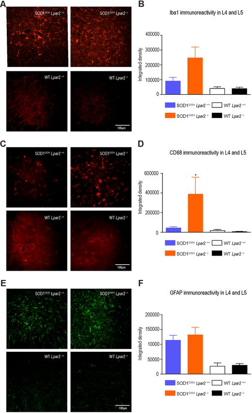
Puigdomenech-Poch et al.                                                                                                           LPA2 in ALS

were placed onto the rotarod at a constant speed of 14 revolutions        Imagen, Merial) and xylazine (10 mg/kg i.m.; Rompun,
per minute. Animals were trained before the beginning of the              Bayer). The right hindlimb was shaved and disinfected,
experiment. 180 s was chosen as the arbitrary cut-off time.               and then, an incision in the skin and muscles was done to
Rotarod test was then performed weekly from 8 to 20 or 22 weeks           expose the sciatic nerve. The sciatic nerve was cut above the
of age at the same speed. The time for which each animal could            trifurcation and ligated to prevent axon regeneration and muscle
remain on the rotating cylinder was measured. Each animal                 reinnervation. The wound was closed and disinfected, and the
was given three trials and the longest latency without falling            mice were kept in a warm environment until complete recovery
was recorded.                                                             from anesthesia. Buprenorphine (0.01 mg/kg i.p.; Buprex,
   The time of disease onset was defined at the time when                 Indivior) was administrated intraperitoneally, twice a day, for
mice could not remain in the rotarod for at least 180 s. The              3 days.
endpoint was determined when mice were not able to right
themselves within 30 s of being placed on their side. At the              Quantitative Real-Time PCR (qPCR)
endpoint, mice were euthanized by an overdose of pentobarbital            SOD1G93A mice were transcardially perfused with sterile saline
sodium (Dolethal, Vetoquinol) following the requirements of the           at 8 (pre-symptomatic), 12 (disease onset), 16 (symptomatic),
Animal Experimentation Ethical Committee of the Universitat               and 20 weeks of age (late-symptomatic), and lumbar spinal
Autònoma de Barcelona.                                                    cord, sciatic nerve, and gastrocnemius muscle were harvested
                                                                          and immediately frozen in liquid nitrogen. The same tissues
Electrophysiological Test                                                 were also harvested from WT littermates at 20 weeks of age.
Compound Muscle Action Potential (CMAP)                                   Samples were homogenized in QIAzol lysis reagent (Qiagen)
Animals were anesthetized with pentobarbital sodium (50 mg/kg,            using a tissue rupture, and RNA was isolated using RNeasy
i.p, Sigma) and placed prone over a heating pad that maintains            Lipid Tissue kit (Qiagen), according to the manufacturer0 s
body temperature. Needle electrodes were placed deep into                 protocol. One microgram of RNA was retro-transcribed using
the sciatic nerve notch and the sciatic nerve was stimulated              the Omniscript RT kit (Qiagen). Quantitative PCRs (qPCRs)
using single pulses of 0.02 ms duration (Grass S88) Recording             were done using Brilliant III Ultra-Fast SYBR (Bio-Rad) and
microneedles were inserted superficially in the tibial anterior           custom-designed primers. Primer sequences were the following
muscle. The recording needles were placed using a magnifier lens          Lpar2 forward 50 -CTCACTGGTCAATGCAGTGGTATAT-30 ,
and guided by anatomical landmarks, to ensure reproducibility             Lpar2 reverse 50 -GAAGGCGGCGGAAGGT-30 . Glyceraldehyde
of needle location on all animals. Reference and ground                   3-phosphate dehydrogenase (GAPDH); Gapdh forward 50 -
electrodes were inserted at the third toe and the base of                 TCAACAGCAACTCCCACTCTTCCA-30 , Gapdh reverse 50 -
the paw, respectively. CMAPs were recorded from the tibialis              ACCCTGTTGCTGTAGCCGTATTCA-30 .
anterior and gastrocnemius muscle. Signals were bandpass                     RNA from human spinal cord samples was isolated using
filtered (3 Hz to 3 kHz), amplified 100× for gastrocnemius                RNeasy Mini Kit (Qiagen) and retro-transcribed using cDNA
and tibialis (P511AC amplifiers, Grass), and digitized with a             Reverse Transcription kit (Applied Biosystems, Foster City, CA,
Power Lab recording system (PowerLab16SP, ADInstruments)                  USA) following manufacturers’ kit instructions. qPCR was done
at 20 kHz. The amplitude of the M wave was studied                        using TaqMan-designed primers (Thermo Fisher Scientific, MA,
from the difference between the baseline to the maximal                   USA) for gene expression of LPA1 (Hs00173500_m1), LPA2
negative peak.                                                            (Hs01109356_m1), and HPRT1 (Hs02800695_m1).
                                                                             Mouse and human qPCR data were analyzed using the
Motor Unit Number Estimation (MUNEs)                                      double-delta cycle threshold (∆∆Ct) method on a MyiQ Single-
Animals were anesthetized as described above. Electrodes were             Color Real-Time PCR Detection System (Bio-Rad). Gapdh and
placed in the same place as for the CMAP registers and the                HRPT1 were used as housekeeping genes for mouse and human
amplitude for the M wave was recorded. For the MUNE                       samples, respectively.
assessment, the protocol used consists of the incremental
technique. Starting from the subthreshold intensity, the sciatic          Histology
nerves were stimulated with single pulses of gradually increased          SOD1G93A Lpar2+/+ , SOD1G93A -Lpar2−/− , Lpar2 null mice and
intensity until the first response appeared, representing the first       wild-type littermates were euthanized at 16 weeks of age with
motor unit recruited. With the next stimuli, quantal increases            an overdose of pentobarbital sodium (Dolethal, Vetoquinol)
in the response were recorded. The data were represented                  and transcardially perfused with 4% paraformaldehyde in 0.1 M
as the frequency distribution of the single motor unit size               phosphate buffer (PB). Also, Lpar2 deficient mice and wildtype
(SU); as the mean of consistent increases (increments >100                littermates with sciatic nerve injury were perfused at 3 weeks
µV) single motor unit potential (SMUP) and finally, MUNE                  after the lesion. Samples were post-fixed in 4% PFA for 2 h
results from the CMAP maximal amplitude divided by the                    and cryoprotected with 30% sucrose in 0.1 M PB at 4◦ C for
average SMUP.                                                             48 h. The spinal cords and muscles were fast-frozen at -20◦ C
                                                                          in a cryo embedding compound (Tissue-Tek OCT, Sakura),
Sciatic Nerve Injury                                                      cut on a cryostat (Leica), and serially picked up in gelatine
Lpar2−/− mice and wild-type littermates were deeply                       coated slides. The ventral root of the spinal nerve (L4) that
anesthetized with a mixture of ketamine (90 mg/kg i.m.;                   fixed into 3% glutaraldehyde, 3% PFA in 0.1 PB at 4◦ C for

Frontiers in Cellular Neuroscience | www.frontiersin.org              3                                 March 2021 | Volume 15 | Article 600872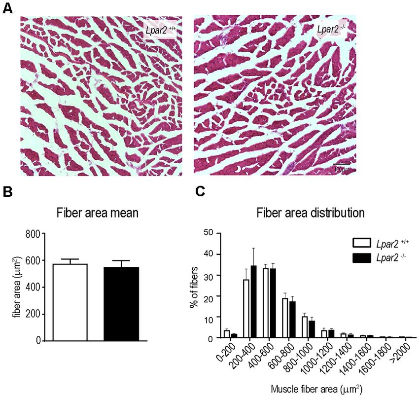
Puigdomenech-Poch et al.                                                                                                            LPA2 in ALS

1 week, immersed in 2% osmium tetroxide in 0.1 M PB                       RESULTS
for 2 h, dehydrated through ethanol series and embedded in
Epon resin. Semithin sections (0.5 µm thick) were cut by an               LPA2 Transcripts Are Increased in Human
ultramicrotome (Leica).                                                   and Mouse ALS Samples
    To analyze the preservation of motoneurons, transversal               LPA exerts its biological effects by binding to 6 G protein-
sections of the spinal cord (L4 and L5) were placed in                    coupled receptors, known as LPA1–6 . We have recently
a hotplate for 30 min, rehydrated, and stained in cresyl                  demonstrated that among the different LPA receptors, LPA1
violet. The number of motoneurons was manually quantified                 (Santos-Nogueira et al., 2015) and LPA2 (López-Serrano et al.,
in both ventral horns. To ensure reproducibility, only those              2019) mediate demyelination and functional impairments in
neurons that were within the laminae IX, were larger than                 spinal cord injury. Therefore, we first examined the RNA levels
20 µm and had a polygonal shape and prominent nucleoli                    of LPA1 and LPA2 in postmortem spinal cord samples of sALS
were counted.                                                             patients and age-matched controls. LPA1 and LPA2 transcripts
    For immunofluorescence, spinal cord and muscle samples                were found in the spinal cord of control and sALS individuals
were placed on a hotplate at 37◦ C for 30 min, then rehydrated            (Figures 1A,B). However, only the levels of LPA2 significantly
in 0.1 PBS for 5 min and blocked with a blocking buffer                   increased in sALS samples (Figures 1A,B).
of 5% normal donkey serum (NDS) in 0.3% triton-PBS                           Based on these data, we focused our interest on LPA2 and
for 1 h at RT. The sections were incubated overnight at                   characterized its expression in the spinal cord, sciatic nerve, and
4◦ C with primary antibodies against Iba1 (for microglia and              muscle of SOD1G93A mice. QPCR revealed that the expression
macrophages; 1:500; Abcam), CD68 (1:100; Abcam), GFAP                     of Lpar2 in these three tissues was similar in SOD1G93A mice
(for astrocytes; 1:500; Millipore). The sections were then                at pre-symptomatic stages of the disease (8 weeks of age) than
washed in 0.3% triton-PBS and further incubated for 1 h                   in wild-type littermates (Figures 2A–C). RNA levels of Lpar2 in
at RT with the specific secondary antibodies tagged to an                 the spinal cord of SOD1G93A mice tended to increase once they
Alexa-594 or Alexa-488 fluorochrome (1:200, Invitrogen). After            show marked clinical symptoms of the disease (16–20 weeks of
washing several times in 0.3% Triton-PBS and PBS, slides                  age), although this result did not reach statistical significance
were incubated for 1 min in a solution containing DAPI                    (Figure 2A). The transcripts of Lpar2 in the sciatic nerve and
(1 µg/ml, Sigma).                                                         gastrocnemius muscle of SOD1G93A mice increased significantly
    Microgliosis and astrogliosis were analyzed by quantifying            over disease progression, peaking at the age of 16 weeks
the integrated density of Iba, CD68, and GFAP immunostaining              (Figures 2B,C). At this time point, the RNA levels of lpar2 in
at the spinal cord ventral horns of L4–L5 segments.                       both tissues were ∼2.5 fold higher in SOD1G93A mice relative to
Macrophage counts in the gastrocnemius were done                          wildtype littermates (Figures 2B,C).
by manually quantifying the number Iba1 and CD68
positive cells.
                                                                          Genetic Deletion of Lpar2 Modifies ALS
    To analyze the muscle fiber area, cryostat transversal sections
of gastrocnemius muscle were hydrated in water and stained                Disease Course
for hematoxylin (Fluka, Sigma) and eosin (Merck, Millipore)               To determine the contribution of LPA2 in ALS, we crossed
for 5 min. The sections were dehydrated with series of graded             SOD1G93A mice with Lpar2 deficient mice (SOD1G93A -
ethanol rinses and mounted with DPX (Fluka).                              Lpar2−/− ). We observed that the absence of Lpar2 significantly
    To study motor axon preservation, ventral root (L4) semithin          delayed ALS disease onset by 4 and 5 weeks in female and male
sections, were stained with 1% Borax (Sigma) and 1% Toluidine             mice, respectively (Figures 3A–F). In this line, the rotarod
Blue (Fluka) in distillate water. The number of spared motor              test also revealed that the genetic deletion of Lpar2 markedly
axons, with intact myelin sheaths and those with some degree of           slowed the neurological decline of ALS mice (Figures 3A,D).
myelin damage were counted.                                               Unexpectedly, the lack of Lpar2 did not increase the lifespan of
    Samples were visualized with an Olympus BX51 microscope               SOD1G93A , despite its absence markedly protected against the
and the images were captured with Olympus DP50 digital                    clinical signs of the disease (Figures 3G–J). Indeed, deficiency
camera. All Histological quantifications were done using ImageJ           of Lpar2 shortened the lifespan of ALS mice by 21 and 10 days,
software (NIH, Bethesda, MD, USA).                                        in males and females, respectively (Figures 3H,J). Therefore,
                                                                          these observations suggest that the lack of LPA2 signaling slows
Statistical Analysis                                                      disease onset and progression but shortens survival.
Data are shown as mean ± standard error of the mean (SEM).
Rotarod test results were analyzed using two-way repeated-                LPA2 Does Not Contribute to Motoneuron
measures ANOVA with post-hoc Bonferroni’s for multiple                    Death in ALS
comparisons. Disease onset and the survival data were analyzed            To gain insights into the early detrimental actions of LPA2
using the Mantel–Cox test. Gene expression, histological                  in ALS, we focused our experiments on SOD1G93A mice at
analysis, and electrophysiological results were analyzed by               16 weeks of age, when neurological outcomes were markedly
using one-way ANOVA with post-hoc Bonferroni’s for multiple               enhanced in ALS mice lacking lpar2. Since LPA mediates gliosis
comparisons of unpaired t-test. Statistical significance was              and we recently reported that microglial cells become cytotoxic
considered at p < 0.05.                                                   upon LPA2 stimulation (López-Serrano et al., 2019), we first

Frontiers in Cellular Neuroscience | www.frontiersin.org              4                                  March 2021 | Volume 15 | Article 600872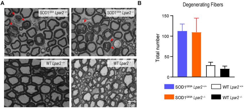
Puigdomenech-Poch et al. LPA2 in ALS
FIGURE 1 | Characterization of Lysophosphatidic acid 1 (LPA1 ) and LPA2 expression in the spinal cord of sporadic amyotrophic lateral sclerosis (sALS) patients.
(A,B) Plots showing RNA levels of LPA1 (A) and LPA2 (B) in the spinal cord of sALS patients (n = 22) and controls (n = 17). Unpaired t-test. Data are shown as
mean ± SEM. ***p < 0.001.
FIGURE 2 | Dynamic changes in Lpa2 expression in SOD1G93A mice. (A–C) Graphs showing the changes in Lpar2 RNA levels in the spinal cord (A), sciatic nerve
(B), and gastrocnemius muscle (C) of SOD1G93A at 8, 12, 16, and 20 weeks of age relative to wild-type littermate mice. (A) n = 5 per group and time point. One-way
ANOVA with Bonferroni’s post hoc test. *p < 0.05, **p < 0.01 vs. WT mice. Data are shown as mean ± SEM.
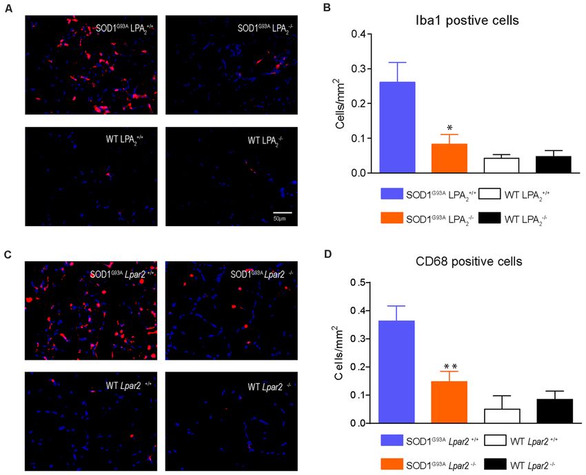
evaluated whether LPA2 signaling contributed to microgliosis mice deficient in Lpar2 at 16 weeks of age (Figures 5C–F). These
and astrogliosis in the lumbar spinal cord of SOD1G93A results indicated that neuromuscular integrity was enhanced
mice. Histological analysis revealed that immunoreactivity for in SOD1G93A mice lacking Lpar2 but this was not due to
Iba1, a marker for microglial cells, tended to be increased detrimental actions of LPA2 on motoneuron. These observations,
in the spinal cord ventral horn of ALS mice deficient in therefore, suggest that the initial harmful effects of LPA2
Lpar2, although this result did not reach statistical significance in ALS may be mediated in the peripheral nerve and/or
(Figures 4A,B). However, CD68 expression in microglial cells, skeletal muscle.
which is considered a marker of activated cells, was significantly
increased in ALS mice deficient in Lpar2 (Figures 4C,D), LPA2 Does Not Promote Myelin Damage in
suggesting that although the lack of Lpar2 signaling did not Motor Axons
increase microgliosis it resulted in greater activation of this glial LPA contributes to demyelination in the spinal cord by signaling
cell subset. Immunoreactivity for GFAP, a marker for astrocytes, via LPA1 (Santos-Nogueira et al., 2015) and LPA2 (López-
did not differ in the spinal cord of both ALS experimental groups Serrano et al., 2019). Considering that Lpar2 transcripts peaked
(Figures 4E,F). in the sciatic nerve of ALS mice at 16 weeks of age, we assessed
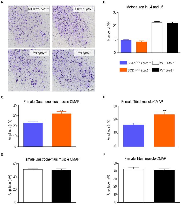
Since degeneration of motoneurons is the main feature in whether LPA2 led to demyelination and/or degeneration of
ALS mice and patients, we assessed whether LPA2 contributed motor axons. For this purpose, we quantified the number of
to motoneuron death. Histopathological analysis of the lumbar fibers that showed some signs of Wallerian degeneration. These
spinal cord (L4 and L5) revealed that the lack of Lpar2 did analyses revealed that the counts of motor fibers that showed
not have any effect on motoneuron survival in ALS mice signs of myelin breakdown did not differ between both ALS
(Figures 5A,B) despite electrophysiological tests revealing that groups (Figures 6A,B). These results, therefore, reveal that LPA2
the amplitude of the CMAPs was significantly increased in ALS does not promote demyelination in ALS.
Frontiers in Cellular Neuroscience | www.frontiersin.org 5 March 2021 | Volume 15 | Article 600872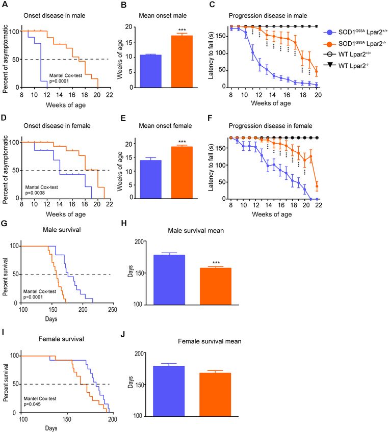
Puigdomenech-Poch et al.                                                                                                                                 LPA2 in ALS

  FIGURE 3 | Dual effects of LPA2 in ALS disease. (A–E) Graphs showing the functional evaluation of ALS mice in the rotarod test over time. (A,B) Lpar2 deficiency
  delays the onset of the disease in 5 weeks in male ALS mice (A,B) and 4 weeks in females (D,E). Graphs showing the functional progression of neurological deficits
  represented by the latency to fall off the rotarod test in males (C) and females (F). (G–J) Effects of Lpar2 gene deletion in the lifespan of ALS mice. Two-way RM
  ANOVA, Bonferroni’s post hoc test in (A,B). Mantel-Cox test in (B,E,G,I). Unpaired t-test in (C,F,H,J). (A–C) Males (n = 9 SOD1G93A Lpar2+/+ , n = 11 SOD1G93A
  Lpar2−/− , n = 4 WT Lpar2+/+ , n = 4 WT Lpar2−/− ). (D–E) Females (n = 10 SOD1G93A Lpar2+/+ , n = 11 SOD1G93A Lpar2−/− , n = 4 WT Lpar2+/+ , n = 4 WT Lpar2−/− ).
  (G–H) Males (n = 14 SOD1G93A Lpar2+/+ , n = 15 SOD1G93A Lpar2−/− ). (I–J) Females (n = 14 SOD1G93A Lpar2+/+ , n = 14 SOD1G93A Lpar2−/− ). **p < 0.01,
  ***p < 0.001, vs. SOD1G93A LPA2 +/+ . Data are shown as mean ± SEM.

The Absence of Lpar2 Prevents Muscle                                                  mice peaked at the age of 16 weeks (Figure 2C). We found
Atrophy in SOD1G93A Mice                                                              that cross tissue sections of the gastrocnemius muscle stained
Since the data reported above suggested that initial detrimental                      with hematoxylin and eosin revealed that the mean muscle fiber
actions of LPA2 in SOD1G93A mice were unlikely to be mediated                         area was decreased in SOD1G93A mice as compared to wildtype
in the spinal cord or peripheral nerves, we then studied the effects                  mice, indicating clear signs of muscle atrophy (Figures 7A,B).
of Lpar2 deficiency in the muscle of ALS animals. Highlight                           The mean muscle fiber area was slightly enhanced in ALS
that lpar2 transcripts in the gastrocnemius muscle of SOD1G93A                        mice lacking Lpar2, although this result did not reach statistical

Frontiers in Cellular Neuroscience | www.frontiersin.org                          6                                        March 2021 | Volume 15 | Article 600872
Puigdomenech-Poch et al.                                                                                                                         LPA2 in ALS

                                                                                        of the neuromuscular junctions (NMJ) in the gastrocnemius
                                                                                        muscle. Electrophysiological estimation of the motor unit
                                                                                        number (MUNE), the mean amplitude of single motor unit
                                                                                        potential (SMUA), and the distribution of motor unit action
                                                                                        potential (MUAP) amplitude revealed that enhanced collateral
                                                                                        sprouting was not the mechanism that prevented muscle atrophy
                                                                                        in ALS mice lacking Lpar2 (Figure 8).
                                                                                            Previous work revealed that muscle was a primary target of
                                                                                        mutant SOD1 protein (SOD1G93A ) and that selective expression
                                                                                        of SOD1G93A in skeletal muscle cells induced atrophy and
                                                                                        functional impairments (Dobrowolny et al., 2008). Therefore,
                                                                                        the attenuation of muscle atrophy and neurological decline
                                                                                        observed in ALS mice lacking Lpar2 could be due to the
                                                                                        deleterious actions of this LPA receptor on innervated muscle
                                                                                        fibers. However, the possibility that LPA2 signaling could be
                                                                                        required for the atrophy of denervated muscle fibers cannot be
                                                                                        discarded. To dissect out whether LPA2 signaling is crucial for
                                                                                        the atrophy of denervated fibers, we transected the sciatic nerve
                                                                                        of Lpar2 knockout mice and wildtype littermates to promote
                                                                                        muscle denervation and ligated the injured nerve to prevent
                                                                                        axon regeneration and muscle reinnervation. Histological cross-
                                                                                        sections of gastrocnemius muscles harvested at 21 after
                                                                                        lesion revealed that the lack of Lpar2 signaling did not
                                                                                        alter muscle atrophy caused by the denervation of the nerve
                                                                                        (Figures 9A–C). These data, therefore, indicated that LPA2
                                                                                        is not required for the atrophy of denervated fiber, and
                                                                                        suggested, that the reduced atrophy observed in ALS mice
                                                                                        lacking Lpar2 was due to greater protection of innervated
                                                                                        muscle fibers.
                                                                                            Recent studies reveal that immune cells infiltrate into the
                                                                                        muscles of SOD1G93A animals and that they contribute to
                                                                                        disease progression (Van Dyke et al., 2016; Trias et al., 2017).
                                                                                        Since LPA is involved in triggering inflammation (Choi and
                                                                                        Chun, 2013; Yung et al., 2014) we assessed whether LPA2
                                                                                        signaling is involved in regulating muscle inflammation in
  FIGURE 4 | The lack of Lpar2 does not reduce astrogliosis and microgliosis
  in the lumbar spinal cord of SOD1G93A mice. (A) Representative images of the
                                                                                        ALS. Histological analysis of gastrocnemius muscle showed
  lumbar spinal cord stained against Iba1. (B) Plot showing the quantification of       that the number of macrophages (Iba1+ cells) was significantly
  the Iba1 immunoreactivity in the L4 and L5 spinal cord segments.                      reduced in SOD1G93A mice lacking Lpar2 (Figures 10A,B).
  (C) Representative images of the lumbar spinal cord showing stained against           In this line, the expression of CD68, which is found in
  CD68. (D) Graph showing quantification of CD68 immunoreactivity in the
                                                                                        activated macrophages, was also reduced in the muscles of
  L4 and L5 segments. (E) Representative images of the lumbar spinal cord
  showing stained against GFAP. (F) Graph showing quantification of GFAP                ALS mice deficient in Lpar2 (Figures 10C,D), suggesting
  immunoreactivity in the L4 and L5 segments. One-way ANOVA, Bonferroni’s               that LPA2 signaling contributes to muscle inflammation
  post hoc test. *p < 0.05 vs. SOD1G93A LPA2 +/+ (males; n = 3 SOD1G93A                 in ALS.
  Lpar2+/+ , n = 4 SOD1G93A Lpar2−/− , n = 4 WT Lpar2+/+ , n = 4 WT Lpar2−/− ).
  Data are shown as mean ± SEM.
                                                                                        Discussion
                                                                                        In the present study, we assessed the effects of LPA2 on
significance (Figures 7A,B). However, further analysis revealed                         ALS. We showed that LPA2 mRNA levels are increased
that the distribution of the muscle fiber according to their size                       in post-mortem spinal cord samples of ALS patients, as
was shifted towards the right in ALS mice deficient for Lpar2.                          well as in the sciatic nerve and gastrocnemius muscle of
These data indicated that the proportion of fibers with larger                          ALS mice. We also identified a biphasic role of LPA2 in
diameter was increased in ALS lacking lpar2, whereas SOD1G93A                           ALS. Initially, LPA2 signaling accelerates disease onset and
Lpar2+/+ animals displayed a greater proportion of small muscle                         neurological deficits, whereas later, LPA2 extends survival. In
fibers (Figure 7C). These data, therefore, indicate that LPA2                           the search for the mechanisms underlying the harmful actions
signaling contributes to muscle atrophy in ALS.                                         of LPA2 at the early stages of ALS disease, we revealed this
   To determine whether protection against muscle atrophy                               receptor did not promote spinal cord gliosis, motoneuron
observed in ALS mice deficient in Lpar2 was due to increased                            death, motor axon demyelination, but it increased muscle
collateral sprouting of motor axons, we assessed the integrity                          atrophy and inflammation. Overall, this work provides, for

Frontiers in Cellular Neuroscience | www.frontiersin.org                            7                                 March 2021 | Volume 15 | Article 600872
Puigdomenech-Poch et al. LPA2 in ALS FIGURE 5 | The lack of Lpar2 does not increase motoneuron survival but preserves neuromuscular integrity in ALS mice at 16 weeks of age. (A) Representative micrograph of lumbar spinal cord showing motoneurons stained with cresyl violet. (B) Quantification of motoneurons in the L4 and L5 spinal cord segments (males; n = 6 SOD1G93A Lpar2+/+ , n = 9 SOD1G93A Lpar2−/− , n = 7 WT Lpar2+/+ , n = 8 WT Lpar2−/− ). (C,D) Plots showing the compound muscle potential amplitude of the gastrocnemius (C,E) and tibialis anterior (D,F) muscle at 16 weeks of age (males; n = 14 SOD1G93A Lpar2+/+ , n = 14 SOD1G93A Lpar2−/− , n = 5 WT Lpar2+/+ , n = 5 WT Lpar2−/− ). **p < 0.01 vs. SOD1G93A LPA2 +/+ . One-way ANOVA, Bonferroni’s post hoc test. Data are shown as mean ± SEM. the first time, evidence for the involvement of LPA2 in models (Philips and Robberecht, 2011), as well as, in most ALS physiopathology. neurological conditions including Alzheimer’s, Parkinson and Inflammation is a hallmark of neurodegenerative diseases, frontotemporal lobe dementia among others (De Virgilio et al., typified by reactive morphology of astrocytes and microglia, 2016; Newcombe et al., 2018). Although most of the ALS accompanied by the release of inflammatory mediators in the reports have focused on studying the inflammatory response parenchyma (Ransohoff, 2016). This physiological response in the CNS, it is currently known that in ALS there is also has been shown in ALS in both patients and animal an invasion of immune cells into the peripheral nerves and Frontiers in Cellular Neuroscience | www.frontiersin.org 8 March 2021 | Volume 15 | Article 600872
Puigdomenech-Poch et al.                                                                                                                                 LPA2 in ALS

  FIGURE 6 | Lpar2 deletion does alter Wallerian degeneration of motor axons in SOD1G93A mice. (A) Representative cross-section images of the spinal nerve ventral
  root (L4) stained in toluidine blue. Note the presence of fiber with disrupted myelin sheaths the motor roots of ALS mice (arrowhead). (B) Graph showing the
  quantification of fibers with some signs of myelin degeneration at 16 weeks of age in male mice (n = 3 SOD1G93A Lpar2−/− , n = 5 SOD1G93A Lpar2+/+ , n = 4 WT
  LPA2 −/− , n = 3 WT Lpar2+/+ ). Two-way ANOVA, Bonferroni’s post hoc test. Data are presented as mean ± SEM.

  FIGURE 7 | Lpar2 gene deletion prevents muscle atrophy in SOD1G93A mice. (A) Representative images of gastrocnemius muscle stained in hematoxylin and eosin
  at 16 weeks of age in male mice. (B) Quantification of the muscle cross-section area at 16 weeks of age in male mice. (C) Plot showing the distribution of muscle
  fiber width (males; n = 4 per group). One-way ANOVA, Bonferroni’s post hoc test. # p < 0.05 vs. WT Lpar2+/+ ; ns, not significant vs. WT Lpar2-/- in (B); Two-way
  ANOVA, Bonferroni’s post hoc test in (C). **p < 0.01 vs. SOD1G93A Lpar2+/+ . Data are presented as mean ± SEM.

muscles that contributes to disease progression (Martínez-                            response. Among them, lipids, such as the lysophospholipids
Muriana et al., 2016; Van Dyke et al., 2016; Trias et al., 2017;                      (LPLs; Chun et al., 2014), have demonstrated inflammatory
Trias et al., 2018, 2020).                                                            effects that are both detrimental and beneficial, including
    Studies done over the last years have demonstrated that                           within the CNS (David et al., 2012). LPA is an LPL that
lipids play a crucial role in the regulation of the inflammatory                      acts as an extracellular and intracellular signaling molecule

Frontiers in Cellular Neuroscience | www.frontiersin.org                          9                                        March 2021 | Volume 15 | Article 600872
Puigdomenech-Poch et al. LPA2 in ALS
FIGURE 9 | Lpar2 deficiency does not prevent muscle atrophy after sciatic
nerve injury. (A) Representative images of gastrocnemius muscle cross tissue
sections at 21 days after sciatic nerve transection. (B,C) Plot showing the
FIGURE 8 | The absence of Lpar2 does not enhance collateral sprouting. quantification of the muscle cross-section area (B) and the distribution of
(A–C) Electrophysiological estimation of motor unit number (A), mean muscle fiber width (C). Lpar2−/− mice (n = 4) Lpar2+/+ mice (n = 6). Unpaired
amplitude of single motor unit potential (B), and single size of motor units (C) t-student in (B); Two-way RM ANOVA with Bonferroni’s post hoc test in (C).
of the gastrocnemius muscle at 16 weeks of age in male mice. Note that the Data shown as mean ± SEM.
absence of Lpar2 does not modify any of these parameters (males;
n = 5 SOD1G93A Lpar2−/− , n = 8 SOD1G93A Lpar2+/+ , n = 5 WT Lpar2−/− ,
n = 3 WT Lpar2+/+ ). One-way ANOVA with Bonferroni’s post hoc test in (A,B);
Two-way RM ANOVA with Bonferroni’s post hoc test in (C). Data shown as levels observed in the ALS animal model. We also found
mean ± SEM. that Lpar2 RNA levels were up-regulated in the sciatic
nerve and the gastrocnemius muscle of ALS mice, both
peaking at 16 weeks of age. The source of LPA2 in ALS
and controls a wide range of physiological responses. The was not studied due to the lack of specificity of current
effects of LPA are mediated by 6 G protein-coupled receptors, antibodies. However, Lpar2 transcripts in the CNS have
LPA1 –LPA6 (Choi and Chun, 2013; Kihara et al., 2014; been described in neurons, oligodendrocytes, microglia, and
Yung et al., 2015). Earlier studies demonstrated that this infiltrating macrophages, whereas in the peripheral nervous
bioactive lipid has important actions in the CNS after injury system and skeletal muscle have been shown in Schwann cells
and disease. LPA activates astrocytes and microglia, causes and muscle fibers, respectively (Frohnert et al., 2003; Jean-
axonal retraction, induces brain blood barrier permeability Baptiste et al., 2005; Zhang et al., 2014).
among others (Sorensen et al., 2003; Choi et al., 2010). To assess whether LPA2 signaling contributes to ALS, we
We previously reported that LPA1 and LPA2 that are crossed mice lacking Lpar2 with SOD1G93A mice. This double-
normally associated with myelination (Weiner et al., 1998, transgenic animal revealed for the first time that the absence
2001; Weiner and Chun, 1999; Anliker et al., 2013) also of Lpar2 delayed the onset of ALS disease in both males and
cause demyelination and functional impairments after females, and slowed the deterioration of locomotor performance.
spinal cord injury, in part, by activating microglial cells Despite the detrimental actions of LPA2 at the initial stages
(Santos-Nogueira et al., 2015; López-Serrano et al., 2019). of the disease, deficiency in Lpar2 shortened the lifespan of
In the present work, we found that LPA1 and LPA2 SOD1G93A mice. These data suggest that LPA2 signaling has
RNA transcripts were expressed in post-mortem human a dual role in ALS, being detrimental at the early stages
spinal cord samples and that the transcripts of LPA2 , but of the disease and later, being protective. However, we do
not LPA1 , were increased ∼2 fold in sALS patients. The not discard that LPA2 signaling could play an important
expression of Lpar2 in the spinal cord of SOD1G93A mice physiological role for neuronal function, and its genetic removal
tended to increase once the clinical signs of the disease were could increase their vulnerability to stress conditions. Therefore,
evident, although this up-regulation did not reach statistical the development of LPA2 agonist is critical to elucidate
significance. Since ALS mouse samples were not harvested whether activation of this receptor confers protection at late
at the disease endpoint, unlike the spinal cords of sALS stages of ALS disease. Highlight that lifespan of SOD1G93A
patients, this may account for the lower Lpar2 transcript mice was about 2 weeks increased than that reported from
Frontiers in Cellular Neuroscience | www.frontiersin.org 10 March 2021 | Volume 15 | Article 600872Puigdomenech-Poch et al. LPA2 in ALS
FIGURE 10 | Gene deletion of Lpar2 reduces macrophage invasion in the muscle of ALS mice. (A) Representative images of gastrocnemius muscle cross tissue
sections stained against Iba1. (B) Plot showing the quantification of the Iba1+ cells. (C) Representative images of gastrocnemius muscle cross tissue sections
stained against CD68. (D) Plot showing the quantification of the CD68+ cells. n = 4 per group. One-way ANOVA with Bonferroni’s post hoc test. ∗ p < 0.05,
∗∗
p < 0.01 vs. SOD1G93A Lpar2+/+ . Data are presented as mean ± SEM.
Jackson Laboratories. This could be due to the background increase the activation of microglial cells in the lumbar
acquired by ALS mice after being bred with the LPA2 mice. spinal cord of ALS mice, as revealed the CD68 expression.
However, other factors related to the loss of SOD1G93A gene Although microglia and astroglia activation at later stages
copies or the housing conditions of the animal facility, cannot of the disease was not studied in the present work, the
be discarded. increased microglia reactivity observed in ALS mice deficient
To elucidate the early pathological role of LPA2 in ALS, in Lpar2 might indicate that this receptor could put the brakes
we assessed its effects at different levels in which motor on microglia activation in advanced stages of the disease,
function is involved (spinal cord, peripheral nerve, and muscle). which may account for the shorter survival of ALS mice
We first focused on the effects of LPA2 in the spinal lacking Lpar2.
cord of ALS animals, where lower motoneurons are located In ALS, axonopathy precedes and underlies skeletal muscle
(Contos et al., 2002; Mancuso et al., 2014; McCampbell weakness and progressive paralysis. Indeed, peripheral axons
et al., 2018). Despite having previously reported that activation degenerate before the death of cell bodies in the CNS,
of LPA2 exerts deleterious effects after spinal cord injury according to evidence in ALS patients and murine models
(López-Serrano et al., 2019), we observed that the lack of (Fischer and Glass, 2007; Moloney et al., 2014; Trias et al.,
Lpar2 did not modify the number of motoneurons in ALS 2020). Unlike peripheral nerve injury, axonal growth of
mice, suggesting that the harmful effects of LPA2 in ALS degenerated axons is limited in ALS, leading to chronic and
were independent on motoneuron death. We also found progressive axon degeneration, recruitment of immune cells,
that the lack of Lpar2 did not attenuate astrogliosis but and promotion of inflammation (Trias et al., 2020). In this
Frontiers in Cellular Neuroscience | www.frontiersin.org 11 March 2021 | Volume 15 | Article 600872Puigdomenech-Poch et al. LPA2 in ALS
line, treatment with a CSF1R antagonist attenuated the influx above, mutant SOD1 protein causes a direct toxic effect
of macrophages into the nerves of ALS mice, that together in the skeletal muscle cell and leads to muscle atrophy
with its effects on minimizing microgliosis, extended mice (Dobrowolny et al., 2008). It is now known that different
survival (Martínez-Muriana et al., 2016). Little is known immune cell subsets infiltrate the skeletal muscle of ALS
about the specific functions of LPA2 in the sciatic nerve, but animals, which is likely due to the damage of muscle
similar to LPA1 , this receptor is found in Schwann cells and fibers, and contribute to disease pathogenesis (Van Dyke
its expression appears shortly before maturation/myelination et al., 2016; Trias et al., 2018). Interestingly, we observed
(Weiner and Chun, 1999; Weiner et al., 2001; García-Díaz that the infiltration of macrophages in skeletal muscles of
et al., 2015). Indeed, activation of LPA2 in myelinating cells SOD1G93A mice was markedly attenuated in the lack of
seems to play a key role in myelin formation (Weiner Lpar2. These findings suggest that muscle damage caused by
and Chun, 1999; Weiner et al., 2001; Anliker et al., 2013; mutant SOD1 favors the recruitment of macrophages into
García-Díaz et al., 2015). the muscle, in part, by LPA2 -dependent mechanisms, and
Previous works revealed that LPA2 activation contributes causes further muscle deterioration, and consequently, motor
to demyelination after spinal cord injury (Santos-Nogueira function decline.
et al., 2015; López-Serrano et al., 2019) suggesting that the To summarize, this work provides the first evidence that
upregulation of Lpar2 in the peripheral nerves of ALS mice could LPA2 signaling contributes to ALS. Initial activation of LPA2
mediate demyelination and/or damage in motor axons. However, accelerates ALS progression by promoting muscle atrophy, which
histological analysis of the spinal cord motor roots of ALS mice, is likely due to the inflammatory actions of this receptor in the
where motor axons are present, revealed that the absence of muscle, whereas later, it promotes beneficial effects on survival.
Lpar2 did not prevent degeneration of the axons or their myelin
sheaths, and suggested that the detrimental actions of LPA2 in DATA AVAILABILITY STATEMENT
ALS could be mediated in the skeletal muscle.
Skeletal muscle atrophy in ALS has been interpreted as a The raw data supporting the conclusions of this article will be
consequence of degeneration of motor axons. Nevertheless, it made available by the authors, without undue reservation.
has been recently postulated that skeletal myocytes also have
an active role in muscle atrophy in ALS, independent from ETHICS STATEMENT
motoneuron loss. This is clear from experiments using mice with
selective expression of SOD1G93A in muscle cells, which revealed The studies involving human participants were reviewed and
that skeletal muscle is a primary target of SOD1G93A -mediated approved by the Clinical Research Ethics Committee (CEIC)
toxicity and causes progressive muscle atrophy (Dobrowolny of the Bellvitge University Hospital. The patients/participants
et al., 2008). In this view, numerous studies have attempted provided their written informed consent to participate in
to palliate muscle pathology to counterbalance the effects of this study. The animal study was reviewed and approved
motoneuron degeneration (Derave et al., 2003; Gifondorwa et al., by Animal Experimentation Ethical Committee of the
2007; Yoo and Ko, 2012; Halon et al., 2014; Loeffler et al., 2016). Universitat Autònoma de Barcelona (CEEAH 1188R3-DMAH
Here, we observed that Lpar2 deficiency protected from muscle 3131)/(2969).
atrophy in ALS animals.
Collateral axonal sprouting is a natural mechanism observed AUTHOR CONTRIBUTIONS
in ALS disease and nerve injury, among other disorders, in
which surviving motoneurons reinnervate denervated end-plates RL-V and MP-P designed the study. MP-P, AM-M, PA-B, IF, and
within the same muscle, enabling to compensate for the loss RL-V performed the research, analyzed, or interpreted results.
of functional motor units and prevent muscle atrophy (Siu and JC created LPA2 mutant mice. MP-P, JC, and RL-V wrote the
Gordon, 2003). However, we revealed using electrophysiological manuscript. All authors contributed to the article and approved
techniques that deficiency in Lpar2 did not enhance collateral the submitted version.
axonal sprouting in ALS. We also found that LPA2 signaling was
not required for the events that lead to atrophy of denervated FUNDING
muscle fibers, since muscle atrophy caused by sciatic nerve
injury was not altered in Lpar2 null mice. These observations This work was supported by TERCEL, the Spanish Ministry of
suggested that LPA2 signaling contributed to skeletal muscle Economy and Competitiveness (MINECO, Ministerio de Ciencia
atrophy of ALS mice by promoting deterioration of innervated y Tecnología; SAF2016-79774-R), by the European Regional
muscle fibers. Development Fund (ERDF) to RL-V, and by U.S. Department of
A recent study revealed that administration of LPA after Defense W81XWH-17-1-0455 and National Institutes of Health
muscle injury worsens atrophy (Davies et al., 2017). The (NIH) R01 NS084398 to JC.
same work demonstrated that the detrimental effects of LPA
in muscle injury were likely due to the actions of LPA on ACKNOWLEDGMENTS
inflammation since this biolipid increased the expression of
pro-inflammatory cytokines in the muscle and increased the We thank Jèssica Jaramillo and Mónica Espejo for
invasion of immune cells (Davies et al., 2017). As reported technical assistance.
Frontiers in Cellular Neuroscience | www.frontiersin.org 12 March 2021 | Volume 15 | Article 600872Puigdomenech-Poch et al. LPA2 in ALS
REFERENCES Gifondorwa, D. J., Robinson, M. B., Hayes, C. D., Taylor, A. R., Prevette, D. M.,
Oppenheim, R. W., et al. (2007). Exogenous delivery of heat shock protein
Anliker, B., Choi, J. W., Lin, M.-E., Gardell, S. E., Rivera, R. R., Kennedy, G., 70 increases lifespan in a mouse model of amyotrophic lateral sclerosis.
et al. (2013). Lysophosphatidic acid (LPA) and its receptor, LPA1, influence J. Neurosci. 27, 13173–13180. doi: 10.1523/JNEUROSCI.4057-07.2007
embryonic schwann cell migration, myelination, and cell-to-axon segregation. Gilhus, N. E., and Deuschl, G. (2019). Neuroinflammation—a common thread in
Glia 61, 2009–2022. doi: 10.1002/glia.22572 neurological disorders. Nat. Rev. Neurol. 15, 429–430. doi: 10.1038/s41582-019-
Appel, S. H., Beers, D. R., and Henkel, J. S. (2010). T cell-microglial dialogue in 0227-8
Parkinson’s disease and amyotrophic lateral sclerosis: are we listening? Trends Goldshmit, Y., Matteo, R., Sztal, T., Ellett, F., Frisca, F., Moreno, K., et al.
Immunol. 31, 7–17. doi: 10.1016/j.it.2009.09.003 (2012). Blockage of lysophosphatidic acid signaling improves spinal cord injury
Bräuer, A. U., Savaskan, N. E., Kühn, H., Prehn, S., Ninnemann, O., and Nitsch, R. outcomes. Am. J. Pathol. 181, 978–992. doi: 10.1016/j.ajpath.2012.06.007
(2003). A new phospholipid phosphatase, PRG-1, is involved in axon growth González-Gil, I., Zian, D., Vázquez-Villa, H., Hernández-Torres, G.,
and regenerative sprouting. Nat. Neurosci. 6, 572–578. doi: 10.1038/nn1052 Martínez, R. F., Khiar-Fernández, N., et al. (2020). A novel agonist of the
Bright, F., Werry, E. L., Dobson-Stone, C., Piguet, O., Ittner, L. M., Halliday, G. M., type 1 lysophosphatidic acid receptor (LPA1), UCM-05194, shows efficacy in
et al. (2019). Neuroinflammation in frontotemporal dementia. Nat. Rev. neuropathic pain amelioration. J. Med. Chem. 63, 2372–2390. doi: 10.1016/j.
Neurol. 15, 540–555. doi: 10.1038/s41582-019-0231-z ejogrb.2020.06.039
Broggini, T., Nitsch, R., and Savaskan, N. E. (2010). Plasticity-related gene 5 Halon, M., Kaczor, J. J., Ziolkowski, W., Flis, D. J., Borkowska, A., Popowska, U.,
(PRG5) induces filopodia and neurite growth and impedes lysophosphatidic et al. (2014). Changes in skeletal muscle iron metabolism outpace amyotrophic
acid- and nogo-A-mediated axonal retraction. Mol. Biol. Cell 21, 521–537. lateral sclerosis onset in transgenic rats bearing the G93A hmSOD1 gene
doi: 10.1091/mbc.e09-06-0506 mutation. Free Radic. Res. 48, 1363–1370. doi: 10.3109/10715762.2014.955484
Choi, J. W., and Chun, J. (2013). Lysophospholipids and their receptors in the Hardiman, O., Al-Chalabi, A., Chio, A., Corr, E. M., Logroscino, G.,
central nervous system. Biochim. Biophys. Acta 1831, 20–32. doi: 10.1016/j. Robberecht, W., et al. (2017). Amyotrophic lateral sclerosis. Nat. Rev. Dis.
bbalip.2012.07.015 Primers 3:17071. doi: 10.1038/nrdp.2017.71
Choi, J. W., Herr, D. R., Noguchi, K., Yung, Y. C., Lee, C.-W., Mutoh, T., et al. Hecht, J. H., Weiner, J. A., Post, S. R., and Chun, J. (1996). Ventricular zone gene-1
(2010). LPA receptors: subtypes and biological actions. Annu. Rev. Pharmacol. (vzg-1) encodes a lysophosphatidic acid receptor expressed in neurogenic
Toxicol. 50, 157–186. doi: 10.1146/annurev.pharmtox.010909.105753 regions of the developing cerebral cortex. J. Cell Biol. 135, 1071–1083.
Chun, J., Hla, T., Moolenaar, W., and Spiegel, S. E. (2014). Lysophospholipid doi: 10.1083/jcb.135.4.1071
Receptors: Signaling and Biochemistry. Hoboken, NJ: John Wiley &Sons, Inc. Hwang, S. H., Shin, E.-J., Shin, T.-J., Lee, B.-H., Choi, S.-H., Kang, J., et al. (2012).
Chun, J., Hla, T., Spiegel, S., and Moolenaar, W. (2013). Lysophospholipid Gintonin, a ginseng-derived lysophosphatidic acid receptor ligand, attenuates
Receptors. Hoboken, NJ: John Wiley & Sons. Alzheimer’s disease-related neuropathies: Involvement of non-amyloidogenic
Contos, J. J. A., Ishii, I., Fukushima, N., Kingsbury, M. A., Ye, X., Kawamura, S., processing. J. Alzheimers Dis. 31, 207–223. doi: 10.3233/JAD-2012-120439
et al. (2002). Characterization of lpa2 (Edg4) and lpa1/lpa2 (Edg2/Edg4) Jean-Baptiste, G., Yang, Z., Khoury, C., and Greenwood, M. T. (2005).
lysophosphatidic acid receptor knockout mice: signaling deficits without Lysophosphatidic acid mediates pleiotropic responses in skeletal muscle cells.
obvious phenotypic abnormality attributable to lpa2. Mol. Cell. Biol. 22, Biochem. Biophys. Res. Commun. 335, 1155–1162. doi: 10.1016/j.bbrc.2005.
6921–6929. doi: 10.1128/mcb.22.19.6921-6929.2002 08.011
David, S., López-Vales, R., and Wee Yong, V. (2012). ‘‘Harmful and beneficial Kihara, Y., Maceyka, M., Spiegel, S., and Chun, J. (2014). Lysophospholipid
effects of inflammation after spinal cord injury,’’ in Handbook of Clinical receptor nomenclature review: IUPHAR Review 8. Br. J. Pharmacol. 171,
Neurology, eds Joost Verhaagen, and John W. McDonald III (Netherlands: 3575–3594. doi: 10.1111/bph.12678
Elsevier), 485–502. Kingsbury, M. A., Rehen, S. K., Contos, J. J. A., Higgins, C. M., and Chun, J. (2003).
Davies, M. R., Lee, L., Feeley, B. T., Kim, H. T., and Liu, X. (2017). Non-proliferative effects of lysophosphatidic acid enhance cortical growth and
Lysophosphatidic acid-induced RhoA signaling and prolonged macrophage folding. Nat. Neurosci. 6, 1292–1299. doi: 10.1038/nn1157
infiltration worsens fibrosis and fatty infiltration following rotator cuff tears. Kwon, J. H., Gaire, B. P., Park, S. J., Shin, D. Y., and Choi, J. W. (2018).
J. Orthop. Res. 35, 1539–1547. doi: 10.1002/jor.23384 Identifying lysophosphatidic acid receptor subtype 1 (LPA1) as a novel factor
De Virgilio, A., Greco, A., Fabbrini, G., Inghilleri, M., Rizzo, M. I., Gallo, A., to modulate microglial activation and their TNF-α production by activating
et al. (2016). Parkinson’s disease: autoimmunity and neuroinflammation. ERK1/2. Biochim. Biophys. Acta 1863, 1237–1245. doi: 10.1016/j.bbalip.2018.
Autoimmun. Rev. 15, 1005–1011. doi: 10.1016/j.autrev.2016.07.022 07.015
Derave, W., Van Den Bosch, L., Lemmens, G., Eijnde, B. O., Robberecht, W., and López-Serrano, C., Santos-Nogueira, E., Francos-Quijorna, I., Coll-Miró, M.,
Hespel, P. (2003). Skeletal muscle properties in a transgenic mouse model for Chun, J., and López-Vales, R. (2019). Lysophosphatidic acid receptor type
amyotrophic lateral sclerosis: effects of creatine treatment. Neurobiol. Dis. 13, 2 activation contributes to secondary damage after spinal cord injury in mice.
264–272. doi: 10.1016/s0969-9961(03)00041-x Brain Behav. Immun. 76, 258–267. doi: 10.1016/j.bbi.2018.12.007
Dobrowolny, G., Aucello, M., Rizzuto, E., Beccafico, S., Mammucari, C., López-Vales, R., and David, S. (2012). ‘‘Bioactive lipids in inflammation after
Boncompagni, S., et al. (2008). Skeletal muscle is a primary target of central nervous system injury,’’ in Advances in Experimental Medicine and
SOD1G93A-mediated toxicity. Cell Metab. 8, 425–436. doi: 10.1016/j.cmet. Biology, eds Andres Trostchansky, and Homero Rubbo (New York, NY:
2008.09.002 Springer), 181–194.
Fischer, L. R., and Glass, J. D. (2007). Axonal degeneration in motor neuron Lin, M.-E., Rivera, R. R., and Chun, J. (2012). Targeted deletion of LPA5 identifies
disease. Neurodegener. Dis. 4, 431–442. doi: 10.1159/000107704 novel roles for lysophosphatidic acid signaling in development of neuropathic
Frakes, A. E., Ferraiuolo, L., Haidet-Phillips, A. M., Schmelzer, L., Braun, L., pain. J. Biol. Chem. 287, 17608–17617. doi: 10.1074/jbc.M111.330183
Miranda, C. J., et al. (2014). Microglia induce motor neuron death via Loeffler, J. P., Picchiarelli, G., Dupuis, L., and Gonzalez De Aguilar, J. L.
the classical NF-κB pathway in amyotrophic lateral sclerosis. Neuron 81, (2016). ‘‘The role of skeletal muscle in amyotrophic lateral sclerosis,’’ in Brain
1009–1023. doi: 10.1016/j.neuron.2014.01.013 Pathology, ed Markus Glatzel (USA: Wiley-Blackwell Publishing), 227–236.
Frohnert, P. W., Stonecypher, M. S., and Carroll, S. L. (2003). Lysophosphatidic Mancuso, R., del Valle, J., Modol, L., Martinez, A., Granado-Serrano, A. B.,
acid promotes the proliferation of adult Schwann cells isolated from Ramirez-Núñez, O., et al. (2014). Resveratrol improves motoneuron function
axotomized sciatic nerve. J. Neuropathol. Exp. Neurol. 62, 520–529. and extends survival in SOD1G93A ALS mice. Neurotherapeutics 11, 419–432.
doi: 10.1093/jnen/62.5.520 doi: 10.1007/s13311-013-0253-y
García-Díaz, B., Riquelme, R., Varela-Nieto, I., Jiménez, A. J., de Diego, I., Martínez-Muriana, A., Mancuso, R., Francos-Quijorna, I., Olmos-Alonso, A.,
Gómez-Conde, A. I., et al. (2015). Loss of lysophosphatidic acid receptor Osta, R., Perry, V. H., et al. (2016). CSF1R blockade slows the progression
LPA1 alters oligodendrocyte differentiation and myelination in the mouse of amyotrophic lateral sclerosis by reducing microgliosis and invasion
cerebral cortex. Brain Struct. Funct. 220, 3701–3720. doi: 10.1007/s00429-014 of macrophages into peripheral nerves. Sci. Rep. 6:25663. doi: 10.1038/
-0885-7 srep25663
Frontiers in Cellular Neuroscience | www.frontiersin.org 13 March 2021 | Volume 15 | Article 600872You can also read