Circadian control of brain glymphatic and lymphatic fluid flow - Nature
←
→
Page content transcription
If your browser does not render page correctly, please read the page content below
ARTICLE
                  https://doi.org/10.1038/s41467-020-18115-2                 OPEN

                  Circadian control of brain glymphatic
                  and lymphatic fluid flow
                  Lauren M. Hablitz 1 ✉, Virginia Plá 1, Michael Giannetto1, Hanna S. Vinitsky 1, Frederik Filip Stæger2,
                  Tanner Metcalfe1, Rebecca Nguyen1, Abdellatif Benrais1 & Maiken Nedergaard1,2 ✉
1234567890():,;

                  The glymphatic system is a network of perivascular spaces that promotes movement of
                  cerebrospinal fluid (CSF) into the brain and clearance of metabolic waste. This fluid transport
                  system is supported by the water channel aquaporin-4 (AQP4) localized to vascular endfeet
                  of astrocytes. The glymphatic system is more effective during sleep, but whether sleep timing
                  promotes glymphatic function remains unknown. We here show glymphatic influx and
                  clearance exhibit endogenous, circadian rhythms peaking during the mid-rest phase of mice.
                  Drainage of CSF from the cisterna magna to the lymph nodes exhibits daily variation opposite
                  to glymphatic influx, suggesting distribution of CSF throughout the animal depends on time-
                  of-day. The perivascular polarization of AQP4 is highest during the rest phase and loss of
                  AQP4 eliminates the day-night difference in both glymphatic influx and drainage to the lymph
                  nodes. We conclude that CSF distribution is under circadian control and that AQP4 supports
                  this rhythm.

                  1 Center for Translational Neuromedicine, University of Rochester Medical Center, Rochester, NY 14642, USA. 2 Center for Basic and Translational Neuroscience,

                  Faculty of Health and Medical Sciences, University of Copenhagen, 2200 Copenhagen, Denmark. ✉email: Lauren_Hablitz@urmc.rochester.edu;
                  Nedergaard@urmc.rochester.edu

                  NATURE COMMUNICATIONS | (2020)11:4411 | https://doi.org/10.1038/s41467-020-18115-2 | www.nature.com/naturecommunications                                    1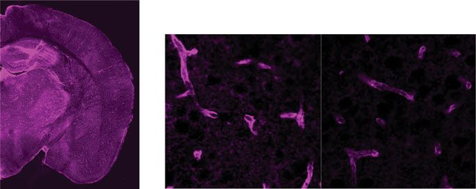
ARTICLE                                                                  NATURE COMMUNICATIONS | https://doi.org/10.1038/s41467-020-18115-2

S
      leep is an evolutionarily conserved biological function,               confirmed similar results (Mann–Whitney test: U = 4, p = 0.0260;
      clearing the brain of harmful metabolites such as amyloid              Supplementary Fig. 1a, b).
      beta that build up during wakefulness1 and consolidating                  After establishing a day/night difference of influx in vivo, we
memory2. Acute sleep deprivation impairs cognitive function3,4,              generated a full time course of glymphatic influx under KX
and sleep disruption is often an early correlate of neurodegen-              anesthesia (Fig. 1a, d). CM injections of CSF tracer were
erative disease5. Sleep quality is highly dependent on both sleep            performed every 4 hours across a standard 24 h light cycle, for
deficit and sleep timing controlled by circadian rhythms, 24 hour             a total of six time points. Using ex vivo tissue processing, we
cycles in gene transcription, cell signaling, physiology, and                prepared 100 µm thick coronal sections through the brains and
behavior6. Understanding how circadian rhythms contribute to                 quantified mean pixel fluorescence intensity across six slices
sleep quality is necessary to promote long-term brain health.                collected at 600 µm intervals starting at 1.2 mm anterior to
   During sleep, there is increased cerebrospinal fluid (CSF)                 bregma, to calculate the mean total tracer influx per entire brain
movement into the glymphatic system, promoting waste clear-                  (n = 10–12 mice per time point; Supplementary Fig. 1b, Supple-
ance1. The glymphatic system is a network of perivascular tunnels            mentary Fig. 2). Cosinor analysis of mean pixel intensity of brains
wrapped by astrocyte endfeet7. The localization of the water                 across a 24 h cycle revealed a significant rhythm in glymphatic
channel aquaporin-4 (AQP4) to astrocyte vascular endfeet pro-                influx with a peak at ZT6.2, around mid-day (cosinor analysis: F
motes CSF movement through the brain8. This fluid movement is                 (2, 67) = 5.949, p = 0.004; R2 = 0.151). The difference in influx
also dependent upon arterial pulsation9,10, and correlated to                between the day and night was ~22% (average day influx: 7.91 ±
decreased heart rate11. Although the cardiovascular system and               0.4 MPI, average night influx: 6.20 ± 0.3 MPI). Because these
sleep, two main influencers of the glymphatic system, are tightly             experiments were all performed under KX anesthesia, and some
regulated by circadian rhythms12, it remains unknown whether                 anesthetics have potential to shift circadian timing18, we
glymphatic fluid movement is under circadian control.                         generated full time courses under two additional anesthetics,
   CSF moves not only into the brain and spinal cord via the                 pentobarbital and 2,2,2-tribromoethanol (also known as avertin)
glymphatic system, but also drains out of the central nervous                (Fig. 1d; n = 7–11 mice per time point for each anesthetic).
system to the lymphatic system of the body13,14. In fact, lym-               Anesthesia impacted overall tracer influx, as reported pre-
phatic drainage may prevail when the animals are awake and                   viously11, where KX had significantly more influx (average influx
glymphatic influx is low15. The immune system is also regulated               under KX: 7.08 ± 0.3 MPI) when compared with pentobarbital
by circadian rhythms16,17, yet whether lymphatic drainage of CSF             (4.42 ± 0.1 MPI) or tribromoethanol (3.47 ± 0.1 MPI;
to the lymph nodes is under clock control has to be determined.              Kruskal–Wallis H(3) = 105.2, p < 0.0001). More importantly, we
   Here, we test the hypothesis that glymphatic influx of CSF                 show significant rhythms under both pentobarbital (cosinor
tracer from the cisterna magna (CM) into the brain, and clear-               analysis: F(2,52) = 5.527, p = 0.0069; R2 = 0.175) and 2,2,2-
ance of solute from the brain varies across the day independent of           tribromoethanol (cosinor analysis: F(2,49) = 6.479, p = 0.0035;
arousal state. We also test whether AQP4 may have a role in                  R2 = 0.209) with acrophases around mid-day (ZT7.8 and ZT6.6,
rhythmic glymphatic function. We show that glymphatic influx                  respectively) and similar amplitudes (Fig. 1e, f).
and clearance of a small tracer from the parenchyma is increased                In addition to using a cosinor analysis to determine
during the day compared with night in mice independent of the                rhythmicity and circadian parameters of glymphatic influx, we
type of anesthesia or light/dark cycle, corresponding to day/night           also analyzed the data using CircWave19–21. This re-analysis
changes in AQP4 localization. These rhythms in influx, clearance,             confirmed our previous findings, where the simplest fit of the data
and AQP4 persist after 10 days in constant light, indicating that            were a single-harmonic sinusoid curve that accounted for ~20%
the glymphatic system is under circadian control. AQP4 knock-                of the overall variance. We also compared the Center of Gravity
out (AQP4 KO) animals exhibit no day/night difference in                     (CoG), a measure of phase and mesor of the data sets, and found
glymphatic influx. In fully anesthetized animals, in vivo imaging             similar results to our 95% confidence intervals above, where
of mandibular lymph nodes showed increased tracer outflow                     anesthetic changed the mesor but each phase estimate was
during the night compared with day, when entry of CSF to the                 approximately mid-day (Supplementary Fig. 1c–e). These experi-
brain is low. This day/night difference in lymph node filling                 ments demonstrate that regardless of anesthesia, glymphatic
persists in constant conditions, and is absent in animals                    influx of CSF tracer into the brain exhibits diurnal variation with
without AQP4.                                                                peak influx around mid-day.
                                                                                Although glymphatic influx is significantly higher in the
                                                                             sleeping1 or anesthetized11 brain, we tested whether awake mice
Results                                                                      showed day/night variation of CSF influx. We found no
The glymphatic system exhibits diurnal variation. First, we                  significant difference of CSF tracer between the day (ZT4–8,
tested the hypothesis that there was a day/night difference in               n = 7 mice) and night (ZT16–20, n = 9 mice) in total influx,
influx of CSF tracer along the perivascular spaces around the                 anterior to posterior distribution, or in distinct subregions
middle cerebral artery (MCA) of the brain. Mice were anesthe-                (Supplementary Fig. 1f–h). This was unsurprising as the awake
tized with ketamine/xylazine (KX) at mid-day or mid-night (ZT6               brain has limited glymphatic function1.
or ZT18, respectively; ZT0 is lights on). In order to measure                   Glymphatic influx does not reflect function of the whole
glymphatic function, fluorescent tracer was injected into the CSF             glymphatic system, therefore we measured clearance of Evans
pool in the CM and allowed to circulate for 30 min prior to brain            blue (EB), a small molecule (960 Da) that freely diffuses through
collection. During circulation, fluorescent tracer movement was               the brain and binds tightly to albumin in the blood, from the
imaged through the skull of anesthetized mice (Fig. 1a–c, Sup-               brain to the femoral vein in anesthetized animals during the day
plementary Videos 1 and 2). We found ~53% more CSF tracer                    (n = 9 mice) and night (n = 6 mice). We chose this method
influx to the brain during the day compared with night in vivo                because although we cannot measure where in the vasculature EB
along the MCA (mean pixel intensity (MPI)) at 30 min during the              enters and binds to albumin, any fluorescence detected in the
day: 53.68 ± 8.5 MPI, n = 6 mice, night: 24.56 ± 7.1 MPI, n = 6              femoral vein had to be cleared from the brain parenchyma. Thus,
mice; t test: t(10) = 2.617, p = 0.0257). Because there is also              this approach enabled us to test the hypothesis that net clearance
increased CSF tracer in areas outside of cortex, we re-analyzed the          of EB from the brain is under diurnal control. Mice were
images using an ROI more tightly confined to the MCA and                      implanted with a microdialysis cannula into the striatum 24–36 h

2                NATURE COMMUNICATIONS | (2020)11:4411 | https://doi.org/10.1038/s41467-020-18115-2 | www.nature.com/naturecommunications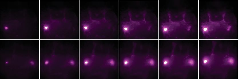
NATURE COMMUNICATIONS | https://doi.org/10.1038/s41467-020-18115-2 ARTICLE
a b 10 min 15 min 20 min 25 min 30 min c
ZT 2 6 10 14 18 22
100
80
* Day
Night
Mean Intensity (A.U.)
60 60
Day
40
40 20
0
30 min
20
Night
CM Tracer Tracer Distribution
Injection Analysis
0
0 10 20 30 (min)
d Pentobarbital Avertin
e f Phase Amplitude Mesor
Ketamine/Xylazine Amplitude
18 8 7
Mean Intensity (A.U.)
Mean Intensity (A.U.)
Mean Intensity (A.U.)
95% CI 95% CI 95% CI
Mean Intensity (A.U.)
Mesor 2.5 8
15 7 6 1
KX 2.0 7
Phase
12 6 5
0 1.5 6
9 5 4 P
KX 1.0 5
6 4 3 Pentobarbital
–1 A 0.5 4
3 Avertin
3 2 0.0 3
0 4 8 12 16 20 24 0 4 8 12 16 20 24 0 4 8 12 16 20 24 0 4 8 12 16 20 24 3 6 9 12
A
A
P
P
KX
KX
Zeitgeber Time (h) Zeitgeber Time (h) Zeitgeber Time (h) Zeitgeber Time (h) ZT (h)
Fig. 1 Glymphatic influx is higher during the day. a Schematic of experiment. b Representative images for in vivo recordings of CSF tracer influx after
injection in the CM either during the day (orange) or night (blue) under ketamine/xylazine (KX) anesthesia. Dotted white line indicates region of
quantification in c. White scale bar: 2 mm. c Mean pixel intensity in arbitrary units (A.U.) over 30 min post CM injection. Thick lines indicate group means
with SEM outlined in shaded regions. The inset is the magnitude of fluorescence at 30 min with boxplots, minima is minimum value, maxima is maximum
value, center is median and quartiles shown by box and whiskers, individual animals represented as colored dots, two-sided t test, p = 0.0257. Time 0 is
tracer infusion start. n = 6 mice per group. Asterisk indicates p < 0.05. d Time course of mean pixel intensity from ex vivo coronal sections under KX
(violet, n = ZT22: 10 mice; ZT18: 11 mice; ZT2, ZT6, ZT10, and ZT14: 12 mice), pentobarbital (red, n = ZT2: 7 mice; ZT6, ZT10, and ZT22: 9 mice; ZT18: 10
mice; ZT14: 11 mice), and Avertin (gray, n = ZT22: 7 mice; ZT2 and ZT6: 8 mice; ZT14: 9 mice; ZT10 and ZT18: 10 mice). Each colored point is an animal,
black points are mean ± SEM for each time bin. Dotted line indicates estimated cosinor curve. The light cycle is indicated by orange (day) and blue (night)
coloring in the background. Zeitgeber Time (ZT) in hours, where ZT0 is lights on and ZT12 is lights off. e Estimated cosine curves aligned at the mesor for
KX, pentobarbital (P), and avertin (A), with an inset depicting measurements from a cosine curve. f 95% confidence intervals for estimates of phase,
amplitude, and mesor for each anesthetic. Midline is the model generated estimate, bars indicate 95% range around the estimate. Source data are provided
as a Source Data file.
a b c d e Day f
100
*
Day Night 80 15
ns
60
Mean Intensity (A.U.)
60
40
% Area EB
10
45 20
0 Night
90 min
30
5
15 Day
Night
0 0
0 15 30 45 60 75 90 Day Night
EB injection evaluation
Time (min)
Fig. 2 Interstitial fluid clearance is higher during the day. a Schematic of experiment. b Representative Cy5 images of the femoral vein from animals
90 min after evans blue (EB) injection during the day (orange) or night (blue). White scale bar: 1 mm. c Mean pixel intensity in arbitrary units (A.U.) over
90 min post EB injection. Thick lines indicate group means with SEM shaded, individual points indicate time image was taken. The inset is a boxplot of the
magnitude of fluorescence at 90 min, two-sided t test, p = 0.0256. Time 0 is tracer infusion start. Day: n = 9 mice, night: n = 6 mice. Asterisk indicates p <
0.05. d Schematic of processing brains to check injection localization and specificity. e Representative striatal EB injection sites. Blue circle indicates probe
end. White scale bar: 1 mm. f Boxplot of percent area of EB quantified in a subset of brains during the day (n = 6 mice) and night (n = 4 mice). All boxplots:
minima is minimum value, maxima is maximum value, center is median and quartiles shown by box and whiskers, with individual animals shown as colored
dots, ns: not significant. Source data are provided as a Source Data file.
prior to experiments. At the time of experiment, the microdialysis Influx of CSF into the brain parenchyma is highly dependent
probe was inserted in the cannula, the femoral vein exposed, and upon localization of the water channel AQP4 to the endfeet of
placed under the macroscope (Fig. 2a). Imaging began at the astrocytes that surround brain vasculature7,8,22–24. Subsequently,
beginning of infusion of EB (4% in ACSF, 1 μL, 0.2 μL min−1). EB we asked if AQP4 localization to the vasculature exhibits diurnal
was significantly (t test: t(13) = 2.605, p = 0.0218) higher in the variation. Using immunohistochemistry in brain slices taken at
femoral vein as early as 15 min post infusion (day: 17.96 ± 1.1 MPI; ZT6 (n = 11 mice, 99 vessels) or ZT18 (n = 12 mice, 108 vessels),
night: 14.00 ± 0.8 MPI). By 90 min there was 37% more EB during we found increased polarization of AQP4 around vascular
the day than the night (t test: t(13) = 2.519, p = 0.0256; Fig. 2b, c). structures across the cortex during the day (Fig. 3a–c; t test:
There was no significant difference in percent area covered by EB t(21) = 4.173, p = 0.0004), with clear trends persisting in both the
in the brain after the experiment (Mann–Whitney test: U = 9, p = hypothalamus (n = 4 mice per group; t test: t(6) = 2.342, p =
0.6095; Fig. 2d–f), indicating that infusion volumes were similar in 0.0577) and CA1 of the hippocampus (n = 4 mice per group;
each group. We conclude that, similar to glymphatic influx, Welch’s t test: t(3.109) = 2.139, p = 0.1188) (Supplementary
clearance of solutes from the brain is increased during the day and Fig. 3). We next asked whether this change of localization
decreased during the night, supporting the notion that global corresponded to a change in total amounts of AQP4. Using
glymphatic function is upregulated during the day. western blot to measure protein in whole-brain extract, we found
NATURE COMMUNICATIONS | (2020)11:4411 | https://doi.org/10.1038/s41467-020-18115-2 | www.nature.com/naturecommunications 3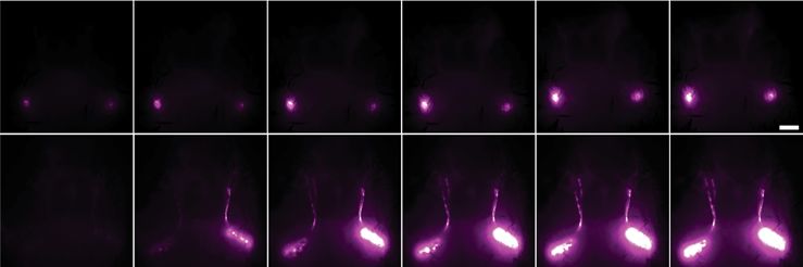
ARTICLE NATURE COMMUNICATIONS | https://doi.org/10.1038/s41467-020-18115-2
a b c d D N
e f
AQP4 Lectin Merge 25 1.1
Day
* 250
2.5
ns
2.0
Polarization Index
150
Day
20 Night ns
Intensity (A.U.)
0.9 100 2.0
Ratio M1/M23
1.5
AQP4 / Actin
15
AQP4 50
0.7 1.5
10 M1 37 1.0
AQP4 Lectin Merge 1.0
0.5 M23 25
5
Night
15 0.5
0.5
0 0.3
–10 0 10 Day Night 50 0.0 0.0
βActin
37 kDa Day Night Day Night
Distance (μm)
g Dystrophin-Associated Complex (DAC) h 1
Control DAC Putative DAC
Perivascular space Day
Dystroglycan Aquaporin-4
0
Fold change
(DAG1) (AQP4) Night
–1
ns ns ns
–2 ns ns
Dystrophin ns
(DMD)
α-syntrophin * * *
Dystrobrevin (SNTA1)
–3 * *
Astrocyte endfoot
(DTNA)
Arntl1 Arnt Dag Dmd Dtna Snta Aqp4 Gja1 Gjb6 Mlc1 Slc4a4
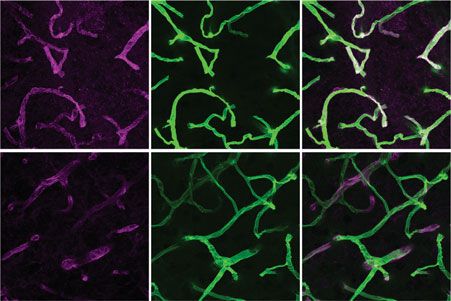
Fig. 3 Diurnal variation in AQP4 protein and localization. a Representative ×63 magnification confocal images during the day (ZT6) and night (ZT18).
Purple is AQP4, green is vascular staining, white is overlap between both AQP4 and vascular staining. White scale bar: 50 µm. b Average intensity of
AQP4 staining centered on vasculature in the cortex during the day (orange, for b, c: n = 11 mice, 99 vessels) and night (blue, for b, c: n = 12 mice, 108
vessels), with mean ± SEM indicated by the thick line (mean) with shading (SEM). c Average polarization index boxplot. Polarization index equals peak
vascular end foot fluorescence minus 10 µm baseline. All values were normalized to the highest signal for ease of visualization. Two-sided t test, p =
0.0004. d Representative western blot for AQP4 and βActin loading control during the day (orange) and night (blue). Molecular weights are indicated to
the right. M1 and M23 splice variants of AQP4 monomers are indicated on the left. e Bloxplot of mean AQP4 densitometry normalized to the loading
control. For e, f: n = 15 mice per group. f Bloxplot of the ratio of M1/M23 densitometry, both normalized to loading control. g Schematic of key components
of the dystrophin-associated complex. h Bar plot of day (ZT6) and night (ZT18) mRNA expression of target genes assessed by RT-qPCR means with error
bars indicating SEM. Gray dots are individual mice. n = 5 mice per group. Two-sided Welch’s t tests: Arntl1, p = 0.037; Dag, p = 0.0483; Dtna, p = 0.0283;
Aqp4, p = 0.038; Gja1, p = 0.045. DTNA, DMD, DAG1, AQP4, GJA1, GJB6, MLC1, and SLC4A4 are all upregulated in astrocytes at minimum two times
more than any other cell type30. All boxplots: minima is minimum value, maxima is maximum value, center is median and quartiles shown by box and
whiskers, with individual animals shown as colored dots. Asterisk indicates p < 0.05; ns: not significant. Source data are provided as a Source Data file.
no difference in total AQP4 between ZT6 and ZT18 (Fig. 3d, e, and putatively associated with the DAC27. We found only Gja1
day: n = 15 mice, night: n = 15 mice), or in higher molecular exhibited day/night differences in gene expression, with a
weight bands of AQP4 complexes (Supplementary Fig. 4). In reduction during the night (Welch’s t test, t(7.201) = 2.43, p =
mice, M1 or M23 splice variants can determine localization of 0.0445). This may indicate that temporal localization of
AQP4, with higher levels of M23 at astrocytic endfeet25,26. connexin-43 to astrocytic endfeet is regulated by the DAC.
Although we found no difference in intensity of the M1 or M23 Increased gene expression of key components of the DAC,
bands between day and night (Supplementary Fig. 4), we found a including AQP4, match the increased localization of AQP4 and
trend in the ratio of M1 to M23 being lower during the day (day: increased glymphatic influx of CSF into the brain during the day.
0.67 ± 0.04 M1/M23; night: 0.82 ± 0.07 M1/M23; Welch’s t test, t
(22.41) = 1.912, p = 0.0688; Fig. 3f), supporting the hypothesis Influx clearance and AQP4 rhythms persist in constant light.
that AQP4 localization to perivascular endfeet during the day Next, we tested the hypothesis that diurnal changes in glymphatic
promotes increased glymphatic influx. CSF tracer influx are not driven by cycles of light and dark (LD).
AQP4 localization to the vascular endfeet of astrocytes is We chose constant light (LL) over constant dark (DD) to avoid
dependent upon the dystrophin-associated complex (DAC)27–29, effects of light pulses that are inevitable during live-animal sur-
components of which exhibit highly enriched gene expression in gery. The surgery takes ~10 min, more than enough to phase shift
astrocytes (Dtna, Dmd, Dag1, Aqp4) according to a cell-specific mice in constant darkness32,33. Mice were housed in constant
transcriptome data base30. We hypothesized that day/night light (LL) for 10 days with continuous activity monitoring to
changes in gene expression for components of this complex ensure chi-squared periodogram estimates of free-running period
may support rhythmic localization of AQP4 to perivascular were accurate34. Because long-term (>30 days) LL induces
endfeet of astrocytes. Brains were harvested at ZT6 (n = 5 mice) behavioral arrhythmicity and altered SCN organization35,36, we
and ZT18 (n = 5 mice), then reverse transcription PCR (RT- wanted to ensure that general cage activity was unchanged in LL
qPCR) was used to measure gene expression. First, we measured (n = 6 cages of 2 mice) compared with DD (n = 10 cages of two
the clock gene Arntl1 and the non-clock gene homolog Arnt31. As mice). Using chi-squared periodogram analysis we found animals
expected, there was a significant day/night difference in Arntl1 in DD and LL had similar variability in periodicity, and every
(Welch’s t test, t(7.912) = 2.504, p = 0.037; Fig. 3h), and no cage had significant Qp values from a chi-squared periodogram
difference in Arnt. Next, we measured gene expression of key analysis (Supplementary Fig. 5a–c), though the amplitude of the
components of the DAC: Dag1, Dmd, Dtna, Snta1, and Aqp4 Qp was reduced in the constant light group (DD: 1016.0 ± 61 Qp,
(Fig. 3g, h). We found significant increased expression of Dag, LL: 757.7 ± 85 Qp; Welch’s t test: t(10.13) = 2.477, p = 0.0325).
Dtna, and Aqp4 during the day compared to the night (Welch’s Similar to previous reports, mice in LL had an average period of
t tests, Dag: t(7.874) = 2.335, p = 0.0483; Dtna: t(6.272) = 2.837, 25.35 ± 0.1 h. Using average activity profiles for 10 days in LL and
p = 0.0283; Aqp4: t(7.973) = 0.038). Both Dmd and Snta1 had DD, we found no evidence of splitting in LL (Supplementary
similar trends of expression, but the day/night differences were Fig. 5d). Although there was a trend toward decreased activity,
not significant (Fig. 3g). In addition to AQP4, several genes specifically in the active phase in LL (t test: t(14) = 2.089, p =
encoding gap junctions (Gja1, Gjb6), cation channels (Mlc1), and 0.0555; Supplementary Fig. 5e, f), there was no significant dif-
bicarbonate transporters (Slc4a4) are upregulated in astrocytes30, ference in the average amount of activity between light cycles
4 NATURE COMMUNICATIONS | (2020)11:4411 | https://doi.org/10.1038/s41467-020-18115-2 | www.nature.com/naturecommunications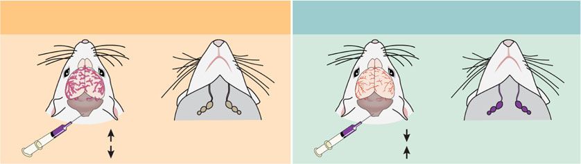
NATURE COMMUNICATIONS | https://doi.org/10.1038/s41467-020-18115-2 ARTICLE
a Rest
b c
Tracer distribution Rest Active
analysis 60 *
12
Mean Intensity (A.U.)
Activity Counts
45
KX 10
0 10 days CM injection 30 8
Days in constant light
1
2 Active
3 15 6
4 AQP4 IHC
5
4
6
7
0
8 KX 0 4 8 12 16 20 24 2
9 Rest Active
0 24 48 CM injection Circadian time (h)
Time (h)
d e f g
20 Rest
DC 1.1 *
Polarization Index
Rest Active
Intensity (A.U.)
16 Active
12 0.9
LC
8
0.7
4
0 0.5
VC –10 0 10 Rest Active
Distance (µm)
h i j k
150 * ns
25
120
Mean Intensity (A.U.)
100
Rest Active 20
% Area EB
90 50 15
60 90 min
10
30 Rest 5
Active
0 0
0 15 30 45 60 75 90 Rest Active
Time (min)
Fig. 4 Differences in the glymphatic system persist in constant light. a Experimental outline with representative double-plotted actogram of two animals
in constant light (LL). Black tick marks: beam-breaks. b Average activity profile of animals housed in LL for 10 days. Black line and gray error bars: group
mean ± SEM, thin gray lines: individual cage activity profiles. Yellow: rest phase, green: active phase. Circadian time (CT) 12 is activity onset. n = 7 cages,
14 mice. c Boxplot of mean intensity (arbitrary units, A.U.) from ex vivo tissue fluorescence. Mann–Whitney test: p = 0.0033, n = 13 mice rest, 14 mice
active. d Representative slice of AQP4 staining. White boxes indicate areas of dorsal, lateral, and ventral cortex (DC, LC, VC, respectively) for AQP4
localization quantification. White scale bar: 500 µm. e Representative ×40 images stained for AQP4 (magenta). White scale bar: 50 µm. f Average
intensity of AQP4 centered on vasculature in cortex during the rest (yellow, for e, g: n = 5 mice, 45 vessels) and active (green, for f, g: n = 7 mice,
63 vessels) phase, mean ± SEM is indicated by shading (SEM) around thick line (mean). g Boxplot of polarization index for staining in ventral, dorsal and
lateral cortex. Polarization index equals peak fluorescence minus 10 µm baseline. All values normalized to the highest signal for better visualization. Two-
sided t test: p = 0.0288. h Schematic of clearance experiment. i Representative Cy5 images of the femoral vein 90 min after evans blue (EB) injection
during rest (yellow) or active (green) phases. White scale bar: 1 mm. j Mean intensity over time. Thick lines: group means, outlines: SEM, individual points:
image acquisition. Inset: boxplot of fluorescence at 90 min. Two-sided t test: p = 0.0051. Time 0 is tracer infusion start. For j, k: n = 5 mice per group.
k Boxplot of percent area EB during rest and active phases. All boxplots minima: minimum value, maxima: maximum value, center: median, and quartiles:
box and whiskers, individual animals: colored dots. Asterisk indicates p < 0.05; ns: not significant. Source data are provided as a Source Data file.
(t test: t(14) = 1.727, p = 0.1062). Thus, 10 days in LL does not rhythm in glymphatic function is under circadian control, as
drastically alter circadian behavior, indicating the circadian sys- opposed to being driven by changes in the light cycle.
tem is intact.
On day 11, animals were anesthetized with KX, received a CM Lymphatic drainage rhythms are opposite to glymphatic influx.
injection of CSF tracer, and ex vivo glymphatic influx analyses Glymphatic function is increased during sleep1 and drainage of
were performed during the mid-rest (circadian time 4–8, CT 4–8, CSF to the lymph nodes is increased when awake15. Because we
where CT 12 is activity onset) or mid-active phase (CT 16–20) of observed a strong underlying circadian rhythm in glymphatic
the animal’s behavior (Fig. 4a, b). Influx was increased during the influx, our next question was whether CSF outflow via the cer-
rest phase compared with the active phase of animal behavior vical lymphatic system showed circadian variation. Mice received
(Fig. 4c; rest: 10.10 ± 0.2 MPI, active: 8.00 ± 0.6 MPI, n = 13–14 CM injections of tracer under KX anesthesia, as discussed above,
mice per group; Mann–Whitney test: U = 32, p = 0.0033). In during the day (n = 12 mice) or night (n = 15 mice). The man-
addition, AQP4 polarization to vascular endfeet was increased dibular lymph nodes were then exposed and imaged for 50 min
during rest phase of the animal (rest: n = 5 mice, 45 vessels, post CM. We found that filling of the submandibular lymph
active: n = 7 mice, 63 vessels; t test: t(10) = 2.552, p = 0.0288; nodes was 46% lower during the day compared with the night
Fig. 4d–g). Finally, we performed EB clearance assays, as (Fig. 5a–e; t test: t(25) = 2.25, p = 0.0335). This effect persisted
described above, during the rest phase (CT 2–7, n = 5 mice) or after 10 days in constant light (Fig. 5f–j; rest: n = 8 mice, active:
active phase (CT 14–19, n = 5 mice). Similar to LD, clearance was n = 6 mice; t test: t(12) = 2.603, p = 0.0231). Appearance of tracer
significantly increased by 55% during the rest phase compared in the lymph node occurs as early as the first frame of recording
with the active phase at 90 min (t test: t(8) = 3.823, p = 0.0051), (on average 8 min post tracer infusion start; Fig. 5e, g), similar to
with no significant differences in percent coverage of EB in the first detectable tracer movement along the perivascular space of
brain (Fig. 4h–k). These data support the hypothesis that the the MCA (5 min post injection; Fig. 1c), suggesting that the CSF
NATURE COMMUNICATIONS | (2020)11:4411 | https://doi.org/10.1038/s41467-020-18115-2 | www.nature.com/naturecommunications 5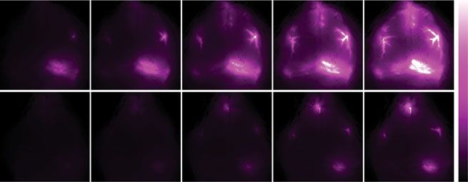
ARTICLE NATURE COMMUNICATIONS | https://doi.org/10.1038/s41467-020-18115-2
a Day
b c d e 0 min 10 min 20 min 30 min 40 min 50 min
90 Day
Mean Intensity (A.U.)
*
Mean Intensity / min
5
Mean Intensity (A.U.)
Night 160 *
Day
60 4
120
3
80
Night 30 2
40 1
Night
0 0 0
0 10 20 30 40 50 Day Night Day Night
Time (min)
f Rest
g h i j 0 min 10 min 20 min 30 min 40 min 50 min
150 Rest
Mean Intensity (A.U.)
ns
Active * 8
Mean Intensity (A.U.)
180
Mean Intensity / min
Rest
100 135 6
Active 90 4
50
45 2
Active
0
0 10 20 30 40 50 0 0
Rest Active Rest Active
Time (min)
k l m Summary – Circadian regulation of CSF distribution
Contractions per minute 8 ns
Subjective day Subjective night
6
ΔF
30 sec 4
2
0 Brain Brain
ΔF
Day Night Rest Active Lymph nodes Lymph nodes
30 sec
Fig. 5 Lymph node drainage exhibits circadian variation. a Experimental outline. Lymph nodes in purple, black dashed square: area imaged. White dashed
lines: regions of interest (ROI) around lymph nodes. b Mean intensity (arbitrary units, A.U.) of lymph node fluorescence during day (orange, for b–e: n = 12
mice) and night (blue, for b–e: n = 15 mice). Time 0 on all graphs indicate start of imaging, on average 8 min after tracer infusion start. Thick line: mean,
shading: SEM. c Boxplot of lymph node intensity at minute 50. Two-sided t test, p = 0.0335. d Boxplot of the rate of lymph node filling calculated from the
first 20 min recording. Mann–Whitney test, p = 0.0321. e Representative images from in vivo lymph node imaging across 50 min for day (orange) and
night (blue). White scale bar: 2 mm. f Experimental outline. g Average fluorescence over time (thick line: mean, SEM: shading). h Boxplot of fluorescence in
lymph nodes at 50 min, during active (green, for f–j: n = 6 mice) and rest (yellow, for f–j n = 8 mice) phases. Two-sided t test, p = 0.0231. i Boxplot of rate
of lymph node filling. j Representative time lapse images for animals housed in constant light for 10 days. White scale bar: 2 mm. k Diagram of experiment
for lymph node contraction rate after FITC-dextran cheek injection, black dashed square: imaging area (left) with representative lymph vessel and trace of
contractions from two regions of interest over 2 min (right). White scale bar: 1 mm. White dotted lines: area of measurement for traces. l Boxplot of
contractions per minute of lymph vessels during the day (n = 5 mice) and night (n = 7 mice) in LD, and active (n = 7 animals) and rest (n = 7 animals)
after 10 days in constant light. m Summary diagram of circadian regulation of CSF distribution. All boxplots minima: minimum value, maxima: maximum
value, center: median, box and whiskers: quartiles, individual animals: colored dots. Asterisk indicates p < 0.05; ns: not significant. Source data are provided
as a Source Data file.
evident in the mandibular lymph nodes has not entered the glym- night n = 5 mice) and in 10 days LL (rest: n = 7 mice; active: n =
phatic system. If CSF had entered the glymphatic system, we would 7 mice) we found no significant differences in contractions per
have expected a delay between influx into the brain and lymphatic minute between the rest and active phases (two-way analysis of
drainage we measure in the lymph nodes. We also calculated the rate variance (ANOVA), main effect of phase: F(1, 22) = 0.603, p =
of lymph node filling during the first 20 min of recording, before the 0.4457; interaction light*phase: F(1, 22) = 0.173, p = 0.6815). We
majority of recordings reached a plateau of fluorescent intensity conclude that intrinsic contraction rate of the mandibular lymph
(Fig. 5d, i). We found lymph nodes filled significantly faster during node vessels is not responsible for circadian drainage of CSF to
the night than the day (night: 2.04 ± 0.4 MPI min−1, day 0.87 ± 0.3 the lymph nodes.
MPI min−1; Mann–Whitney test: U = 46, p = 0.0321) with a trend
for this pattern to persist in constant conditions (active: 3.60 ± 1.0 Loss of AQP4 eliminates circadian CSF distribution. We per-
MPI min−1, rest: 1.81 ± 0.6 MPI min−1; Mann–Whitney test: U = formed CM injections of CSF tracer (BSA647) into AQP4 KO
12, p = 0.1419), indicating there are diurnal timing mechanisms for mice during the day (ZT4–8; n = 10 mice) or night (ZT14–20;
how fast the CSF can distribute into different compartments. We n = 10 mice) to test whether this day/night difference in AQP4 is
conclude that CSF drainage to the cervical lymph nodes is under necessary for day/night differences in glymphatic influx. AQP4
circadian control with a peak during the active phase that temporally KO animals did not have a day/night difference in glymphatic
is in antiphase to CSF entry into the glymphatic system of the brain influx, as measured by average slice intensity (t test: t(18) = 1.511,
(Fig. 5m). p = 0.1481; Fig. 6a–c). Because lymphatic drainage shows circa-
Lymphatic vessels contract to draw fluid into the lymph dian variation that was temporally antiphase to glymphatic influx,
nodes37. We next tested whether circadian variation in mandib- we did the same lymph node drainage assay described above in
ular lymph node filling was dependent on circadian differences in the AQP4 KO mice during the day (n = 6 mice) and night (n = 7
lymph vessel contraction rate (Fig. 5k, l). KX anesthetized mice mice). There was no difference in lymph node drainage after
were injected subcutaneously in the cheek with fluorescein 50 min between day and night (t test: t(11) = 1.103, p = 0.2937;
isothiocyanate (FITC)-conjugated 3 kD dextran (0.25% in normal n = 6–7 mice), nor in the slope of drainage calculated from the
saline, 20 µL per cheek), then placed on their backs with the first 20 min of recording (t test: t(11) = 0.9202, p = 0.3772;
mandibular lymph nodes exposed via skin resection. Imaging Fig. 6d–h).
acquisition began following lymph vessel filling (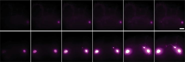
NATURE COMMUNICATIONS | https://doi.org/10.1038/s41467-020-18115-2 ARTICLE
a b 10 c distribution, we next pooled LM with C57 values at 30 min post
pump start (Fig. 5) (WT day: n = 15 mice, WT night: n = 18
Day ns
Mean Intensity (A.U.)
Day Night
8
mice). Overall, drainage to the mandibular lymph nodes was
Night
6 different between groups (Kruskal–Wallis test: H(3) = 8.464, p =
4 0.037; Fig. 6b). Prior to Bonferroni correction, WT day vs. night,
2 and WT night vs. KO night comparisons were significantly
Day Night
different (p = 0.020 and p = 0.011, respectively) supporting
d e f g increased drainage only during the night in WT mice, though
Day
this effect was gone after correction.
Mean Intensity (A.U.)
Mean Intensity (A.U.)
ns ns
Mean Intensity / min
60 Day 120 2.5
40
Night
90 2.0 Overall, our observations support the hypothesis that daily
Night 60
1.5
changes in glymphatic function and drainage of CSF to the lymph
20 1.0
30 0.5
nodes is supported by daily rhythms in polarization of AQP4 to
0
0 10 20 30 40 50 0 0.0 the vascular endfeet of astrocytes.
Time (min) Day Night Day Night
h 0 min 10 min 20 min 30 min 40 min 50 min
Discussion
Here, we show the difference in glymphatic function is not solely
Day
based on arousal state, but exhibits a daily rhythm that peaks
mid-day when mice are mostly likely to sleep. Glymphatic influx
under three different anesthesia paradigms (ketamine-xylazine,
Night
pentobarbital, and avertin), and an increase in clearance of EB
from the brain parenchyma to the periphery indicate that
Fig. 6 AQP4 KO animals lack a rhythm in glymphatic influx. glymphatic system function peaks during the day. Conversely,
a Experimental outline for b. b Boxplot of mean pixel intensity of slices drainage of CSF tracer to the mandibular lymph nodes is highest
from AQP4 KO animals during the day (orange) and night (blue). n = 10 in anesthetized mice during the night, when animal activity will
mice per group. c Population-based averaging of slice intensity in AQP4 be at its peak. Day/night differences in glymphatic influx, solute
KO animals in the day and night. d Experimental outline. e Average clearance, and CSF drainage to the lymph nodes persists under
fluorescence over time, mean is the thick line, SEM indicated by shading. constant light, supporting the hypothesis that these are endo-
f Boxplot of fluorescence in the lymph nodes at 50 min. g Boxplot of the genous circadian oscillations. Circadian glymphatic function is
rate of lymph vessel filling for the first 20 min. h Time lapse imaging across supported by circadian regulation of AQP4 polarization in
50 min in AQP4 KO mice during the day (orange, for d–h: n = 6 mice) and astrocytes, and genetic deletion of AQP4 effectively eliminates the
night (blue, for d–h: n = 7 mice). White scale bar: 2 mm. Time 0 on all circadian regulation of CSF distribution detected as an absence of
graphs indicate start of imaging, on average 8 min after tracer infusion start. day/night differences both in cortical tracer influx and in drainage
All boxplots: minima is minimum value, maxima is maximum value, center of tracer to the lymph node.
is median and quartiles shown by box and whiskers, with individual animals Recent work has provided evidence that astrocytes in the clock
shown as colored dots. ns: not significant. Source data are provided as a center of the brain, the suprachiasmatic nucleus (SCN), may be
Source Data file. responsible for setting periodicity of circadian behavior38. In
addition, restoring molecular clock rhythms in SCN astrocytes is
sufficient to drive rhythmic sleep/wake behavior in animals that
published7,8. Next, we tested whether this persists during the are genetically arrhythmic39. Our data provide evidence sup-
night, and whether there are LM/KO day/night differences in porting the idea that astrocytic regulation of glymphatic rhythms
drainage to the lymph nodes. Using the ex vivo tissue processing occurs at least in part via AQP4 vascular polarization in the
as described above, we found no significant difference in cortex, which is supported by rhythmic gene expression of key
nighttime influx (t test: t(14) = 0.2208, p = 0.8284) between LM components in the DAC. Deletion of the molecular clock in
(n = 3 mice) and KO animals (n = 13 mice; three mice in astrocytes induces mis-regulation of AQP4 gene expression,
addition to those shown in Fig. 6b) (Supplementary Fig. 6a). supporting our hypothesis that the DAC is under molecular clock
Previous work has shown that LM controls for AQP4 KO animals control40. Circadian regulation of proteins in the DAC beyond
have similar day-time influx compared to C57BL6 (C57) AQP4 may provide insight into conserved astrocyte clock func-
animals8. Because our animals are on a C57 background, we tion controlling the release and buffering of extracellular signals
grouped the littermate controls with the nighttime C57 under KX such as glutamate, as in the SCN41, or regulating sleep- and wake-
from Fig. 1. All three LM fit within the median quartiles of the promoting extracellular ion concentrations and fluid transport
C57 data, indicating they are comparable. Though trending throughout the cortex1,42. Our data show that expression of
toward decreased influx in KOs, there was no significant connexin-43 (Cx43, Gja1) is diurnally regulated in a pattern
difference between WT (n = 37 mice) and KO influx in the similar to genes in the DAC, consistent with Cx43 being highly
brain (t test: t(48) = 1.962, p = 0.0556). enriched in the vascular endfeet of astrocytes43, and provide
To measure lymph node drainage, we compared fluorescence additional mechanisms for clock-controlled astrocytic signaling.
in the mandibular lymph nodes after 30 min of CSF circulation These observations support the notion that astrocytes stabilize
during the day and night in LM (n = 3 mice per time point) and and perhaps drive circadian behavior, in part by regulating bulk
KO (day: n = 9 mice, night: n = 10 mice; three additional mice fluid movement through and CSF/ISF exchange across the brain
per group pooled with 30 min from pump start in Fig. 6) to entrain circadian rhythms.
(Supplementary Fig. 6b). With the small sample size, we found no CSF is produced in the choroid plexus (CP) of the brain, an
significant differences between genotypes (two-way ANOVA: F epithelial layer of cells located within the ventricles. The CP
(1,21) = 0.184, p = 0.672), time (F(1,21) = 0.191, p = 0.667), or exhibits robust cycling of the molecular clock in vitro, and can tune
the interaction between the two factors (F(1,21) = 0.902, p = periodicity of molecular rhythms in SCN co-cultures and in
0.353). Because our AQP4 KO line is backcrossed to C57, and the behavior44. In addition, CSF production in humans may be
majority of LM fell within the two median quartiles of drainage rhythmic with peak CSF production during the night45,46, though
NATURE COMMUNICATIONS | (2020)11:4411 | https://doi.org/10.1038/s41467-020-18115-2 | www.nature.com/naturecommunications 7
ARTICLE                                                                    NATURE COMMUNICATIONS | https://doi.org/10.1038/s41467-020-18115-2

results may be inconclusive47. This poses at least two potential               Methods
paths the CP might regulate glymphatic rhythms: first, by reg-                  Animals. Male and female C57BL/6 mice (aged 3–5 months, weight between 25 g
ulating concentration of signaling molecules in the CSF and second             and 30 g) were acquired from Charles River Laboratories (Wilmington, MA) in
                                                                               equal numbers for each experimental group to control for any potential sex dif-
by mechanically supporting glymphatic influx by providing a larger              ferences. Male and female AQP4 KO mice62 were bred in the University of
volume of CSF during the rest phase. When improved metho-                      Rochester vivarium, and backcrossed to C57BL/6 mice for 20+ generations before
dology for CSF production in rodents becomes available48,49, it                use. A minimum of five mice were used in each group, with exact animal numbers
should be investigated whether CSF production regulates glym-                  stated in the results and figure legends. Mice were group-housed either in a 12:12
                                                                               light/dark cycle or under constant light with ad libitum access to food and water.
phatic CSF influx along the perivascular spaces of the brain.                   All experiments were approved by the University of Rochester Medical Center
   Proulx and colleagues recently proposed a model whereby                     Committee on Animal Resources. All of the University of Rochester’s animal
rapid CSF turnover through lymphatics precludes significant bulk                holding rooms are maintained within temperature (18–26 degrees Celsius) and
flow into the brain15. This was based on two main observations:                 humidity ranges (30–70%) described in the ILAR Guide for the Care and Use of
                                                                               Laboratory Animals (1996). All efforts were made to keep animal usage to a
no tracer enters perivascular spaces of penetrating arterioles prior           minimum.
to death based on macroscopic imaging, and lymphatic outflow is                     For constant light experiments, animals were housed two per cage to reduce risk
increased in wakefulness, whereas glymphatic influx is decreased                of hypothermia63. Activity was monitored continuously in 5 min bins via the
along the MCA. Our laboratory has already demonstrated CSF                     Comprehensive Lab Animal Monitoring System (Columbus Instruments).
                                                                               Circadian behavioral analysis was completed with ActogramJ. Free-running period
tracer alongside penetrating arterioles in live animals using two-             of each cage was determined using at least 10 days of activity and a χ2 periodogram,
photon microscopy1,7,9, which has considerably higher spatial                  which was used to confirm behavioral rhythmicity and estimate experimental times
resolution than macroscopic imaging10,50. In addition, we have                 in conjunction with activity onset of the cage.
used rapid decapitation followed by drop fixation to process all of
our brain samples since 2017 to reduce the death15 and perfusion               Drugs. Anesthesia was administered as follows: 100 mg kg−1 racemic ketamine and
artifact10 of CSF tracer localization in the brain. Here, we col-              20 mg kg−1 xylazine kg i.p. (KX); pentobarbital 60 mg kg−1 i.p.; 2,2,2-tri-
lected both in vivo and ex vivo data in fully anesthetized mice and            bromoethanol (also known as Avertin) 120 mg kg−1 i.p.. Depth of anesthesia was
                                                                               determined by the pedal reflex test; if the mouse responded to toe pinch, an
observe changes in glymphatic and lymphatic CSF distribution                   additional one-tenth the initial dosage was given and the tracer experiment delayed
dependent upon circadian timing, with underlying molecular                     until full unconsciousness was obtained. Directly prior to CM infusion, the animal
rhythms in the DAC and AQP4 polarization. Based on these data,                 received an additional one-tenth the initial dosage, and the pedal reflex was tested
we propose that the lack of glymphatic influx is not simply a                   every 5–10 min during the tracer circulation time to ensure proper anesthesia
                                                                               throughout the study. Animals under avertin anesthesia were most likely to need
downstream effect of the lymphatic system drainage of CSF                      supplemental dosing.
before it reaches the brain. Instead, the awake brain actively
suppresses periarterial influx10, has less perivascular AQP4, and
                                                                               Intracisternal CSF tracer infusion. Fluorescent CSF tracer (bovine serum albu-
prevents CSF/ISF exchange by reducing the interstitial space                   min, Alexa FluorTM 647 conjugate; 66 kDa; Invitrogen, Life Technologies, Eugene,
volume1. This would cause CSF to follow alternative, pre-existing              OR) was formulated in artificial CSF at a concentration of 0.5% weight by volume.
routes out of the subarachnoid space around the brain and spinal               Anesthetized mice were fixed in a stereotaxic frame, the CM surgically exposed,
cord, such as into the mandibular lymph nodes, meningeal                       and a 30 gauge needle connected to PE10 tubing filled with the tracer was inserted
                                                                               into the CM. Ten microliters of CSF tracer was infused at a rate of 2 μl min−1 for
lymphatics, and/or the deep cervical lymph nodes13,51. In short,               5 min with a syringe pump (Harvard Apparatus)10. For the awake animal cohort:
CSF distribution will change based on fluid changes and space                   mice were anesthetized with 1–2% isoflurane, and 0.25% bupivicane was applied
availability within the brain.                                                 topically to the surgery site at the beginning of and after recovery from the pro-
   The mechanisms behind brain-regulated suppression of                        cedure. Mice were allowed to resurface from anesthesia before pump start. Total
                                                                               volume of CSF tracer was increased to 12 µL to compensate for decreased glym-
glymphatic activity during wakefulness remain unknown. EEG                     phatic function, pump speed remained the same (2 μl min−1).
activity tightly correlates to glymphatic influx and the patterns of                To visualize tracer movement from the cisternal compartments into the brain
EEG activity that resemble wakefulness strongly suppress CSF                   parenchyma, the animals were killed by decapitation and the brain removed 30 min
influx11. One hypothesis is that the synchronous, slow wave firing               after the start of intracisternal infusion (note that the needle was left in place after
                                                                               the infusion to prevent backflow of CSF). The brain was fixed overnight by
characteristics of non-REM sleep drive consolidated movement of                immersion in 4% paraformaldehyde in phosphate-buffered saline (PBS). Coronal
extracellular ions across cortex, supporting pulsating inflow of                vibratome slices (100 μm) were cut and mounted. Tracer influx into the brain was
CSF into the neuropil. This process would not be compatible with               imaged ex vivo by macroscopic whole-brain and whole-slice conventional
the asynchronous patterns of neuronal activity that characterize               fluorescence microscopy (Olympus; Stereo Investigator Software).
the awake brain.                                                                   Tracer influx was quantified by a blinded investigator using ImageJ software11.
                                                                               The cerebral cortex in each slice was manually outlined, and the mean fluorescence
   There are multiple circadian rhythms that may interact to                   intensity within the cortical ROIs was measured. An average of fluorescence
promote rhythmic glymphatic function. Recent work hypothe-                     intensity was calculated between six slices for a single animal, resulting in a single
sized increased glymphatic clearance during the sleep phase,                   biological replicate (Supplementary Fig. 2). Equivalent slices were used for all
driving rhythmic fluid flow in the brain52. Cortical neuronal                    biological replicates.
activity as measured by EEG has strong circadian components in
humans53,54. The cardiovascular system as a whole is under tight               Transcranial imaging. Animals were anesthetized with KX at ZT6 or ZT18 and
                                                                               were prepared for transcranial imaging64,65. In brief, animals were placed in a
circadian control55, and arterial pulsatility is a key driving factor in       stereotaxic frame and the skin was removed from the top of the head to expose the
transport of CSF along the penetrating arteries9,10. In flies, there is         skull. The animal was then placed under the macroscope (MVX10 Research Macro
evidence of a circadian-clock in the equivalent of the blood–brain             Zoom Microscope, Olympus). A CM injection was then performed as described
barrier56,57. Finally, immune system functionality is regulated by             above, with imaging beginning at pump start. Image acquisition was once per
                                                                               minute on the Cy5 channel, 30 min total (31 images per animal).
circadian timing16,17,58 and we describe a clear interaction of CSF
entry to the brain and lymphatic clearance of CSF. Although
glymphatic function has yet to be studied in models of circadian               Parenchymal clearance assay. All experiments occurred between CT 2–7 or CT
                                                                               14–19 depending on if it was the rest or active phase of the animal, respectively.
disruption, such as in shiftwork, it has been established that shift           Cannulas (guide cannula: 26 G, C315G SPC, 4.5 mm bellow pedestal, dummy:
workers are at increased risk for neurodegenerative disorders,                 33 G, C315DC/SP, 0.1 mm projection; PlasticsOne, Roanoke, VA) were implanted
cardiovascular disease, and exhibit increased markers of systemic              into mice 24–36 h prior to experimentation into striatum (AP: +0.6 from Bregma,
inflammation16,59–61. Understanding how these rhythms, all with                 LM: −2 and DV: −3.25 mm). At the appropriate experimental time points, mice in
                                                                               either LD or 10 days LL were anesthetized with KX and fitted with a microdialysis
different timing and biological functions, interact to affect glym-            probe (inner cannula: 33 G, C315I/SP, 0.1 mm projection), had skin over the
phatic function and lymphatic drainage may help prevent mor-                   femoral vein resected to better visualize EB in the blood stream, and EB was
bidity associated with circadian misalignment.                                 injected into the brain (4% in ACSF, 1 μL, 0.2 μL min−1). Images of the femoral

8                  NATURE COMMUNICATIONS | (2020)11:4411 | https://doi.org/10.1038/s41467-020-18115-2 | www.nature.com/naturecommunications
NATURE COMMUNICATIONS | https://doi.org/10.1038/s41467-020-18115-2                                                                                        ARTICLE

vein were taken under a macroscope (MVX10 Research Macro Zoom Microscope,             manufacturer’s instructions and quantified on a nanodrop spectrophotometer.
Olympus) using the Cy5 filter set, for EB visualization, and GFP, for venous           First-strand cDNA was synthetized using TaqMan Reverse Transcription Reagents
structure visualization, once every 15 min for 90 min (14 images per animal)          (Applied Biosystems, USA). Real-time PCR samples were prepared in triplicate
beginning at the start of EB injection. Brains were collected upon experiment end,    with 5 ng of RNA in FastStart Universal SybrGreen Mastermix (Roche Diagnostics,
drop-fixed overnight in paraformaldehyde (PFA) and sliced the next day in the          Germany) and amplified on a CFX Connect Real-Time System Thermocycler (Bio-
same manner as the CSF influx experiments. Slices were used to ensure par-             Rad, USA). Primer sequences are listed in Supplementary Table 1. Melting-curve
enchymal injections did not hit the ventricle and volume of injection, as measured    analysis was performed following each PCR to confirm reaction specificity. Results
by % area, was similar between animals. All images were analyzed in ImageJ. Mean      were normalized within samples to 18s gene expression. Fold changes were cal-
pixel intensity in the femoral vein was quantified on the Cy5 channel, with the GFP    culated using the ΔΔCt method66.
channel used to ensure placement of the intra-venous region of interest.
                                                                                      Lymph node imaging. Mice were anesthetized with KX between CT 4–8 or CT
AQP4 immunohistochemistry and quantification. Equivalent 100 µm thick brain            16–20 and received a CM injection as described above. Upon completion of the
sections between −1.2 mm to −1.8 mm from Bregma were selected for staining for        tracer infusion (5 min) the mice were placed supine under the macroscope
each mouse. Slices were permeablilized with 0.1% Triton-X-100 in PBS, blocked         (MVX10 Research Macro Zoom Microscope, Olympus) with all skin over the neck
with 7% normal donkey serum (Jackson Immunoresearch) in PBS with 0.03%                resected to reveal the superficial lymph nodes. Images were taken on the Cy5
Triton-X-100 and incubated with primary antibody overnight, followed by three         channel for 50 minutes at a rate of one image per minute, 51 images total (ORCA
washes in PBS and incubation with the fluorophore-linked secondary antibodies          Flash 4.0 CMOS Camera, Hammamatsu). The speed of the lymph node filling
(Invitrogen) for 2 hours. Stained slices were mounted with Fluoromount G              (fluorescent intensity per min) was evaluated by the slope of a linear regression on
(Thermofisher Scientific). Primary antibody used was rabbit anti-AQP4 (AB3594,          the first 20 minutes of each recording (20 images).
Millipore), secondary antibody used was Alexa 594 donkey anti-rabbit (A21207,
Invitrogen, 1:500 dilution), cell nuclei were identified using DAPI (D1306, Invi-      Lymph vessel contractility assay. Mice were anesthetized with KX between CT
trogen). A subset of animals were cardiac-perfused with wheat germ agglutinin         4–8 or CT 16–20. Mice had superficial lymph nodes exposed and were placed
conjugated with Alexa Fluor 488 (Thermofisher Scientific) at a working con-             under the macroscope, as described above. FITC-conjugated 3 kD dextran (Invi-
centration of 15 µg mL−1 in PBS, followed by 4% PFA to enable better visualization    trogen, 0.25% in normal saline, 20 µL per cheek) was injected into each cheek of the
of the vasculature. The brains were harvested and processed for immunohis-            mouse. Imaging acquisition began following lymph vessel filling (
ARTICLE                                                                                  NATURE COMMUNICATIONS | https://doi.org/10.1038/s41467-020-18115-2

4.    Kreutzmann, J. C., Havekes, R., Abel, T. & Meerlo, P. Sleep deprivation and            35. Pittendrigh, C. S. & Daan, S. A functional analysis of circadian pacemakers in
      hippocampal vulnerability: changes in neuronal plasticity, neurogenesis and                nocturnal rodents. V. Pacemaker structure: A clock for all seasons. J. Comp.
      cognitive function. Neuroscience 309, 173–190 (2015).                                      Physiol. 106, 333–355 (1976).
5.    Vanderheyden, W. M., Lim, M. M., Musiek, E. S. & Gerstner, J. R. Alzheimer’s           36. Ohta, H., Yamazaki, S. & McMahon, D. G. Constant light desynchronizes
      disease and sleep-wake disturbances: amyloid, astrocytes, and animal models.               mammalian clock neurons. Nat. Neurosci. 8, 267–269 (2005).
      J. Neurosci. 38, 2901–2910 (2018).                                                     37. Bachmann, S. B., Proulx, S. T., He, Y., Ries, M. & Detmar, M. Differential
6.    Borbely, A. A., Daan, S., Wirz-Justice, A. & Deboer, T. The two-process model              effects of anaesthesia on the contractility of lymphatic vessels in vivo. J.
      of sleep regulation: a reappraisal. J. Sleep Res. 25, 131–143 (2016).                      Physiol. 597, 2841–2852 (2019).
7.    Iliff, J. J. et al. A paravascular pathway facilitates CSF flow through the brain       38. Tso, C. F. et al. Astrocytes regulate daily rhythms in the suprachiasmatic
      parenchyma and the clearance of interstitial solutes, including amyloid beta.              nucleus and behavior. Curr. Biol. 27, 1055–1061 (2017).
      Sci. Transl. Med. 4, 147ra111 (2012).                                                  39. Brancaccio, M. et al. Cell-autonomous clock of astrocytes drives circadian
8.    Mestre, H. et al. Aquaporin-4-dependent glymphatic solute transport in the                 behavior in mammals. Science 363, 187–192 (2019).
      rodent brain. Elife 7, e40070 (2018).                                                  40. Lananna, B. V. et al. Cell-autonomous regulation of astrocyte activation by the
9.    Iliff, J. J. et al. Cerebral arterial pulsation drives paravascular CSF-interstitial       circadian clock protein BMAL1. Cell Rep. 25, 1–9.e5 (2018).
      fluid exchange in the murine brain. J. Neurosci. 33, 18190–18199 (2013).                41. Brancaccio, M., Patton, A. P., Chesham, J. E., Maywood, E. S. & Hastings, M.
10.   Mestre, H. et al. Flow of cerebrospinal fluid is driven by arterial pulsations and          H. Astrocytes control circadian timekeeping in the suprachiasmatic nucleus
      is reduced in hypertension. Nat. Commun. 9, 4878 (2018).                                   via glutamatergic signaling. Neuron 93, 1420–1435.e1425 (2017).
11.   Hablitz, L. M. et al. Increased glymphatic influx is correlated with high EEG           42. Ding, F. et al. Changes in the composition of brain interstitial ions control the
      delta power and low heart rate in mice under anesthesia. Sci. Adv. 5, eaav5447             sleep-wake cycle. Science 352, 550–555 (2016).
      (2019).                                                                                43. Simard, M., Arcuino, G., Takano, T., Liu, Q. S. & Nedergaard, M. Signaling at
12.   Thosar, S. S., Butler, M. P. & Shea, S. A. Role of the circadian system in                 the gliovascular interface. J. Neurosci. 23, 9254–9262 (2003).
      cardiovascular disease. J. Clin. Investig. 128, 2157–2167 (2018).                      44. Myung, J. et al. The choroid plexus is an important circadian clock
13.   Da Mesquita, S., Fu, Z. & Kipnis, J. The meningeal lymphatic system: a new                 component. Nat. Commun. 9, 1062 (2018).
      player in neurophysiology. Neuron. 100, 375–388 (2018).                                45. Nilsson, C., Stahlberg, F., Gideon, P., Thomsen, C. & Henriksen, O. The
14.   Ma, Q., Ineichen, B. V., Detmar, M. & Proulx, S. T. Outflow of cerebrospinal                nocturnal increase in human cerebrospinal fluid production is inhibited by a
      fluid is predominantly through lymphatic vessels and is reduced in aged mice.               beta 1-receptor antagonist. Am. J. Physiol. 267, R1445–R1448 (1994).
      Nat. Commun. 8, 1434 (2017).                                                           46. Nilsson, C. et al. Circadian variation in human cerebrospinal fluid production
15.   Ma, Q. et al. Rapid lymphatic efflux limits cerebrospinal fluid flow to the                   measured by magnetic resonance imaging. Am. J. Physiol. 262, R20–R24
      brain. Acta Neuropathol. 137, 151–165 (2019).                                              (1992).
16.   Labrecque, N. & Cermakian, N. Circadian clocks in the immune system. J.                47. Takahashi, H., Tanaka, H., Fujita, N., Murase, K. & Tomiyama, N. Variation
      Biol. Rhythms 30, 277–290 (2015).                                                          in supratentorial cerebrospinal fluid production rate in one day: measurement
17.   Scheiermann, C., Kunisaki, Y. & Frenette, P. S. Circadian control of the                   by nontriggered phase-contrast magnetic resonance imaging. Jpn J. Radio. 29,
      immune system. Nat. Rev. Immunol. 13, 190–198 (2013).                                      110–115 (2011).
18.   Poulsen, R. C., Warman, G. R., Sleigh, J., Ludin, N. M. & Cheeseman, J. F.             48. Oreskovic, D., Klarica, M., Vukic, M. & Marakovic, J. Evaluation of ventriculo-
      How does general anaesthesia affect the circadian clock? Sleep Med. Rev. 37,               cisternal perfusion model as a method to study cerebrospinal fluid formation.
      35–44 (2018).                                                                              Croatian Med. J. 44, 161–164 (2003).
19.   van der Spek, R., Fliers, E., la Fleur, S. E. & Kalsbeek, A. Daily gene expression     49. Oreskovic, D. & Klarica, M. Measurement of cerebrospinal fluid formation
      rhythms in rat white adipose tissue do not differ between subcutaneous and                 and absorption by ventriculo-cisternal perfusion: what is really measured?
      intra-abdominal depots. Front. Endocrinol. 9, 206 (2018).                                  Croatian Med. J. 55, 317–327 (2014).
20.   Keller, M. et al. A circadian clock in macrophages controls inflammatory                50. Mestre, H. et al. Cerebrospinal fluid influx drives acute ischemic tissue
      immune responses. Proc. Natl Acad. Sci. USA 106, 21407–21412 (2009).                       swelling. Science. https://doi.org/10.1126/science.aax7171 (2020).
21.   Oster, H., Damerow, S., Hut, R. A. & Eichele, G. Transcriptional profiling in           51. Sun, B. L. et al. Lymphatic drainage system of the brain: a novel target for
      the adrenal gland reveals circadian regulation of hormone biosynthesis genes               intervention of neurological diseases. Prog. Neurobiol. 163–164, 118–143
      and nucleosome assembly genes. J. Biol. Rhythms 21, 350-3-61 (2006).                       (2018).
22.   Kress, B. T. et al. Impairment of paravascular clearance pathways in the aging         52. Cai, X. et al. Imaging the effect of the circadian light-dark cycle on the
      brain. Ann. Neurol. 76, 845–861 (2014).                                                    glymphatic system in awake rats. Proc. Natl. Acad. Sci. USA 117, 668–676
23.   Munk, A. S. et al. PDGF-B is required for development of the glymphatic                    (2020).
      system. Cell Rep. 26, 2955–2969.e2953 (2019).                                          53. Ly, J. Q. et al. Circadian regulation of human cortical excitability. Nat.
24.   Lundgaard, I. et al. Beneficial effects of low alcohol exposure, but adverse                Commun. 7, 11828 (2016).
      effects of high alcohol intake on glymphatic function. Sci. Rep. 8, 2246 (2018).       54. Olbrich, E., Landolt, H. P. & Achermann, P. Effect of prolonged wakefulness
25.   Smith, A. J., Jin, B. J., Ratelade, J. & Verkman, A. S. Aggregation state                  on electroencephalographic oscillatory activity during sleep. J. Sleep Res. 23,
      determines the localization and function of M1- and M23-aquaporin-4 in                     253–260 (2014).
      astrocytes. J. Cell Biol. 204, 559–573 (2014).                                         55. Portaluppi, F. et al. Circadian rhythms and cardiovascular health. Sleep Med.
26.   Ciappelloni, S. et al. Aquaporin-4 surface trafficking regulates astrocytic                 Rev. 16, 151–166 (2012).
      process motility and synaptic activity in health and autoimmune disease. Cell          56. Cuddapah, V. A., Zhang, S. L. & Sehgal, A. Regulation of the blood-brain
      Rep. 27, 3860–3872.e3864 (2019).                                                           barrier by circadian rhythms and sleep. Trends Neurosci. 42, 500–510 (2019).
27.   Simon, M. J., Murchison, C. & Iliff, J. J. A transcriptome-based assessment of         57. Zhang, S. L., Yue, Z., Arnold, D. M., Artiushin, G. & Sehgal, A. A circadian
      the astrocytic dystrophin-associated complex in the developing human brain.                clock in the blood-brain barrier regulates Xenobiotic efflux. Cell 173, 130–139.
      J. Neurosci. Res. 96, 180–193 (2018).                                                      e110 (2018).
28.   Amiry-Moghaddam, M. et al. An alpha-syntrophin-dependent pool of AQP4                  58. Phillips, D. J., Savenkova, M. I. & Karatsoreos, I. N. Environmental disruption
      in astroglial end-feet confers bidirectional water flow between blood and                   of the circadian clock leads to altered sleep and immune responses in mouse.
      brain. Proc. Natl. Acad. Sci. USA 100, 2106–2111 (2003).                                   Brain Behav. Immun. 47, 14–23 (2015).
29.   Simon, M. J. et al. Transcriptional network analysis of human astrocytic               59. Jorgensen, J. T., Karlsen, S., Stayner, L., Andersen, J. & Andersen, Z. J. Shift
      endfoot genes reveals region-specific associations with dementia status and tau             work and overall and cause-specific mortality in the Danish nurse cohort.
      pathology. Sci. Rep. 8, 12389 (2018).                                                      Scand. J. Work Environ. Health 43, 117–126 (2017).
30.   Zhang, Y. et al. An RNA-sequencing transcriptome and splicing database of              60. Ferri, P. et al. The impact of shift work on the psychological and physical
      glia, neurons, and vascular cells of the cerebral cortex. J. Neurosci. 34,                 health of nurses in a general hospital: a comparison between rotating night
      11929–11947 (2014).                                                                        shifts and day shifts. Risk Manag. Health. Policy 9, 203–211 (2016).
31.   Bunger, M. K. et al. Mop3 is an essential component of the master circadian            61. Pauley, S. M. In Med Hypotheses. 63, 588–596 (2004 Elsevier Ltd., 2004).
      pacemaker in mammals. Cell 103, 1009–1017 (2000).                                      62. Thrane, A. S. et al. Critical role of aquaporin-4 (AQP4) in astrocytic Ca2+
32.   Pittendrigh, C. S. & Daan, S. A functional analysis of circadian pacemakers in             signaling events elicited by cerebral edema. Proc. Natl. Acad. Sci. USA 108,
      nocturnal rodents. II. The variability of phase response curves. J. Comp.                  846–851 (2011).
      Physiol. 106, 253–266 (1976).                                                          63. Kalliokoski, O., Teilmann, A. C., Jacobsen, K. R., Abelson, K. S. & Hau, J. The
33.   Morin, L. P. & Studholme, K. M. Light pulse duration differentially regulates              lonely mouse - single housing affects serotonergic signaling integrity measured
      mouse locomotor suppression and phase shifts. J. Biol. Rhythms 29, 346–354                 by 8-OH-DPAT-induced hypothermia in male mice. PLoS ONE 9, e111065
      (2014).                                                                                    (2014).
34.   Zielinski, T., Moore, A. M., Troup, E., Halliday, K. J. & Millar, A. J. Strengths      64. Sweeney, A. M. et al. In vivo imaging of cerebrospinal fluid transport through
      and limitations of period estimation methods for circadian data. PLoS ONE 9,               the intact mouse skull using fluorescence macroscopy. J. Vis. Exp. https://doi.
      e96462 (2014).                                                                             org/10.3791/59774 (2019).

10                      NATURE COMMUNICATIONS | (2020)11:4411 | https://doi.org/10.1038/s41467-020-18115-2 | www.nature.com/naturecommunications
You can also read