Aromatic L-amino acid decarboxylase deficiency: a patient-derived neuronal model for precision therapies - Oxford ...
←
→
Page content transcription
If your browser does not render page correctly, please read the page content below
Aromatic L-amino acid decarboxylase deficiency: a patient-
               derived neuronal model for precision therapies

   Giada Rossignoli,1,2 Karolin Krämer,1 Eleonora Lugarà,3 Haya Alrashidi,4 Simon Pope,5
   Carmen De La Fuente Barrigon,4 Katy Barwick,1 Giovanni Bisello,2 Joanne Ng,1,6 John
 Counsell,1 Gabriele Lignani,3 Simon J. R. Heales,5,7 Mariarita Bertoldi,2 Serena Barral1 and
                                                 Manju A. Kurian1,8

                                                                                                                                  Downloaded from https://academic.oup.com/brain/advance-article/doi/10.1093/brain/awab123/6178275 by guest on 25 April 2021
Abstract
Aromatic L-amino acid decarboxylase (AADC) deficiency is a complex inherited neurological
disorder of monoamine synthesis which results in dopamine and serotonin deficiency. The
majority of affected individuals have variable, though often severe cognitive and motor delay,
with a complex movement disorder and high risk of premature mortality. For most, standard
pharmacological treatment provides only limited clinical benefit. Promising gene therapy
approaches are emerging, though may not be either suitable or easily accessible for all patients.
In order to better characterize the underlying disease pathophysiology and guide precision
therapies, we generated a patient-derived midbrain dopaminergic (mDA) neuronal model of
AADC deficiency from induced pluripotent stem cells (iPSCs). The neuronal model
recapitulates key disease features, including absent AADC enzyme activity and dysregulated
dopamine metabolism. We observed developmental defects affecting synaptic maturation and
neuronal electrical properties, which were improved by lentiviral gene therapy. Bioinformatic
and biochemical analyses on recombinant AADC predicted that the activity of one variant
could be improved by L-3,4-dihydroxyphenylalanine (L-DOPA) administration; this
hypothesis was corroborated in the patient-derived neuronal model, where L-DOPA treatment
leads to amelioration of dopamine metabolites. Our study has shown that patient-derived
disease modelling provides further insight into the neurodevelopmental sequelae of AADC
deficiency, as well as a robust platform to investigate and develop personalised therapeutic
approaches.

Author affiliations:

 © The Author(s) (2021). Published by Oxford University Press on behalf of the Guarantors of Brain.
 This is an Open Access article distributed under the terms of the Creative Commons Attribution License
 (http://creativecommons.org/licenses/by/4.0/), which permits unrestricted reuse, distribution, and reproduction in any medium,
 provided the original work is properly cited.

             ScholarOne, 375 Greenbrier Drive, Charlottesville, VA, 22901 Support (434) 964 4100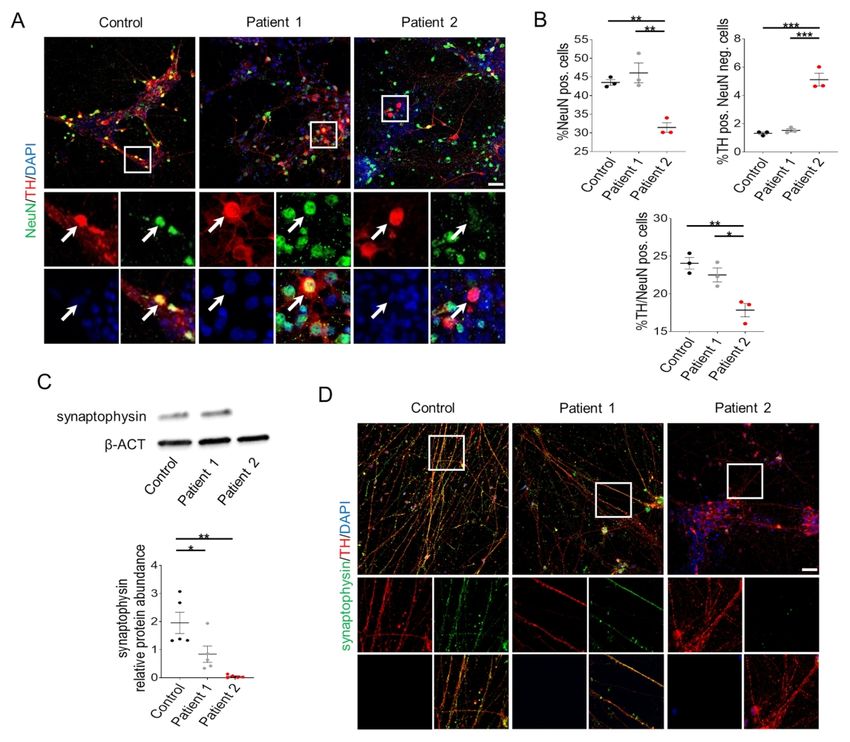
1 Developmental Neurosciences, GOS Institute of Child Health, University College London,
London, UK

2 Biological Chemistry, NBM Department, University of Verona, Verona, Italy

3 Clinical and Experimental Epilepsy, Queen Square Institute of Neurology, University
College London, London, UK

                                                                                                Downloaded from https://academic.oup.com/brain/advance-article/doi/10.1093/brain/awab123/6178275 by guest on 25 April 2021
4 Genetics and Genomic Medicine, GOS Institute of Child Health, University College London,
London, UK

5 Neurometabolic Unit, National Hospital for Neurology and Neurosurgery, Queen Square,
London, UK

6 Gene Transfer Technology Group, EGA-Institute for Women's Health, University College
London, UK

7 Centre for Inborn Errors of Metabolism, GOS Institute of Child Health, University College
London, London, UK

8 Department of Neurology, Great Ormond Street Hospital, London, UK

Correspondence to: Prof. Manju Kurian

Zayed Centre for Research, UCL – GOS Institute of Child Health, 20 Guilford St, WC1N 1DZ,
London, UK

E-mail: manju.kurian@ucl.ac.uk

Correspondence may also be addressed to: Prof. Mariarita Bertoldi

Room 1.24, Biological Chemistry Section, Dep. of Neuroscience, Biomedicine and Movement
Sciences, Strada le Grazie 8, 37134, Verona, Italia

E-mail: mita.bertoldi@univr.it

Running title: Neuronal models for precision therapies

Keywords: induced pluripotent stem cells; dopaminergic neurons; aromatic L-amino acid
decarboxylase deficiency; neurodevelopment; personalized medicine

          ScholarOne, 375 Greenbrier Drive, Charlottesville, VA, 22901 Support (434) 964 4100
Abbreviations

3-OMD          3-O-methyldopa

AADC           Aromatic L-amino acid decarboxylase

AP             action potential

DEGs           differentially expressed genes

                                                                                                     Downloaded from https://academic.oup.com/brain/advance-article/doi/10.1093/brain/awab123/6178275 by guest on 25 April 2021
DOPAC          3,4-Dihydroxyphenylacetic acid

HPLC           High Performance Liquid Chromatography

HVA            homovanillic acid

iPSCs          induced pluripotent stem cells

L-DOPA         L-3,4-dihydroxyphenylalanine

mDA            midbrain dopaminergic

PLP            pyridoxal 5’-phosphate

sEPSCs         spontaneous excitatory postsynaptic currents

Introduction

Neurodevelopmental processes are commonly disrupted in the vast majority of inborn errors
of metabolism, resulting in a wide repertoire of clinical manifestations from severe cognitive,
neuropsychiatric, and motor problems to more subtle learning difficulties.1 Aromatic L-amino
acid decarboxylase (AADC) deficiency is a rare inborn error of neurotransmitter metabolism
due to bi-allelic mutations in DDC, which encodes the enzyme that catalyzes the final step of
serotonin and dopamine synthesis.2 The resultant enzyme deficiency leads to combined
serotonin and catecholamine (dopamine, norepinephrine, epinephrine) deficiency.3 Although
there is a wide phenotypic spectrum,4,5 the majority of affected patients show many of the
typical features seen in recessively inherited, early-onset neurotransmitter disorders6, including
severe global neurodevelopmental delay, oculogyric crises, a complex movement disorder
(characterised by central and peripheral hypotonia with commonly features of dystonia/chorea)
and symptoms of dysautonomia, as well as secondary gastrointestinal, respiratory and
orthopedic complications.7,8 As a result, the majority of patients have significant disability and

          ScholarOne, 375 Greenbrier Drive, Charlottesville, VA, 22901 Support (434) 964 4100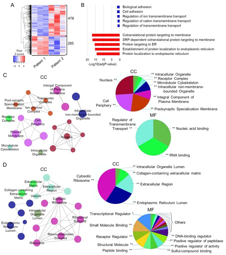
high risk of premature mortality. AADC deficiency is associated with a characteristic CSF
monoamines profile, with reduced 5-hydroxyindoleacetic acid, homovanillic acid (HVA), and
3,4-dihydroxyphenylacetic       acid    (DOPAC),        and    a    concomitant      increase    in   5-
hydroxytryptophan, L-3,4-dihydroxyphenylalanine (L-DOPA), and 3-O-methyldopa (3-
OMD). Definitive diagnosis is ideally achieved by confirming a decrease or absence of plasma
AADC enzymatic activity, and DDC gene sequencing. To date, there are no clear correlations

                                                                                                           Downloaded from https://academic.oup.com/brain/advance-article/doi/10.1093/brain/awab123/6178275 by guest on 25 April 2021
between patient genotype, CSF monoamine profile, AADC enzyme activity and phenotype.

A recently published consensus guideline outlines recommendations for the diagnosis and
management of AADC deficiency.8 Pharmacological therapy provides some, though often
limited, clinical benefit and patients often show variable drug response. It has been postulated
that the variability in disease severity and medication response may be partly attributed to
genotype9,10 and as a result, a number of studies have focused on characterising the underlying
molecular defects caused by different pathogenic variants.11–15 More recently, promising gene
therapy approaches are emerging for AADC deficiency, with a number of clinical trials
evaluating the safety and efficacy of targeted intraparenchymal delivery of AAV2-based
vectors.16–18 It is hoped that with time, these studies may clarify the effect of patient genotype,
age at surgery, pre-treatment motor function and target delivery site on overall therapeutic
efficacy. Although early clinical studies on AADC gene therapy are encouraging, it is likely
that this therapeutic strategy may not be either viable, suitable or easily accessible for a
proportion of patients. Moreover, with advances in diagnostic testing, the global incidence and
prevalence of AADC deficiency continues to increase,19 and the need for alternative precision
therapies is increasingly apparent.

Recently, patient-derived cellular models of neurodevelopmental disorders have proven to be
a valuable experimental system to unravel disease mechanisms and test novel therapeutic
strategies with translational potential.20 As such, we have developed a humanized neuronal
model of AADC deficiency, by reprogramming patient fibroblasts into induced pluripotent
stem cells (iPSCs) for differentiation into midbrain dopaminergic (mDA) neurons. This model
system has allowed us to gain further insight into the neurodevelopmental consequences of
AADC deficiency, with effects on synaptic maturation and neuronal function. Moreover, it has
also provided a suitable platform to evaluate the effects of precision medicine approaches at a
cellular level, demonstrating the potential for rational development of patient-specific
strategies in such rare monogenic disorders.

           ScholarOne, 375 Greenbrier Drive, Charlottesville, VA, 22901 Support (434) 964 4100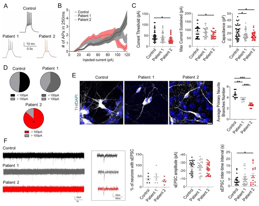
Materials and methods

iPSCs generation and maintenance

Generation of iPSCs from patient dermal fibroblasts was approved by the Local Research
Ethics Committee (Reference 13/LO/0171). Written informed consent was obtained from all
patients. Age-matched healthy control fibroblasts were collected from the MRC Centre for
Neuromuscular Disorders Biobank. Patient fibroblasts were isolated from skin biopsies and

                                                                                                       Downloaded from https://academic.oup.com/brain/advance-article/doi/10.1093/brain/awab123/6178275 by guest on 25 April 2021
maintained in DMEM (Gibco), 10% fetal bovine serum (Gibco), 2 mM L-glutamine (Gibco),
1% MEM non-essential amino acids (Gibco), and 1% penicillin/streptomycin (P/S, Gibco), and
tested for mycoplasma contamination. Reprogramming was performed using the commercially
available CytoTune®-iPS 2.0 Sendai Reprogramming kit (Invitrogen), following manufacturer
instructions. Fibroblast were transduced at 80% confluence (1-1.5x10⁵ cells/well). After 6 days,
infected cells were harvested with TrypLETM (Invitrogen) and 8,000 cells/well were seeded
onto gamma-irradiated mouse embryonic fibroblasts. After 24 hours, cells were cultured into
KO-DMEM (Gibco), 20% serum replacement (Gibco), 2 Mm L-glutamine, 50 µM 2-
mercaptoethanol, 1% MEM non-essential amino acids, 1% P/S, and 10 ng/ml basic fibroblast
growth factor (Gibco). 13 days post-transfection, cells were cultured in gamma-irradiated
mouse embryonic fibroblasts-conditioned medium. Around day 30 post-transduction, 8-10
independent colonies with iPSCs-like morphology were collected and expanded using ReLeSR
(Stemcelltm technologies). Between passage 15 and 20, 3 colonies were converted to mTeSR1
medium (Stemcelltm technologies) on Matrigel® (Corning®) coated plates. Derived iPSC lines
were     maintained      in     mTeSR1/matrigel          system,      regularly      passaged   with
ethylenediaminetetraacetic acid, 0.02% solution (Sigma-Aldrich) and again tested for
mycoplasma infection, as previously. Two iPSC lines for each patient (Patient 1-04, Patient 1-
10; Patient 2-01, Patient 2-06) and the age-matched healthy control (Control-05, Control-03)
were characterized at the iPSCs stage and further differentiated into mDA neurons to exclude
clonal variability. Given the relative homogeneity reported in clonal lines with respect to
transcriptome, growth, and capability of germ layer formation,21,22 one clone per patient
(Patient 1-04; Patient 2-01) and age-matched healthy control (Control-05) were then used for
downstream experiments.

Differentiation of iPSCs into mDA neurons

iPSCs were differentiated into mDA dopaminergic neurons as previously described.23 Briefly,
iPSCs were harvested using TrypLETM (Invitrogen), and plated onto non-adherent bacterial

          ScholarOne, 375 Greenbrier Drive, Charlottesville, VA, 22901 Support (434) 964 4100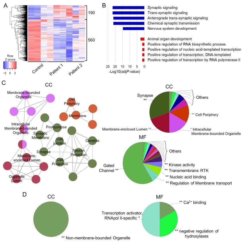
dishes in a concentration of 1.5x10⁵ per cm2 in DMEM/F12:Neurobasal (1:1), N2 (1:100) and
B27 minus vitamin A (1:50) supplements (Invitrogen), 2 mM L-glutamine and ROCK-inhibitor
for the first two days. EBs were plated at day 4 onto polyornithine (PO; 15 μg/ml; Sigma),
fibronectin (FN; 5 μg/ml Gibco) and laminin (LN; 5 μg/ml; Sigma) coated dishes in
DMEM/F12:Neurobasal (1:1), N2 (1:200), B27 minus vitamin A (1:100), 2 mM L-glutamine.
From day 0 to day 9, medium was supplemented with: 10 μM SB431542 (Tocris Bioscience),

                                                                                                               Downloaded from https://academic.oup.com/brain/advance-article/doi/10.1093/brain/awab123/6178275 by guest on 25 April 2021
100 nM LDN193189 (Stemgent Inc.), 0.8 µM CHIR99021 (Tocris Biosceince) and 100 ng/ml
hSHH-C24-II (R&D Systems). On day 2, 0.5 μM purmorphamine (Cambridge Bioscience) was
added. SB431542 was withdrawn on day 6. On day 11, cells were either processed for mDA
precursors analysis or harvested with Accumax and re-plated on PO/FN/LN coated dishes in
droplets of 1-1.5 x10⁴ cells/µl in Neurobasal/B27 minus vitamin A (1:50), 2 mM L-glutamine,
0.2 mM ascorbic acid (AA) and 20ng/ml BDNF (Miltenyi Biotech). On day 14 of
differentiation, 0.5 mM dibutyryl c-AMP (Sigma-Aldrich) and 20ng/ml GDNF (Miltenyi
Biotech) were added. On day 30 of differentiation, cells were re-plated as describe above onto
PO/FN/LN coated dishes or Labteck slides (NuncTM), and γ-secretase inhibitor DAPT (10 μM,
Tocris) was added until final differentiation at day 65. Cells were then harvested or processed
for further analysis.

AADC activity assay

AADC enzyme assay was performed using the refined method developed in                       24,   from   25.

Neuronal cultures at day 65 in phenol red free medium were harvested and lysed by snap
freezing twice in liquid nitrogen in 100 µl of 10 mM Tris pH 7.4 (Sigma-Aldrich), 1 mM
ethylenediaminetetraacetic acid, 320 mM sucrose (Sigma-Aldrich) and protease inhibitor
cocktail (Roche). 50 μl of cell lysate was incubated with 70 μM pyridoxal 5’-phosphate (PLP,
Sigma-Aldrich) in assay buffer composed by 500 mM sodium phosphate pH 7.0, 0.167 mM
ethylenediaminetetraacetic acid, and 39 mM dithiothreitol (Sigma-Aldrich) for 120 min at
37°C, and subsequently 2 mM final concentration of L-DOPA (Sigma-Aldrich) was added and
incubated for 20 min at 37°C. The reaction was stopped with 250 μl of 0.8 M perchloric acid
(final concentration 0.4 M) for 10 min at room temperature and centrifuged at 12.000×g for 5
min at 4°C. A substrate blank with no L-DOPA and a sample blank without cell lysate were
performed for each sample. Dopamine in the supernatant was then quantified by High
Performance Liquid Chromatography (HPLC, see below).

HPLC for quantification of activity assay and metabolic profile

           ScholarOne, 375 Greenbrier Drive, Charlottesville, VA, 22901 Support (434) 964 4100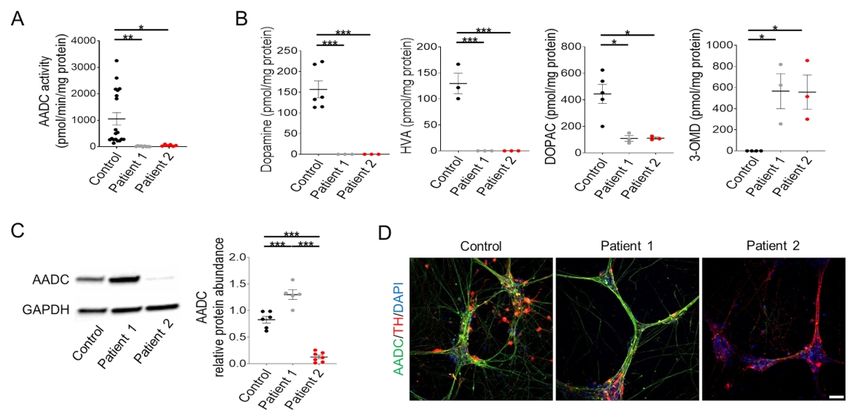
Dopamine produced in the activity assay was separated by reverse-phase HPLC using a HiQSil
C18 column 250x4.6mm (Kya technologies) and detected by coulometric electrochemical
detection using a Coulochem III detector (ESA) with 5010 analytical cell (ESA) setting the
detector electrode at 350 mV and the screening electrode at 20 mV. The mobile phase consisted
of     50   mM    sodium     phosphate      pH    3.6,   5   mM      octaensulfonic     acid,     67   μM
ethylenediaminetetraacetic acid, 43 mM orthophosphoric acid and 230 ml/l methanol diluted

                                                                                                            Downloaded from https://academic.oup.com/brain/advance-article/doi/10.1093/brain/awab123/6178275 by guest on 25 April 2021
in 18.2 Ω HPLC grade water, at a flow rate of 1.2 ml/min at 25 °C. Dopamine was quantified
with Azur software package using a 1000 nM external standard and enzymatic activity was
expressed as pmol/min/mg protein.

HPLC analysis of metabolic profile in derived mature cultures was performed on the phenol
red-free medium incubated for 48h on day 65 mDA neurons. 1:1 medium was mixed with
perchloric acid to a final concentration of 0.4 M, incubated 10 min at 4°C in the dark,
centrifuged at 12000×g for 5 min at 4°C, and supernatant was collected for analysis by HPLC
26.   Metabolites were separated by reverse-phase HPLC using a C:18HS column 250 mm×4.5
mm (Kromatek) and detected by coulometric electrochemical detection using a Coulochem II
detector (ESA) with 5010A analytical cell (Thermo Fisher Scientific) setting the detector
electrode at 450 mV and the screening electrode at 20 mV. Mobile phase consisted of 20 mM
sodium acetate trihydrate pH 3.45, 12.5 mM citric acid monohydrate, 100 μM
ethylenediaminetetraacetic acid, 3.35 mM octaensulfonic acid and 16% methanol diluted in
18.2 Ω HPLC grade water, at a flow rate of 1.5 ml/min at 27 °C. Metabolites were quantified
with EZChrom EliteTM chromatography software (JASCO) using a 500 nM external standard
mixture, and expressed as pmol/mg protein.

Bulk RNA-Seq analysis

Total RNA was isolated using the RNeasy mini kit (Qiagen) following manufacturer’s
instructions. RNA libraries were prepared from 100 ng of total RNA using KAPA mRNA
HyperPrep kit (Roche) according to manufacturer’s protocol and sequenced with Illumina
NextSeq 500 Mid Output 75bp paired-end (~22M reads/sample). FASTQ obtained files were
uploaded to Galaxy web platform, and the public server at usegalaxy.org was used for
downstream analyses.27 FASTQ-files were filtered with Trimmomatic (v.0.38), with
SLIDINGWINDOW trimming and low quality (phread score
edgeR (v.3.24.1), filtering low counts with 0.35 minimum CPM in at least 3 samples,31 and
comparing disease status (patients vs control) and disease-specific genotype (patient 2 vs
patient 1). DEGs with a p-value2 were considered as
statistically significant. Heatmaps were generated from the row-scaled z-score of DEGs
normalised counts obtained by EdgeR with complete-linkage Euclidean hierarchical clustering.
GO enrichment analyses were performed using ShinyGO v0.6132 for biological process, and
Downloaded from https://academic.oup.com/brain/advance-article/doi/10.1093/brain/awab123/6178275 by guest on 25 April 2021
ClueGO v.2.5.733 for cellular component and molecular function enrichments and groupings,
with Benjamini-Hochberg p-value correction of FDR-50mV), bridge-
balance >20MΩ and/or holding current >200pA were discarded. Bridge balance compensation
was applied in current clamp and the resting membrane potential was held at -70mV. Current
steps protocol was used to evoke APs injecting 250ms long depolarizing current steps of
increasing amplitude (Δ10pA). APs were triggered holding the neurons around -60mV/-55mV.
Neurons with repetitive spontaneous APs and repetitive evoked APs were considered to be
functional mature mDA neurons. Recordings were acquired using a Multiclamp 700A
amplifier (Axon Instruments, Molecular Devices) at 10kHz and filtered at 2kHz (Bessel) using
WinEDR (John Dempster, University of Strathclyde). Recording were not corrected for liquid
junction potentials. The approximate cell capacitance was computed as capacitance=tau/Ri,
whereby the time constant tau was found by fitting a single exponential function to the time
points where the membrane voltage was between 10% and 95% of the initial charging decay
slope of a negative hyperpolarizing current step. Input resistance was calculated fitting ΔV/ΔI
at two hyperpolarising steps (-20 and -10pA) and a positive one (+10pA). APs were identified
when the voltage signal crossed 0V. sEPSCs were recorded in voltage clamp and automatically
detected with a template-based algorithm using Clampfit (Molecular Device).
Treatment with L-DOPA and cytotoxicity assay
ScholarOne, 375 Greenbrier Drive, Charlottesville, VA, 22901 Support (434) 964 4100Neuronal cultures at day 65 of differentiation were treated with 80 µM L-DOPA in phenol red-
free medium for 24 h. The medium was subsequently removed and analysed by HPLC, as
described above. Dead-cell proteases release measurement was quantified using CytoTox-
Glo™ Cytotoxicity Assay (Promega) according to manufacturer instructions.
Statistical Analysis
Two-tailed Student’s t-test for single comparisons and statistical one-way ANOVA followed
Downloaded from https://academic.oup.com/brain/advance-article/doi/10.1093/brain/awab123/6178275 by guest on 25 April 2021
by Tukey’s multiple comparisons test were performed using GraphPad Prism. Results are
reported as mean SEM from at least three independent biological replicates, the exact number
of which is stated for each experiment in each figure legend. Significance levels were
determined by p-value, and shown on graphs with asterisks. One asterisk (*) represents p-
values between 0.05 and 0.01, two asterisks (**) represent p-values between 0.01 and 0.001,
and three asterisks (***) represent p-values of less than 0.001.
Data availability
Data supporting the findings of this study are available from the corresponding authors, upon
reasonable request.
Results
Patient-derived mDA neurons show disease-specific loss of AADC enzymatic activity and
dysregulated dopamine synthesis.
Dermal fibroblasts were obtained from two patients with AADC deficiency (Table 1). Patient
1 (homozygous missense variant c.1039C>G, p.R347G) presented with classical infantile onset
disease, with early hypotonia, oculogyric crises and neurodevelopmental delay.15 He is
currently 6½ years of age, and although he continues to make neurodevelopmental progress,
remains non-ambulant and non-verbal. Patient 2 (compound heterozygous variants c.19C>T,
p.Arg7*; c.299G>C, p.C100S) had a classical infantile-onset presentation of disease with
severe global developmental delay, oculogyric crises and hypoglycaemia, but over time
showed a positive response to therapy and had an overall milder disease course. Once AADC
deficiency was diagnosed at 3½ years of age, the instigation of dopaminergic medication and
other specific AADC deficiency treatments was associated with neurodevelopmental progress;
independent ambulation was achieved by 4½ years and spoken language by 5½ years. From
10-18 years, adjunct therapies were needed to combat side effects from long-term use of the
ScholarOne, 375 Greenbrier Drive, Charlottesville, VA, 22901 Support (434) 964 4100original treatments to maintain basic motor and verbal function. Now aged 22 years, he has
ongoing learning difficulties, mild motor impairments, behavioral issues, autistic traits and
neuropsychiatric symptoms of anxiety and intermittent low mood. iPSC lines were generated
from dermal fibroblasts of both patients and from an age-matched healthy individual (Control).
Sequencing of genomic DNA confirmed that patient iPSC lines retained their specific DDC
mutation (Supplementary Fig. 1A). All iPSCs lines showed clearance of viral transgenes,
Downloaded from https://academic.oup.com/brain/advance-article/doi/10.1093/brain/awab123/6178275 by guest on 25 April 2021
genomic integrity (Supplementary Fig. 1B-C), and true pluripotency (Supplementary Fig. 2A-
D).
iPSCs were then differentiated into mDA neurons, and both patient and control iPSC lines
showed similar differentiation efficiency. After 11 days of differentiation, all lines showed high
levels of mDA progenitors and typical midbrain precursors gene expression profile
(Supplementary Fig. 3A-C). By 65 days of differentiation, both control and patient lines
comparably matured into neurons, in particular with dopaminergic identity (Supplementary
Fig. 4A-B). Both control and patient-derived neuronal cultures showed upregulation of
midbrain-related genes (Supplementary Fig. 4C). Whole-cell patch clamp electrophysiology
confirmed that iPSC-derived mDA neurons were functional and exhibited continuous and
rhythmic pacemaker-like activity (Supplementary Fig. 4D). Derived neuronal cultures were
almost devoid of serotonergic neurons, restricting all further analyses specifically to the mDA
neuronal subtype (Supplementary Fig. 5A).
We first investigated the effect of patient mutations on AADC enzyme activity and protein
expression. Measurement of AADC activity showed significantly lower enzymatic function in
patients when compared to control-derived neurons (Fig. 1A). HPLC analysis of extracellular
metabolites showed a disease-specific absence of dopamine and HVA with significantly
reduced levels of DOPAC. In contrast, 3-OMD, a downstream metabolite of the AADC
substrate L-DOPA, was significantly increased in patient-derived neurons (Fig. 1B). Analysis
of AADC protein levels showed an increase in Patient 1 neuronal cultures when compared to
the Control; in contrast, a significant reduction of AADC protein was detected in Patient 2
neuronal cultures (Fig. 1C-D and Supplementary Fig. 5B), in line with the second heterozygous
early stop codon variant predicted to result in nonsense mediated mRNA decay.
We then explored whether the aberrant AADC protein levels in patients could be linked to a
difference in intrinsic protein stability. Recombinant AADC proteins were produced for the
homozygous R347G variant (Patient 1) and C100S variant (Patient 2). Circular dichroism and
ScholarOne, 375 Greenbrier Drive, Charlottesville, VA, 22901 Support (434) 964 4100dynamic light scattering analyses showed comparable values for both mutant and wild-type
AADC protein, inferring similar intrinsic protein stability (Supplementary Table 1).
To investigate whether aberrant AADC protein levels related to DDC gene expression, qRT-
PCR studies were undertaken. In line with protein expression data, we observed a statistically
significant increase in DDC expression in Patient 1 when compared to the Control
(Supplementary Figure 5C). For Patient 2, we observed comparable levels of DDC expression
Downloaded from https://academic.oup.com/brain/advance-article/doi/10.1093/brain/awab123/6178275 by guest on 25 April 2021
to the Control (Supplementary Figure 5C), despite the predicted nonsense-mediated decay of a
proportion of transcripts. We also observed an increase in tyrosine hydroxylase (TH) gene and
protein expression in both patient lines when compared to the control (Supplementary Figure
5D-E).
AADC deficiency has mutation-specific effects on neuronal synaptic maturation and
connectivity.
We then sought to investigate the neurodevelopmental consequences of AADC deficiency in
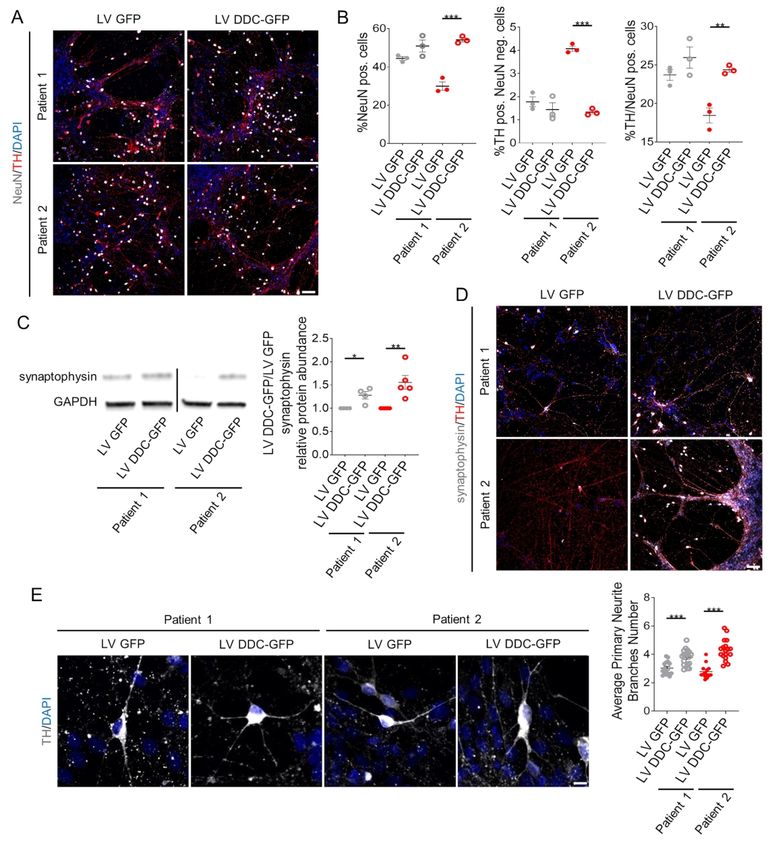
our in vitro model. Immunofluorescence analysis of the mature neuronal marker NeuN showed
comparable levels in Patient 1 and Control mDA neurons, while Patient 2 cultures showed a
significant decrease in NeuN positivity when compared to both Control and Patient 1 lines (Fig.
2A-B). Moreover, analysis of the vesicular protein synaptophysin revealed a significant
decrease in protein levels for both Patient 1 and 2 when compared to control-derived neuronal
cultures (Fig. 2C-D).
In order to further investigate the neurodevelopmental effects of AADC deficiency, we
undertook bulk RNA sequencing for analysis of differentially expressed genes (DEGs) between
patient and control-derived neurons, with a particular focus on protein-coding genes. In a
combined analysis of Patient 1 and Patient 2-derived neuronal cultures, we identified 750
DEGs (75% underexpressed and 25% overexpressed) when compared to the Control (Fig. 3A).
Gene Ontology (GO) analysis of underexpressed DEGs revealed a strong enrichment in
synaptic transmission-related biological processes and nervous system development, whilst
overexpressed DEGs mainly enriched protein transcription and general organ developmental
processes (Fig. 3B). Furthermore, underexpressed DEGs were associated with membranous
cellular compartments (in particular the cell periphery and synaptic region), and enriched in
gated channels and regulators of membrane transport (Fig. 3C). In contrast, overexpressed
DEGs were associated with non-membrane-bounded cell compartments (nucleus), with
enrichment in transcriptional regulator proteins (Fig. 3D).
ScholarOne, 375 Greenbrier Drive, Charlottesville, VA, 22901 Support (434) 964 4100Considering the previously detected differences between the two patient lines (Fig. 2), single-
comparison RNA sequencing analysis was also performed. We identified 842 protein-coding
DEGs for Patient 1 compared to the Control (Supplementary Fig. 6A) and 871 protein-coding
DEGs for Patient 2 compared to the Control (Supplementary Fig. 7A). For both analyses,
underexpressed genes showed common enrichment for synaptic transmission (Supplementary
Fig. 6B and 7B) - reflected in the significant P-values observed in the combined analysis (Fig.
Downloaded from https://academic.oup.com/brain/advance-article/doi/10.1093/brain/awab123/6178275 by guest on 25 April 2021
3B) - representing genes encoding proteins mainly localized at the cell periphery or synapses,
and associated with ion channel function (Patient 1 and Patient 2) and gated channel function
(for Patient 2 in particular) (Supplementary Fig. 6C and 7C). Differences in separate single
Patient 1 and Patient 2 comparisons with the Control were mainly detected for upregulated
genes with regard to biological processes and significance (Supplementary Fig. 6B and 7B):
for Patient 1, overexpressed DEGs were enriched for developmental and cell projection
assembly genes (Supplementary Fig. 6B and 6D), whilst for Patient 2 overexpressed DEGs
were enriched for genes encoding endoplasmic reticulum and membrane-targeting processes
and function (Supplementary Fig. 7B and 7D). Despite these inter-patient differences, the
combined analysis reflects a common, disease-specific overexpression of developmental and
transcriptional/translational processes from both single comparisons (Fig. 3B).
We then explored DEGs between the two different patient-derived neuronal cultures. We
identified a total of 763 protein-coding DEGs for Patient 2 when compared to Patient 1 (Fig.
4A). The underexpressed DEGs showed enrichment in cell adhesion and membrane transport-
related processes, while overexpressed DEGs enriched endoplasmic reticulum and membrane-
targeting processes categories (Fig. 4B). Underexpressed DEGs corresponded to proteins
localized both in cell periphery/membrane regions and nuclear compartment, with enrichment
for genes regulating transcription and transmembrane transport (Fig. 4C). Overexpressed
DEGs showed enrichment in both cytosolic transcriptional and extracellular compartments,
with molecular functions mainly linked to structural/binding molecules, and
transcriptional/activity regulators (Fig. 4D), resembling the result from the single comparison
between Patient 2 and Control (Supplementary Fig. 7B and 7D).
Whole-cell patch clamp electrophysiology studies were undertaken to determine whether the
observed differences in gene expression were associated with functional differences in
neuronal activity. The parameters analysed are similar to other studies using iPSC-derived
dopaminergic neurons, with comparable findings for firing pattern, pacemaker and synaptic
activity in controls.34–37 Recordings with increasing current amplitude (Fig. 5A) showed that
ScholarOne, 375 Greenbrier Drive, Charlottesville, VA, 22901 Support (434) 964 4100the current threshold to elicit an action potential (AP) for Patient 2 was significantly lower than
for the Control (Fig. 5B-C) and failed to follow current injection up to 100pA (Fig. 5D). On
investigation of passive neuronal properties, both patients displayed lower capacitance
compared to control neurons without affecting input resistance (Fig. 5C), in accordance with a
decreased average number of primary neurite branches (Fig. 5E). For both control and patient-
derived neurons showing spontaneous excitatory postsynaptic currents (sEPSC), we observed
Downloaded from https://academic.oup.com/brain/advance-article/doi/10.1093/brain/awab123/6178275 by guest on 25 April 2021
no differences in either the percentage of functionally connected neurons or current amplitude,
although the inter-event interval was significantly higher in Patient 2-derived neurons (Fig.
5F).
DDC lentiviral gene-transfer significantly improves neurodevelopmental defects in
patient-derived neurons.
Given that gene therapy is an emerging new treatment for AADC deficiency,16–18 we sought to
investigate the cellular effects of human DDC (hDDC) transgene delivery in our model; in
particular we wished to evaluate whether this therapeutic approach could improve the
neurodevelopment sequelae of AADC deficiency, independent of genetic background. We
generated a lentiviral construct for the delivery of hDDC under the control of the neuronal-
specific promoter human synapsin (hSyn1) (Supplementary Fig. 8). Patient-derived mDA
precursors were transduced at day 24 of differentiation and analysed at day 65. For both patient
lines, lentiviral gene transfer resulted in an increase in AADC protein levels (Supplementary
Fig. 9A-B), and rescued enzymatic activity to levels comparable to those observed in Control
neurons (Supplementary Fig. 9C). Furthermore, Patient 2 transduced neurons showed a
significant increase in the NeuN-positive neuronal population, and in particular mDA neurons,
to levels comparable to Patient 1 (Fig. 6A-B and Supplementary Fig. 10A). hDDC lentiviral
delivery also resulted in a significant increase in synaptophysin protein levels in both patients-
derived neuronal cultures (Fig. 6C) and more specifically in the mDA neuronal subpopulation
(Fig. 6D and Supplementary Fig. 10B), with a significant increase in primary branching (Fig.
6E).
In silico and recombinant biochemical analyses predict mutation-specific L-DOPA
response for Patient 2.
The different mutations harbored by Patient 1 and 2 were further investigated to determine
whether they had differential effects on enzymatic function. For Patient 1, despite
supraphysiological levels of protein expression (Fig. 1C), the homozygous missense
ScholarOne, 375 Greenbrier Drive, Charlottesville, VA, 22901 Support (434) 964 4100substitution R347G significantly impairs catalytic function of AADC leading to undetectable
enzyme activity (Fig. 1A) without impacting the protein structure by a molecular mechanism
extensively investigated in 15. In contrast, despite significantly low levels of AADC protein in
Patient 2-derived neuronal cultures (Fig. 1C), residual enzymatic activity was still detected,
albeit at a fraction of that evident in Control line (Fig. 1A). It is likely that this residual AADC
enzyme activity can be attributed to the p.C100S variant, since the second heterozygous
Downloaded from https://academic.oup.com/brain/advance-article/doi/10.1093/brain/awab123/6178275 by guest on 25 April 2021
mutation leads to an early stop codon at Arg7, predicted to result in nonsense mediated mRNA
decay and absent protein production. The missense mutation C100S results in an amino acid
substitution at the beginning of an essential loop (loop 2, residues 100 to 110) that contains key
hydrophobic active site residues involved in substrate binding, in particular Ile101 and
Phe103.38 The cysteine-to-serine amino acid substitution has the potential to alter the
conformation of loop 2 and consequently the substrate-binding cleft, thereby affecting substrate
affinity (Fig. 7A). AADCC100S was produced in vitro in recombinant form to further
characterize the effects of this mutation through spectroscopic, circular dichroism and
fluorescence analyses, and calculation of kinetic parameters. A minor perturbation of PLP
cofactor microenvironment (in particular for the enolimine tautomer) was observed
(Supplementary Fig. 11A-B). However, PLP binding affinity (Supplementary Table 1) was not
particularly affected, with a KD(PLP) consistently lower than previously reported values for
AADC variants with cofactor binding impairment.13 Calculation of kinetic parameters
(Supplementary Table 1) revealed that AADCC100S retains more residual enzyme activity
than that reported for other AADC variants.12,13 The catalytic activity (kcat) of AADCC100S
was indeed similar to that observed for wild-type, with the actual decrease in overall
AADCC100S catalytic efficiency (kcat/KM) attributed to a slight decrease in L-DOPA affinity
(KM) (Supplementary Table 1). As such, we postulated that dopamine production by
AADCC100S could be enhanced with L-DOPA administration, as demonstrated for other
AADC variants.39
Patient 2-derived mDA neurons specifically respond to L-DOPA administration.
To determine whether the C100S mutation resulted in L-DOPA responsivity, we sought to
investigate the effect of L-DOPA treatment in Patient 2-derived mDA neurons. After 65 days
of differentiation, both patients and control-derived neuronal cultures were incubated with 80
µM L-DOPA for 24 hours, a dose just below that considered to be toxic in neuronal and other
cellular systems.40,41 Subsequent HPLC analysis of extracellular metabolites was then
undertaken. As expected in a system with catalytically competent AADC, HVA levels were
ScholarOne, 375 Greenbrier Drive, Charlottesville, VA, 22901 Support (434) 964 4100significantly higher in treated Control compared to untreated Control neurons (Fig. 7B).
Furthermore, as predicted, there was no detectable HVA in Patient 1-derived neuronal cultures
both pre- and post- L-DOPA treatment. However, for Patient 2, we observed a significant
increase of HVA levels in L-DOPA treated cultures when compared to untreated cultures (Fig.
7B). In order to evaluate any potential toxicity related to L-DOPA administration40 or
dopamine production,42 we measured dead-cell protease release and found no increase in
Downloaded from https://academic.oup.com/brain/advance-article/doi/10.1093/brain/awab123/6178275 by guest on 25 April 2021
membrane permeability for both Patient and Control-derived neuronal cultures treated with 80
µM L-DOPA for 24 hours (Fig. 7C). Moreover, analysis of JNK protein phosphorylation,
which increases in response to toxic levels of dopamine,43 showed a significant increase in the
phosphorylated form of this kinase in treated Control neurons only, while no significant
increase was detected in both treated Patient 1 and 2 cultures (Supplementary Fig. 11C).
Discussion
AADC deficiency is a complex and often pharmacoresistant neurological disorder, with a broad
phenotypic spectrum, variable drug response, substantial burden of disease and significant risk
of premature mortality.7 Improved understanding of the underlying pathogenic mechanisms
and the development of better targeted treatments, such as gene therapy and other personalised
medicine approaches, will be key in modifying disease and long-term outcome. In this study,
we have developed a new humanized model of AADC deficiency. Our in vitro patient-derived
mDA neuronal model of AADC deficiency has provided further insight into mechanisms
governing disease, as well as an ideal system to evaluate the impact of approaches such as gene
therapy at cellular level and a unique research platform to evaluate mutation-specific precision
medicine approaches.
Importantly, our patient-derived mDA model recapitulates key features of the human
phenotype with near-absent AADC enzyme activity and impaired dopamine metabolism. In
our dopaminergic model, we observed a greater degree of residual AADC enzyme activity in
Patient 2, which may relate to the more advanced motor gains observed in this patient. We also
observed patient-specific altered levels of AADC protein. The reasons for this are not entirely
clear, given that little is known about factors that govern AADC enzyme regulation. Our
biochemical investigations did not show a differential intrinsic protein stability between mutant
and wild-type protein. We did however observe a clear patient-specific increase in DDC gene
expression for Patient 1 and higher than expected levels of DDC expression for Patient 2, given
ScholarOne, 375 Greenbrier Drive, Charlottesville, VA, 22901 Support (434) 964 4100the predicted nonsense-mediated decay of a proportion of Patient 2 transcripts. Furthermore
for both patients there was an increase in TH gene and protein expression; interestingly, TH
gene and protein expression has previously been shown to increase in Parkinson’s disease, as
a likely compensatory response to a state of dopamine deficiency in the context of striatonigral
degeneration.44,45 As such, it is plausible that the similar state of dopamine deficiency in
AADC-deficient patient lines drives a positive feedback mechanism to modulate neuronal
Downloaded from https://academic.oup.com/brain/advance-article/doi/10.1093/brain/awab123/6178275 by guest on 25 April 2021
levels of key enzymes driving dopamine synthesis.
Moreover, our study suggests that AADC dysfunction may have widespread effects on gene
expression that may impact neuronal development and functional maturation. As well as its
pivotal role in monoamine neurotransmission, dopamine is postulated to have important
functions in modulating neuronal structure and connectivity.46 The early production of
dopamine in midbrain development suggests that it may have neurodevelopmental influence,47
a notion that is further corroborated by DDC knock-in mice and knockout zebrafish which
show abnormal development.48,49 Interestingly, our patient-derived cell model also shows that
defective AADC enzymatic activity and dysregulated dopamine metabolism affects neuronal
maturity, with altered expression of genes involved in neurodevelopment and synaptic
formation, as well as disruption of electrophysiological properties and functional activity.
Considering that iPSC-derived neurons resemble fetal neurons,50 it is possible that the neuronal
maturation defects observed in our in vitro model correlate with prenatal disease onset in
humans. This is not surprising, given that many affected patients present with their first
clinically discernible symptoms in early infancy. Our results are particularly relevant in the
current climate of emerging gene therapy approaches,18 where neuronal plasticity is considered
to be an important requisite for clinical benefit.51 It is likely that gene therapy within this
‘therapeutic window’ of brain plasticity may predict a more favorable long-term
neurodevelopmental outcome.
In our system, we identified around the same number of differentially expressed genes between
patients (when combined) and control and between the two patients; the latter observation
likely reflects both the biological and clinical differences between patients affected by a disease
with a broad phenotypic continuum. Patient 2-derived cultures showed indeed a greater degree
of neuronal immaturity. Notably, Patient 2 had a number of behavioural issues, significant
autistic traits and prominent neuropsychiatric symptoms, features that were less evident in
Patient 1. Our data may indicate that the greater degree of neuronal immaturity evident in
Patient 2 lines as seen on maturation marker analysis, transcriptome profiling and
ScholarOne, 375 Greenbrier Drive, Charlottesville, VA, 22901 Support (434) 964 4100electrophysiology may contribute to the aforementioned neurodevelopmental symptoms.
Importantly, lentiviral treatment of patient-derived neurons restored AADC protein levels and
enzymatic activity with significant improvement in neuronal maturity. Whether AADC protein
has additional functions in governing neurodevelopment processes, that are independent of its
catalytic activity in dopamine production, remains yet to be determined. Further studies with a
greater number of patient lines and age-matched/isogenic controls, or analysis of multiple
Downloaded from https://academic.oup.com/brain/advance-article/doi/10.1093/brain/awab123/6178275 by guest on 25 April 2021
clones from each line, will help confirm and further delineate the complex neurodevelopmental
biological phenotypes identified in this study. Additional genetic, epigenetic and
environmental factors may also play a role in such phenotypic variability seen in the cell model
and human phenotype; over time, advances in next generation sequencing technologies may
also help further elucidate some of the underlying contributory genetic factors.
Our patient-derived model of AADC deficiency has proven to be a useful tool for evaluating
therapeutic approaches. We have shown recovery of AADC enzyme activity and specific
neuronal maturation defects using a gene therapy approach in patient-derived neurons. In
tandem with other models, such therapeutic testing in iPSC-based systems may in the future
guide and influence clinical trial design. Our model has also demonstrated the potential utility
of L-DOPA treatment for some patients with AADC deficiency. Although L-DOPA is not
traditionally used in the majority of patients,8 it has been previously empirically used in patients
suspected to have L-DOPA responsive AADC deficiency.39 Our study confirms that it may
indeed have a role for patients with specific DDC mutations associated with residual enzymatic
activity due to altered substrate affinity. A planned therapeutic trial will further inform whether
the positive effects of L-DOPA observed in vitro are recapitulated in vivo. More generally, our
study shows that better definition of the physiochemical properties of specific mutations with
subsequent validation in patient-relevant models has great potential in guiding personalised
pharmacological strategies for rare disorders.
In conclusion, as new therapeutic avenues emerge for patients with AADC deficiency, our
study shows the clear utility of an iPSC-based modelling system to elucidate disease
mechanisms and evaluate therapeutic strategies.
Acknowledgments
ScholarOne, 375 Greenbrier Drive, Charlottesville, VA, 22901 Support (434) 964 4100We sincerely thank the AADC Research Trust for inspiring us to undertake this study, and our
patients and their families for participating in this study. We also thank the MRC Centre for
Neuromuscular Disorders Biobank for providing age-matched control fibroblasts.
Funding
Downloaded from https://academic.oup.com/brain/advance-article/doi/10.1093/brain/awab123/6178275 by guest on 25 April 2021
GR, KK, MB and MAK received funding from the AADC Research Trust. EL and GL have
been supported by European Union’s Horizon 2020 research and innovation program under
Marie Skłodowska-Curie grant agreement 642881 and an Epilepsy Research UK Fellowship
(GL: ERUK F1701). HA received funding from Kuwait University. J.N. received funding from
MRC MR/K02342X/1 and MR/R015325/1. J.C. has been supported by Wellcome Innovator
Award (210774/2/18/Z). MB was supported by University of Verona Grant FUR2019. SB and
MAK have received funding support from the Wellcome Trust (MAK: Wellcome Intermediate
Clinical Fellowship WT098524MA) and is from the Rosetrees Trust. MAK is funded by an
NIHR Research Professorship (NIHR-RP-2016-07-019) and the Sir Jules Thorn Award for
Biomedical Research.
This research was supported by the NIHR Great Ormond Street Hospital Biomedical Research
Centre. The views expressed are those of the author(s) and not necessarily those of the NHS,
the NIHR or the Department of Health.
Competing interests
The authors report no competing financial interests.
Supplementary material
o Supplementary Figures
o Supplementary Material and Methods
o Supplementary full-length Blots
ScholarOne, 375 Greenbrier Drive, Charlottesville, VA, 22901 Support (434) 964 4100References
1. Saudubray JM, Garcia-Cazorla A. An overview of inborn errors of metabolism affecting
the brain: From neurodevelopment to neurodegenerative disorders. Dialogues Clin
Neurosci. 2018;20(4):301-325. doi:10.31887/dcns.2018.20.4/jmsaudubray
2. Ng J, Papandreou A, Heales SJ, Kurian MA. Monoamine neurotransmitter disorders -
Clinical advances and future perspectives. Nat Rev Neurol. 2015;11(10):567-584.
doi:10.1038/nrneurol.2015.172
Downloaded from https://academic.oup.com/brain/advance-article/doi/10.1093/brain/awab123/6178275 by guest on 25 April 2021
3. Hyland K, Clayton PT. Aromatic amino acid decarboxylase deficiency in twins. J Inherit
Metab Dis. 1990;13(3):301-304. doi:10.1007/BF01799380
4. Dai W, Lu D, Gu X, Yu Y. Aromatic L-amino acid decarboxylase deficiency in 17
Mainland China patients: Clinical phenotype, molecular spectrum, and therapy
overview. Mol Genet Genomic Med. 2020;8(3). doi:10.1002/mgg3.1143
5. Helman G, Pappa MB, Pearl PL. Widening phenotypic spectrum of aadc deficiency, a
disorder of dopamine and serotonin synthesis. In: JIMD Reports. Vol 17. Springer;
2014:23-27. doi:10.1007/8904_2014_327
6. Kurian MA, Gissen P, Smith M, Heales SJR, Clayton PT. The monoamine
neurotransmitter disorders: An expanding range of neurological syndromes. Lancet
Neurol. 2011;10(8):721-733. doi:10.1016/S1474-4422(11)70141-7
7. Pearson TS, Gilbert L, Opladen T, et al. AADC deficiency from infancy to adulthood:
Symptoms and developmental outcome in an international cohort of 63 patients. J
Inherit Metab Dis. 2020;43(5):1121-1130. doi:10.1002/jimd.12247
8. Wassenberg T, Molero-Luis M, Jeltsch K, et al. Consensus guideline for the diagnosis
and treatment of aromatic l-amino acid decarboxylase (AADC) deficiency. Orphanet J
Rare Dis. 2017;12(1). doi:10.1186/s13023-016-0522-z
9. Cellini B. Biochemical and Computational Approaches to Improve the Clinical
Treatment of Dopa Decarboxylase-Related Diseases: An Overview. Open Biochem J.
2012;6(1):131-138. doi:10.2174/1874091x01206010131
10. Himmelreich N, Montioli R, Bertoldi M, et al. Aromatic amino acid decarboxylase
deficiency: Molecular and metabolic basis and therapeutic outlook. Mol Genet Metab.
2019;127(1):12-22. doi:10.1016/j.ymgme.2019.03.009
11. Montioli R, Battini R, Paiardini A, et al. A novel compound heterozygous genotype
associated with aromatic amino acid decarboxylase deficiency: Clinical aspects and
biochemical studies. Mol Genet Metab. 2019;127(2):132-137.
doi:10.1016/j.ymgme.2019.05.004
12. Montioli R, Bisello G, Dindo M, Rossignoli G, Voltattorni CB, Bertoldi M. New
variants of AADC deficiency expand the knowledge of enzymatic phenotypes. Arch
Biochem Biophys. 2020;682. doi:10.1016/j.abb.2020.108263
13. Montioli R, Dindo M, Giorgetti A, Piccoli S, Cellini B, Voltattorni CB orr. A
comprehensive picture of the mutations associated with aromatic amino acid
decarboxylase deficiency: from molecular mechanisms to therapy implications. Hum
Mol Genet. 2014;23(20):5429-5440. doi:10.1093/hmg/ddu266
14. Montioli R, Janson G, Paiardini A, Bertoldi M, Borri Voltattorni C. Heterozygosis in
aromatic amino acid decarboxylase deficiency: Evidence for a positive interallelic
ScholarOne, 375 Greenbrier Drive, Charlottesville, VA, 22901 Support (434) 964 4100complementation between R347Q and R358H mutations. IUBMB Life. 2018;70(3):215-
223. doi:10.1002/iub.1718
15. Montioli R, Paiardini A, Kurian MA, et al. The novel R347g pathogenic mutation of
aromatic amino acid decarboxylase provides additional molecular insights into enzyme
catalysis and deficiency. Biochim Biophys Acta - Proteins Proteomics.
2016;1864(6):676-682. doi:10.1016/j.bbapap.2016.03.011
16. Chien YH, Lee NC, Tseng SH, et al. Efficacy and safety of AAV2 gene therapy in
children with aromatic L-amino acid decarboxylase deficiency: an open-label, phase 1/2
Downloaded from https://academic.oup.com/brain/advance-article/doi/10.1093/brain/awab123/6178275 by guest on 25 April 2021
trial. Lancet Child Adolesc Heal. 2017;1(4):265-273. doi:10.1016/S2352-
4642(17)30125-6
17. Hwu WL, Muramatsu SI, Tseng SH, et al. Gene therapy for aromatic L-amino acid
decarboxylase deficiency. Sci Transl Med. 2012;4(134).
doi:10.1126/scitranslmed.3003640
18. Kojima K, Nakajima T, Taga N, et al. Gene therapy improves motor and mental function
of aromatic l-amino acid decarboxylase deficiency. Brain. 2019;142(2):322-333.
doi:10.1093/brain/awy331
19. Hyland K, Reott M. Prevalence of Aromatic L-Amino Acid Decarboxylase Deficiency
in At-Risk Populations. Pediatr Neurol. 2020;106:38-42.
doi:10.1016/j.pediatrneurol.2019.11.022
20. Lee KM, Hawi ZH, Parkington HC, et al. The application of human pluripotent stem
cells to model the neuronal and glial components of neurodevelopmental disorders. Mol
Psychiatry. 2020;25(2):368-378. doi:10.1038/s41380-019-0495-0
21. Schuster J, Halvardson J, Pilar Lorenzo L, et al. Transcriptome profiling reveals degree
of variability in induced pluripotent stem cell lines: Impact for human disease modeling.
Cell Reprogram. 2015;17(5):327-337. doi:10.1089/cell.2015.0009
22. Matsa E, Burridge PW, Yu KH, et al. Transcriptome Profiling of Patient-Specific
Human iPSC-Cardiomyocytes Predicts Individual Drug Safety and Efficacy Responses
In Vitro. Cell Stem Cell. 2016;19(3):311-325. doi:10.1016/j.stem.2016.07.006
23. Lehnen D, Barral S, Cardoso T, et al. IAP-Based Cell Sorting Results in Homogeneous
Transplantable Dopaminergic Precursor Cells Derived from Human Pluripotent Stem
Cells. Stem Cell Reports. 2017;9(4):1207-1220. doi:10.1016/j.stemcr.2017.08.016
24. Allen GFG. The neurochemical consequences of aromatic L-amino acid decarboxylase
deficiency. Dr thesis, UCL (University Coll London) . Published online April 28, 2011.
25. Hyland K, Clayton PT. Aromatic L-amino acid decarboxylase deficiency: Diagnostic
methodology. Clin Chem. 1992;38(12):2405-2410. doi:10.1093/clinchem/38.12.2405
26. de la Fuente C, Burke DG, Eaton S, Heales SJR. Inhibition of neuronal mitochondrial
complex I or lysosomal glucocerebrosidase is associated with increased dopamine and
serotonin turnover. Neurochem Int. 2017;109:94-100. doi:10.1016/j.neuint.2017.02.013
27. Afgan E, Baker D, Batut B, et al. The Galaxy platform for accessible, reproducible and
collaborative biomedical analyses: 2018 update. Nucleic Acids Res.
2018;46(W1):W537-W544. doi:10.1093/nar/gky379
28. Bolger AM, Lohse M, Usadel B. Trimmomatic: A flexible trimmer for Illumina
sequence data. Bioinformatics. 2014;30(15):2114-2120.
doi:10.1093/bioinformatics/btu170
ScholarOne, 375 Greenbrier Drive, Charlottesville, VA, 22901 Support (434) 964 410029. Kim D, Langmead B, Salzberg SL. HISAT: A fast spliced aligner with low memory
requirements. Nat Methods. 2015;12(4):357-360. doi:10.1038/nmeth.3317
30. Liao Y, Smyth GK, Shi W. FeatureCounts: An efficient general purpose program for
assigning sequence reads to genomic features. Bioinformatics. 2014;30(7):923-930.
doi:10.1093/bioinformatics/btt656
31. Liu R, Holik AZ, Su S, et al. Why weight? Modelling sample and observational level
variability improves power in RNA-seq analyses. Nucleic Acids Res. 2015;43(15).
doi:10.1093/nar/gkv412
Downloaded from https://academic.oup.com/brain/advance-article/doi/10.1093/brain/awab123/6178275 by guest on 25 April 2021
32. Ge SX, Jung D, Jung D, Yao R. ShinyGO: A graphical gene-set enrichment tool for
animals and plants. Bioinformatics. 2020;36(8):2628-2629.
doi:10.1093/bioinformatics/btz931
33. Bindea G, Mlecnik B, Hackl H, et al. ClueGO: A Cytoscape plug-in to decipher
functionally grouped gene ontology and pathway annotation networks. Bioinformatics.
2009;25(8):1091-1093. doi:10.1093/bioinformatics/btp101
34. Hartfield EM, Yamasaki-Mann M, Ribeiro Fernandes HJ, et al. Physiological
characterisation of human iPS-derived dopaminergic neurons. PLoS One. 2014;9(2).
doi:10.1371/journal.pone.0087388
35. Wakeman DR, Hiller BM, Marmion DJ, et al. Cryopreservation Maintains Functionality
of Human iPSC Dopamine Neurons and Rescues Parkinsonian Phenotypes In Vivo.
Stem Cell Reports. 2017;9(1):149-161. doi:10.1016/j.stemcr.2017.04.033
36. Xi J, Liu Y, Liu H, Chen H, Emborg ME, Zhang SC. Specification of midbrain dopamine
neurons from primate pluripotent stem cells. Stem Cells. 2012;30(8):1655-1663.
doi:10.1002/stem.1152
37. Doi D, Magotani H, Kikuchi T, et al. Pre-clinical study of induced pluripotent stem cell-
derived dopaminergic progenitor cells for Parkinson’s disease. Nat Commun.
2020;11(1). doi:10.1038/s41467-020-17165-w
38. Burkhard P, Dominici P, Borri-Voltattorni C, Jansonius JN, Malashkevich VN.
Structural insight into Parkinson’s disease treatment from drug-inhibited DOPA
decarboxylase. Nat Struct Biol. 2001;8(11):963-967. doi:10.1038/nsb1101-963
39. Chang YT, Sharma R, Marsh JL, et al. Levodopa-Responsive Aromatic L-Amino Acid
Decarboxylase Deficiency. Ann Neurol. 2004;55(3):435-438. doi:10.1002/ana.20055
40. Sabens Liedhegner EA, Steller KM, Mieyal JJ. Levodopa activates apoptosis signaling
kinase 1 (ASK1) and promotes apoptosis in a neuronal model: Implications for the
treatment of Parkinson’s disease. Chem Res Toxicol. 2011;24(10):1644-1652.
doi:10.1021/tx200082h
41. Park KH, Shin KS, Zhao TT, Park HJ, Lee KE, Lee MK. L-DOPA modulates cell
viability through the ERK-c-Jun system in PC12 and dopaminergic neuronal cells.
Neuropharmacology. 2016;101:87-97. doi:10.1016/j.neuropharm.2015.09.006
42. Blum D, Torch S, Lambeng N, et al. Molecular pathways involved in the neurotoxicity
of 6-OHDA, dopamine and MPTP: Contribution to the apoptotic theory in Parkinson’s
disease. Prog Neurobiol. 2001;65(2):135-172. doi:10.1016/S0301-0082(01)00003-X
43. Jiang H, Ren Y, Zhao J, Feng J. Parkin protects human dopaminergic neuroblastoma
cells against dopamine-induced apoptosis. Hum Mol Genet. 2004;13(16):1745-1754.
doi:10.1093/hmg/ddh180
ScholarOne, 375 Greenbrier Drive, Charlottesville, VA, 22901 Support (434) 964 4100You can also read