Neuroscience of affect: brain mechanisms of pleasure and displeasure Kent C Berridge1 and Morten L Kringelbach2,3
←
→
Page content transcription
If your browser does not render page correctly, please read the page content below
Available online at www.sciencedirect.com

Neuroscience of affect: brain mechanisms of pleasure and
displeasure
Kent C Berridge1 and Morten L Kringelbach2,3

Affective neuroscience aims to understand how affect                       as a core process of pleasure or displeasure, has rarely
(pleasure or displeasure) is created by brains. Progress is aided          been the explicit goal of affective neuroscience studies.
by recognizing that affect has both objective and subjective               Consequently, the degree of understanding of how affect
features. Those dual aspects reflect that affective reactions are          per se is created by brain mechanisms has remained
generated by neural mechanisms, selected in evolution based                relatively undeveloped even as brain studies of emotion
on their real (objective) consequences for genetic fitness. We             have multiplied [1]. Yet fortunately, substantial progress
review evidence for neural representation of pleasure in the               has begun to be made in the past few years in under-
brain (gained largely from neuroimaging studies), and evidence             standing brain mechanisms of pleasure and displeasure
for the causal generation of pleasure (gained largely from brain           [2,3,4,5].
manipulation studies). We suggest that representation and
causation may actually reflect somewhat separable                          We will focus here on the prototypical affect of pleasure as
neuropsychological functions. Representation reaches an apex               sensory reward. Pleasure and reward are important, both
in limbic regions of prefrontal cortex, especially orbitofrontal           today and in evolutionary history. Healthy well-being
cortex, influencing decisions and affective regulation.                    requires capacity for normal pleasure reactions. Dysfunc-
Causation of core pleasure or ‘liking’ reactions is much more              tion in reward circuitry can produce affective psycho-
subcortically weighted, and sometimes surprisingly localized.              pathologies ranging from depression to addiction.
Pleasure ‘liking’ is especially generated by restricted hedonic            Evolutionarily, selected pleasure reactions shape beha-
hotspot circuits in nucleus accumbens (NAc) and ventral                    vior toward adaptive goals.
pallidum. Another example of localized valence generation,
beyond hedonic hotspots, is an affective keyboard mechanism                Reward involves multiple neuropsychological com-
in NAc for releasing intense motivations such as either                    ponents together: first, the hedonic affect of pleasure
positively valenced desire and/or negatively valenced dread.               itself (‘liking’); second, motivation to obtain the reward
Addresses                                                                  (‘wanting’ or incentive salience); and third, reward-
1
  Department of Psychology, University of Michigan, Ann Arbor, MI          related learning. Each component likely played key roles
48109-1043, USA                                                            in optimizing the allocation of brain resources necessary
2
  Department of Psychiatry, Warneford Hospital, University of Oxford,
Oxford, UK
                                                                           for evolutionary survival, by helping to initiate, sustain
3
  Centre for Functionally Integrative Neuroscience (CFIN), University of   and switch behavior adaptively among different available
Aarhus, Aarhus, Denmark                                                    options [5–7]. Here, we concentrate on describing the
                                                                           progress made in uncovering brain mechanisms involved
Corresponding author: Berridge, Kent C (berridge@umich.edu)
                                                                           in ‘liking’ or core pleasure reactions, but note that ‘want-
                                                                           ing’ and learning components involve overlapping neural
Current Opinion in Neurobiology 2013, 23:294–303                           systems.
This review comes from a themed issue on Social and emotional
neuroscience                                                               Pleasure arises from hedonic brain systems: subjective
Edited by Ralph Adolphs and David Anderson                                 and objective features
For a complete overview see the Issue and the Editorial                    What is pleasure or core ‘liking’? First, pleasure is never
Available online 31st January 2013
                                                                           merely a sensation. Even a sensory pleasure such as a
                                                                           sweet taste requires the co-recruitment of additional
0959-4388/$ – see front matter, # 2013 Elsevier Ltd. All rights
reserved.
                                                                           specialized pleasure-generating neural circuitry to add
                                                                           the positive hedonic impact to the sweetness that elicits
http://dx.doi.org/10.1016/j.conb.2013.01.017
                                                                           ‘liking’ reactions (described in details below) [4,5,8].
                                                                           Without that pleasure gloss, even a sweet sensation can
                                                                           remain neutral or actually become unpleasant.
Introduction
Affect, the hedonic quality of pleasure or displeasure, is                 Second, pleasure has not only subjective, but also objec-
what distinguishes emotion from other psychological                        tive features. Although the conscious experience of plea-
processes. Affect therefore distinguishes affective neuro-                 sure is its most striking feature, brain systems naturally
science from other branches of neuroscience, and in a                      evolved as objective mechanisms to produce behavior.
sense, all affective neuroscience could be viewed as a                     Pleasure mechanisms were selected and conserved by the
search for affect in the brain. Yet to search for affect itself,           same natural evolutionary pressures that shape any

Current Opinion in Neurobiology 2013, 23:294–303                                                                  www.sciencedirect.com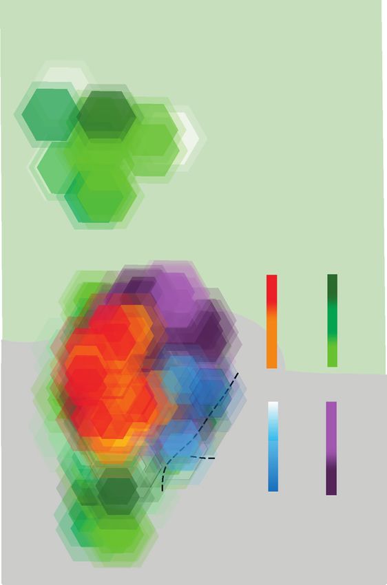
Neuroscience of affect: brain mechanisms of pleasure and displeasure Berridge and Kringelbach 295

psychological function. Hedonic mechanisms require             essentially the same homologous region of deep ventral
millions of neurons arranged into patterns of mesocorti-       anterior cingulate cortex exists in both, but is called the
colimbic circuitry, a combination constituting substantial     subgenual anterior cingulate cortex (Area 25) in humans,
biological investment that was unlikely to have evolved if     and called infralimbic cortex in rodents.
affective reactions did not convey significant objective
benefits [3,9,10,11].                                         Still, some real differences do exist between limbic brains
                                                               of humans and other animals. The most obvious differ-
Our focus on objective affective reactions to identify core    ence is the massive expansion of prefrontal cortex in
hedonic processes takes its lead from Darwin’s original        humans, reflecting greater encephalization. Anatomically,
book on emotion over a century ago [12]. Darwin noted          encephalization also creates greater differentiation among
distinctive affective expressions (facial, bodily, and auto-   prefrontal subregions. This may produce a few human
nomic) in humans and animals in various emotional              cortex subregions that lack any clear homologue in non-
situations. Darwin’s approach is also echoed by Joseph         primates, such as dorsal anterior insula [23]. This may
LeDoux’s recent proposal: ‘‘By focusing on survival func-      also produce some neuronal differences, such as the
tions instantiated in conserved circuits, key phenomena        granular layer in anterior orbitofrontal cortex of humans,
relevant to emotions and feelings are discussed with the       that is, missing in rats.
natural direction of brain evolution in mind (by asking to
what extent are functions and circuits that are present in     Encephalization may also foster greater invasion by des-
other mammals are also present in humans). . .’’ (p. 654)      cending projections from prefrontal cortex into subcorti-
[9]. We similarly suggest that considering animal and         cal structures and functions. A possible human feature is
human studies together allows the best progress to be          greater ‘freeway’ connectivity, or direct projections be-
made in understanding how affective reactions are              tween cortex and deep subcortical structures. By com-
mediated by brain systems.                                     parison, other animals might rely a bit more on ‘local’ road
                                                               connections, which make more frequent intermediate
Concerning human affect, not only can subjective plea-         stops. For example, descending projections from orbito-
sure ratings (liking in the ordinary sense) be assessed in     frontal cortex make more clearly defined connections to
adults, but also objective ‘liking’-related reactions exist    hypothalamus and brainstem structures in primates than
that can be measured in adults and even infants. In adults,    in rats [24]. Conversely, ascending sensory pain and taste
objective affective reactions alone, without any subjec-       signals toward cortex from the brainstem primary visceral/
tive feelings, can occur as unconscious pleasures under        sensory relay, the nucleus of the solitary tract in the
limited circumstances (e.g. as unfelt but behaviorally         medulla, may leap directly to the thalamus in primates,
biasing affective reactions to subliminally brief stimuli)     but make an obligatory stop at the pontine parabrachial
[13–15]. The translation of objective ‘liking’ reaction into   nucleus in rats [23,25]. Psychologically, human encepha-
subjective pleasure feeling probably requires recruitment      lization may consequently result in a greater cortical
of additional brain mechanisms specialized for cognitive       involvement of affect and emotion, expressed as top-
appraisal and conscious experience. An implication of the      down regulation of affective reactions. Still, mesocorti-
objective-subjective distinction is that subjective ratings    colimbic circuits for mediating core affective reactions are
of felt pleasure, while crucial signatures of human affec-     largely similar across all mammals.
tive experience, are interpretive readouts of underlying
affective processes, not always infallible windows into        Many pleasures: one hedonic brain system to mediate
core pleasure reactions themselves. Indeed, ‘liking’ can       them all?
sometimes occur unconsciously, and at other times even         The sensory pleasure of a delicious-tasting food feels
conscious pleasure ratings sometimes detach substan-           different from pleasures of sex or drugs. Even more
tially from core affective reactions (as people concoct        different seem social or cognitive pleasures of seeing a
explanations to themselves for how they think they             loved one or listening to music. But does each psycho-
should feel) [16–19]. Therefore objective measures can         logical pleasure have its own neural circuit? Perhaps not.
be equally as useful as subjective measures for probing        Instead there appears heavy overlap, with a shared meso-
pleasure and displeasure mechanisms.                           corticolimbic circuit or single common neural currency,
                                                               involved in all those diverse pleasures [6,7,26–35]. Neu-
Comparing limbic systems: affect circuitry in humans           roimaging studies often implicate the same list of usual
and animals                                                    culprits as activated by various pleasures. The list
The brain’s circuitry for affective reactions spans from       includes cortical regions (e.g. orbitofrontal, anterior cin-
front to back of nearly the entire brain (Figure 1). Much of   gulate, and insula cortices) and subcortical structures
this circuitry is remarkably similar between humans and        (nucleus accumbens (NAc), ventral pallidum, amygdala,
other mammals [20–22]. Even some apparent differences          and mesolimbic tegmentum). This overlapping pattern
between humans and other species in limbic circuits may        opens the possibility that the same hedonic generating
be more exaggerated in name than in fact. For example,         circuit, embedded in larger mesocorticolimbic systems,

www.sciencedirect.com                                                         Current Opinion in Neurobiology 2013, 23:294–303
296 Social and emotional neuroscience
Figure 1
Hedonic Brain Circuit
‘Liking’ Hotspots are Anatomically Embedded
Infralimbic Cortex/
Brodman’s Area 25 Anterior Cingulate
Thalamus
Cortex
Paraventricular
Prelimbic Interfascicular
Nucleus
Cortex Thompson and Nucleus
Orbitofrontal TS
Swanson, 2010
Cortex
Z Zahm et al., 2012
Z
Glutamate
Z
TS Z TS
Z Glutam
ate
Glut
Dopamine
am
A
at e
AB
G TS Z
TS Z Glutamte
Nac Hotspot TS Z
Z
(Rostrodorsal G A BA
Medial Shell) Z TS
G
Nucleus tam G luta m at e
lu
GABA Z
a te
Accumbens
Medial Shell
Ventral
VP Hotspot Ventral
Pallidum Preoptic Lateral
(Posterior
Hypothalamus Tegmental Parabrachial
Ventral Area Nucleus
Area
Pallidum)
Current Opinion in Neurobiology
Hedonic hotspots and anatomical circuits that distinguish the nucleus accumbens hotspot in rostrodorsal medial shell as a unique site (anatomy based
on Thompson and Swanson (TS symbol in orange boxes; 2010) and on Zahm and colleagues (Z symbol in purple hexagons; 2012). Thompson and
Swanson [66] reported that the nucleus accumbens hotspot of rostrodorsal medial shell is uniquely embedded in its own closed-circuit
corticolimbicpallidal–thalamocortical loop, connecting discrete input subregions and output subregions, and segregated from other parallel loops
passing through other regions of medial shell. Zahm and colleagues suggested additional unique connections for the rostrodorsal hotspot [65].
GABAergic projections are indicated in red, hedonic hotspots are marked in yellow, glutamatergic projections are green, and dopaminergic projections
are marked in blue.
Figure by Daniel Castro, modified from [80].
could give a pleasurable gloss to all such rewards even when (because other nonhedonic features of the experience
the final experience of each seems otherwise unique. remain constant). Additional medial regions of orbitofron-
tal, ventromedial prefrontal, and middle anterior insula
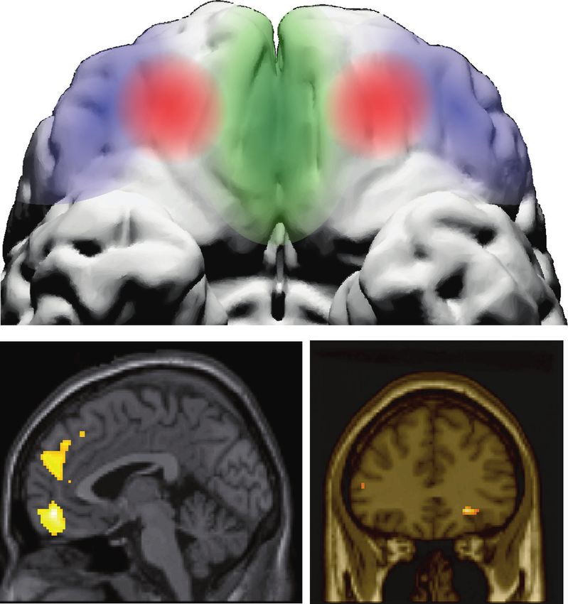
Encoding pure pleasure: apex in orbitofrontal cortex cortices also code aspect of subjective pleasure ratings,
In human neocortex, pleasure appears most faithfully but appear to be more concerned with monitoring and
represented by activity in orbitofrontal cortex, particu- predicting reward value than in pleasure of the experi-
larly in a mid-anterior subregion (Figure 2). Evidence ence per se.
suggests activity of this mid-anterior zone tracks changes
in subjective pleasantness ratings of chocolate and deli- Some studies also indicate lateralization of affect repres-
cious drinks, such as when pleasure intensity is dimin- entation, often as hemispheric differences in positive
ished by switching the taster’s state from hunger to versus negative valence. Most notably, the left hemi-
satiety, and may also encode pleasures of sexual orgasm, sphere of prefrontal cortex often has been implicated
drugs, and music [26,33,36,37]. Subcortically, selective more in positive affect than right hemisphere [42].
hedonic changes also may be tracked by neural firing in For example, individuals who give higher ratings of
NAc and ventral pallidum [38–41]. Such tracking gives subjective well-being may have higher activity in left than
the strongest evidence of pleasure representation right prefrontal cortex, and activation of left subcortical
Current Opinion in Neurobiology 2013, 23:294–303 www.sciencedirect.comNeuroscience of affect: brain mechanisms of pleasure and displeasure Berridge and Kringelbach 297
Figure 2
right x left
+60 +50 +40 +30 +20 +10 0 -10 -20 -30 -40 -50 -60
70 70
60 60
50 50
40 40
y
30 30
20 20
10 10
0 0
Medial OFC Mid-anterior OFC
Pleasure coding sites in human orbitofrontal cortex
Current Opinion in Neurobiology
Subjective pleasure is faithfully coded by orbitofrontal cortex (OFC) activations in people. Sensory pleasures appear most faithfully represented
especially by a mid-anterior OFC site (orange). Pleasant sensations are also coded by activation in a medial strip of OFC (green), but the medial strip
may not as faithfully track changes in pleasure as the orange mid-anterior site [37]. Smaller symbols show results of a large meta-analysis of 267 orbital
areas, which indicated that a medial subregion of orbitofrontal cortex monitored learning and memory of reward values (green area and round blue
dots), whereas a lateral orbitofrontal subregion monitored punishers (purple and orange triangles) [81]. Independently, posterior subregions of OFC
represented complex or abstract reinforcers (such as money) whereas anterior subregions represented sensory rewards such as taste [81].
striatum also may be more tightly linked to pleasantness generating their own different functions, such as cogni-
ratings than right-side [43–45]. However, other studies tive appraisal, memory, decision making, and so on.
have found more equal or bilateral activity patterns, and
so the precise role of lateralization in pleasure still needs Much cortical representation of affect may fit into this
further clarification. ‘other causal’ category. Evidence from humans that cor-
tex is not needed to cause affect includes, for instance,
Subcortical weighting of brain pleasure generators persistence of relatively normal affective reactions such as
The weight of evidence from research on causation of pleasure even after massive damage to prefrontal cortex.
affect suggests that affective reactions may be generated For example, thousands of lobotomy patients in the 1950s
chiefly in subcortical brain structures rather than by any of retained most feelings as far as could be discerned (albeit
the cortical regions discussed above [3,46]. Causation showing impairment in cognitive judgment), despite
may be more anatomically restricted than representation having lost most contributions from their prefrontal cor-
of affect, because only a few of structures that represent tex. More recent strong evidence comes from a beauti-
an affective reaction need also cause that reaction. fully detailed analysis of apparently normal pleasure (and
The other structures may represent affect as a step to other emotions) remaining in a man who lost most of
www.sciencedirect.com Current Opinion in Neurobiology 2013, 23:294–303298 Social and emotional neuroscience
limbic prefrontal cortex due to encephalitis destruction of Pleasure generators identified through objective ‘liking’
bilateral medial orbitofrontal cortex, insula, and ventral reactions
anterior cingulate (plus hippocampus and amygdala) Subcortical brain machinery for actually generating or
[46]. Despite pronounced memory and cognitive defi- causing a ‘liking’ reaction to core pleasure can be probed
cits, this patient retained a rich emotional life for 20 years more extensively via brain manipulations in animals.
as far as could be told, including remarkable capacity to Studies in our laboratory have identified neural pleasure
talk about his feelings. Asked about himself, the patient generators by focusing on the sensory pleasure of sweet-
reported, ‘‘I have a strong feeling of happiness, that we ness. Sweet ‘liking’ is useful because affective facial
are here together working on these wonderful games and expressions of taste pleasure ‘liking’ exist in newborn
feeling happy together. I am glad to be here with you’’. humans and in some animals, aiding the objective
He was also able to develop new Pavlovian learned fears measure of hedonic impact. For example, parents often
of medical syringe needles and noisy fMRI machines and know when their baby expresses a ‘liking’ judgment of
socially learned to prefer a friendly caretaker to a grumpy the deliciousness of a meal. Sweet foods elicit a contented
one. The most parsimonious interpretation of all this is licking of the lips, but bitter tastes instead elicit disgust
that even in humans, pleasures and fears are generated gapes and headshakes. Homologous ‘liking’ orofacial
primarily by subcortical systems that continue to function expressions are elicited also in apes and monkeys, and
strikingly well in the absence of limbic neocortex [3,46]. even in rats and mice [47]. We have used brain manip-
Affective reaction remaining despite prefrontal loss is one ulations of ‘liking’ reactions to identify brain mechanisms
reason to suggest that cortical representation is a quite that generate and enhance such pleasures as sweetness
different function from subcortical causation of affect. (Figure 3).
Figure 3
3.8
Dorsal
4.2
4.6
Hedonic Reactions (sweet)
5.0
5.4
Hedonic Eating
Below skull (mm)
increase increase
5.8
> 800%
> 250%
6.2
> 400%
> 200%
1.9 Sagittal Sections
6.6
> 200%
7.0
Hedonic Aversive
Aversive Reactions (bitter)
decrease decrease
7.4 0.90
< 75% < 75%
7.8
< 50% < 50%
8.2
Ventral
8.6
Rostral Caudal
+2.5 2.0 1.5 1.0 0.5 0.0 -0.5 -1.0 -1.5
Bregma
Current Opinion in Neurobiology
Detail of hedonic hotspot in nucleus accumbens for pleasure generation (sagittal view of medial shell and of neostriatum). This is a causation map:
colors reflect hedonic or motivation consequences (on ‘liking’ reactions or on food intake) of mu opioid agonist microinjections at each site (based
originally on Peciña and Berridge [51]). Red/orange symbols in the rostrodorsal hotspot show sites that caused doubling or higher levels of hedonic
‘liking’ reactions to sucrose taste. By comparison, at caudal sites the same opioid microinjections only suppressed aversive ‘disgust’ reactions to bitter
quinine (purple; e.g. suppressed gapes), or bivalently suppressed both ‘liking’ and ‘disgust reactions (blue). Green sites denote increases in motivation
‘wanting’ to eat without any hedonic change in either ‘liking’ or disgust (enhanced motivation also extended through all red/purple/blue sites in nucleus
accumbens).Modified from [80], based on data from [51].
Current Opinion in Neurobiology 2013, 23:294–303 www.sciencedirect.comNeuroscience of affect: brain mechanisms of pleasure and displeasure Berridge and Kringelbach 299
One surprising finding has been that neural generators of hedonic hotspots have yet been found in neocortex
intense pleasure are much more restricted neurochemi- (though the search continues), but rather only in these
cally than was previously envisioned [48,49,50,51]. For subcortical structures. Continued failure to find a hedo-
instance, mesolimbic dopamine, probably the most pop- nic-enhancing hotspot in prefrontal cortex would be
ular brain neurotransmitter candidate for pleasure two another reason to distinguish between cortical representa-
decades ago, turns out not to cause pleasure or ‘liking’ at tion and subcortical causation of pleasure as different
all. Rather dopamine more selectively mediates a motiva- functions.
tional process of incentive salience, which is a mechanism
for ‘wanting’ rewards but not for ‘liking’ them Each accumbens-pallidum hotspot is only a cubic-milli-
[48,52,53,54–57]. When amplified by addictive drugs meter in volume in rats (a human hotspot equivalent
or by endogenous factors, dopamine helps generate should be approximately a cubic-centimeter, if scaled to
intense levels of ‘wanting’, characteristic of drug addic- whole-brain size). Functionally, hedonic hotspots seem
tion, eating disorders, and related compulsive pursuits quite specialized for intense pleasure generation com-
[44,53,58–61]. Why, then, are dopamine-promoting pared to regions around them. Neurobiologically, hot-
drugs such as cocaine or methamphetamine reportedly spots may have unique anatomical or neurobiological
so pleasant? One possibility is that some psychostimulant features that distinguish them from the rest of their
euphoria comes from the ‘wanting’ component of reward: containing structure, and which perhaps permit the func-
a world that seems more attractive may well carry an aura tional specialization for pleasure causation [65,66,67]
of euphoria. Another potential mechanism is that, distinct (Figure 1).
from raising dopamine in the synapse, such drugs might
also induce secondary recruitment of additional neuro- Integrating neurochemical and anatomical findings, what
biological mechanisms that more directly cause hedonic makes opioid neurotransmitters more hedonic than dopa-
pleasure. For instance, there is evidence to suggest that mine is not that limbic opioid signals always generate
elevation of endogenous opioid signals may be recruited ‘liking’. In most of NAc, neither does. Rather opioid
in limbic structure [62,63]. Such opioid recruitment in stimulation has the special capacity to enhance ‘liking’
accumbens-pallidal hotspots described below would plau- only if the stimulation occurs within an anatomical hot-
sibly generate pleasure ‘liking’ [64]. Conceivably, the spot — whereas dopamine never does anywhere
secondary recruitment of hedonic mechanisms might [48,68,69]. Beyond NAc and ventral pallidum, opioid
become somewhat sluggish with continual drug-taking, stimulation in all regions tested so far for other structures,
therefore requiring higher doses for the sought-after such as neostriatum, amygdala, and so on, at best generate
pleasurable high, even if dopamine-related sensitization enhancement only of motivation ‘wanting’ without
enhanced circuit reactivity to produce more and more enhancing hedonic ‘liking’ [51,70,71]. Overall, the pat-
intense ‘wanting’ [60]. tern indicates not only strong localization of hedonic
function, but also neurochemical specificity of pleasure
Hedonic hotspot network neurotransmitters.
Another surprising finding has been that pleasures gen-
erators are much more anatomically restricted than pre- Functionally, hotspots in NAc and ventral pallidum inter-
viously envisioned, localized to particular subregions. We act together in a single integrated circuit. The two sites
have identified several pleasure generators as small hedo- act as a functional unit for mediating pleasure enhance-
nic hotspots, nestled in subcortical structures [48,49–51]. ments [48,72]. Each hotspot seems able to recruit the
Opioid and endocannabinoid neurochemical signals do other to unanimously generate amplification of ‘liking’.
more effectively generate intense pleasures than dopa- For example, a single opioid microinjection into the NAc
mine — but only within the boundaries of such hotspots. hotspot enhances also responsiveness of ventral pallidum
For example, mu opioid stimulation by DAMGO micro- hotspot neurons, reflected in neuronal firing patterns
injection within a hotspot of NAc (localized in the ros- elicited by a sweet taste or in gene activation, at the
trodorsal quadrant of medial shell), or in another hotspot same time as enhancing behavioral ‘liking’ reactions
of ventral pallidum (in the posterior half of ventral palli- [48,72]. Unanimous recruitment of both hotspots further
dum), more than doubles the intensity of ‘liking’ reac- appears to be required to magnify pleasure. Blocking
tions elicited by sweetness. But the same DAMGO either hotspot with an opioid-antagonist microinjection
microinjections elsewhere in the remaining 90% of completely prevents opioid stimulation of the other hot-
NAc outside the hotspot generate only ‘wanting’ without spot from producing any ‘liking’ enhancement
enhancing ‘liking’ — much like dopamine (i.e. remaining [72].Finally, the ventral pallidum hotspot may be especi-
60% of medial shell and probably entire lateral shell and ally important for maintaining normal levels of pleasure.
core; and even regions of dorsal striatum) [48,49–51] Damage to ventral pallidum can cause even sweet sucrose
(Figures 1 and 3). In addition, in the anterior half of taste to elicit purely negative gapes and other disgust
ventral pallidum, DAMGO microinjection actually causes reactions for days or weeks afterwards (C-Y Ho, The
opposite suppression of ‘liking’ reactions. So far, no ventral pallidum as a limbic pleasure generator, PhD
www.sciencedirect.com Current Opinion in Neurobiology 2013, 23:294–303300 Social and emotional neuroscience
Dissertation, Ann Arbor, University of Michigan, 2010) circuitry, operating in a different mode than for reward.
[8,73]. No other brain lesion of a single site so potently Recent studies have demonstrated existence of an ‘affec-
transforms sensory pleasure into purely negative affect. tive keyboard mechanism’ in the medial shell of NAc for
Of course, other brain structures do help generate intense generating intense dread versus desire. The accumbens
aversive emotions when manipulated in other ways keyboard has remarkable anatomical orderliness in
[9,74–77]. arrangement of valence generators (Figure 4). This mech-
anism releases intense dread-desire motivations following
Affective keyboards in nucleus accumbens: beyond localized disruptions, arranged by valence along a rostro-
desire to dread caudal gradient in shell [76,77]. Just as a musical keyboard
With valence reversal, we arrive at the knotty problem of generates many different notes according to key location,
how brain systems control the balance between positive the affective keyboard can generate many mixtures of
versus negative emotions. Many limbic structures are desire versus fear, each mixture triggered at a different
implicated in both valences, and a given valence is dis- location. The mechanism is probably best viewed as
tributed across several structures. For instance, take the operating via disinhibition: localized shell inhibitions that
negative emotion of fear. Although amygdala is best in turn release downstream efferent targets into exci-
known for fear conditioning [9], active forms of intense tation. That disinhibition interpretation arises because
fear may also be generated via manipulation of NAc suppressive GABAergic neurons project from NAc to
Figure 4
Affective Keyboard for Desire & Dread
Sites in NAc Shell Produce Intense Motivations
% Increase Eating
Appetitive 850
Defensive 600
Mixed 350
No effect
100
Rostral
sites
generate
eating Caudal
sites
generate
fear
% Increase Fear
100
350
600
850
Current Opinion in Neurobiology
Affective keyboard pattern in nucleus accumbens for releasing intense desire and/or dread. The keyboard pattern of intense motivated behaviors is
revealed in the consequences of drug microinjections at various rostrocaudal sites in medial shell. Microinjections of drugs that relatively inhibit
accumbens neurons via amino acid neurotransmitters (e.g. a GABA agonist or a glutamate antagonist) in turn may disinhibit or release motivation
generating circuits in downstream target structures. Rostral green sites released stimulation of eating by up to 600% (desire only). Caudal red sites
released purely increased fearful reactions at levels up to 600% over normal (dread only; escape attempts, distress calls, defensive bite attempts;
spontaneous anti-predator treading/burying). Yellow sites released both desire and dread in the same rats during the same 1-hour test. Just as a
keyboard has many notes, bars reflect the many graded mixtures of affective desire-dread released as microinjection sites move rostrocaudal location
in medial shell (appetitive desire to eat at top; fearful dread reactions at bottom).Modified from [80], based on data from [76,77,79].
Current Opinion in Neurobiology 2013, 23:294–303 www.sciencedirect.comNeuroscience of affect: brain mechanisms of pleasure and displeasure Berridge and Kringelbach 301
targets in ventral pallidum, lateral hypothalamus or brain- same site requires only its D1 dopamine signal (potentially
stem. A motivation-releasing drug microinjection inhibits indicating a more dominant role for neurons of its ‘direct’
NAc neurons within a local 0.5–1.0 mm diameter micro- path to ventral tegmentum) [77,79].
domain corresponding to a single ‘key’ of the keyboard
(via a GABA agonist or AMPA glutamate antagonist). Neurochemical signals similarly switch the operating
Thus drug inhibition of accumbens microdomains pre- modes of a NAc microdomain to generate different affects
vents the endogenous NAc inhibition of targets, and so and motivations. For example, in the rostral NAc hotspot,
releases particular downstream motivation generating opioid stimulation generates intense ‘liking’ plus ‘want-
circuits into final activation to produce the intense motiv- ing’ for reward, but microinjection of dopamine or glu-
ations that are observed. tamate-related drugs generates ‘wanting’ alone.
Conversely, in a caudal NAc shell key, microinjections
The keyboard pattern is revealed by observations that, at inducing opioid or dopamine stimulation generate ‘want-
anterior sites in NAc shell, AMPA-blocking or GABA- ing’, whereas glutamate AMPA blockade instead gener-
stimulating microinjections can double eating and food ates fear, and GABA signals add disgust to the fear [74–
intake amounts, and increase other incentive-related 79]. Thus quite a variety of intense affective states can be
behaviors (Figure 4). At more posterior sites in shell, created by varying the manipulation of a single site.
the same drug microinjections produce increasingly fear-
ful reactions. For example, a normally tame rat will emit Conclusion
frightened distress calls and frantic leaps to escape when The merit of any scientific approach can be judged by
gently touched, and even bite the hand that touches it. whether the approach produces important new insights.
Spontaneously, even when not touched, the rat after In this short review we have shared some evidence
posterior keyboard microinjection also emits fearful anti- produced so far by our approach to finding pleasure in
predator reactions that rodents use to defend against the brain. New insights are emerging into how core
threats in the wild (e.g. rattlesnake), using forepaws to affective reactions of pleasure, disgust or fear are gener-
throw debris toward objects or people seen in the room ated and represented in complex brain systems. Many of
[78]. Corticolimbic inputs from the prefrontal cortex can these new insights could not have been gained without
suppressively regulate these intense motivations, by inhi- aid of the approach sketched here. However, the real
biting general intensity, or by tilting the desire/dread contribution of this approach will hopefully come in
balance in positive direction, though causal generation future, as the new affective neuroscience findings are
of the intense motivations themselves again appears to be applied to help addiction, depression, and other affective
contained more subcortically (e.g. subcortical but not cor- disorders. Our hope is that future generations may be able
tical manipulations are known to cause the intense motiv- to use such scientific insights into brain affective mech-
ations; and even within NAc microinjection of drugs that anisms to create better lives for more people [3].
mimic subcortical GABA signals cause a broader range of
affects than drugs that disrupt corticolimbic glutamate Acknowledgements
signals [i.e. rostral GABA releases ‘liking’ as well as ‘want- Our research is supported by grants from the NIH (MH63644 and
DA015188) to KCB, and from the TrygFonden Charitable Foundation,
ing’, and caudal disgust as well as fear]) [76,77]. Braveheart Charity, Novo Nordisk Foundation to MLK. We thank Daniel
Castro and Dr Jocelyn Richard for redrawing Figures 1, 2 and 4.
Though anatomical location biases the valence of desire
versus dread released by the keyboard, valence at many References and recommended reading
sites can be retuned by psychological factors. The presence Papers of particular interest, published within the period of review,
have been highlighted as:
of a stressfully over-stimulating sensory environment (i.e.
bright lights and loud Iggy Pop music) remaps the accum- of special interest
bens bivalent keyboard by expanding the fear-generating of outstanding interest
zone while shrinking the desire-generating zone [77,79].
Conversely, a comfortable and quiet home-like ambience 1. Lindquist KA, Wager TD, Kober H, Bliss-Moreau E, Barrett LF: The
brain basis of emotion: a meta-analytic review. Behav Brain Sci
remaps in opposite direction, expanding desire and shrink- 2012, 35:121-143.
ing fear. Such psychological top-down remapping can A thoughtful review of neuroimaging studies, concluding that specific
emotions do not have different discrete neural substrates.
actually retune a single site in NAc into releasing opposite
motivations in the different situations, reversing the cir- 2. Leknes S, Tracey I: A common neurobiology for pain and
pleasure. Nat Rev Neurosci 2008, 9:314-320.
cuit’s mode of operation. The switch in operating mode Penetrating review of brain mechanisms involved in both pleasures and
may involve recruiting different neurobiological com- displeasures.
ponents. Fear generation demands endogenous dopamine 3. Berridge KC, Kringelbach ML: Building a neuroscience of
activity at D1 and D2 receptors simultaneously within a pleasure and well-being. Psychol Well Being 2011, 1:1-3.
‘key’ site (implicating roles for both ‘direct’ output path to 4. Smith KS, Mahler SV, Pecina S, Berridge KC: Hedonic hotspots:
generating sensory pleasure in the brain. In Pleasures of the
tegmentum and ‘indirect’ path to ventral pallidum and Brain. Edited by Kringelbach ML, Berridge KC. Oxford University
hypothalamus), whereas positive desire generation by the Press; 2010:27-49.
www.sciencedirect.com Current Opinion in Neurobiology 2013, 23:294–303302 Social and emotional neuroscience
5. Kringelbach ML, Berridge KC (Eds): Pleasures of the Brain. 26. Veldhuizen MG, Rudenga KJ, Small D: The pleasure of taste
Oxford: Oxford University Press; 2010. flavor and food. In Pleasures of the Brain. Edited by Kringelbach
ML, Berridge KC. Oxford University Press; 2010:146-168.
6. Kringelbach ML, Stein A, van Hartevelt TJ: The functional human
27. Zeki S, Romaya JP: The brain reaction to viewing faces of
neuroanatomy of food pleasure cycles. Physiol Behav 2012,
opposite- and same-sex romantic partners. PLoS ONE 2010,
106:307-316.
5:e15802.
7. Georgiadis JR, Kringelbach ML: The human sexual response
cycle: brain imaging evidence linking sex to other pleasures. 28. Xu X, Aron A, Brown L, Cao G, Feng T, Weng X: Reward and
Prog Neurobiol 2012, 98:49-81. motivation systems: a brain mapping study of early-stage
intense romantic love in Chinese participants. Hum Brain Mapp
8. Cromwell HC, Berridge KC: Where does damage lead to 2011, 32:249-257.
enhanced food aversion: the ventral pallidum/substantia
innominata or lateral hypothalamus? Brain Res 1993, 624:1-10. 29. Geogiadis JR, Kortekaas R: The sweetest taboo: functional
neurobiology of human sexuality in relation to pleasure. In
9. LeDoux J: Rethinking the emotional brain. Neuron 2012, Pleasures of the Brain. Edited by Kringelbach ML, Berridge KC.
73:653-676. Oxford University Press; 2010:178-201.
Succinct analysis of brain systems for emotional reactions and evolved
functions. 30. Cacioppo S, Bianchi-Demicheli F, Frum C, Pfaus JG, Lewis JW:
The common neural bases between sexual desire and love: a
10. Damasio AR: Self Comes to Mind: Constructing the Conscious multilevel kernel density fMRI analysis. J Sex Med 2012,
Brain. edn 1. New York: Pantheon Books; 2010. 9:1048-1054.
11. Panksepp J: The basic emotional circuits of mammalian 31. De Groof G, Van der Linden A: Love songs, bird brains and
brains: do animals have affective lives? Neurosci Biobehav Rev diffusion tensor imaging. NMR Biomed 2010, 23:873-883.
2011, 35:1791-1804.
32. Salimpoor VN, Benovoy M, Larcher K, Dagher A, Zatorre RJ:
12. Darwin C: The Expression of the Emotions in Man and Animals Anatomically distinct dopamine release during anticipation
(1998 edition: Revised and with Commentary by p. Ekman). and experience of peak emotion to music. Nat Neurosci 2011,
Oxford: Harper Collins–Oxford University Press; 1872. 14:257-262.
13. Winkielman P, Berridge KC, Wilbarger JL: Unconscious affective 33. Vuust P, Kringelbach ML: The pleasure of music. In Pleasures of
reactions to masked happy versus angry faces influence the Brain. Edited by Kringelbach ML, Berridge KC. Oxford
consumption behavior and judgments of value. Pers Soc University Press; 2010:255-269.
Psychol Bull 2005, 31:121-135.
34. Parsons CE, Young KS, Murray L, Stein A, Kringelbach ML: The
14. Pessiglione M, Schmidt L, Draganski B, Kalisch R, Lau H, Dolan R, functional neuroanatomy of the evolving parent–infant
Frith C: How the brain translates money into force: a relationship. Prog Neurobiol 2010, 91:220-241.
neuroimaging study of subliminal motivation. Science 2007,
316:904-906. 35. Georgiadis JR, Kringelbach ML, Pfaus JG: Sex for fun: a
synthesis of human and animal neurobiology. Nat Rev Urol
15. Childress A, Ehrman R, Wang Z, Li Y, Sciortino N, Hakun J, Jens W, 2012. (online first).
Suh J, Listerud J, Marquez K et al.: Prelude to passion: limbic
activation by ‘‘unseen’’ drug and sexual cues. PLoS ONE 2008, 36. Kringelbach ML, O’Doherty J, Rolls ET, Andrews C: Activation of
3:e1506. the human orbitofrontal cortex to a liquid food stimulus is
correlated with its subjective pleasantness. Cereb Cortex 2003,
16. Schooler JW, Mauss IB: To be happy and to know it: the 13:1064-1071.
experience and meta-awareness of pleasure. In Pleasures of
the Brain. Edited by Kringelbach ML, Berridge KC. Oxford 37. Kringelbach ML: The hedonic brain: a functional
University Press; 2010:244-254. neuroanatomy of human pleasure. In Pleasures of the Brain.
Edited by Kringelbach ML, Berridge KC. Oxford University Press;
17. O’Brien E, Ellsworth PC, Schwarz N: Today’s misery and 2010: 202-221.
yesterday’s happiness: differential effects of current life-
events on perceptions of past wellbeing. J Exp Soc Psychol 38. Loriaux AL, Roitman JD, Roitman MF: Nucleus accumbens shell,
2012, 48:968-972. but not core, tracks motivational value of salt. J Neurophysiol
2011, 106:1537-1544.
18. Dijksterhuis A, Aarts H: Goals, attention, and
(un)consciousness. Annu Rev Psychol 2010, 61:467-490. 39. Roitman MF, Wheeler RA, Tiesinga PH, Roitman JD, Carelli RM:
19. Gilbert DT, Wilson TD: Why the brain talks to itself: sources of Hedonic and nucleus accumbens neural responses to a
error in emotional prediction. Philos Trans R Soc B 2009, natural reward are regulated by aversive conditioning. Learn
364:1335-1341. Mem 2010, 17:539-546.
20. Haber SN, Knutson B: The reward circuit: linking primate 40. Tindell AJ, Smith KS, Pecina S, Berridge KC, Aldridge JW: Ventral
anatomy and human imaging. Neuropsychopharmacology 2010, pallidum firing codes hedonic reward: when a bad taste turns
35:4-26. good. J Neurophysiol 2006, 96:2399-2409.
21. Heimer L, Van Hoesen GW, Trimble M, Zahm DS: Anatomy of 41. Krause M, German PW, Taha SA, Fields HL: A pause in nucleus
Neuropsychiatry: The New Anatomy of the Basal Forebrain and its accumbens neuron firing is required to initiate and maintain
Implications for Neuropsychiatric Illness. Amsterdam: Elsevier/ feeding. J Neurosci 2010, 30:4746-4756.
Academic Press; 2008.
42. Davidson RJ: Well-being and affective style: Neural substrates
22. Murray EA, O’Doherty JP, Schoenbaum G: What we know and do and biobehavioural correlates. Philos Trans R Soc Lond B Biol
not know about the functions of the orbitofrontal cortex after 20 Sci 2004, 359:1395-1411.
years of cross-species studies. J Neurosci 2007, 27:8166-8169.
43. Kuhn S, Gallinat J: The neural correlates of subjective
23. Craig AD: How do you feel — now? The anterior insula and pleasantness. Neuroimage 2012, 61:289-294.
human awareness. Nat Rev Neurosci 2009, 10:59-70.
Influential advocacy of view that human emotional consciousness arises 44. Lawrence NS, Hinton EC, Parkinson JA, Lawrence AD: Nucleus
from neocortex, especially insula. accumbens response to food cues predicts subsequent snack
consumption in women and increased body mass index in
24. Price JL: Definition of the orbital cortex in relation to specific those with reduced self-control. Neuroimage 2012,
connections with limbic and visceral structures and other 63:415-422.
cortical regions. Ann NY Acad Sci 2007, 1121:54-71.
45. Price TF, Harmon-Jones E: Approach motivational body
25. Scott TR, Small DM: The role of the parabrachial nucleus in taste postures lean toward left frontal brain activity.
processing and feeding. Ann NY Acad Sci 2009, 1170:372-377. Psychophysiology 2011, 48:718-722.
Current Opinion in Neurobiology 2013, 23:294–303 www.sciencedirect.comNeuroscience of affect: brain mechanisms of pleasure and displeasure Berridge and Kringelbach 303
46. Damasio A, Damasio H, Tranel D: Persistence of feelings and 64. Tritsch NX, Ding JB, Sabatini BL: Dopaminergic neurons inhibit
sentience after bilateral damage of the insula. Cereb Cortex striatal output through non-canonical release of gaba. Nature
2012. (online first). 2012, 490:262-266.
Powerful case study demonstrating normal emotional feelings in absence
of insula and most other limbic neocortex. 65. Zahm DS, Parsley KP, Schwartz ZM, Cheng AY: On lateral
septum-like characteristics of outputs from the accumbal
47. Steiner JE, Glaser D, Hawilo ME, Berridge KC: Comparative hedonic ‘hotspot’ of Peciña and Berridge with commentary on
expression of hedonic impact: affective reactions to taste by the transitional nature of basal forebrain ‘boundaries’. J Comp
human infants and other primates. Neurosci Biobehav Rev 2001, Neurol 2013, 521:50-68.
25:53-74. Neuroanatomical identification of special septal transition features of
hedonic hotspot in nucleus accumbens.
48. Smith KS, Berridge KC, Aldridge JW: Disentangling pleasure
from incentive salience and learning signals in brain reward 66. Thompson RH, Swanson LW: Hypothesis-driven structural
circuitry. Proc Natl Acad Sci USA 2011, 108:E255-E264. connectivity analysis supports network over hierarchical
Demonstration of neuronal firing codes in ventral pallidum for separable model of brain architecture. Proc Natl Acad Sci USA 2010,
‘liking’, ‘wanting’, and learning signals for reward. 107:15235-15239.
First demonstration of anatomical uniqueness of nucleus accumbens
49. Mahler SV, Smith KS, Berridge KC: Endocannabinoid hedonic hotspot and sophisticated technical tour de force.
hotspot for sensory pleasure: anandamide in nucleus
accumbens shell enhances ‘liking’ of a sweet reward. 67. Kupchik YM, Kalivas PW: The rostral subcommissural ventral
Neuropsychopharmacology 2007, 32:2267-2278. pallidum is a mix of ventral pallidal neurons and neurons from
adjacent areas: an electrophysiological study. Brain Struct
50. Smith KS, Berridge KC: The ventral pallidum and hedonic Funct 2012. (online first).
reward: neurochemical maps of sucrose ‘‘liking’’ and food Neuroanatomical identification of special morphological and electrophy-
intake. J Neurosci 2005, 25:8637-8649. siological differences between neurons in rostral ventral pallidum versus
in caudal ventral pallidum (hedonic hotspot).
51. Peciña S, Berridge KC: Hedonic hot spot in nucleus accumbens
shell: where do mu-opioids cause increased hedonic impact 68. Tindell AJ, Berridge KC, Zhang J, Peciña S, Aldridge JW: Ventral
of sweetness? J Neurosci 2005, 25:11777-11786. pallidal neurons code incentive motivation: amplification by
mesolimbic sensitization and amphetamine. Eur J Neurosci
52. Berridge KC: From prediction error to incentive salience:
2005, 22:2617-2634.
mesolimbic computation of reward motivation. Eur J Neurosci
2012, 35:1124-1143. 69. Peciña S, Cagniard B, Berridge KC, Aldridge JW, Zhuang X:
Hyperdopaminergic mutant mice have higher ‘‘wanting’’ but
53. Saunders BT, Robinson TE: The role of dopamine in the
not ‘‘liking’’ for sweet rewards. J Neurosci 2003, 23:9395-9402.
accumbens core in the expression of pavlovian-conditioned
responses. Eur J Neurosci 2012. (online first). 70. Mahler SV, Berridge KC: What and when to ‘‘want’’? Amygdala-
Demonstration that dopamine blockade in nucleus accumbens impairs based focusing of incentive salience upon sugar and sex.
reward motivation ‘wanting’ but not learning/memories. Psychopharmacology (Berl) 2012, 221:407-426.
54. Flagel SB, Clark JJ, Robinson TE, Mayo L, Czuj A, Willuhn I, 71. Difeliceantonio AG, Mabrouk OS, Kennedy RT, Berridge KC:
Akers CA, Clinton SM, Phillips PE, Akil H: A selective role for Enkephalin surges in dorsal neostriatum as a signal to eat.
dopamine in stimulus-reward learning. Nature 2011, 469:53-57. Curr Biol 2012, 22:1918-1924.
55. Shiner T, Seymour B, Wunderlich K, Hill C, Bhatia KP, Dayan P, 72. Smith KS, Berridge KC: Opioid limbic circuit for reward:
Dolan RJ: Dopamine and performance in a reinforcement interaction between hedonic hotspots of nucleus accumbens
learning task: evidence from parkinson’s disease. Brain 2012, and ventral pallidum. J Neurosci 2007, 27:1594-1605.
135:1871-1883.
73. Smith KS, Tindell AJ, Aldridge JW, Berridge KC: Ventral pallidum
56. Schultz W: Potential vulnerabilities of neuronal reward, risk, roles in reward and motivation. Behav Brain Res 2009, 196:155-167.
and decision mechanisms to addictive drugs. Neuron 2011,
69:603-617. 74. von dem Hagen EA, Beaver JD, Ewbank MP, Keane J,
Passamonti L, Lawrence AD, Calder AJ: Leaving a bad taste in
57. Leyton M: The neurobiology of desire: dopamine and the your mouth but not in my insula. Soc Cogn Affect Neurosci 2009,
regulation of mood and motivational states in humans. In 4:379-386.
Pleasures of the Brain. Edited by Kringelbach ML, Berridge KC.
Oxford University Press; 2010:222-243. 75. Baliki MN, Geha PY, Fields HL, Apkarian AV: Predicting value of
pain and analgesia: nucleus accumbens response to noxious
58. Volkow ND, Wang GJ, Baler RD: Reward, dopamine and the stimuli changes in the presence of chronic pain. Neuron 2010,
control of food intake: implications for obesity. Trends Cogn 66:149-160.
Sci 2011, 15:37-46.
76. Richard JM, Berridge KC: Prefrontal cortex modulates desire
59. Berridge KC, Robinson TE: Drug addiction as incentive and dread generated by nucleus accumbens glutamate
sensitization. In Addiction and Responsibility. Edited by Poland disruption. Biol Psychiatry 2012. (online first).
J, Graham G. MIT Press; 2011:21-54.
77. Richard JM, Berridge KC: Nucleus accumbens dopamine/
60. Robinson TE, Berridge KC: The incentive sensitization theory of glutamate interaction switches modes to generate desire
addiction: some current issues. Philos Trans R Soc Lond B Biol versus dread: D1 alone for appetitive eating but d1 and d2
Sci 2008, 363:3137-3146. together for fear. J Neurosci 2011, 31:12866-12879.
61. O’Sullivan SS, Wu K, Politis M, Lawrence AD, Evans AH, Bose SK, 78. Coss RG, Owings DH: Snake-directed behavior by snake naive
Djamshidian A, Lees AJ, Piccini P: Cue-induced striatal and experienced California ground squirrels in a simulated
dopamine release in parkinson’s disease-associated burrow. Z Tierpsychol J Comp Ethol1978, 48:421-435.
impulsive-compulsive behaviours. Brain 2011,
134:969-978. 79. Reynolds SM, Berridge KC: Emotional environments retune the
valence of appetitive versus fearful functions in nucleus
62. Soderman AR, Unterwald EM: Cocaine-induced mu opioid accumbens. Nat Neurosci 2008, 11:423-425.
receptor occupancy within the striatum is mediated by
dopamine d2 receptors. Brain Res 2009, 1296:63-71. 80. Richard JM, Castro D, DiFeliceantonio AG, Robinson MJF, Berridge
KC: Mapping brain circuits of reward and motivation: in the
63. Colasanti A, Searle GE, Long CJ, Hill SP, Reiley RR, Quelch D, footsteps of Ann Kelley. Neurosci Biobehav Rev 2012. (online first).
Erritzoe D, Tziortzi AC, Reed LJ, Lingford-Hughes AR et al.:
Endogenous opioid release in the human brain reward system 81. Kringelbach ML, Rolls ET: The functional neuroanatomy of the
induced by acute amphetamine administration. Biol Psychiatry human orbitofrontal cortex: evidence from neuroimaging and
2012, 72:371-377. neuropsychology. Prog Neurobiol 2004, 72:341-372.
www.sciencedirect.com Current Opinion in Neurobiology 2013, 23:294–303You can also read