The Effects of Iron Supplementation and Fortification on the Gut Microbiota: A Review - MDPI
←
→
Page content transcription
If your browser does not render page correctly, please read the page content below
Review
The Effects of Iron Supplementation and Fortification
on the Gut Microbiota: A Review
Emma CL Finlayson-Trick 1 , Jordie AJ Fischer 2,3 , David M Goldfarb 1,3,4 and
Crystal D Karakochuk 2,3, *
 1    Faculty of Medicine, University of British Columbia, Vancouver, BC V6T 1Z3, Canada;
      efinlaysontrick@alumni.ubc.ca (E.C.F.-T.); david.goldfarb@cw.bc.ca (D.M.G.)
 2    Department of Food, Nutrition and Health, University of British Columbia, Vancouver, BC V6T 1Z4, Canada;
      jordie.fischer@ubc.ca
 3    British Columbia Children’s Hospital Research Institute, Vancouver, BC V5Z 4H4, Canada
 4    Department of Pathology and Laboratory Medicine, BC Children’s and Women’s Hospital and University of
      British Columbia, Vancouver, BC V6T 1Z7, Canada
 *    Correspondence: crystal.karakochuk@ubc.ca

 Received: 30 August 2020; Accepted: 24 September 2020; Published: 26 September 2020

 Abstract: Iron supplementation and fortification are used to treat iron deficiency, which is often
 associated with gastrointestinal conditions, such as inflammatory bowel disease and colorectal
 cancer. Within the gut, commensal bacteria contribute to maintaining systemic iron homeostasis.
 Disturbances that lead to excess iron promote the replication and virulence of enteric pathogens.
 Consequently, research has been interested in better understanding the effects of iron supplementation
 and fortification on gut bacterial composition and overall gut health. While animal and human
 trials have shown seemingly conflicting results, these studies emphasize how numerous factors
 influence gut microbial composition. Understanding how different iron formulations and doses
 impact specific bacteria will improve the outcomes of iron supplementation and fortification in
 humans. Furthermore, discerning the nuances of iron supplementation and fortification will benefit
 subpopulations that currently do not respond well to treatment.

 Keywords: iron supplementation; gut microbiome; iron metabolism; gastrointestinal homeostasis

1. Introduction
     The majority of living organisms require iron for survival. Iron can exist in one of two oxidation
states, and due to this redox potential, can function in several fundamental processes, such as respiration,
DNA replication, energy production, and cellular proliferation [1]. Humans absorb iron from their
diet in a dynamic, tightly regulated process within the intestine [2]. In addition to controlling the
amount of iron absorbed, this process dictates iron availability for the complex community of bacteria
living in the intestine, hereafter referred to as the gut microbiota. As such, many bacteria have
developed sophisticated systems to obtain, store, and regulate iron. Iron deficiency and excess both
impact gut microbial health and lead to diseases, such as iron deficiency anemia and iron overload,
respectively. Iron deficiency is highly prevalent worldwide and is commonly treated with oral iron
supplements and fortificants [3]. In this review, we cover the effects of oral iron supplementation
and fortification on gut health and disease. We begin with an overview of how the body acquires
and utilizes iron. Then, we discuss the complex relationship between iron homeostasis and the gut
microbiome. Finally, we summarize the microbial changes that occur following iron supplementation
and fortification in animal and human trials, and we identify areas in need of continued research.
In this literature review, we used PubMed and MEDLINE databases to search for articles related to

Gastrointest. Disord. 2020, 2, 327–340; doi:10.3390/gidisord2040030     www.mdpi.com/journal/gastrointestdisord
Gastrointest. Disord. 2020, 2 328
“human iron metabolism”, “bacterial iron metabolism”, “iron and gut flora”, and “the effects of iron on
the gut microbiota/microbiome in animals and/or humans”.
Gastrointest. Disord. 2020, 2 FOR PEER REVIEW 2
2. Overview of Iron Absorption
to search
Humans forapproximately
lose articles related to0.5–2
“human mgiron
of metabolism”,
iron every day “bacterial
from ironskinmetabolism”, “iron and gut
cell desquamation, intestinal
flora”, and “the effects of iron on the gut microbiota/microbiome in animals
epithelial cell (IEC) sloughing, and urine and sweat production [4]. Additional iron may also be lost and/or humans.”
during specific physiological
2. Overview processes, such as menstruation and lactation [5]. To balance this loss,
of Iron Absorption
the human duodenum and proximal jejunum absorb approximately 2 mg of dietary iron daily, a small
Humans lose approximately 0.5–2 mg of iron every day from skin cell desquamation, intestinal
proportion of the total daily dietary intake [6,7]. Iron from the diet is found primarily as heme, derived
epithelial cell (IEC) sloughing, and urine and sweat production [4]. Additional iron may also be lost
from myoglobin and hemoglobin,
during specific or nonheme
physiological processes, such asiron, derived from
menstruation plants and
and lactation [5]. Toiron-fortified foods [6].
balance this loss,
Nonheme theiron
humanexists in two and
duodenum forms as reduced
proximal jejunumferrous
absorb iron or oxidized
approximately 2 mgferric iron.iron
of dietary IECs, known
daily, a as
small proportion of the total daily dietary intake [6,7]. Iron from the diet is
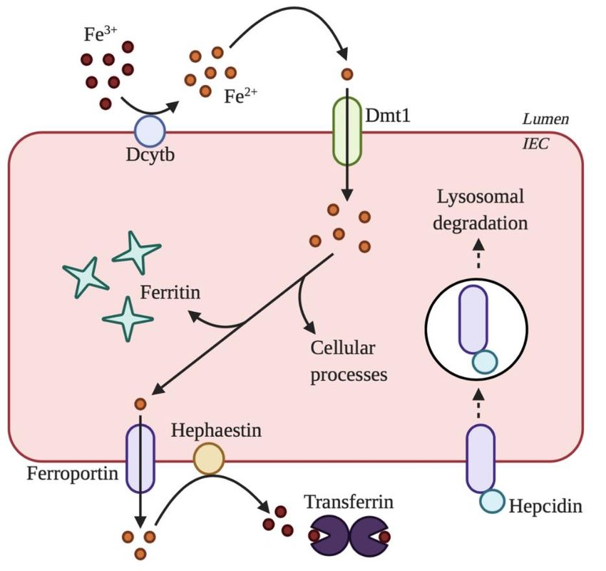
enterocytes, can absorb only ferrous iron (Figure 1). As such, ferric iron is reduced to ferrous iron found primarily as heme,
derived from myoglobin
by the membrane-bound ferricand hemoglobin,
reductase or nonheme
duodenal iron, derived
cytochrome from plants
B (Dcytb) and
that is iron-fortified
expressed on the
foods [6]. Nonheme iron exists in two forms as reduced ferrous iron or oxidized ferric iron. IECs,
apical brush border membrane of IECs [8]. Once in the ferrous form, iron is transported across the
known as enterocytes, can absorb only ferrous iron (Figure 1). As such, ferric iron is reduced to
apical membrane
ferrous ironof enterocytes by the 12 transmembrane
by the membrane-bound ferric reductasedomain
duodenal protein, divalent
cytochrome metal transporter
B (Dcytb) that is 1
(DMT1, also known as Nramp2) [9]. Within enterocytes, iron is stored
expressed on the apical brush border membrane of IECs [8]. Once in the ferrous form, iron isin ferritin, used in a variety
of cellular processes,
transported or transported
across into systemic
the apical membrane circulation
of enterocytes by theby 12 crossing
transmembranethe basolateral membrane
domain protein,
through divalent metal transporter 1 (DMT1,
the 12 transmembrane domainalso known as
protein, Nramp2) [9].
ferroportin Within
[10]. enterocytes,isiron
Ferroportin is stored
also in
expressed on
ferritin, used in a variety of cellular processes, or transported into systemic
macrophages and hepatocytes [10]. On the basolateral membrane, hephaestin oxidizes ferrous iron to circulation by crossing
the basolateral membrane through the 12 transmembrane domain protein, ferroportin [10].
ferric iron, enabling the transportation of iron in the blood by transferrin [5]. In comparison to nonheme
Ferroportin is also expressed on macrophages and hepatocytes [10]. On the basolateral membrane,
iron, heme absorption
hephaestin remains
oxidizes ferrousenigmatic [11].
iron to ferric There
iron, are two
enabling current hypotheses
the transportation of iron infortheintestinal
blood by heme
absorption: Either heme is endocytosed from the apical membrane or transported
transferrin [5]. In comparison to nonheme iron, heme absorption remains enigmatic [11]. There are through a specific
receptor two
intocurrent
the cytosol [12]. for intestinal heme absorption: Either heme is endocytosed from the apical
hypotheses
membrane or transported through a specific receptor into the cytosol [12].
Absorption
Figure 1.Figure of nonheme
1. Absorption of nonhemeironiron
by by
intestinal
intestinalepithelial cells(IECs).
epithelial cells (IECs). Ferric
Ferric iron iron isreduced
is first first reduced
to ferrousto iron byiron
ferrous duodenal cytochrome
by duodenal cytochromeB (Dcytb)
B (Dcytb)on on the
the apical membrane.
apical membrane. Then,Then,
ferrousferrous
iron isiron is
transported
transported across
across the the apical
apical membrane
membrane byby divalentmetal
divalent metal transporter
transporter 1 (DMT1).
1 (DMT1).OnceOnceinsideinside
the cell,the cell,
iron is
iron is stored instored in ferritin,
ferritin, transported
transported acrossacross the basolateral
the basolateral membrane
membrane by by ferroportin,ororused
ferroportin, usedininaavariety
variety of cellular processes. After transport across the basolateral membrane, ferrous iron is oxidized
of cellular processes. After transport across the basolateral membrane, ferrous iron is oxidized to ferric
to ferric iron by hephaestin. Ferric iron is then transported by transferrin in circulation. Iron
iron by hephaestin. Ferric iron is then transported by transferrin in circulation. Iron absorption is
reduced when hepcidin binds to ferroportin because hepcidin causes the internalization and degradation
of ferroportin. The figure created with www.BioRender.com.Gastrointest. Disord. 2020, 2 329
3. Maintenance of Systemic Iron Homeostasis
Humans have no active iron excretory mechanism; therefore, systemic iron homeostasis is
primarily regulated at the point of absorption. Hepcidin, a peptide hormone produced by the liver,
is considered the master regulator of systemic iron homeostasis [13]. Hepcidin binds to and degrades
ferroportin, which consequently impacts how iron is recycled by macrophages, absorbed by IECs,
and stored by hepatocytes [14,15]. Hepcidin expression is upregulated when iron stores are adequate
or high, or in response to inflammation, infection, or injury. Conversely, hepcidin expression is
downregulated to improve iron absorption when iron stores are low, as in the case of iron deficiency
or instances of certain genetic hemoglobinopathies, such as β-thalassemia [6,16,17]. As an aside,
low hepcidin levels, as seen in some thalassemias and hereditary hemochromatosis, increase the risk
of iron overload, due to increased intestinal iron absorption [18,19]. In hereditary hemochromatosis,
a disease related to mutations in iron metabolism genes, excess iron is deposited throughout the
body in the heart, pancreas, and liver, as well as the skin and joints [20]. Regardless of etiology,
iron overload can result in several different diseases related to specific organ damage from oxidative
stress. Iron overload also is known to increase the risk of infection.
Altered hepcidin levels are also associated with a variety of gastrointestinal conditions, including
colorectal cancer (CRC) and inflammatory bowel disease (IBD). In CRC, hepcidin production is increased,
which enables the tumor to retain more iron as tumoral ferroportin expression is decreased [21].
Intratumoral iron promotes oncogene activation, inflammation, and tumor growth [21]. In comparison,
hepcidin levels in IBD do not follow a clear pattern, despite many patients with active disease
experiencing reduced iron absorption [22–25]. Nevertheless, when examined using a dextran sulfate
sodium (DSS) induced colitis mouse model, hepcidin levels were found to be reduced [26]. For both
CRC and IBD, current research is focused on developing treatments and management strategies that
reduce hepcidin levels. Vitamin D administration and anti-TNF-α monoclonal antibody therapy,
for example, have shown promising results for the treatment and management of anemia in IBD [23,27].
4. Role of the Gut Microbiota in Maintaining Iron Homeostasis
Along the length of the intestine, there are physiological gradients (e.g., pH, oxygen, nutrient,
etc.) that produce not only distinct bacterial habitats, but also influence the solubility and availability
of iron [2]. The iron that is not absorbed by the duodenum passes into the colon, where it is
thought to be metabolized by gut bacteria, as well as other microorganisms, such as parasites and
fungi [1,28]. Iron is known to be a growth-limiting nutrient for both human cells and bacteria alike [29].
Accordingly, bacteria have developed two main strategies to obtain iron from their environment.
The most prevalent mechanism involves the synthesis and secretion of siderophores, which are
high-affinity ferric iron chelators [30]. Escherichia coli, Pseudomonas aeruginosa, Klebsiella pneumoniae,
Staphylococcus aureus, and Mycobacterium tuberculosis are just some of the bacteria that use siderophores
to acquire iron [31,32]. In addition to benefiting the bacterium, siderophores also appear to assist the
host. As an example, Qi and Han (2018) observed in their study that enterobactin, an archetypical
siderophore produced by many Gram-negative bacteria, promoted mitochondrial iron uptake and
homeostasis by binding to the ATP synthase α subunit [33]. The results from this study emphasize
that the battle for iron between microbes and host is far more complicated than currently understood,
and opens the field for continued exploration of cooperative iron-mediated host-microbe interactions.
The second strategy involves specific receptors that enable bacteria to acquire iron directly from host
proteins, such as heme, transferrin, and lactoferrin [34]. Finally, some potentially beneficial gut bacteria,
such as Lactobacillus plantarum, do not require iron at all but instead depend on manganese [35].
Therefore, in the presence of iron, iron-independent bacteria do not increase at a rate proportional to
iron-dependent (and possibly pathogenic) bacteria [36].
Commensal gut bacteria play a vital role in maintaining iron homeostasis. In a recent study
by Das et al. (2020), gut microbial metabolites were shown to suppress intestinal hypoxia-inducible
factor 2-α activity (a master transcription factor of intestinal iron absorption) and upregulate ferritinGastrointest. Disord. 2020, 2 330
expression [37]. Additionally, Das et al. (2020) identified Lactobacillus species as the key sensors of
intestinal iron [37]. Several studies have so far attempted to characterize the relationship between
lactobacilli and iron. Using a mouse knockout model for iron regulatory protein 2, which increases
fecal iron concentration, Buknik-Rosenblau et al. (2012) observed an increase in fecal Lactobacillus
species abundance [38]. It remains to be seen whether the increase in lactobacilli was due to greater
iron bioavailability or due to changes in the bacterial community that allowed lactobacilli to proliferate.
Nevertheless, the work by Buknik-Rosenblau et al. (2012) demonstrates that deletions or mutations
of iron metabolism genes affect the intestinal bacterial composition, which has clinically important
implications. In human trials, a study by Balamurugan et al. (2010) found that young women in
South India with iron deficiency had a low abundance of gut lactobacilli [39]. In comparison, the study
by Kalipatnapu et al. (2017) observed an inverse relationship between fecal iron concentration and
Lactobacillus species in rural children in India [40]. Regardless of the exact mechanism, several groups
are testing whether specific Lactobacillus species improve iron absorption and status. In a meta-analysis
of eight studies, Vonderheld et al. (2019) observed that Lactobacillus plantarum 299v significantly
improved nonheme dietary iron absorption in humans [41]. Improvement in iron absorption may
be due to an increase in Dcytb activity as Sandberg et al. (2018) observed activation of this axis
following treatment with an L. plantarum 299v supplement in their human intestinal co-culture model of
enterocytes and goblet cells [42]. In contrast, Rosen et al. found that L. plantarum 299v did not enhance
iron absorption in iron-deficient pediatric patients treated with ferrous sulfate [43]. To improve the
efficacy of L. plantarum 229v as a probiotic, future studies need to further assess the effects of dose,
formulation, the timing of administration, and diet. In addition to Lactobacillus, other bacteria have
been examined for their probiotic properties, and those studies are summarized in the recent review by
Rusu et al. (2020) [44]. The use of prebiotics and synbiotics for the treatment of iron deficiency is also
summarized in the same Rusu et al. (2020) review.
When the gut bacterial composition is altered or when gut bacteria are absent, iron homeostasis
is disturbed. In experiments using germ-free mice [45] and rats [46], iron uptake and storage was
reduced within IECs. In their study, Deschemin et al. (2016) also reported a decrease in ferroportin
expression by IECs in germ-free mice, providing a mechanism for the observed reduction in iron
absorption [45]. Similarly, iron absorption was reduced in rats [47] and rabbits [48] treated with
antibiotics. These results, however, seem to conflict with a more recent study in mice that found
iron absorption increased following antibiotic treatment [37]. These findings suggest that antibiotic
administration may improve iron absorption in patients with iron deficiency.
While gut bacteria are important for maintaining systemic iron homeostasis, iron can also
promote the replication and virulence of enteric pathogens, such as Salmonella spp., Shigella spp.,
and Campylobacter spp. [49,50]. When iron is abundant, bacteria proliferate, and form biofilms
readily, which is hypothesized to be one of the reasons why individuals with iron overload are
more susceptible to infection [7]. It has been shown that humans with iron overloading syndromes,
including hemochromatosis and refractory anemias are more susceptible to bacterial infections,
including Yersinia spp., Listeria monocytogenes, and Vibrio vulnificus [51–53]. Iron limitation, therefore,
serves as an innate immune defence mechanism termed “nutritional immunity” [54]. Mediated by
hepcidin, iron withholding strategies, such as hypoferraemia, denies iron to invading pathogens [55].
As an aside, iron retention within cells, such as macrophages, promotes the virulence of intracellular
pathogens like Salmonella enterica [56]. Parmanand et al. (2019) observed in their in vitro colonic
fermentation study that iron chelation resulted in a lower relative abundance of potentially pathogenic
bacteria [57]. Similarly, Kortman et al. (2015) observed using a mouse model that dietary iron limitation
reduced disease pathology upon oral challenge with Citrobacter rodentium, a well-established model
for infectious gastroenteritis [58]. Finally, some of the pathogens that benefit from increased iron
availability are procarcinogenic [59]. Specifically, Streptococcus bovis, Bacteroides, Enterococcus faecalis,
and Clostridia are all implicated in carcinogenesis as these bacteria promote inflammation through the
production of genotoxic metabolites [60]. These gut microbiota can contribute to the start and/or theGastrointest. Disord. 2020, 2 331
progression of colorectal cancer. With this knowledge in mind, there needs to be a reassessment as to
whether providing oral iron therapy to colorectal cancer patients with iron deficiency is the best route
of administration.
5. Effect of Iron Supplementation and Fortification on the Gut Microbiota
Iron supplementation and fortification are two different methods used to address iron deficiency.
Iron supplementation is considered the more effective method, while iron fortification is often considered
the safer method (as iron is delivered in smaller doses and is more amenable to physiological uptake
when combined with food) [61]. While supplementation is a population-specific approach, iron-fortified
foods, such as cereal products, milk, meal replacements, and infant foods, is a public health strategy to
enhance the nutritional quality of diets in a population.
Iron salts are widely used in oral iron supplementation programs. As an inexpensive
iron supplement, ferrous sulfate (FeSO4 ) is one of the most commonly used iron salts.
Unfortunately, ferrous sulfate is well known to irritate the stomach lining, causing gastrointestinal side
effects, including stomach pain, nausea, diarrhea, and constipation, which makes supplementation
adherence challenging [62,63]. Ferrous gluconate, another type of iron salt, appears to have fewer
side-effects than ferrous sulfate [64]. Iron absorption from oral supplements is typically low,
with less than 20% absorbed in the duodenum and the remainder passing unabsorbed into the
colon [65]. Of the commonly used iron supplements, ferrous fumarate has the most iron per gram [64].
Emerging iron preparations, such as ferrous bisglycinate are marketed as having greater gastrointestinal
tolerance, bioavailability, and protection against dietary iron inhibitors (e.g., phytates), as iron amino
acid chelate does not form insoluble compounds with substances containing high content of iron
absorption inhibitors, commonly found in cereal-based foods [66,67]. Nagpal and Choudhury (2004)
previously conducted a comprehensive review of ferrous salts, ferric salts, iron amino acid chelates,
iron polymaltose complex, and carbonyl iron; therefore, this review will not discuss the characteristics
of these iron forms [68]. In comparison to iron supplementation, there are three primary forms of
iron fortification: The fortificant can be added during food processing (e.g., flour) or during food
preparation (e.g., multiple micronutrient powders—MNPs), or food can be genetically engineered to
contain more iron (e.g., biofortified cereals) [17,69–71].
5.1. Animal Studies
In animal studies, microbial composition and metabolite production are altered by varying
colonic iron availability. Mice weaned onto iron-deficient diets for eight weeks experienced
a decrease in microbial richness compared to their baseline measurements and the control diet
group [72]. Furthermore, iron repletion could not fully restore microbial richness [72]. In rats,
iron deprivation increased Lactobacillus species while concomitantly reducing Bacteroides species and
Roseburia species/Eubacterium rectale [73]. These compositional changes were associated with decreased
levels of fecal propionate and butyrate (two types of short-chain fatty acids [SCFAs] used to fuel the gut)
and were partially restored following ferrous iron repletion [73]. Roseburia species are found in a high
abundance within the gut microbiota and are significant butyrate producers [74]. Similar findings
were reported by Dostal et al. (2012) in their in vitro colonic fermentation models inoculated with
immobilized fecal microbiota from a child [75]. Under very low iron conditions (0.9 mg Fe/L),
Roseburia species/E. rectale decreased, as did butyrate levels, while Lactobacillus species increased [75].
The results from this experiment were subsequently confirmed in another Dostal et al. (2015) study that
additionally showed that Roseburia intestinalis grown in low iron conditions preferentially produced
lactate over butyrate [76]. Under high iron conditions, R. intestinalis increased expression of genes
involved in butyrate production compared to results from the normal iron condition [76]. Another study
in iron deprived rats also observed an increase in lactobacilli, in addition to total fecal anaerobes
and Enterococcus species [77]. In comparison to iron deprivation, iron supplementation enabled
bacterial taxa within the Clostridia class to proliferate in mice [78]. It should be noted that while ironGastrointest. Disord. 2020, 2 332
metabolism in mice is similar in many ways to humans, mice do not absorb iron, as well as humans;
mice lose more iron relative to what they store, and thus, derive ~50% of their daily iron turnover
from their diet, and mice have a more active iron excretory system that humans [78,79]. The fact
that bacteria respond to iron bioavailability has led some researchers to pursue whether changes
in fecal bacterial composition could provide unintrusive hints towards host iron status. Liu et al.
(2020) showed in mice that five key bacterial taxa (Porphyromonadaceae Parabacteroides, Clostridiales
Peptostreptococcaceae, Akkermansia muciniphila, Clostridium perfringens, and Clostridia Clostridiales) could
be used as biomarkers to predict tissue iron levels in the small intestine and the liver (R2 = 99.7% and
99.6%, respectively) [80]. Still, to be validated in humans, this method potentially offers a non-invasive
way to diagnose iron-related diseases and monitor nutritional intervention. Not all studies, however,
have observed changes to bacterial composition following changes to the availability of iron. Work by
Alexeev et al. (2017) found that iron supplementation had no profound impact on individual rat pup
bacterial operational taxonomic units [81].
Several studies have also examined the impact of iron supplementation on gut bacteria in animal
models of IBD. Mahalhal et al. (2018) observed in their DSS-treated mouse model that any changes
to dietary iron concentration (either an increase or decrease from standard) exacerbated the severity
of colitis [82]. Interestingly, DSS-treated mice fed high iron diets did not lose as much weight as
mice fed low iron diets, but had worse intestinal inflammation as measured by fecal calprotectin [82].
These same mice fed high iron diets experienced an increase in Proteobacteria, and a concomitant
decrease in Firmicutes and Bacteroidetes [82]. Within the gut, Firmicutes are the most predominant
producers of SCFAs, which have been shown to have anti-inflammatory properties [83]. In their DSS
mouse model, Constante et al. (2017) observed worsened colitis in mice fed the iron preparation
known as FEDTA (ferrous sulfate, ferrous bisglycinate, and ferric ethylenediaminetetraacetic acid)
in comparison to the mice fed ferrous bisglycinate [84]. These mice had a reduced abundance of
Roseburia species, which are known butyrate producers (a type of SCFA) [84]. Constante et al. (2017)
also observed that iron supplementation with iron sulfate had a modest yet significant protective effect
in mice treated with DSS by increasing survival [84]. These results are supported by research that
shows that the severity of trinitrobenzene sulfonic acid-induced colitis can be reduced with oral ferric
iron oral [85]. Still, these results conflict with research showing that ileitis in mice can be prevented by
depleting luminal iron [86]. The discrepancy between studies may be a result of the different animal
models as TNF-alpha ∆ARE mice are susceptible to ileitis [86]; whereas, DSS- and trinitrobenzene
sulfonic acid-treatment in mice results in colitis [84,85]. Werner et al. (2011) also observed that iron
repletion by way of injection maintained the protective effect of the iron sulfate free diet, suggesting
a role for luminal iron in the pathogenesis of chronic ileitis [86].
5.2. Human Studies
Iron supplementation and fortification have varying effects on human gut bacterial composition.
Dostal et al. (2014) observed that South African children with iron deficiency who received high-dose
iron supplements for 38 weeks (50 mg iron as FeSO4 for four days/week) did not have significantly
different concentrations of dominant gut bacterial groups or fecal SCFAs compared to children with
no iron deficiency [87]. Similarly, Tang et al. (2016) observed in their study with American infants
that iron supplementation did not create a more pathogenic microbial profile, rather, the abundance of
Escherichia species decreased [88]. Likewise, Nitert et al. (2018) found no significant difference in the
fecal microbiome at any taxonomic level between pregnant women receiving low-dose (0–10 mg Fe/day)
or high-dose (>60 mg Fe/day) iron supplements [89]. Contrasting these studies, Jaeggi et al. (2015) and
Tang et al. (2017) noted increased pathogen abundance in Kenyan infants receiving iron-containing
micronutrient powder (12.5 mg/day) [90,91]. In healthy, non-anemic Swedish infants, consumption of
high-iron formula (6.6 mg Fe/day) for 45 days did not increase the growth of pathogenic bacteria;
however, the relative abundance of Bifidobacterium decreased [92]. In the same study, infants who
received iron drops (6.6 mg Fe/day) had a lower relative abundance of Lactobacillus species thanGastrointest. Disord. 2020, 2 333
infants who received high-iron formula. Despite the comparable doses, this study suggests that
form of administration (i.e., as formula vs. drop) differentially influences gut microbial composition.
Furthermore, as iron drops lead to a decrease in lactobacilli, which are important commensal bacteria,
iron drops may increase susceptibility to infection. The varying microbial responses to iron reinforce
the idea that multiple factors influence the gut bacterial composition. Future studies need to analyze
how varying concentrations of iron influence fecal bacteria diversity and abundance. In addition,
ethnicity and geography can influence gut microbial composition; therefore, future studies also need
to investigate more disparate human populations [93]. These investigations will contribute to the
developing field of microbiome-based personalized medicine [94]. Finally, another avenue for future
research is examining the association between iron supplementation and exacerbation of infections
such as malaria. In the infamous Pemba trial, iron supplementation in a malaria-endemic area was
shown to increase the incidence of severe adverse events, including hospitalizations, due to malaria
and other infections [95]. The potential mechanism for the worsening of malaria infection is thought to
be excessive iron suppressing ferroportin, an iron exporter preventing iron excess in red blood cells,
protecting against infection [96].
Human studies have also focused on characterizing the effects of iron supplementation and
fortification on gut health as measured by inflammatory markers and incidence of diarrhea. Dostal et al.
(2014) observed that the iron-deficient children on iron supplementation (study described in the previous
paragraph) did not experience an increase in gut inflammation [87]. In contrast, Zimmermann et al.
(2010) found that anemic children in Côte d’Ivoire who were iron-fortified biscuits (2 biscuits containing
20 mg Fe 4 times/week) presented with increased levels of fecal calprotectin, indicating increased
levels of gastrointestinal inflammation. Several other studies in infants and toddlers from around
the world also demonstrated increased intestinal inflammation following iron supplementation or
fortification [88,90,97,98]. In their systematic review, Chanchi et al. (2019) examined 19 studies to
evaluate the impact of oral iron supplementation and fortification on diarrhea incidence among
children aged 4–59 months [99]. In 12 of the 19 studies, they found iron not to affect diarrheal
incidence [90,100–110]. In the remaining studies, four recorded a significant increase in diarrheal
incidence [111–114], and three recorded an increase within a specific subpopulation [115–117].
While most studies suggest iron supplementation and fortification do not induce diarrhea, there are
two leading hypotheses to explain the sometimes-observed effect. Firstly, iron can produce reactive
oxygen species within the intestine (through Haber-Weiss and Fenton reactions), which can cause
intestinal damage and lead to inflammatory diarrhea [118]. This hypothesis is supported by in vitro
experiments where intestinal epithelial cell lose their integrity following iron exposure [119,120].
Secondly (as previously discussed), iron can alter gut bacterial composition creating a more
inflammatory environment [90,121]. In their review, Lönnerdal (2017) describes the other effects
excess iron can have on children, such as impairing cognitive and motor development [29].
6. Conclusions
Iron supplementation and fortification studies have demonstrated that there are several factors,
including diet, hygiene, inflammation status, disease burden, and genetics, that influence the complex
interplay between iron and the gut. As the need for effective treatments for iron deficiency is
indisputable, there are three main areas to explore related iron supplementation and fortification.
Firstly, due to the previously mentioned complex interplay, future studies could benefit from a more
thorough assessment and description of the study population to identify patterns and compare
populations. Secondly, as newer iron preparations have been shown to have higher bioavailability and
have been associated with fewer gastrointestinal side effects, there is a need to investigate the effects of
varying doses, periodicity, and forms of iron supplementation on the gut microbiome. Similarly, as iron
fortification within complex dietary matrices needs to overcome iron inhibitors, there is a need to
examine the potential effects of iron-fortified foods on the gut microbiota. Finally, while this review
has focused on oral iron supplementation and fortification, future reviews should assess how gutGastrointest. Disord. 2020, 2 334
microbial health is impacted by other iron therapies, including iron delivered intravenously and
through biofortification.
Author Contributions: E.C.F.-T. developed the concept for this review; E.C.F.-T. and J.A.F. drafted the manuscript;
E.C.F.-T. developed the figures; D.M.G. and C.D.K. provided revisions until the final version; All authors have
read and agreed to the published version of the manuscript.
Funding: This research received no external funding.
Conflicts of Interest: The authors declare no conflict of interest.
References
1. Cassat, J.E.; Skaar, E.P. Iron in Infection and Immunity. Cell Host Microbe 2013, 13, 509–519. [CrossRef]
[PubMed]
2. Yilmaz, B.; Li, H. Gut Microbiota and Iron: The Crucial Actors in Health and Disease. Pharmaceuticals 2018,
11, 98. [CrossRef] [PubMed]
3. James, S.L.; Abate, D.; Abate, K.H.; Abay, S.M.; Abbafati, C.; Abbasi, N.; Abbastabar, H.; Abd-Allah, F.;
Abdela, J.; Abdelalim, A.; et al. Global, regional, and national incidence, prevalence, and years lived with
disability for 354 diseases and injuries for 195 countries and territories, 1990–2017: A systematic analysis for
the Global Burden of Disease Study 2017. Lancet 2018, 392, 1789–1858. [CrossRef]
4. Abbaspour, N.; Hurrell, R.; Kelishadi, R. Review on iron and its importance for human health. J. Res. Med. Sci.
2014, 19, 164–174. [PubMed]
5. Winter, W.E.; Bazydlo, L.A.L.; Harris, N.S. The Molecular Biology of Human Iron Metabolism. Lab. Med.
2014, 45, 92–102. [CrossRef]
6. Gulec, S.; Anderson, G.J.; Collins, J.F. Mechanistic and regulatory aspects of intestinal iron absorption. Am. J.
Physiol. Gastrointest. Liver Physiol. 2014, 307, G397–G409. [CrossRef]
7. Ganz, T. Hepcidin, a key regulator of iron metabolism and mediator of anemia of inflammation. Blood 2003,
102, 783–788. [CrossRef]
8. McKie, A.T. An Iron-Regulated Ferric Reductase Associated with the Absorption of Dietary Iron. Science
2001, 291, 1755–1759. [CrossRef]
9. Garrick, M.D.; Dolan, K.G.; Horbinski, C.; Ghio, A.J.; Higgins, D.; Porubcin, M.; Moore, E.G.; Hainsworth, L.N.;
Umbreit, J.N.; Conrad, M.E.; et al. DMT1: A mammalian transporter for multiple metals. BioMetals 2003, 16,
41–54. [CrossRef]
10. Drakesmith, H.; Nemeth, E.; Ganz, T. Ironing out Ferroportin. Cell Metab. 2015, 22, 777–787. [CrossRef]
11. Anderson, G.J.; Frazer, D.M. Current understanding of iron homeostasis. Am. J. Clin. Nutr. 2017, 106,
1559S–1566S. [CrossRef] [PubMed]
12. West, A.R.; Oates, P.S. Mechanisms of heme iron absorption: Current questions and controversies.
World J. Gastroenterol. 2008, 14, 4101. [CrossRef] [PubMed]
13. Wallace, D.F. The Regulation of Iron Absorption and Homeostasis. Clin. Biochem. Rev. 2016, 37, 51–62.
14. Conway, D.; Henderson, M.A. Iron metabolism. Anaesth. Intensive Care Med. 2019, 20, 175–177. [CrossRef]
15. Aschemeyer, S.; Qiao, B.; Stefanova, D.; Valore, E.V.; Sek, A.C.; Ruwe, T.A.; Vieth, K.R.; Jung, G.; Casu, C.;
Rivella, S.; et al. Structure-function analysis of ferroportin defines the binding site and an alternative
mechanism of action of hepcidin. Blood 2018, 131, 899–910. [CrossRef]
16. Jones, E.; Pasricha, S.-R.; Allen, A.; Evans, P.; Fisher, C.A.; Wray, K.; Premawardhena, A.; Bandara, D.;
Perera, A.; Webster, C.; et al. Hepcidin is suppressed by erythropoiesis in hemoglobin E β-thalassemia and
β-thalassemia trait. Blood 2015, 125, 873–880. [CrossRef] [PubMed]
17. Paganini, D.; Zimmermann, M.B. The effects of iron fortification and supplementation on the gut microbiome
and diarrhea in infants and children: A review. Am. J. Clin. Nutr. 2017, 106, 1688S–1693S. [CrossRef]
18. Taher, A.T.; Saliba, A.N. Iron overload in thalassemia: Different organs at different rates. Hematology 2017,
2017, 265–271. [CrossRef]
19. Papanikolaou, G.; Tzilianos, M.; Christakis, J.I.; Bogdanos, D.; Tsimirika, K.; MacFarlane, J.; Goldberg, Y.P.;
Sakellaropoulos, N.; Ganz, T.; Nemeth, E. Hepcidin in iron overload disorders. Blood 2005, 105, 4103–4105.
[CrossRef]
20. Porter, J.L.; Rawla, P. Hemochromatosis. In StatPearls; StatPearls Publishing: Treasure Island, FL, USA, 2020.Gastrointest. Disord. 2020, 2 335
21. Xue, X.; Shah, Y. Intestinal Iron Homeostasis and Colon Tumorigenesis. Nutrients 2013, 5, 2333–2351.
[CrossRef]
22. Nielsen, O.H.; Ainsworth, M.; Coskun, M.; Weiss, G. Management of Iron-Deficiency Anemia in Inflammatory
Bowel Disease: A Systematic Review. Medicine 2015, 94, e963. [CrossRef] [PubMed]
23. Moran-Lev, H.; Galai, T.; Yerushalmy-Feler, A.; Weisman, Y.; Anafy, A.; Deutsch, V.; Cipok, M.; Lubetzky, R.;
Cohen, S. Vitamin D Decreases Hepcidin and Inflammatory Markers in Newly Diagnosed Inflammatory
Bowel Disease Paediatric Patients: A Prospective Study. J. Crohns Colitis 2019, 13, 1287–1291. [CrossRef]
[PubMed]
24. Krawiec, P.; Mroczkowska-Juchkiewicz, A.; Pac-Kożuchowska, E. Serum Hepcidin in Children with
Inflammatory Bowel Disease. Inflamm. Bowel Dis. 2017, 23, 2165–2171. [CrossRef] [PubMed]
25. Paköz, Z.B.; Çekiç, C.; Arabul, M.; Sarıtaş Yüksel, E.; İpek, S.; Vatansever, S.; Ünsal, B. An Evaluation
of the Correlation between Hepcidin Serum Levels and Disease Activity in Inflammatory Bowel Disease.
Gastroenterol. Res. Pract. 2015, 2015, 1–4. [CrossRef] [PubMed]
26. Shanmugam, N.K.N.; Trebicka, E.; Fu, L.; Shi, H.N.; Cherayil, B.J. Intestinal Inflammation Modulates
Expression of the Iron-Regulating Hormone Hepcidin Depending on Erythropoietic Activity and the
Commensal Microbiota. J. Immunol. 2014, 193, 1398–1407. [CrossRef]
27. Shu, W.; Pang, Z.; Xu, C.; Lin, J.; Li, G.; Wu, W.; Sun, S.; Li, J.; Li, X.; Liu, Z. Anti-TNF-α Monoclonal Antibody
Therapy Improves Anemia through Downregulating Hepatocyte Hepcidin Expression in Inflammatory
Bowel Disease. Mediat. Inflamm. 2019, 2019, 1–13. [CrossRef]
28. Caza, M.; Kronstad, J.W. Shared and distinct mechanisms of iron acquisition by bacterial and fungal pathogens
of humans. Front. Cell. Infect. Microbiol. 2013, 3. [CrossRef]
29. Lönnerdal, B. Excess iron intake as a factor in growth, infections, and development of infants and young
children. Am. J. Clin. Nutr. 2017, 106, 1681S–1687S. [CrossRef]
30. Page, M.G.P. The Role of Iron and Siderophores in Infection, and the Development of Siderophore Antibiotics.
Clin. Infect. Dis. 2019, 69, S529–S537. [CrossRef]
31. Wilson, B.R.; Bogdan, A.R.; Miyazawa, M.; Hashimoto, K.; Tsuji, Y. Siderophores in Iron Metabolism: From
Mechanism to Therapy Potential. Trends Mol. Med. 2016, 22, 1077–1090. [CrossRef]
32. Golonka, R.; Yeoh, B.S.; Vijay-Kumar, M. The Iron Tug-of-War between Bacterial Siderophores and Innate
Immunity. J. Innate Immun. 2019, 11, 249–262. [CrossRef] [PubMed]
33. Qi, B.; Han, M. Microbial Siderophore Enterobactin Promotes Mitochondrial Iron Uptake and Development
of the Host via Interaction with ATP Synthase. Cell 2018, 175, 571–582.e11. [CrossRef] [PubMed]
34. Mosbahi, K.; Wojnowska, M.; Albalat, A.; Walker, D. Bacterial iron acquisition mediated by outer membrane
translocation and cleavage of a host protein. Proc. Natl. Acad. Sci. USA 2018, 115, 6840–6845. [CrossRef]
[PubMed]
35. Archibald, F. Lactobacillus plantarum, an organism not requiring iron. FEMS Microbiol. Lett. 1983, 19, 29–32.
[CrossRef]
36. Weinberg, E.D. The Lactobacillus Anomaly: Total Iron Abstinence. Perspect. Biol. Med. 1997, 40, 578–583.
[CrossRef]
37. Das, N.K.; Schwartz, A.J.; Barthel, G.; Inohara, N.; Liu, Q.; Sankar, A.; Hill, D.R.; Ma, X.; Lamberg, O.;
Schnizlein, M.K.; et al. Microbial Metabolite Signaling Is Required for Systemic Iron Homeostasis. Cell Metab.
2020, 31, 115–130.e6. [CrossRef]
38. Buhnik-Rosenblau, K.; Moshe-Belizowski, S.; Danin-Poleg, Y.; Meyron-Holtz, E.G. Genetic modification of
iron metabolism in mice affects the gut microbiota. BioMetals 2012, 25, 883–892. [CrossRef]
39. Balamurugan, R.; Mary, R.R.; Chittaranjan, S.; Jancy, H.; Shobana Devi, R.; Ramakrishna, B.S. Low levels of
faecal lactobacilli in women with iron-deficiency anaemia in south India. Br. J. Nutr. 2010, 104, 931–934.
[CrossRef]
40. Kalipatnapu, S.; Kuppuswamy, S.; Venugopal, G.; Kaliaperumal, V.; Ramadass, B. Fecal total iron concentration
is inversely associated with fecal Lactobacillus in preschool children: Fecal iron is inversely associated with
Fecal Lactobacillus. J. Gastroenterol. Hepatol. 2017, 32, 1475–1479. [CrossRef]
41. Vonderheid, S.C.; Tussing-Humphreys, L.; Park, C.; Pauls, H.; OjiNjideka Hemphill, N.; LaBomascus, B.;
McLeod, A.; Koenig, M.D. A Systematic Review and Meta-Analysis on the Effects of Probiotic Species on
Iron Absorption and Iron Status. Nutrients 2019, 11, 2938. [CrossRef]Gastrointest. Disord. 2020, 2 336
42. Sandberg, A.-S.; Önning, G.; Engström, N.; Scheers, N. Iron Supplements Containing Lactobacillus plantarum
299v Increase Ferric Iron and Up-regulate the Ferric Reductase DCYTB in Human Caco-2/HT29 MTX
Co-Cultures. Nutrients 2018, 10, 1949. [CrossRef] [PubMed]
43. Rosen, G.M.; Morrissette, S.; Larson, A.; Stading, P.; Griffin, K.H.; Barnes, T.L. Use of a Probiotic to Enhance
Iron Absorption in a Randomized Trial of Pediatric Patients Presenting with Iron Deficiency. J. Pediatr. 2019,
207, 192–197.e1. [CrossRef] [PubMed]
44. Rusu, I.G.; Suharoschi, R.; Vodnar, D.C.; Pop, C.R.; Socaci, S.A.; Vulturar, R.; Istrati, M.; Moros, an, I.;
Fărcas, , A.C.; Kerezsi, A.D.; et al. Iron Supplementation Influence on the Gut Microbiota and Probiotic Intake
Effect in Iron Deficiency—A Literature-Based Review. Nutrients 2020, 12, 1993. [CrossRef]
45. Deschemin, J.; Noordine, M.; Remot, A.; Willemetz, A.; Afif, C.; Canonne-Hergaux, F.; Langella, P.; Karim, Z.;
Vaulont, S.; Thomas, M.; et al. The microbiota shifts the iron sensing of intestinal cells. FASEB J. 2016, 30,
252–261. [CrossRef] [PubMed]
46. Reddy, B.S.; Pleasants, J.R.; Wostmann, B.S. Effect of Intestinal Microflora on Iron and Zinc Metabolism,
and on Activities of Metalloenzymes in Rats. J. Nutr. 1972, 102, 101–107. [CrossRef]
47. Forrester, R.H.; Conrad, M.E.; Crosby, W.H. Measurement of total body iron in animals using whole-body
liquid scintillation detectors. Exp. Biol. Med. 1962, 111, 115–119. [CrossRef]
48. Stern, P.; Košak, R.; Misirlija, A. The problem of iron resorption. Experientia 1954, 10, 227. [CrossRef]
49. Sanchez, K.K.; Chen, G.Y.; Schieber, A.M.P.; Redford, S.E.; Shokhirev, M.N.; Leblanc, M.; Lee, Y.M.; Ayres, J.S.
Cooperative Metabolic Adaptations in the Host Can Favor Asymptomatic Infection and Select for Attenuated
Virulence in an Enteric Pathogen. Cell 2018, 175, 146–158.e15. [CrossRef]
50. Kortman, G.A.M.; Boleij, A.; Swinkels, D.W.; Tjalsma, H. Iron Availability Increases the Pathogenic Potential
of Salmonella Typhimurium and Other Enteric Pathogens at the Intestinal Epithelial Interface. PLoS ONE
2012, 7, e29968. [CrossRef]
51. Abbott, M.; Galloway, A.; Cunningham, J.L. Haemochromatosis presenting with a double yersinia infection.
J. Infect. 1986, 13, 143–145. [CrossRef]
52. Van Asbeck, B.S.; Verbrugh, H.A.; van Oost, B.A.; Marx, J.J.; Imhof, H.W.; Verhoef, J. Listeria monocytogenes
meningitis and decreased phagocytosis associated with iron overload. BMJ 1982, 284, 542–544. [CrossRef]
[PubMed]
53. Moura; Verheul; Marx A functional defect in hereditary haemochromatosis monocytes and monocyte-derived
macrophages. Eur. J. Clin. Invest. 1998, 28, 164–173. [CrossRef] [PubMed]
54. Núñez, G.; Sakamoto, K.; Soares, M.P. Innate Nutritional Immunity. J. Immunol. 2018, 201, 11–18. [CrossRef]
55. Barton, J.C.; Acton, R.T. Hepcidin, iron, and bacterial infection. In Vitamins and Hormones; Elsevier:
Amsterdam, The Netherlands, 2019; Volume 110, pp. 223–242. ISBN 978-0-12-817842-3.
56. Chen, G.Y.; Ayres, J.S. Beyond tug-of-war: Iron metabolism in cooperative host–microbe interactions.
PLoS Pathog. 2020, 16, e1008698. [CrossRef] [PubMed]
57. Parmanand, B.A.; Kellingray, L.; Le Gall, G.; Basit, A.W.; Fairweather-Tait, S.; Narbad, A. A decrease in iron
availability to human gut microbiome reduces the growth of potentially pathogenic gut bacteria; an in vitro
colonic fermentation study. J. Nutr. Biochem. 2019, 67, 20–27. [CrossRef]
58. Kortman, G.A.M.; Mulder, M.L.M.; Richters, T.J.W.; Shanmugam, N.K.N.; Trebicka, E.; Boekhorst, J.;
Timmerman, H.M.; Roelofs, R.; Wiegerinck, E.T.; Laarakkers, C.M.; et al. Low dietary iron intake restrains
the intestinal inflammatory response and pathology of enteric infection by food-borne bacterial pathogens:
Immunity to infection. Eur. J. Immunol. 2015, 45, 2553–2567. [CrossRef] [PubMed]
59. Phipps, O.; Al-Hassi, H.O.; Quraishi, M.N.; Kumar, A.; Brookes, M.J. Influence of Iron on the Gut Microbiota
in Colorectal Cancer. Nutrients 2020, 12, 2512. [CrossRef] [PubMed]
60. Ng, O. Iron, microbiota and colorectal cancer. Wien. Med. Wochenschr. 2016, 166, 431–436. [CrossRef]
61. Baltussen, R.; Knai, C.; Sharan, M. Iron Fortification and Iron Supplementation are Cost-Effective Interventions
to Reduce Iron Deficiency in Four Subregions of the World. J. Nutr. 2004, 134, 2678–2684. [CrossRef]
62. World Health Organization. Nutritional Anaemias: Tools for Effective Prevention and Control; WHO:
Geneva, Switzerland, 2017.
63. Patil, S.S.; Khanwelkar, C.C.; Patil, S.K.; Thorat, V.M.; Jadhav, S.A.; Sontakke, A.V. Comparison of efficacy,
tolerability, and cost of newer with conventional oral iron preparation. Al Ameen. J. Med. Sci. 2013, 6, 29–33.Gastrointest. Disord. 2020, 2 337
64. Cancelo-Hidalgo, M.J.; Castelo-Branco, C.; Palacios, S.; Haya-Palazuelos, J.; Ciria-Recasens, M.; Manasanch, J.;
Pérez-Edo, L. Tolerability of different oral iron supplements: A systematic review. Curr. Med. Res. Opin.
2013, 29, 291–303. [CrossRef] [PubMed]
65. Tondeur, M.C.; Schauer, C.S.; Christofides, A.L.; Asante, K.P.; Newton, S.; Serfass, R.E.; Zlotkin, S.H.
Determination of iron absorption from intrinsically labeled microencapsulated ferrous fumarate (sprinkles)
in infants with different iron and hematologic status by using a dual-stable-isotope method. Am. J. Clin. Nutr.
2004, 80, 1436–1444. [CrossRef] [PubMed]
66. Bagna, R.; Spada, E.; Mazzone, R.; Saracco, P.; Boetti, T.; Cester, E.A.; Bertino, E.; Coscia, A. Efficacy of
Supplementation with Iron Sulfate Compared to Iron Bisglycinate Chelate in Preterm Infants. Curr. Pediatr. Rev.
2018, 14, 123–129. [CrossRef] [PubMed]
67. Milman, N.; Jønsson, L.; Dyre, P.; Pedersen, P.L.; Larsen, L.G. Ferrous bisglycinate 25 mg iron is as effective as
ferrous sulfate 50 mg iron in the prophylaxis of iron deficiency and anemia during pregnancy in a randomized
trial. J. Perinat. Med. 2014, 42. [CrossRef] [PubMed]
68. Nagpal, J.; Choudhury, P. Iron formulations in pediatric practice. Indian Pediatr. 2004, 41, 807–815. [PubMed]
69. Ramsay, L.C.; Charles, C.V. Review of Iron Supplementation and Fortification. In Topics in Public Health;
Claborn, D., Ed.; InTech: London, UK, 2015; ISBN 978-953-51-2132-9.
70. De-Regil, L.M.; Suchdev, P.S.; Vist, G.E.; Walleser, S.; Peña-Rosas, J.P. Home fortification of foods with multiple
micronutrient powders for health and nutrition in children under two years of age. Cochrane Database
Syst. Rev. 2011. [CrossRef]
71. Pachón, H.; Spohrer, R.; Mei, Z.; Serdula, M.K. Evidence of the effectiveness of flour fortification programs
on iron status and anemia: A systematic review. Nutr. Rev. 2015, 73, 780–795. [CrossRef]
72. Pereira, D.I.A.; Aslam, M.F.; Frazer, D.M.; Schmidt, A.; Walton, G.E.; McCartney, A.L.; Gibson, G.R.;
Anderson, G.J.; Powell, J.J. Dietary iron depletion at weaning imprints low microbiome diversity and this is
not recovered with oral nano Fe(III). Microbiol. Open 2015, 4, 12–27. [CrossRef]
73. Dostal, A.; Chassard, C.; Hilty, F.M.; Zimmermann, M.B.; Jaeggi, T.; Rossi, S.; Lacroix, C. Iron Depletion and
Repletion with Ferrous Sulfate or Electrolytic Iron Modifies the Composition and Metabolic Activity of the
Gut Microbiota in Rats. J. Nutr. 2012, 142, 271–277. [CrossRef]
74. Louis, P.; Flint, H.J. Diversity, metabolism and microbial ecology of butyrate-producing bacteria from the
human large intestine. FEMS Microbiol. Lett. 2009, 294, 1–8. [CrossRef]
75. Dostal, A.; Fehlbaum, S.; Chassard, C.; Zimmermann, M.B.; Lacroix, C. Low iron availability in continuous
in vitro colonic fermentations induces strong dysbiosis of the child gut microbial consortium and a decrease
in main metabolites. FEMS Microbiol. Ecol. 2013, 83, 161–175. [CrossRef] [PubMed]
76. Dostal, A.; Lacroix, C.; Bircher, L.; Pham, V.T.; Follador, R.; Zimmermann, M.B.; Chassard, C. Iron Modulates
Butyrate Production by a Child Gut Microbiota In Vitro. mBio 2015, 6, e01453-15. [CrossRef] [PubMed]
77. Tompkins, G.R.; O’Dell, N.L.; Bryson, I.T.; Pennington, C.B. The Effects of Dietary Ferric Iron and Iron
Deprivation on the Bacterial Composition of the Mouse Intestine. Curr. Microbiol. 2001, 43, 38–42. [CrossRef]
78. La Carpia, F.; Wojczyk, B.S.; Annavajhala, M.K.; Rebbaa, A.; Culp-Hill, R.; D’Alessandro, A.; Freedberg, D.E.;
Uhlemann, A.-C.; Hod, E.A. Transfusional iron overload and intravenous iron infusions modify the mouse
gut microbiota similarly to dietary iron. NPJ Biofilms Microbiomes 2019, 5, 26. [CrossRef] [PubMed]
79. Coffey, R.; Ganz, T. Iron homeostasis: An anthropocentric perspective. J. Biol. Chem. 2017, 292, 12727–12734.
[CrossRef]
80. Liu, B.; Pan, X.; Liu, Z.; Han, M.; Xu, G.; Dai, X.; Wang, W.; Zhang, H.; Xie, L. Fecal microbiota as a noninvasive
biomarker to predict the tissue iron accumulation in intestine epithelial cells and liver. FASEB J. 2020, 34,
3006–3020. [CrossRef]
81. Alexeev, E.E.; He, X.; Slupsky, C.M.; Lönnerdal, B. Effects of iron supplementation on growth, gut microbiota,
metabolomics and cognitive development of rat pups. PLoS ONE 2017, 12, e0179713. [CrossRef]
82. Mahalhal, A.; Williams, J.M.; Johnson, S.; Ellaby, N.; Duckworth, C.A.; Burkitt, M.D.; Liu, X.; Hold, G.L.;
Campbell, B.J.; Pritchard, D.M.; et al. Oral iron exacerbates colitis and influences the intestinal microbiome.
PLoS ONE 2018, 13, e0202460. [CrossRef]
83. Lucas López, R.; Grande Burgos, M.J.; Gálvez, A.; Pérez Pulido, R. The human gastrointestinal tract and oral
microbiota in inflammatory bowel disease: A state of the science review. APMIS 2017, 125, 3–10. [CrossRef]Gastrointest. Disord. 2020, 2 338
84. Constante, M.; Fragoso, G.; Lupien-Meilleur, J.; Calvé, A.; Santos, M.M. Iron Supplements Modulate Colon
Microbiota Composition and Potentiate the Protective Effects of Probiotics in Dextran Sodium Sulfate-induced
Colitis. Inflamm. Bowel Dis. 2017, 23, 753–766. [CrossRef]
85. Ettreiki, C. Juvenile ferric iron prevents microbiota dysbiosis and colitis in adult rodents. World J. Gastroenterol.
2012, 18, 2619. [CrossRef] [PubMed]
86. Werner, T.; Wagner, S.J.; Martinez, I.; Walter, J.; Chang, J.-S.; Clavel, T.; Kisling, S.; Schuemann, K.; Haller, D.
Depletion of luminal iron alters the gut microbiota and prevents Crohn’s disease-like ileitis. Gut 2011, 60,
325–333. [CrossRef]
87. Dostal, A.; Baumgartner, J.; Riesen, N.; Chassard, C.; Smuts, C.M.; Zimmermann, M.B.; Lacroix, C. Effects
of iron supplementation on dominant bacterial groups in the gut, faecal SCFA and gut inflammation:
A randomised, placebo-controlled intervention trial in South African children. Br. J. Nutr. 2014, 112, 547–556.
[CrossRef]
88. Tang, M.; Frank, D.N.; Sherlock, L.; Ir, D.; Robertson, C.E.; Krebs, N.F. Effect of Vitamin E with Therapeutic
Iron Supplementation on Iron Repletion and Gut Microbiome in US Iron Deficient Infants and Toddlers.
J. Pediatr. Gastroenterol. Nutr. 2016, 63, 379–385. [CrossRef] [PubMed]
89. Nitert, M.D.; Gomez-Arango, L.F.; Barrett, H.L.; McIntyre, H.D.; Anderson, G.J.; Frazer, D.M.; Callaway, L.K.
Iron supplementation has minor effects on gut microbiota composition in overweight and obese women in
early pregnancy. Br. J. Nutr. 2018, 120, 283–289. [CrossRef] [PubMed]
90. Jaeggi, T.; Kortman, G.A.M.; Moretti, D.; Chassard, C.; Holding, P.; Dostal, A.; Boekhorst, J.; Timmerman, H.M.;
Swinkels, D.W.; Tjalsma, H.; et al. Iron fortification adversely affects the gut microbiome, increases pathogen
abundance and induces intestinal inflammation in Kenyan infants. Gut 2015, 64, 731–742. [CrossRef]
91. Tang, M.; Frank, D.; Hendricks, A.; Ir, D.; Esamai, F.; Liechty, E.; Hambidge, K.; Krebs, N. Iron in Micronutrient
Powder Promotes an Unfavorable Gut Microbiota in Kenyan Infants. Nutrients 2017, 9, 776. [CrossRef]
92. Sjödin, K.S.; Domellöf, M.; Lagerqvist, C.; Hernell, O.; Lönnerdal, B.; Szymlek-Gay, E.A.; Sjödin, A.; West, C.E.;
Lind, T. Administration of ferrous sulfate drops has significant effects on the gut microbiota of iron-sufficient
infants: A randomised controlled study. Gut 2019, 68, 2095–2097. [CrossRef]
93. Gupta, V.K.; Paul, S.; Dutta, C. Geography, Ethnicity or Subsistence-Specific Variations in Human Microbiome
Composition and Diversity. Front. Microbiol. 2017, 8, 1162. [CrossRef]
94. Behrouzi, A.; Nafari, A.H.; Siadat, S.D. The significance of microbiome in personalized medicine.
Clin. Transl. Med. 2019, 8, 16. [CrossRef]
95. Sazawal, S.; Black, R.E.; Ramsan, M.; Chwaya, H.M.; Stoltzfus, R.J.; Dutta, A.; Dhingra, U.; Kabole, I.; Deb, S.;
Othman, M.K.; et al. Effects of routine prophylactic supplementation with iron and folic acid on admission
to hospital and mortality in preschool children in a high malaria transmission setting: Community-based,
randomised, placebo-controlled trial. Lancet 2006, 367, 133–143. [CrossRef]
96. Zhang, D.-L.; Wu, J.; Shah, B.N.; Greutélaers, K.C.; Ghosh, M.C.; Ollivierre, H.; Su, X.; Thuma, P.E.;
Bedu-Addo, G.; Mockenhaupt, F.P.; et al. Erythrocytic ferroportin reduces intracellular iron accumulation,
hemolysis, and malaria risk. Science 2018, 359, 1520–1523. [CrossRef] [PubMed]
97. Ma, J.; Sun, Q.; Liu, J.; Hu, Y.; Liu, S.; Zhang, J.; Sheng, X.; Hambidge, K.M. The Effect of Iron Fortification on
Iron (Fe) Status and Inflammation: A Randomized Controlled Trial. PLoS ONE 2016, 11, e0167458. [CrossRef]
[PubMed]
98. Qasem, W.; Azad, M.B.; Hossain, Z.; Azad, E.; Jorgensen, S.; Castillo San Juan, S.; Cai, C.; Khafipour, E.;
Beta, T.; Roberts, L.J.; et al. Assessment of complementary feeding of Canadian infants: Effects on microbiome
& oxidative stress, a randomized controlled trial. BMC Pediatr. 2017, 17, 54. [CrossRef]
99. Ghanchi, A.; James, P.T.; Cerami, C. Guts, Germs, and Iron: A Systematic Review on Iron Supplementation,
Iron Fortification, and Diarrhea in Children Aged 4–59 Months. Curr. Dev. Nutr. 2019, 3. [CrossRef]
100. Chen, K.; Zhang, X.; Li, T.; Chen, L.; Wei, X.; Qu, P.; Liu, Y. Effect of vitamin A, vitamin A plus iron and
multiple micronutrient-fortified seasoning powder on infectious morbidity of preschool children. Nutrition
2011, 27, 428–434. [CrossRef]
101. Javaid, N.; Haschke, F.; Pietschnig, B.; Schuster, E.; Huemer, C.; Shebaz, A.; Ganesh, P.; Steffan, I.; Hurrel, R.;
Secretin, M.C. Interactions between infections, malnutrition and iron nutritional status in Pakistani infants.
A longitudinal study. Acta Paediatr. Scand. Suppl. 1991, 374, 141–150. [CrossRef]Gastrointest. Disord. 2020, 2 339
102. Barth-Jaeggi, T.; Moretti, D.; Kvalsvig, J.; Holding, P.A.; Njenga, J.; Mwangi, A.; Chhagan, M.K.; Lacroix, C.;
Zimmermann, M.B. In-home fortification with 2.5 mg iron as NaFeEDTA does not reduce anaemia but
increases weight gain: A randomised controlled trial in Kenyan infants: Low-dose iron fortification in
Kenyan infants. Matern. Child. Nutr. 2015, 11, 151–162. [CrossRef]
103. Christofides, A.; Schauer, C.; Sharieff, W.; Zlotkin, S.H. Acceptability of micronutrient sprinkles: A new
food-based approach for delivering iron to First Nations and Inuit children in Northern Canada.
Chronic Dis. Can. 2005, 26, 114–120.
104. Lemaire, M.; Islam, Q.S.; Shen, H.; Khan, M.A.; Parveen, M.; Abedin, F.; Haseen, F.; Hyder, Z.;
Cook, R.J.; Zlotkin, S.H. Iron-containing micronutrient powder provided to children with moderate-to-severe
malnutrition increases hemoglobin concentrations but not the risk of infectious morbidity: A randomized,
double-blind, placebo-controlled, noninferiority safety trial. Am. J. Clin. Nutr. 2011, 94, 585–593. [CrossRef]
105. Paganini, D.; Uyoga, M.A.; Kortman, G.A.M.; Cercamondi, C.I.; Moretti, D.; Barth-Jaeggi, T.; Schwab, C.;
Boekhorst, J.; Timmerman, H.M.; Lacroix, C.; et al. Prebiotic galacto-oligosaccharides mitigate the adverse
effects of iron fortification on the gut microbiome: A randomised controlled study in Kenyan infants. Gut
2017, 66, 1956–1967. [CrossRef] [PubMed]
106. Giovannini, M.; Sala, D.; Usuelli, M.; Livio, L.; Francescato, G.; Braga, M.; Radaelli, G.; Riva, E. Double-Blind,
Placebo-Controlled Trial Comparing Effects of Supplementation with Two Different Combinations of
Micronutrients Delivered as Sprinkles on Growth, Anemia, and Iron Deficiency in Cambodian Infants.
J. Pediatr. Gastroenterol. Nutr. 2006, 42, 306–312. [CrossRef] [PubMed]
107. Witzig, R.S.; Black, R.E.; Caulfield, L.E.; Zavaleta, N.; Shankar, A.H.; Richard, S.A. Zinc and Iron
Supplementation and Malaria, Diarrhea, and Respiratory Infections in Children in the Peruvian Amazon.
Am. J. Trop. Med. Hyg. 2006, 75, 126–132. [CrossRef]
108. Luabeya, K.-K.A.; Mpontshane, N.; Mackay, M.; Ward, H.; Elson, I.; Chhagan, M.; Tomkins, A.; den Broeck, J.V.;
Bennish, M.L. Zinc or Multiple Micronutrient Supplementation to Reduce Diarrhea and Respiratory Disease
in South African Children: A Randomized Controlled Trial. PLoS ONE 2007, 2, e541. [CrossRef] [PubMed]
109. Chen, K.; Chen, X.; Zhang, L.; Luo, H.; Gao, N.; Wang, J.; Fu, G.-Y.; Mao, M. Effect of simultaneous
supplementation of vitamin A and iron on diarrheal and respiratory tract infection in preschool children in
Chengdu City, China. Nutrition 2013, 29, 1197–1203. [CrossRef] [PubMed]
110. Rosado, J.L.; López, P.; Muñoz, E.; Martinez, H.; Allen, L.H. Zinc supplementation reduced morbidity, but
neither zinc nor iron supplementation affected growth or body composition of Mexican preschoolers. Am. J.
Clin. Nutr. 1997, 65, 13–19. [CrossRef] [PubMed]
111. Abdelrazik, N.; Al-Haggar, M.; Al-Marsafawy, H.; Abdel-Hadi, H.; Al-Baz, R.; Mostafa, A.-H. Impact of
long-term oral iron supplementation in breast-fed infants. Indian J. Pediatr. 2007, 74, 739–745. [CrossRef]
[PubMed]
112. Dewey, K.G.; Domellöf, M.; Cohen, R.J.; Landa Rivera, L.; Hernell, O.; Lönnerdal, B. Iron supplementation
affects growth and morbidity of breast-fed infants: Results of a randomized trial in Sweden and Honduras.
J. Nutr. 2002, 132, 3249–3255. [CrossRef]
113. Baqui, A.H.; Zaman, K.; Persson, L.A.; El Arifeen, S.; Yunus, M.; Begum, N.; Black, R.E. Simultaneous
weekly supplementation of iron and zinc is associated with lower morbidity due to diarrhea and acute lower
respiratory infection in Bangladeshi infants. J. Nutr. 2003, 133, 4150–4157. [CrossRef]
114. Chang, S.; El Arifeen, S.; Bari, S.; Wahed, M.A.; Rahman, K.M.; Rahman, M.T.; Mahmud, A.B.A.; Begum, N.;
Zaman, K.; Baqui, A.H.; et al. Supplementing iron and zinc: Double blind, randomized evaluation of
separate or combined delivery. Eur. J. Clin. Nutr. 2010, 64, 153–160. [CrossRef]
115. Menon, P.; Ruel, M.T.; Loechl, C.U.; Arimond, M.; Habicht, J.-P.; Pelto, G.; Michaud, L. Micronutrient
Sprinkles reduce anemia among 9- to 24-mo-old children when delivered through an integrated health and
nutrition program in rural Haiti. J. Nutr. 2007, 137, 1023–1030. [CrossRef] [PubMed]
116. Soofi, S.; Cousens, S.; Iqbal, S.P.; Akhund, T.; Khan, J.; Ahmed, I.; Zaidi, A.K.M.; Bhutta, Z.A. Effect of
provision of daily zinc and iron with several micronutrients on growth and morbidity among young children
in Pakistan: A cluster-randomised trial. Lancet. 2013, 382, 29–40. [CrossRef]
117. Mitra, A.K.; Akramuzzaman, S.M.; Fuchs, G.J.; Rahman, M.M.; Mahalanabis, D. Long-term oral
supplementation with iron is not harmful for young children in a poor community of Bangladesh. J. Nutr.
1997, 127, 1451–1455. [CrossRef]You can also read