Surface Plasmon Resonance (SPR)- and Localized SPR (LSPR)-Based Virus Sensing Systems: Optical Vibration of Nano- and Micro-Metallic Materials for ...
←
→
Page content transcription
If your browser does not render page correctly, please read the page content below
biosensors
Review
Surface Plasmon Resonance (SPR)- and Localized SPR
(LSPR)-Based Virus Sensing Systems: Optical Vibration of
Nano- and Micro-Metallic Materials for the Development of
Next-Generation Virus Detection Technology
Kenshin Takemura

                                          Sensing System Research Center, The National Institute of Advanced Industrial Science and Technology,
                                          07-1 Shuku-Machi, Tosu, Saga 841-0052, Japan; takemura.kenshin@aist.go.jp; Tel.: +81-942-81-3619

                                          Abstract: The global damage that a widespread viral infection can cause is evident from the ongoing
                                          COVID-19 pandemic. The importance of virus detection to prevent the spread of viruses has been
                                          reaffirmed by the pandemic and the associated social and economic damage. Surface plasmon
                                          resonance (SPR) in microscale and localized SPR (LSPR) in nanoscale virus sensing systems are
                                          thought to be useful as next-generation detection methods. Many studies have been conducted on
                                          ultra-sensitive technologies, especially those based on signal amplification. In some cases, it has
                                          been reported that even a low viral load can be measured, indicating that the virus can be detected
                                          in patients even in the early stages of the viral infection. These findings corroborate that SPR and
                                          LSPR are effective in minimizing false-positives and false-negatives that are prevalent in the existing
                                          virus detection techniques. In this review, the methods and signal responses of SPR and LSPR-based
                                virus detection technologies are summarized. Furthermore, this review surveys some of the recent

                                          developments reported and discusses the limitations of SPR and LSPR-based virus detection as the
Citation: Takemura, K. Surface            next-generation detection technologies.
Plasmon Resonance (SPR)- and
Localized SPR (LSPR)-Based Virus
                                          Keywords: surface plasmon resonance; localized SPR; biosensing; virus sensing; micro-scale; nanoscale
Sensing Systems: Optical Vibration of
Nano- and Micro-Metallic Materials
for the Development of
Next-Generation Virus Detection
                                          1. Introduction
Technology. Biosensors 2021, 11, 250.
https://doi.org/10.3390/bios11080250            The current COVID-19 pandemic caused by SARS-CoV-2 has revealed how highly in-
                                          fectious emerging viruses spread in modern society and the gravity of social consequences
Received: 1 July 2021                     such infections can cause [1–3]. Viral infections can spread rapidly, especially via droplet
Accepted: 23 July 2021                    transmission, as in the case of SARS-CoV-2 and similar viruses [4]. Nowadays, where
Published: 26 July 2021                   people travel around the world with much ease, viruses capable of spreading via droplet
                                          transmission are one of the most dangerous causes of infectious diseases. Outbreaks of
Publisher’s Note: MDPI stays neutral      emerging viruses have been attributed mainly to adaptive outbreaks [5,6]. Genetic muta-
with regard to jurisdictional claims in   tions that allow infection of previously uninfected hosts can lead to explosive pandemics [7].
published maps and institutional affil-   These facts imply that emerging viral outbreaks and pandemics will continue to occur with
iations.                                  high transmissibility. One way to prevent or control such pandemics is to develop a virus
                                          detection technology that enables the prevention of the global spread of viruses.
                                                Polymerase chain reaction (PCR)-based detection methods form the gold standard
                                          methods for virus detection [8]. However, the cost of equipment, the need for skilled
Copyright: © 2021 by the author.          technicians, and the time required for detection are their limitations [9]. For the SARS-
Licensee MDPI, Basel, Switzerland.        CoV-2 pandemic, the problem was almost solved by expanding the number of sites with
This article is an open access article    PCR facilities and active research and development worldwide [10–12]. In recent years, the
distributed under the terms and           shortage of skilled personnel, which is the most important limitation of PCR, is also being
conditions of the Creative Commons        solved by automating the PCR system to detect the extracted viral RNA [13,14]. Therefore,
Attribution (CC BY) license (https://     next-generation technologies should have a reliable single-step detection of the target virus
creativecommons.org/licenses/by/          and require minimum time for detection.
4.0/).

Biosensors 2021, 11, 250. https://doi.org/10.3390/bios11080250                                           https://www.mdpi.com/journal/biosensors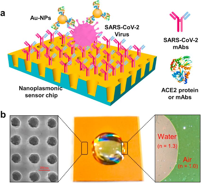
Biosensors 2021, 11, 250                                                                                             2 of 18

                                On the surface of a solid material such as a metal with a free charge, the surface charge
                           (mostly electrons) oscillates collectively due to light irradiation. This phenomenon is called
                           surface plasmon resonance (SPR). [15]. SPR-induced surface charge oscillations are coupled
                           with electromagnetic waves under certain conditions. The quantum of these oscillations is
                           described as the surface plasmon polariton (SPP), and the excitation of SPP is an essential
                           step in SPR biosensors [16–18]. SPR significantly increases the surface sensitivity of the
                           substrates to react with the target material in certain spectroscopic measurement techniques,
                           as characterized by the conditions of resonance between the irradiated light and the
                           substrate [19,20]. The increased sensitivity of SPR has also been applied in virus detection
                           techniques [21–25]. Compared to SPR on an individual substrate such as a thin metal film,
                           the plasmon phenomenon similarly produced by irradiating light on metal nanoparticles
                           is described as localized SPR (LSPR) [26]. The plasmon phenomenon generated on the
                           nanoparticle surface generates a strong electric field in the vicinity of the nanoparticles [27].
                           Within this plasmon region, the interaction of light with molecules and other types of
                           fluorescent nanomaterials is enhanced [28]. Furthermore, when nanoparticles that form an
                           electric field are in close proximity to each other or bind together, a significantly enhanced
                           electric field is formed between the particles, and in this electric field, Raman scattering of
                           certain chemical species is electromagnetically enhanced [29]. Surface-enhanced infrared
                           absorption (SEIRA) has also been reported as an LSPR-based application [30,31]. The
                           potential for application in biosensing is high, because infrared absorption can be induced
                           in the infrared region by adjusting the shape and size of the nanoparticles. [32]. The
                           plasmon resonance effect can be applied to various biosensing fields because it produces a
                           strong optical response and signal enhancement at the micro/nanoscale.
                                In this review, virus detection techniques and their performances based on signal
                           amplification characteristics assessed by SPR and LSPR on a micro/nanoscale basis are
                           explained. SPR uses as a signal the optical changes that occur when a virus is coupled to a
                           sensor that has a prism bonded to a metal film to couple the light. For LSPR, the strong
                           plasmons generated locally on the nanoparticles are used as a signal or as an enhancer
                           for intensity of fluorescence material (Figure 1). In SPR-based detection systems, the
                           properties of the thin metal film used are important for a highly sensitive detection of
                           viruses. The LSPR-based virus detection technique basically consists of an optimized
                           thin metal film modified on a prism for coupling light. On the contrary, LSPR requires
                           meticulous nanoparticle control technology to detect virus particles because more localized
                           plasmons emit highly strong signal responses. It is also worth mentioning that, as SPR
                           does not require a prism, it is possible to construct a detection system using only a light
                           source and a detector that can irradiate specific light. I have also discussed the elements
                           and developments that are required for the next-generation virus detection technology by
                           describing recent examples of research in SPR and LSPR.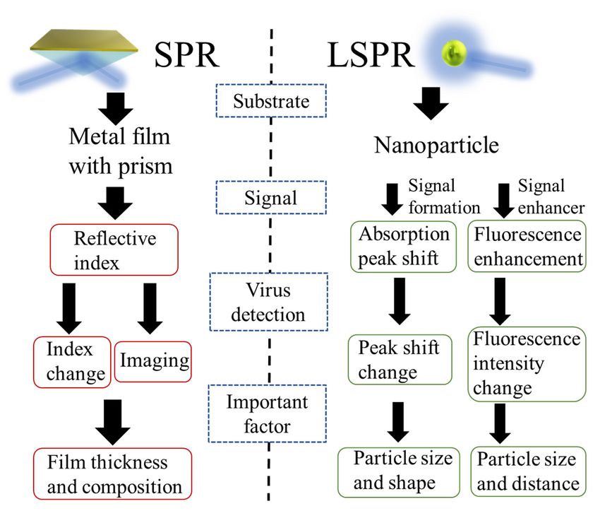
Biosensors2021,
Biosensors 2021,11,
11,250
x FOR PEER REVIEW 33 of 18
18
Figure 1. Diagram
Figure 1. Diagram summarizing
summarizing thethe foundation,
foundation, signals
signals to
to be
be used,
used, signal
signal measurement
measurement methods,
methods,
and
and critical
critical factors
factors as
as building
building elements
elements for
for aa virus
virus detection
detectionsystem
systemusing
usingSPR
SPRand
andLSPR.
LSPR.
2. Current Advancements in SPR-Based Sensor to Detect Virus Particles
2. Current Advancements in SPR-Based Sensor to Detect Virus Particles
2.1. Basic Design Method of Virus Detection Technology Based on SPR
2.1. Basic Design Method of Virus Detection Technology Based on SPR
Metal substrates show strong SPP when irradiated by an appropriate laser [33]. The
most Metal
typicalsubstrates
and effective show strongbased
example SPP whenon thisirradiated
phenomenon by anisappropriate
the Kretschmann laser [33]. The
configu-
most typical and effective example based on this phenomenon is
ration that uses thin films of gold, silver, or a material with other plasmonic properties, suchthe Kretschmann config-
uration
as Pt andthat uses thin
Ti, coated on films
a prism of asgold, silver, orfor
a substrate a material
SPR [34].with When other plasmonic
optimal light is properties,
irradiated,
evanescent waves penetrate the thin metal film and cause plasmon excitation on thelight
such as Pt and Ti, coated on a prism as a substrate for SPR [34]. When optimal outer is
irradiated, evanescent waves penetrate the thin metal film and
surface of the film. The SPP excitation occurs outside the metal film with the medium. This cause plasmon excitation
on the outer
excited state issurface
sensitiveof the film.
to the The SPP
changes excitation
in the refractiveoccurs
index outside
of the the metal and
medium, filmthus,with the
the
medium. This excited state is sensitive to the changes in the
effects of objects adsorbed on the medium can be measured with high sensitivity. In the refractive index of the me-
dium, and thus,
Kretschmann the effects of
configuration, theobjects adsorbed
thin metal on the
film on the prism
medium thatcan be measured
induces SPR andwith high
interacts
sensitivity.
with the lightIndetermines
the Kretschmann configuration,
the performance the thin
of the sensor metal
[35]. film onthe
Therefore, thethickness
prism that in-
of the
duces
thin SPRfilm,
metal andtheinteracts
material with
used, theandlight
thedetermines
roughness of the theperformance
thin film surface of thearesensor
important[35].
Therefore,
to considerthe whenthickness
designingof the anthin metal film,
SPR-based the material
sensor [36]. Gwon used, etand the roughness
al. deposited thin of the
gold
thin film surface are important to consider when designing
films of different thicknesses (30, 52, and 70 nm) on a prism and analyzed the reflectance, an SPR-based sensor [36].
Gwon difference,
phase et al. deposited thin goldfield
and magnetic filmsintensity
of differentunder thicknesses (30, 52, at
light irradiation and63270nm. nm)Theyon a
prism and
found that analyzed the reflectance,
the 50 nm-thick film provided phase the difference, and magnetic
most sensitive field intensity
measurement of the phaseunder
light irradiation
difference at 632
after light nm. They[37].
irradiation foundThethat the 50 nm-thick
formation of a thin filmfilm of provided
optimalthe most sen-
thickness on
sitive
the measurement
substrate for SPRof is the phase
directly difference
related to theafter light irradiation
sensitivity of the sensor [37]. The
and theformation
reliabilityofofa
thinsignal
the film of optimalwhen
response thickness
appliedon the substrate
to virus for SPR
detection is directly
(Figure related to the sensitivity
2) [38,39].
of theSPR imaging (SPRi) has also been actively studied as a methodtotovirus
sensor and the reliability of the signal response when applied applydetection
SPR in
(Figure 2) [38,39].
biosensing. SPRi is broadly classified into spectral, intensity, phase, and polarization
contrast
SPRdepending
imaging (SPRi) on the hassignal used actively
also been [40]. The various
studied as optical
a method responses
to applyobtained
SPR in bio- by
SPR are captured
sensing. as images
SPRi is broadly by a CCD
classified intocamera
spectral, and analyzed.
intensity, As aand
phase, specific application,
polarization the
contrast
interaction
depending of onlabel-free
the signal biomolecules
used [40]. The bound to a substrate
various such as a obtained
optical responses thin metalby film
SPRin are
an
array format has been reported [41,42]. Proteins with nanoscale,
captured as images by a CCD camera and analyzed. As a specific application, the interac- granular, or rod-shaped
structures, such asbiomolecules
tion of label-free virus proteins, are also
bound to aeasy to measure
substrate such as using
a thin SPRi.
metal film in an array
format has been reported [41,42]. Proteins with nanoscale, granular, or rod-shaped struc-
tures, such as virus proteins, are also easy to measure using SPRi.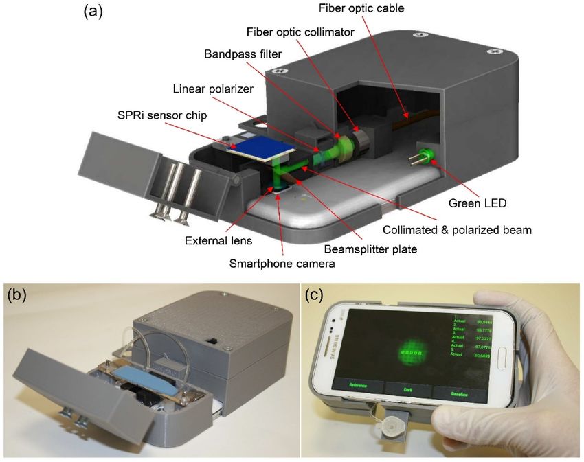
Biosensors 2021, 11, 250 4 of 18
Biosensors 2021, 11, x FOR PEER REVIEW 4 of 18
Figure 2. Schematic diagram of SPR-applied virus detection. The reflected light from the light
Figure 2. Schematic diagram of SPR-applied virus detection. The reflected light from the light source
source irradiated
irradiated on the Kretschmann
on the Kretschmann configuration
configuration is captured
is captured by theby the detector
detector or CCDor CCD camera
camera and
and pro-
processed and analyzed as a signal.
cessed and analyzed as a signal.
2.2. Application of Reflection Angle Change by SPR to Virus Detection
2.2. Application of Reflection Angle Change by SPR to Virus Detection
In order to effectively utilize the properties of SPR, a method to specifically bind the
In order to effectively utilize the properties of SPR, a method to specifically bind the
virus to the metal film is necessary. Antibodies are suitable as trapping materials because
virus to the metal film is necessary. Antibodies are suitable as trapping materials because
they can be modified based on the substrate using chemical cross-linking, and by selecting
they can be modified based on the substrate using chemical cross-linking, and by selecting
a structure with high specificity, a highly selective sensor can be easily constructed [43].
aTable
structure with highthe
1 summarizes specificity,
reports ona SPR-based
highly selective sensor can
virus detection be easily
systems thatconstructed [43].
use the antigen–
Table 1 summarizes the reports on SPR-based virus detection systems
antibody reaction, focusing on the materials used for the thin films, their thickness, and that use the anti-
gen–antibody
virus detectionreaction, focusing
sensitivity. Some of onthe
thereferences
materials used usedafor the thinstructure
two-layer films, their withthickness,
different
and virus detection sensitivity. Some of the references used a
metal substrates, but the total thickness of the thin films was generally around 50 nm. two-layer structure with
different metal substrates, but the total thickness of the thin films
The sensitivity of a rapid diagnostic kit for influenza virus (IFV) based on the prin-was generally around
50 nm.of immunochromatography, which is widely developed as a commercial test using
ciple
The sensitivityreactions,
antigen–antibody of a rapidwasdiagnostic
103 plaque kit for influenza
forming unit virus
(PFU)/mL, (IFV) based on the princi-
104 copies/mL, and
ple of immunochromatography, which is widely developed
100 pg/mL [44–46]. The most basic IFV detection using a thin gold film showed as a commercial test using
lower
antigen–antibody
sensitivity than thereactions, was 10kit
rapid detection
3 plaque forming unit (PFU)/mL, 10⁴ copies/mL, and
reported in the case study. However, using a special
100
thin film structure, the virus could beIFV
pg/mL [44–46]. The most basic detection
detected with using a thinsensitivity
an average gold film100 showed lower
times higher
sensitivity
than that ofthan the the rapidtechniques.
existing detection kit reported
From in the
this point ofcase
view, study.
it canHowever,
be considered usingthat
a spe-
the
cial thin film structure,
advancement the films
of thin metal virusiscould be detected
essential with anapplication
for the practical average sensitivity
of SPR-based100 times
virus
higher
detectionthan that of Several
systems. the existing techniques.
reported cases of From this point
successful of view,
detection it can be considered
of biomaterials using SPR
that the advancement of thin metal films is essential for the practical
with high sensitivity are attributable to the advancement of thin metal films [47–49]. application of SPR-
basedSu virus detection systems. Several reported cases of successful
et al. and Chang et al. reported a bilayer structure of silver and gold thin detection of biomateri-
als using
films SPR with
[50,51]. Thesehigh
aresensitivity
examples are attributable
of how the thintofilm
the advancement
structure improves of thinthemetal films
sensitiv-
[47–49].
ity of virus detection using SPR. The best feature of this structure is that the silver film,
which Suisetanal.excellent
and Chang SPR et al. reported amaterial
signal-response bilayer structure
but can beofeasilysilver and gold
oxidized, thin
was films
covered
[50,51]. Thesefilm
with a gold are examples
to provideofan how the thin film
antioxidant structure
effect. improves
However, whenthe gold sensitivity of virus
thin films were
detection
formed asusing SPR.films
ultrathin The best featureofof
(thickness this
less structure
than 10 nm),isstructural
that the silverchanges film, which
over timeisandan
formation
excellent SPRof signal-response
nanoparticles were reported
material but can[52]. beThe stability
easily oxidized, in long-term
was covered storage
with aisgold
also
an important
film to providefactor for effective
an antioxidant virusHowever,
effect. detection.when Although
gold thinthe bilayer
films were structure
formed of as
a thin
ul-
metal film
trathin filmsimproves
(thickness the
ofsensitivity
less than 10ofnm),
the sensor,
structural thechanges
advanced overstructure
time and offormation
the film has of
some disadvantages.
nanoparticles SPR-based
were reported [52].virus detectioninusing
The stability antigen–antibody
long-term storage is also reaction is more
an important
sensitive
factor for than immunochromatographic
effective virus detection. Although or ELISA methods.
the bilayer To make
structure of athis
thintechnology
metal filmmoreim-
practical, the stability and uniformity of the base and higher sensitivity
proves the sensitivity of the sensor, the advanced structure of the film has some disad- of the detection
method are
vantages. required. virus detection using antigen–antibody reaction is more sensitive
SPR-based
than immunochromatographic or ELISA methods. To make this technology more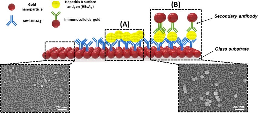
Biosensors 2021, 11, 250                                                                                                      5 of 18

                           Table 1. Comparison layer material and thickness related with limit of detection (LOD) for virus
                           sensing using SPR.

                                      Layer Structure              Thickness of Layer     Target Virus *       LOD         References
                                       Gold thin film                   ~50 nm               IFV            193.3 ng/mL      [53]
                                       Gold thin film                   ~50 nm               DV               0.08 pM        [54]
                                       Gold thin film                    50 nm              EBoV             0.5 pg/mL       [55]
                                    Gold/Silver thin film               8/37 nm              IFV            30 PFU/mL        [50]
                                    Gold/Silver thin film              10/35 nm              IFV           144 copies/mL     [51]
                             Platinum-di-selenide/Gold thin film       2/48 nm             COVID-19           1.95 nM        [56]
                           * IFV: Influenza virus, DV: Dengue virus, EBoV: Ebola virus.

                                In recent years, SPR-based virus detection using aptamers, which can bind to the
                           target antigens and antibodies, has been reported [57–59]. The use of aptamers is also
                           effective in SPR-based virus detection technology because of the ease of functionality, as
                           many SPR-based platforms use gold, which exhibits high antioxidant properties. Aptamers
                           are promising as basic materials for biosensors because they can be synthesized in large
                           quantities at a low cost once the sequence is determined. In contrast, hybridization of viral
                           DNA/RNA is also required in detecting the virus in an SPR-based system. It has been
                           reported that SPR can also be used to monitor DNA hybridization [60,61]. Therefore, it
                           is possible to detect viruses with high sensitivity by hybridizing viral DNA/RNA with
                           a target DNA/RNA-specific probe modified on the surface of the SPR substrate. In this
                           section, the focus was on virus sensors using antibodies as capture materials. However, the
                           strength of SPR as a sensor principle is that versatile and highly sensitive detection systems
                           using materials that capture the target virus at the interface can be constructed.

                           2.3. Application of SPR Signal Response to Imaging during Virus Detection
                                 Wang et al. reported the successful measurement of IFV with a sensitivity of 1 ag
                           (0.2 fg/mL) using SPRi [62]. The authors used intensity as the signal response of SPR and
                           imaging with a CCD camera to capture the surface plasmon diffusion that occurs when
                           IFV binds to anti-influenza antibodies modified on the substrate surface and accurately
                           detected the mass and size of the virus particles. The IFV particle size is approximately
                           100 nm, which is relatively large among viruses, while the size of mosquito-borne viruses
                           such as SARS-CoV-2, norovirus, and dengue virus is approximately 30 nm [63–66]. In
                           SPRi, the molecular weight and particle size of the proteins have a significant effect on
                           the sensitivity. The sensitivity obtained in their study suggests that even a small number
                           of virus particles can be detected. SPRi has high potential as a next-generation virus
                           detection technology. Sun et al. also succeeded in detecting a single T4 phage virus using
                           polystyrene nanoparticles by applying surface plasmon diffusion [67]. Not many examples
                           of effective applications of SPRi for virus detection are presented in this review. However,
                           the principle of SPRi in detecting a single virus particle per measurement is potent. The
                           technical challenge for the next-generation virus detection technology is that it requires
                           more sophisticated equipment compared to the commercially available rapid diagnostic
                           kits [68].
                                 Regarding the improvement of the SPRi device, Guner et al. reported the development
                           of a very efficient system [69]. The authors successfully designed a low-cost SPR-based
                           sensor chip based on the commercially available optical storage disks and developed a
                           compact, smartphone-mountable device using LEDs as the light source (Figure 3). Imaging
                           and detecting antigens captured by the antibodies modified on a thin silver and gold bilayer
                           film were performed. The authors concluded that using a two-layered metallic thin film
                           that can utilize visible light led to the development of a compact and effective biosensing
                           smartphone device. The results also showed that the detection sensitivity was comparable
                           to that of the commercially available SPRi systems, and the authors discussed the future
                           potential of the system as a basis for successful miniaturization and cost reduction.
Biosensors 2021, 11, x FOR PEER REVIEW 6 of 18
Biosensors 2021, 11, 250 comparable to that of the commercially available SPRi systems, and the authors discussed
6 of 18
the future potential of the system as a basis for successful miniaturization and cost reduc-
tion.
Figure3.3.Surface
Figure Surfaceplasmon
plasmonresonance
resonanceimaging
imagingplatform
platformintegrated
integratedwith
withaasmartphone.
smartphone.(a)
(a)Schematic
Schematic
illustration and (b) photograph of the imaging apparatus. (c) Custom developed
illustration and (b) photograph of the imaging apparatus. (c) Custom developed smartphonesmartphone
application for real-time and on-site monitoring of multiple sensing spots. Reprinted permission
application for real-time and on-site monitoring of multiple sensing spots. Reprinted permission
obtained from [69].
obtained from [69].
2.4.Current
2.4. CurrentApplication
ApplicationofofSPR
SPRfor
forVirus
VirusSensing
Sensing
Uptotothis
Up thissection,
section,thethepotential
potentialofofSPR SPRand andSPRi-based
SPRi-basedsystems
systemsasasvirus
virusdetection
detection
methodshas
methods hasbeen
beendiscussed.
discussed.ItItisisalso
alsoclear
clearthat
thatapplied
appliedresearch
researchisisneeded
neededtotomakemakethe the
methodsimple
method simpleand andeffective
effectiveforforimplementation
implementationas asaanext-generation
next-generationsensingsensingsystem.
system.YooYoo
etetal. demonstrated aa reusable
al. demonstrated reusabledetection
detectionsubstrate
substratefor fora anew
new type
type of of SPR-based
SPR-based virus
virus de-
detection system[70].
tection system [70].AAsystem
systemdesign
designthat that allows
allows thethe SPR platform to to be
be used
usedrepeatedly
repeatedly
without
withoutthe theneed
needforforantibody
antibodymodifying
modifyingprocesses
processeswill willbebeaasignificant
significantstepsteptoward
towardthe the
future implementation of virus sensors. In their study, the virus was captured
future implementation of virus sensors. In their study, the virus was captured by antibod- by antibodies
conjugated
ies conjugatedto magnetic
to magneticbeads at specific
beads at specificsites onon
sites thethe
substrate
substrate (Figure
(Figure 4).4).The
Themagnetic
magnetic
nanoparticles
nanoparticles were weretransported
transported by byan anexternal
externalmagnetic
magneticfield fieldtotothe
thesubstrate
substratewherewhereaa
gold/nickel
gold/nickel (10 nm/50nm/50nm) nm)thin
thinfilm
filmwaswaspatterned
patternedonon a gold/chromium
a gold/chromium (45(45
nm/5nm/5
nm)nm)thin
thin film, which was the SPR sensor site (Figure 4A,B). The antibodies
film, which was the SPR sensor site (Figure 4A,B). The antibodies were modified on the were modified
on the magnetic
magnetic nanoparticles
nanoparticles at the at the patterning
patterning site bysite by cross-linking
cross-linking the carboxyl
the carboxyl groups groups
modi-
modified
fied on the magnetic nanoparticles with the amino groups of the antibodies, using ausing
on the magnetic nanoparticles with the amino groups of the antibodies, chem-
aical
chemical cross-linking
cross-linking agent toagent to provide
provide reactivity
reactivity with the with the(Figure
virus virus (Figure
4C,D). It 4C,D). It was
was demon-
demonstrated
strated that the thatantibody-modified
the antibody-modified magnetic
magnetic nanoparticles
nanoparticles bound
bound to antigens
to antigens could
could be
be easily removed from the substrate after virus detection by applying
easily removed from the substrate after virus detection by applying an external magnetic an external magnetic
field
fieldininthe
theopposite
oppositedirection
directionofofthe thedeposition
deposition(Figure
(Figure4E).4E).Several
Severalstudies
studieshad hadalready
already
reported
reported that magnetic nanoparticles are suitable materials for signal enhancementinin
that magnetic nanoparticles are suitable materials for signal enhancement
sensing
sensingby by SPR
SPR [71,72]. Thecontribution
[71,72]. The contributionofofmagnetic
magneticnanoparticles
nanoparticles toto
thethe sensitivity
sensitivity is
is not
not discussed in this review; however, the compact system has been
discussed in this review; however, the compact system has been successfully used for lin- successfully used for
linear, wide-rangeIFV
ear, wide-range IFVnucleic
nucleic protein
protein detection
detection in in the
the range
rangeofof300300ng/mL
ng/mLtoto1010µg/mL.
µg/mL.ItIt
was suggested that magnetic nanoparticles show a signal response in this system, at least
was suggested that magnetic nanoparticles show a signal response in this system, at least
on the SPR platform. In addition, authors have demonstrated that magnetic nanoparticle
on the SPR platform. In addition, authors have demonstrated that magnetic nanoparticle
removal and detection can be repeated up to seven times on the same substrate, making
the author’s idea a more practical SPR-based virus detection technique.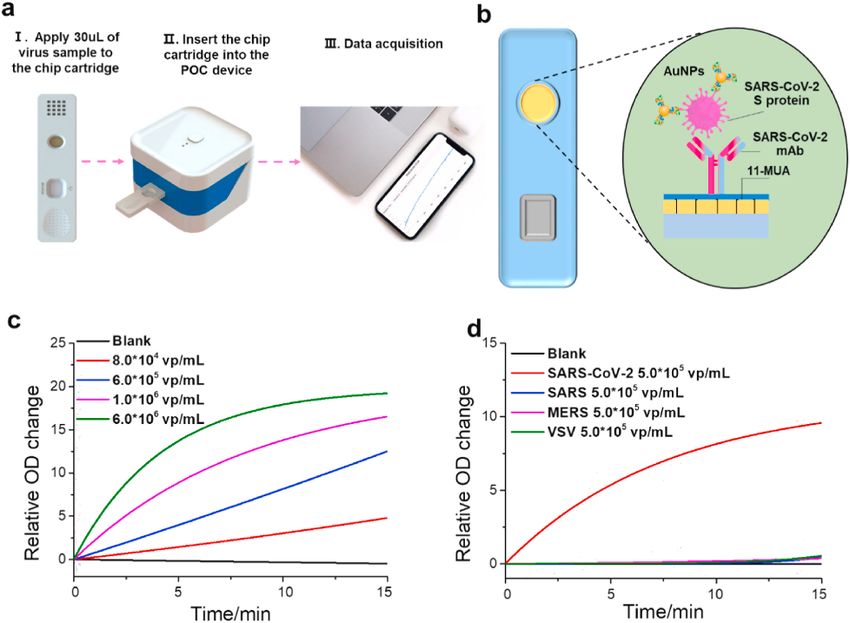
Biosensors 2021, 11, x FOR PEER REVIEW                                                                                                7 of 18

Biosensors 2021, 11, 250                                                                                                               7 of 18
                                  removal and detection can be repeated up to seven times on the same substrate, making
                                  the author’s idea a more practical SPR-based virus detection technique.

                                  Figure 4. Schematic diagram depicting the cyclic process for the repeated sensing measurements
                                 Figure 4. Schematic diagram depicting the cyclic process for the repeated sensing measurements
                                  using reusable SPR biosensor chip. (A) A reusable SPR chip including ferromagnetic nickel patterns
                                 using reusable SPR biosensor chip. (A) A reusable SPR chip including ferromagnetic nickel patterns
                                  on a conventional SPR chip structure. (B) Trapping of magnetic particles on the reusable SPR chip
                                 on a conventional
                                  through           SPRmagnetic
                                           an external    chip structure.   (B) Immobilization
                                                                    field. (C)   Trapping of magnetic    particles
                                                                                                  of antibodies  ononmagnetic
                                                                                                                       the reusable  SPR chip
                                                                                                                                particles using
                                 through  an external magnetic    field. (C)  Immobilization     of antibodies  on magnetic
                                  EDC-NHS coupling in a conventional SPR system. (D) Detection of target molecules. (E) Removalparticles using
                                 EDC-NHS    coupling
                                  of magnetic         in abyconventional
                                               particles      an external SPR     system.
                                                                             magnetic      (D)inDetection
                                                                                        field             of target
                                                                                                 an opposite        molecules.
                                                                                                               direction  to that(E)
                                                                                                                                  forRemoval
                                                                                                                                      trapping.
                                 ofReprinted
                                    magneticpermission
                                              particles by   an external
                                                          obtained          [70].
                                                                     frommagnetic      field in an opposite direction to that for trapping.
                                 Reprinted permission obtained from [70].
                                        Huang et al. applied their low-cost plasmon nanoarray SPR chip design to detect
                                      Huang et (Figure
                                  SARS-CoV-2      al. applied    their low-cost
                                                           5a) [73–75].           plasmoncharacteristics
                                                                          The structural    nanoarray SPR  of chip  design toincluded
                                                                                                              the substrate     detect
                                 SARS-CoV-2     (Figure   5a)  [73–75]. The   structural characteristics of the  substrate
                                  the uniform and tight formation of a special nanosized cup structure, which made it pos-  included
                                 the uniform
                                  sible         and the
                                        to observe    tightplasmon
                                                             formation    of a special
                                                                      resonance        nanosized
                                                                                  wavelength   andcup    structure,
                                                                                                    intensity        which
                                                                                                                changes      made the
                                                                                                                          without   it
                                 possible  to  observe   the  plasmon    resonance   wavelength   and  intensity   changes
                                  need for external optics because of the extraordinary optical transmission [76]. A further without
                                 the needisfor
                                  feature       external
                                              that         optics because
                                                    gold nanoparticles     areoffunctionalized
                                                                                 the extraordinary
                                                                                               with optical   transmission
                                                                                                     ACE2 protein      to bind[76]. A
                                                                                                                                 SARS-
                                 further feature   is that  gold  nanoparticles   are functionalized  with   ACE2    protein
                                  CoV-2. It has been reported that the sandwich structures of SPR substrate-virus particles-  to bind
                                 SARS-CoV-2. It has been reported that the sandwich structures of SPR substrate-virus
                                 particles-gold nanoparticles enhance the SPR signal response [77]. In this research, high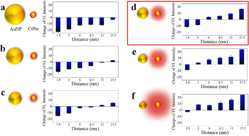
Biosensors 2021, 11, 250 8 of 18
Biosensors 2021, 11, x FOR PEER REVIEW 8 of 18
sensitivity was achieved
gold nanoparticles by applying
enhance the characteristics
the SPR signal response [77].ofInthe thisgold thin film
research, highstructure
sensitivity
and
wasmetal nanoparticles
achieved rather
by applying than
the using a metal
characteristics ofbilayer
the gold structure.
thin filmAnstructure
importantandaspect
metal
ofnanoparticles
the application of thin films with such special structures is to maintain
rather than using a metal bilayer structure. An important aspect of the ap- a uniform
surface structure.
plication The uniformity
of thin films of the gold-nanoparticle
with such special structures is to maintain cup fabricated
a uniformsubstrate
surface was
struc-
observed by scanning electron microscopy, and it was demonstrated
ture. The uniformity of the gold-nanoparticle cup fabricated substrate was observed that water drops onby
the substrate
scanning showed
electron different color
microscopy, from
and it wasthose in the air that
demonstrated (Figure 5b).drops
water The authors noted
on the substrate
that the characteristic
showed spectral
different color from changes made
those in the air it(Figure
possible 5b).toThe
detect the virus
authors notedby SPR
that thewith
char-
high sensitivity.
acteristic spectral changes made it possible to detect the virus by SPR with high sensitivity.
Figure5. 5. Label-free
Figure Label-free detection
detection of
of SARS-CoV-2
SARS-CoV-2 pseudovirus
pseudoviruswithwitha ananoplasmonic
nanoplasmonicsensor.
sensor.(a)
Schematic diagram of the nanoplasmonic resonance sensor for determination of
(a) Schematic diagram of the nanoplasmonic resonance sensor for determination of SARS-CoV- SARS-CoV-2
pseudovirus concentration. (b) Photograph (Middle) of one piece of Au nanocup array chip with a
2 pseudovirus concentration. (b) Photograph (Middle) of one piece of Au nanocup array chip with
drop of water on top. Scanning electron microscopy image (Left) shows the replicated nanocup
a drop of water on top. Scanning electron microscopy image (Left) shows the replicated nanocup
array. Transmission microscopy image (Right) shows that air and water on the device surface
array. Transmission
exhibit microscopy
different colors, greenimage (Right)pink,
and far-red shows that air andReprinted
respectively. water on the device surface
permission exhibit
obtained from
different
[73]. colors, green and far-red pink, respectively. Reprinted permission obtained from [73].
The SPR-based sensor chip reported in this study could be incorporated into a 96-well
The SPR-based sensor chip reported in this study could be incorporated into a 96-
plate or cartridge [73]. This section focuses especially on the cartridge-type detection
well plate or cartridge [73]. This section focuses especially on the cartridge-type detection
systems that can be used for on-site detection and other applications. The cartridge SPR
systems that can be used for on-site detection and other applications. The cartridge SPR
system controlled by a smartphone application could measure the dynamic absorption
system controlled by a smartphone application could measure the dynamic absorption
spectra by simply inserting the antibody-modified SPR nanocup placed on a dedicated
spectra by simply inserting the antibody-modified SPR nanocup placed on a dedicated
cartridge (Figure 6a,b). The SPR-based sensor chip site was chemically cross-linked with
cartridge (Figure 6a,b). The SPR-based sensor chip site was chemically cross-linked with
virus-specific antibodies on a nanocup structure coated with gold thin film to provide
virus-specific antibodies on a nanocup structure coated with gold thin film to provide
reactivity with SARS-CoV-2. The detection results presented by the authors demonstrated
reactivity with SARS-CoV-2. The detection results presented by the authors demonstrated
that the SARS-CoV-2 concentration-dependent signal response in this transportable SARS-
that reservoir
CoV-2 the SARS-CoV-2
was highly concentration-dependent signal response
selective, with no signal response specific toinother
thisvirus
transportable
species.
SARS-CoV-2 reservoir was highly selective, with no signal response specific to
Furthermore, the lower limit of quantitative detection was set at approximately 4000 viruses, other virus
species.
and Furthermore,
it is expected that thethe lower
lower limit
limit of quantitative
of quantitative detection
detection willwas
be thesetsame
at approximately
as that of a
large device with further sensitivity improvements (Figure 6c,d). In this study, thewill
4000 viruses, and it is expected that the lower limit of quantitative detection be the
authors
same as that of a large device with further sensitivity improvements (Figure
chemically cross-linked antibodies to the substrate by forming a self-assembled monolayer 6c,d). In this
study, the authors chemically cross-linked antibodies to the substrate
using 11-mercapto-undecanoic acid (11-MUA) for antibody modification. This is a well-by forming a self-
assembled monolayer using 11-mercapto-undecanoic acid (11-MUA) for antibody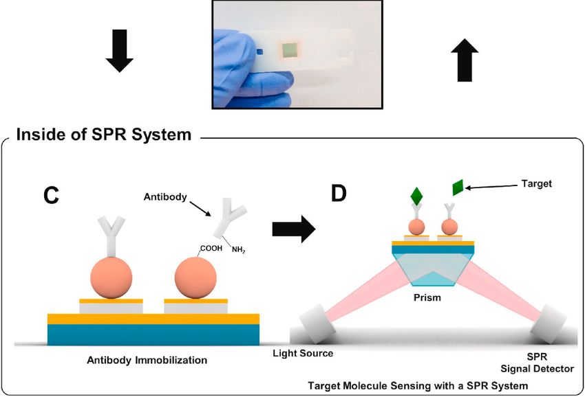
Biosensors 2021, 11, 250 9 of 18
Biosensors 2021, 11, x FOR PEER REVIEW 9 of 18
known chemical
modification. cross-linking
This method,
is a well-known and it cross-linking
chemical has succeeded in yielding
method, and it highly efficient
has succeeded
antibody modification on the surface [78,79]. However, one area that remains
in yielding highly efficient antibody modification on the surface [78,79]. However, one controversial
isarea
the that
randomness of the antibody
remains controversial conjugation
is the randomness sites. Since
of the the gold
antibody film had asites.
conjugation nanocup
Since
structure with a diameter of more than 200 nm, the surface modification
the gold film had a nanocup structure with a diameter of more than 200 nm, the surface by 11-MUA was
likely carried out uniformly into the cup. It is unlikely that antibodies, which
modification by 11-MUA was likely carried out uniformly into the cup. It is unlikely that are structures
smaller than which
antibodies, 10–15 nm, bind only to
are structures the surface
smaller layer of
than 10–15 thebind
nm, goldonly
nanocups
to the[80]. Thelayer
surface signalof
generated
the gold nanocups [80]. The signal generated by the binding of the virus particle to be
by the binding of the virus particle to the antibody modified in the cup might the
different
antibodyfrom the signal
modified generated
in the cup might bybethe bindingfrom
different of the virus
the to the
signal antibody
generated byconjugated
the binding
onof the
the top surface;
virus to the as the sensitivity
antibody conjugatedof the
ondetection systemas
the top surface; increases, the magnitude
the sensitivity of
of the detec-
randomness caused by such antibody modification methods increases
tion system increases, the magnitude of randomness caused by such antibody modifica- [81]. Therefore,
modifying
tion methods antibodies
increases only
[81].atTherefore,
selective sites is important
modifying to minimize
antibodies the difference
only at selective in
sites is im-
signal response between virus detection sites when the substrate forms a complex shape.
portant to minimize the difference in signal response between virus detection sites when
Site-specific conjugation of the antibody can be achieved only under special conditions.
the substrate forms a complex shape. Site-specific conjugation of the antibody can be
There is no report about site-specific conjugation of antibodies on a single material-based
achieved only under special conditions. There is no report about site-specific conjugation
nanostructural substrate, which requires further innovation to fabricate the next-generation
of antibodies on a single material-based nanostructural substrate, which requires further
SPR-based virus sensors.
innovation to fabricate the next-generation SPR-based virus sensors.
Figure6.6.Detection
Figure Detectionofof SARS-CoV-2
SARS-CoV-2 pseudovirus
pseudovirus withwith nanoplasmonic
nanoplasmonic sensor
sensor chipschips
by a by a point-of-
point-of-care
care device. (a) Schematic of nanoplasmonic sensor chip cartridge detecting SARS-CoV-2 pseudo-
device. (a) Schematic of nanoplasmonic sensor chip cartridge detecting SARS-CoV-2 pseudovirus
virus with a low-cost handheld point-of-care testing device. (b) The illustration shows the detection
with a low-cost handheld point-of-care testing device. (b) The illustration shows the detection process
process of the sensor chip cartridge for specific SARS-CoV-2 detection. (c) Dynamic binding curves
ofofthe sensor
virus and chip cartridge
antibody for specific
interaction SARS-CoV-2
with different detection. (c)
concentrations Dynamic
of the bindingpseudovirus
SARS-CoV-2 curves of virus
over
and antibody interaction with different concentrations of the SARS-CoV-2 pseudovirus over the
the range 0–6.0 × 10⁶ virus particles (vp)/mL at the resonance wavelength. (d) Specificity verification
range 0–6.0 × 10⁶
test: Dynamic virus curves
binding particles
of(vp)/mL at theantibodies
SARS-CoV-2 resonanceinteraction
wavelength. (d)different
with Specificity verificationof
pseudovirus
test: Dynamic SARS,
SARS-CoV-2, binding curves
MERS, andof VSV
SARS-CoV-2 antibodies interaction
at the concentration of 5.0 × 10⁵with different
vp/mL. pseudovirus
Reprinted with per-
ofmission obtained
SARS-CoV-2, [73]. and VSV at the concentration of 5.0 × 10⁵ vp/mL. Reprinted with
fromMERS,
SARS,
permission obtained from [73].
In this section, two different types of currently reported SPR sensors for virus detec-
tionInwere
this section, two different
introduced. types of
The common currently
thread reported
among SPR sensors
the studies for virus detection
was developing a more
were introduced. The common thread among the studies was developing a more
practical design of SPR-based virus detection systems with a certain level of sensitivity. practical
design of SPR-based
The research virus
focus in detection
recent years onsystems
viruswith a certain
detection level of sensitivity.
technologies has oftenThe research
been on sen-
focus in recent years on virus detection technologies has often been on
sitivity and speed, and for on-site detection technologies, whether the system can sensitivity andbe
speed, and for on-site detection technologies, whether the system can be
handheld or wireless and employs reusable methods [82–86]. As for the research on virus handheld or
wireless
detection and employs using
technology reusable
SPR,methods [82–86].
the major pointsAs for theinresearch
required on are
the future virus
notdetection
high sen-
technology using SPR, the major points required in the future are not high
sitivity and rapidity, but thin film formation and suitable construction systems to enhancesensitivity
the practicality and applicability.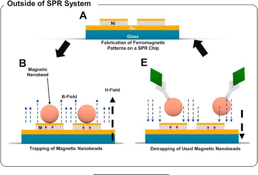
of the nanoparticles, based on the size, shape, and material used [87–89]. Therefore, the
size and shape of the nanoparticles need to be precisely controlled to obtain the desired
optical response. The most fundamental application of optical signal response by LSPR is
the change in the peak absorption wavelength due to the local refractive index change on
Biosensors 2021, 11, 250 the nanoparticle surface (plasmon peak shift) and the fluorescence enhancement 10 of 18
effect
caused by using near-field light. (Figure 7). In this section, the focus is on virus detection
techniques that use the basic optical response of LSPR. Further, the two types of responses
that are still being applied to virus detection are summarized in this section.
and rapidity, but thin film formation and suitable construction systems to enhance the
LSPR is adapted for transmission measurements, whereas SPR can measure the re-
practicality and applicability.
flectance change when some material is attached to a single particle [90–92]. The ad-
vantage
3. LSPRof the former ison
Phenomenon that the measurement
Nanoscale Systems andsystem can be simply
Application of LSPRconstructed without
to Virus Sensing
the need for movable elements necessary for reflection
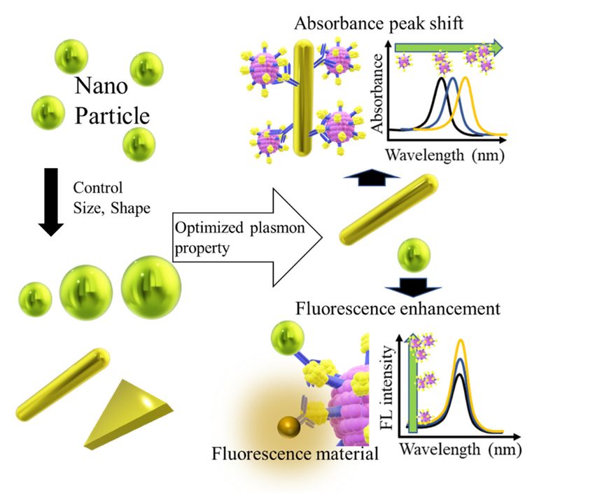
3.1. Signal Response Produced by LSPR for Virus Detection angle measurement [93]. The trans-
missive signal response has already been applied in various virus detection applications
After LSPR, an enhancement of electrolysis and light quenching occurs in the vicinity
as
ofthe
theplasmon peak shift
nanoparticles, of nanoparticles
based sensorand
on the size, shape, compared
materialwith
usedan[87–89].
LSPR-based reflective
Therefore, the
signal sensor [94].
size and shape of the nanoparticles need to be precisely controlled to obtain the desired
Theresponse.
optical near-fieldThe lightmost
formed near the noble
fundamental metal nanoparticles
application by LSPR
of optical signal enhances
response by LSPRthe
interaction of the light with the fluorescent material present at a certain
is the change in the peak absorption wavelength due to the local refractive index change distance in the
region
on the [95–97].
nanoparticleThe resulting significant
surface (plasmon peakfluorescence
shift) and theenhancement
fluorescenceeffect of the fluoro-
enhancement effect
phores is widely used in the development of highly sensitive sensing techniques,
caused by using near-field light. (Figure 7). In this section, the focus is on virus detection as the
effect enables
techniques theuse
that emittance
the basicofoptical
signal responses
response ofeven from
LSPR. small amounts
Further, of sample.
the two types These
of responses
observations render LSPR a potential signal amplification method
that are still being applied to virus detection are summarized in this section. for virus sensing [98].
Figure
Figure7.7.Basic
Basicdevelopment
development method
method of
of virus detection technology
virus detection technologybased
basedononLSPR.
LSPR.Optimal
Optimalmaterials
materi-
als with the desired plasmonic properties are obtained by controlling the shape and size of the na-
with the desired plasmonic properties are obtained by controlling the shape and size of the nanopar-
noparticles. The optimal material is capable of generating a sensitive signal response in the presence
ticles. The optimal material is capable of generating a sensitive signal response in the presence of a
of a virus.
virus.
3.2. Optical Absorbance Peak Shift Application for LSPR-Based Virus Sensing
LSPR is adapted for transmission measurements, whereas SPR can measure the re-
flectance change when some material is attached to a single particle [90–92]. The advantage
of the former is that the measurement system can be simply constructed without the need
for movable elements necessary for reflection angle measurement [93]. The transmissive
signal response has already been applied in various virus detection applications as the
plasmon peak shift of nanoparticles sensor compared with an LSPR-based reflective signal
sensor [94].
The near-field light formed near the noble metal nanoparticles by LSPR enhances
the interaction of the light with the fluorescent material present at a certain distance in
the region [95–97]. The resulting significant fluorescence enhancement effect of the fluo-
rophores is widely used in the development of highly sensitive sensing techniques, as the
effect enables the emittance of signal responses even from small amounts of sample. These
observations render LSPR a potential signal amplification method for virus sensing [98].Biosensors 2021, 11, 250 11 of 18
Biosensors 2021, 11, x FOR PEER REVIEW 11 of 18
3.2. Optical Absorbance Peak Shift Application for LSPR-Based Virus Sensing
To measure the local refractive index change as an optical response at the nanoscale
the strategy
level, the strategy used
usedforforconstructing
constructingLSPR LSPRsensors
sensorsisissimilar
similartoto that
that ofofSPRSPR sensors.
sensors. It
It is important to modify the material to capture the target on the nanomaterials
is important to modify the material to capture the target on the nanomaterials with high with
high efficiency
efficiency whilewhile maintaining
maintaining the structural
the structural properties
properties of theofnanoparticles
the nanoparticles [99]. [99]. The
The shift
shift in plasmon peak value due to the LSPR of plasmon particles
in plasmon peak value due to the LSPR of plasmon particles after virus binding is also aafter virus binding
is also a signal
sensitive sensitive signal[100].
response response
Kim et[100]. Kim et aal.structure
al. designed designed a structure
in which a virusinwaswhich
sand-a
virus was
wiched sandwiched
between between
two different twoofdifferent
sizes AuNP on sizes of AuNP on an
an AuNP-laden AuNP-laden
substrate (Figuresubstrate
8) [101].
(Figure 8) [101]. In this method, two gold nanoparticles in close proximity
In this method, two gold nanoparticles in close proximity repel each other in a plasmon repel each
other in a plasmon resonance state in the presence of the virus, resulting
resonance state in the presence of the virus, resulting in a stronger peak shift effect than in a stronger
peakin
that shift
theeffect than that in
non-sandwich the The
state. non-sandwich state. The signal
signal enhancement enhancement
effect of the sandwich effect of the
structure
sandwich structure has been demonstrated, and a 100-fold increase in
has been demonstrated, and a 100-fold increase in the sensitivity has been successfully the sensitivity has
been successfully achieved compared to the virus captured by AuNP-laden
achieved compared to the virus captured by AuNP-laden substrate only. Furthermore, substrate only.
Furthermore,
the sensitivitythe sensitivity
of the sandwichof the sandwich
structure structure
differed differedon
depending depending
the size ofonthethegold
size nano-
of the
gold nanoparticles
particles on the secondary
on the secondary antibodyantibody
site. Thesite. The the
smaller smaller the particle,
particle, the stronger
the stronger the
the signal
signal response, even at low sample concentrations. This work demonstrated that particle
response, even at low sample concentrations. This work demonstrated that particle size
size has a significant effect on the use of LSPR for developing application-based systems.
has a significant effect on the use of LSPR for developing application-based systems.
Figure 8.8.Detection
Figure Detectionofof HBsAg
HBsAg by by single
single assayassay
LSPRLSPR sensing
sensing chip format
chip format (A) and(A) and modified
modified heteroassembled
heteroassembled AuNP
AuNP sandwich
sandwich immunoassay LSPR chip format using immunocolloid AuNPs (B). Changes in the spectrum peak at different
immunoassay LSPR chip format using immunocolloid AuNPs (B). Changes in the spectrum peak at different concentrations
concentrations of HBsAg (1 pg/mL to 1 µg/mL HBsAg), reacted on the chip. Insert image is presenting the immunological
of HBsAg (1 pg/mL to 1 µg/mL HBsAg), reacted on the chip. Insert image is presenting the immunological reaction
reaction occurring on the active site of LSPR sensing chip. 15, 30 and 50 nm of immunocolloid AuNPs as signal enhancers.
occurring
1 ng/mL to on100
the fg/mL
active site of LSPR
HBsAg sensing
reacted the chip.
LSPR15, 30 and
chip. All 50 nm of immunocolloid
experiments AuNPs
were conducted in as
sixsignal enhancers. and
measurements, 1 ng/mL
data
to 100 fg/mL HBsAg reacted the LSPR chip. All experiments were conducted in six measurements, and data
represent the mean ± standard deviation. The coefficient of variation (% CV) is below 10%. Reprinted with permission represent
the ± standard
meanfrom
obtained [101]. deviation. The coefficient of variation (% CV) is below 10%. Reprinted with permission obtained
from [101].
There are other techniques of virus detection based on absorbance measurement and
There are such
nanoparticles, otherastechniques of virus detection
the agglomeration based oninabsorbance
of nanoparticles measurement
the presence of a target
[102,103]. The peak change of the plasmon band was measured for the structuralof
and nanoparticles, such as the agglomeration of nanoparticles in the presence a tar-
change
get [102,103]. The peak change of the plasmon band was measured for the structural change
due to the aggregation of particles [103]. In this section, the optical response analogous to
due to the aggregation of particles [103]. In this section, the optical response analogous to
SPR and the plasmon peak shift due to local plasmon response on the nanoscale caused
SPR and the plasmon peak shift due to local plasmon response on the nanoscale caused by
by the binding of nanoparticles to target materials were introduced.
the binding of nanoparticles to target materials were introduced.
3.3.
3.3. LSPR
LSPR Fluorescence
Fluorescence Enhancement
Enhancement for
for Virus
Virus Detection
Detection
Electric field enhancement around the nanoparticles by
Electric field enhancement around the nanoparticles LSPR produces
by LSPR produces near-field
near-field
light, which can enhance fluorescence by improving the interaction of
light, which can enhance fluorescence by improving the interaction of light withlight with the fluo-
the
rescent materials placed at an appropriate distance. The distance between the
fluorescent materials placed at an appropriate distance. The distance between the plasmon plasmon
particle
particle and
and the
the target
target fluorescent material is
fluorescent material is the
the most
most important
important factor
factor in
in the
the fluorescence
fluorescence
enhancement effect of LSPR. Chowdhuly et al. used gold nanoparticles as a plasmon am-
plifier and quantum dots (QDs) as fluorescence materials to analyze the fluorescenceBiosensors 2021, 11, 250 12 of 18
Biosensors 2021, 11, x FOR PEER REVIEW 12 of 18
enhancement effect of LSPR. Chowdhuly et al. used gold nanoparticles as a plasmon
amplifier and quantum dots (QDs) as fluorescence materials to analyze the fluorescence
enhancement
enhancementeffect
effectofofLSPR
LSPRon onfluorescent
fluorescentmaterials
materialswith
withrespect
respecttotoplasmon
plasmonparticle
particlesize
size
and
and interparticle distance (Figure 9a–f) [104]. The fluorescence of QDs placed bychemical
interparticle distance (Figure 9a–f) [104]. The fluorescence of QDs placed by chemical
cross-linking
cross-linkingininthe
thevicinity
vicinityofofgold
goldnanoparticles,
nanoparticles,withwithparticle
particlesizes
sizesranging
rangingfrom
from1515toto
30
30nm,
nm,proved
provedtotobe
befluorescence-enhancing
fluorescence-enhancingfor foran
aninterparticle
interparticledistance
distanceofofup
uptoto15.5
15.5nm,
nm,
except
exceptfor
forthe
thenearest
nearestneighbor
neighbordistance
distanceofof1.8
1.8nm.
nm.The
Thefluorescence
fluorescenceenhancement
enhancementeffecteffect
decreased
decreasedwith
withdistance
distance and asas
and thethe
size of of
size thethe
gold nanoparticles
gold nanoparticlesapproached 100 100
approached nm nm(micro-
(mi-
order), the effect was replaced by a strong trend of quenching effect of the QDs (Figure
cro-order), the effect was replaced by a strong trend of quenching effect of the QDs (Figure 9a–f).
Their
9a–f).results
Their showed the distance
results showed to be maintained
the distance betweenbetween
to be maintained the electromagnetic fields
the electromagnetic
formed in the vicinity of the plasmon particle by LSPR and the fluorescent material.
fields formed in the vicinity of the plasmon particle by LSPR and the fluorescent material.
Figure9.9. Fluorescence
Figure Fluorescence spectral
spectralbar
bardiagram
diagram of of
CdSeCdSeQDs in various
QDs CdSeCdSe
in various QD-peptide-AuNP
QD-peptide-AuNPnano-
conjugates. Six different sizes of AuNPs at 80 (a), 60 (b), 45 (c), 35 (d), 25 (e), and 15 nm
nanoconjugates. Six different sizes of AuNPs at 80 (a), 60 (b), 45 (c), 35 (d), 25 (e), and 15 nm (f) combined
with
(f) six different
combined with peptide lengths
six different produced
peptide various
lengths fluorescence
produced variousintensity changes.
fluorescence Reprinted
intensity with
changes.
permission obtained from [104].
Reprinted with permission obtained from [104].
Manystudies
Many studieshavehavereported
reportedthetheapplication
applicationofofthe thefluorescence
fluorescenceenhancement
enhancementeffect effect
of LSPR for virus detection. Nasrin et al. recently reported a nanocomposite
of LSPR for virus detection. Nasrin et al. recently reported a nanocomposite in which the in which the
plasmon particles and QDs were combined by chemical cross-linking
plasmon particles and QDs were combined by chemical cross-linking and modified with and modified with
antibodiesfor
antibodies forsupplementation,
supplementation,therebytherebyproviding
providingreactivity
reactivityagainst
againstviruses
viruses[105].
[105].TheThe
distancebetween
distance betweenthe theparticles
particleswas
wasdesigned
designedtotobe beoptimal
optimalforforfluorescence
fluorescenceenhancement
enhancement
throughantigen–antibody
through antigen–antibodyreactionreactionon onthethevirus.
virus.The
Thevirus
virusdetection
detectiontechniques
techniquesbasedbasedon on
thefluorescence
the fluorescenceenhancement
enhancementeffecteffectofoffluorescent
fluorescentmaterials
materialsbybyLSPRLSPRare aresummarized
summarizedinin
Table2,2,focusing
Table focusingon onthe
thesensitivity
sensitivityofofthe
thetechnique,
technique,the thematerials
materialsused,used,andandwhether
whetherthe the
particledistance
particle distancewas wascontrolled
controlledprecisely
preciselyor orroughly.
roughly.EvenEvenwhen
whenusingusinggoldgoldnanoparti-
nanoparti-
cles,which
cles, whichare arethe
themost
mostbasic
basicplasmon
plasmonparticles,
particles,aa10-fold
10-folddifference
differenceininsensitivity
sensitivitywaswas
obtained
obtaineddepending
dependingon onwhether
whetherthethedistance
distancetotothethefluorescent
fluorescentmaterial
materialwas wasaccurately
accurately
controlled
controlled[106,107].
[106,107].In Inaddition,
addition,aasensor
sensordesigned
designedfor forZika
Zikavirus
virusRNA RNAsuccessfully
successfullyde- de-
tected
tectedeven
evenaasingle
singleviral
viralRNA
RNAmolecule
moleculeby bymaintaining
maintaininga aconstant
constantinterparticle
interparticledistance
distance
through
throughchemical
chemicalbonding.
bonding.The Theinterparticle
interparticledistance
distancewaswasshown
showntotosignificantly
significantlyaffect
affectthe
the
sensitivity of the LSPR fluorescence-enhanced virus detection technique
sensitivity of the LSPR fluorescence-enhanced virus detection technique [98]. As for the [98]. As for the
structure
structureofofthetheplasmon
plasmon particles used,
particles used,particles with
particles a bimetallic
with a bimetallic structure andand
structure composite
compo-
nanomaterials
site nanomaterialsshowed very high
showed verysensitivity. Hence,Hence,
high sensitivity. the structure of composite
the structure nanomateri-
of composite nano-
als is likelyistolikely
materials rendertomore
rendersensitive LSPR andLSPR
more sensitive SPR responses. The most The
and SPR responses. striking
most example
striking
isexample
a compositeis a of magnetic nanoparticles,
composite gold nanoparticles,
of magnetic nanoparticles, gold and graphene, which
nanoparticles, success-
and graphene,
fully detected IFV from an ultra-low concentration of 7.27 fg/mL.
which successfully detected IFV from an ultra-low concentration of 7.27 fg/mL. In this study, the authors
In this
not only enhanced the LSPR effect by complexing with graphene,
study, the authors not only enhanced the LSPR effect by complexing with graphene, but also achieved direct
but
also achieved direct virus detection in clinical specimens containing high levels of con-
taminants by magnetic separation of plasmonic nanoparticles [108].You can also read