MC1R: Front and Center in the Bright Side of Dark Eumelanin and DNA Repair - MDPI
←
→
Page content transcription
If your browser does not render page correctly, please read the page content below
International Journal of
           Molecular Sciences

Review
MC1R: Front and Center in the Bright Side of Dark
Eumelanin and DNA Repair
Viki B. Swope and Zalfa A. Abdel-Malek *
 Department of Dermatology, University of Cincinnati, 231 Albert Sabin Way, Cincinnati, OH 45267, USA;
 swopevk@ucmail.uc.edu
 * Correspondence: abdelmza@ucmail.uc.edu; Tel.: +1-513-558-6246

 Received: 20 August 2018; Accepted: 3 September 2018; Published: 8 September 2018

 Abstract: Melanin, the pigment produced by specialized cells, melanocytes, is responsible for
 skin and hair color. Skin pigmentation is an important protective mechanism against the DNA
 damaging and mutagenic effects of solar ultraviolet radiation (UV). It is acknowledged that
 exposure to UV is the main etiological environmental factor for all forms of skin cancer, including
 melanoma. DNA repair capacity is another major factor that determines the risk for skin cancer.
 Human melanocytes synthesize eumelanin, the dark brown form of melanin, as well as pheomelanin,
 which is reddish-yellow in color. The relative rates of eumelanin and pheomelanin synthesis by
 melanocytes determine skin color and the sensitivity of skin to the drastic effects of solar UV.
 Understanding the complex regulation of melanocyte function and how it responds to solar UV has
 a huge impact on developing novel photoprotective strategies to prevent skin cancer, particularly
 melanoma, the most fatal form, which originates from melanocytes. This review provides an overview
 of the known differences in the photoprotective effects of eumelanin versus pheomelanin, how these
 two forms of melanin are regulated genetically and biochemically, and their impact on the DNA
 damaging effects of UV exposure. Additionally, this review briefly discusses the role of paracrine
 factors, focusing on α-melanocortin (α-melanocyte stimulating hormone; α-MSH), in regulating
 melanogenesis and the response of melanocytes to UV, and describes a chemoprevention strategy
 based on targeting the melanocortin 1 receptor (MC1R) by analogs of its physiological agonist α-MSH.

 Keywords: human melanocytes; eumelanin; pheomelanin; photoprotection; melanocortin 1 receptor;
 paracrine factors; DNA repair

1. The Evolution of Skin Pigmentation: An Important Adaptation to the Environment
     Pigmentation is a complex trait that is regulated by a plethora of genes that are either extrinsic or
intrinsic to melanocytes. These genes regulate melanocyte function and homeostasis by maintaining
the proper expression and activity of receptors and their signaling pathways, transcription factors,
melanogenic enzymes and structural proteins. In humans, the diversity of skin pigmentation evolved
as an adaptation to the variations in geographic locations and the environment [1]. In particular,
skin pigmentation is an adaptation to differences in the extent of solar ultraviolet radiation (UV) in
different areas around the globe. Populations in equatorial regions that receive extensive UV have dark
skin, which reduces the drastic effects of UV, such as skin cancer and folate degradation [1]. In contrast,
populations of areas of higher latitudes with low UV evolved to have a light skin color that allows
penetration of UV rays through the skin as an adaptation to synthesize optimal levels of vitamin D3.
This adaptation is essential for the maintenance of bone health, the prevention of rickets, to insure
reproductive capacity, and hence the preservation of the species [1].

Int. J. Mol. Sci. 2018, 19, 2667; doi:10.3390/ijms19092667                       www.mdpi.com/journal/ijms
Int. J. Mol. Sci. 2018, 19, 2667 2 of 16
2. Photoprotective Role of Melanin, Comparison of the Properties of Eumelanin Versus
Pheomelanin
Melanocytes play a central role in conferring protection to the skin from the photocarcinogenic
and photoaging effects of solar UV exposure [2–4]. It is unequivocal that solar UV is the main
etiological factor for skin cancers, including melanoma, the most fatal [5–7]. By synthesizing the
pigment melanin within specialized organelles, melanosomes, and transferring them to surrounding
epidermal keratinocytes, melanocytes provide the skin with uniform pigmentation that reduces the
genotoxic effects of UV [8]. Melanosomes localize in the perinuclear area of keratinocytes, forming
“supranuclear caps” that reduce the extent of UV rays that penetrate through the epidermal layers and
reach nuclear DNA, thereby shielding genomic DNA from excessive damage and genotoxicity [9,10].
It is established that the diversity of cutaneous pigmentation among humans determines the extent of
photoprotection, and melanin content correlates inversely with the risk of sun-induced skin cancers
and the extent of photoaging [11,12].
Human melanocytes, regardless of ethnicity or phototype, synthesize two main forms of melanin:
eumelanin, which is dark brown/black in color, and pheomelanin, which is reddish-yellowish [13].
Chemical analysis of eumelanin and pheomelanin in human skin ex vivo and in primary melanocyte
cultures derived from donors with different skin pigmentation revealed that eumelanin is the main
determinant of the extent of pigmentation, and correlates directly with total melanin content [14,15].
Eumelanin is more stable and less prone to photodegradation than pheomelanin [16–18]. In dark skin,
melanosomes enriched with eumelanin persist in keratinocytes throughout the epidermal layers [19,20].
In contrast, in lightly pigmented skin, melanosomes with low eumelanin content are degraded,
and only “melanin dust”, presumed to be the degradation product of pheomelanin, is evident in the
epidermis. Pheomelanin contains the amino acid cysteine, which gives it its distinctive red-yellow
color [21]. The sulfhydryl bonds in cysteine are prone to oxidation, which reduces the stability of
pheomelanin. Additionally, eumelanin and pheomelanin differ in their ability to quench reactive
oxygen species (ROS) [22]. Eumelanin, but not pheomelanin, is highly efficient as a scavenger of ROS.
Due to the significant clinical implications of the differences between eumelanin and pheomelanin
on the sensitivity to UV exposure, risk for skin cancer and photoaging, there is great interest in
investigating in depth their impact on the response of melanocytes to solar UV.
The photoprotective role of pigmentation was demonstrated in an in vivo study comparing the
extent of UV-induced DNA damage in the skin of subjects with different skin pigmentation and ethnic
origin [23]. The results showed an inverse correlation between melanin content and the levels of
cyclobutane pyrimidine dimers (CPD), the major form of DNA photoproducts. In vitro experiments
comparing the responses of human melanocyte cultures derived from donors with different skin
phototypes and different total melanin and eumelanin contents to the same doses of UV confirmed the
inverse relationship between eumelanin content and the generation of CPD [24].
3. Oxidative Stress, Impact of Eumelanin Versus Pheomelanin
There is increasing evidence that oxidative stress contributes to the genetic instability of
melanocytes, and that melanoma is an oxidative-stress driven cancer [25,26]. Studies on the
physico-chemical properties of eumelanin and pheomelanin concluded that eumelanin is superior
to pheomelanin in reducing ROS. Additionally, stimulation of pheomelanin synthesis was reported
to be accompanied by reduction in the levels of glutathione, the first-line-of-defense antioxidant [27].
This might be due to consumption of cysteine during pheomelanin synthesis, making it less available
for the synthesis of glutathione. Additionally, glutathione itself might be utilized for the synthesis
of 5-S-gluthatione-L-DOPA, a reaction catalyzed by the enzyme tyrosinase, and then converted to
5-S-L-cysteinyl-glycine-L-DOPA by γ-glutamyl transpeptidase, and finally to 5-S-L-cysteinyl-L-DOPA
and glycine [28]. It was found that dysplastic nevi, a known risk factor for melanoma, have higher
levels of pheomelanin, produce more ROS, and have greater oxidative DNA damage than melanocytes
from normal skin of the same donor [27,29]. The pro-oxidant effect of pheomelanin was confirmed byInt. J. Mol. Sci. 2018, 19, 2667 3 of 16
the demonstration that it depletes glutathione, as well as NADPH, and induces the auto-oxidation of
melanin precursors [30]. Further support for the role of pheomelanin in oxidative stress came from the
report that in human melanocytes, the levels and activity of the antioxidant enzyme catalase correlate
inversely with pheomelanin content, and melanocytes with a low melanin (i.e., low eumelanin) content
are more sensitive to treatment with a pro-oxidant relative to their counterparts with high melanin
content [31].
Additionally, an earlier study found that in mice expressing the activating BRAFv600E mutation,
the most common driver mutation in melanoma [32], those with yellow coat color (that only synthesize
pheomelanin due to loss-of-function mutation in mc1r) had the highest levels of oxidative DNA and
lipid damage than mice with black coat color, or even albino mice that are totally devoid of pigment [33].
Surprisingly, in the absence of any exogenous chemical carcinogen or exposure to UV, the BRAFv600E
mutant yellow mice spontaneously developed melanoma tumors [33]. The authors hypothesized that
the pro-oxidant and oncogenic effects of pheomelanin might be attributed to (i) generation of ROS
by pheomelanin itself, or (ii) depletion of antioxidant defenses by pheomelanin synthesis, thereby
increasing the vulnerability of melanocytes to cellular ROS [34]. The presence of sulfur in pheomelanin
makes it more likely to be involved in the generation of ROS than eumelanin. Although hydroxyl
radicals produced by pheomelanin do not travel beyond 10 Å, they can nevertheless overwhelm
the antioxidant capacity of melanocytes. Such an astounding oncogenic effect of pheomelanin is
not clear in humans, and unlike yellow mice that exclusively synthesize pheomelanin, humans with
red hair due to loss-of-function MC1R variants still synthesize low levels of eumelanin, in addition
to pheomelanin [15,35]. Despite these species differences, the results reported by Mitra et al. [33]
are significant in that they demonstrate the detrimental effect of pheomelanin, in the absence of the
protective effect of eumelanin, on the susceptibility of melanocytes expressing BRAFv600E , to malignant
transformation. It is noteworthy that this activating BRAF mutation is commonly expressed in nevi,
some of which can be precursors to melanoma [36].
An intriguing observation was the continuous generation of CPD in mouse skin irradiated with
ultraviolet A (UVA) for hours after the cessation of irradiation [37]. These latent CPD were coined
“dark CPD”. Interestingly, the levels of immediate, as well as dark CPD were twice as high in the skin of
recessive yellow (mc1re/e ) mice that synthesize only pheomelanin than in wild type black mice, indicating
the poor ability of pheomelanin to shield UVA rays. The role of pheomelanin synthesis in the formation
of these DNA photoproducts was further confirmed by the finding that incubation of plasmid DNA
with 5-S-cysteinyl-DOPA, an intermediate of the pheomelanin synthetic pathway, resulted in the
generation of CPD without any UV exposure. It is known that UVA induces indirect damage to DNA
by increasing the generation of ROS [38,39]. Dark CPD were caused by the generation of superoxide
and nitric oxide that excite an electron in degradation products of melanin, creating a quantum triplet
state and energy transfer to DNA [37]. Since increased DNA damage can reduce the efficiency of DNA
repair and increase the chance for mutagenesis, dark CPD can potentially overwhelm the DNA repair
capacity of melanocytes, and contribute to somatic mutations that drive malignant transformation.
It will be important to confirm the induction of dark CPD in human melanocytes, and to investigate if
their levels depend on the relative eumelanin and pheomelanin contents. Such studies will define the
role of ROS and nitrogen species, as well as the pro-oxidant effects of pheomelanin, in the genotoxic
effects of UV that drive malignant transformation of human melanocytes to melanoma.
4. Genetic and Biochemical Regulation of Eumelanin vesus Pheomelanin Synthesis
The significance of eumelanin versus pheomelanin in determining skin pigmentation and
the response to UV sparked the interest in elucidating the genetic and biochemical regulation
of synthesis of these two forms of melanin by melanocytes. It has been known for decades that
the extension locus in mice, which codes for the melanocortin 1 receptor (mc1r), is a central
regulator of eumelanin synthesis [40,41]. The mc1r is a membrane-bound Gs protein-coupled receptor
expressed on melanocytes [42]. Activation of the mc1r by binding of its physiological agonistInt. J. Mol. Sci. 2018, 19, 2667 4 of 16
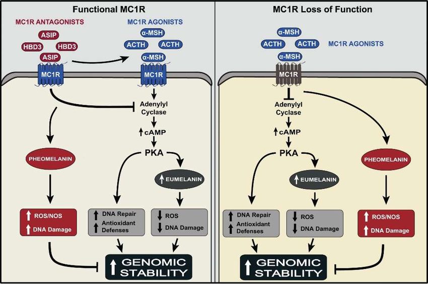
α-melanocyte stimulating hormone (α-melanocortin; α-MSH) increases the synthesis of eumelanin in a
cAMP-dependent manner (Figure 1) [40,43,44]. The recessive yellow mutation in extension, a frame-shift
mutation that causes loss of function of the mc1r, results in a yellow coat color in mice, due to exclusive
synthesis of pheomelanin and failure of melanocytes to synthesize eumelanin [41,43]. Accordingly,
pheomelanin synthesis was proposed to be a default pathway: when melanocytes fail to synthesize
eumelanin, they can still produce pheomelanin. This notion was supported by the biochemical
findings that eumelanin synthesis requires high levels and activity of tyrosinase, the rate-limiting
enzyme of melanin synthesis, as well as high levels of tyrosinase related (Tyrp)-1 and -2 (dopachrome
tautomerase) [45,46]. In contrast, pheomelanin synthesis proceeds in the presence of low levels and
activity of tyrosinase, and in the absence of Tyrp-1 and Tyrp-2. In human melanocytes, eumelanin
synthesis is regulated similarly to mouse melanocytes by activation of MC1R by α-MSH. Treatment
of human melanocytes with the potent melanocortin analog [Nle4 , D-Phe7 ]-α-MSH (NDP-MSH),
or treatment of mice with agents that increase the levels of cAMP, e.g., forskolin, results in increased
eumelanin synthesis [47,48]. In humans, loss-of-function allelic variants of the MC1R are strongly
associated with red hair color and fair skin, due to inhibition of eumelanin synthesis [35,49,50].
Additionally, mutations in the pro-opiomelanocortin (POMC) gene, which codes for the precursor of
α-MSH, is associated with red hair phenotype, in addition to metabolic abnormalities [51]. The human
MC1R recognizes both α-MSH and the structurally-related adrenocorticotropic hormone (ACTH) as
agonists, and binds both ligands with the same affinity [52,53]. This explains the hyperpigmentation
associated with over-production of ACTH, as in Addison’s disease [54]. Collectively, these results lend
strong support to the role of MC1R, its agonists, and its signaling pathway in regulating the synthesis
of eumelanin.
The main physiological antagonist for the mc1r is agouti signaling protein (ASIP), which is
expressed in the mouse hair follicles in a temporal fashion, resulting in agouti phenotype, characterized
by hairs with dark (eumelanin-containing) bands, interrupted by a yellow (pheomelanin-containing)
band [55–57]. Mutations that cause overproduction of ASIP result in a yellow coat color of mice,
a pigmentary phenotype similar to that caused by the recessive yellow mutation in mc1r. The human
Agouti gene was cloned in human skin, and its product was shown to function as an inverse agonist
of MC1R in human melanocytes [58,59]. Treatment of cultured human melanocytes with human
ASIP resulted in displacement of α-MSH from MC1R, and abrogation of α-MSH-induced increase
in cAMP levels and tyrosinase activity (Figure 1) [59]. Therefore, the MC1R/α-MSH/ASIP axis
functions in human epidermal melanocytes, as in mouse follicular melanocytes, to regulate the
eumelanin/pheomelanin switch.
Another physiological modulator of mc1r activity is human beta defensin 3 (HBD3), best known
for its antimicrobial effects, and is synthesized by keratinocytes [60,61]. A deletion mutation in CBD103,
the ortholog of HBD3, was first reported to result in a black coat color in dogs [62]. It was proposed
that this mutation results in black pigmentation in dogs and transgenic mice via competing with,
and inhibiting the binding of ASIP to the mc1r. We found that HBD3 acts as an antagonist of the
human MC1R, blocking the effects of α-MSH on cAMP and tyrosinase activity in human melanocytes
(Figure 1) [63]. Collectively, these studies underscore the significance of the MC1R, and defines its
physiological agonists and antagonists and their function in regulating eumelanin and pheomelanin
synthesis by human melanocytes.
In addition to MC1R, agouti, and HBD3, there are other genes that contribute to the regulation of
eumelanin and pheomelanin synthesis. For example, mahogany and mahoganoid function as negative
modifiers of agouti in mice, inhibiting its effects, thereby increasing eumelanin synthesis. Both mahogany
and mahoganoid are thought to be downstream of agouti and upstream of mc1r [64]. The pink eyed-dilution
(p) gene, which codes for a protein with 12 membrane spanning domains, is thought to be a
melanosomal transmembrane protein that is involved in melanosome biogenesis, and functions as an
anion transporter that regulates melanosomal pH, and the stability of tyrosinase-Tyrp-1 and Tyrp-2
complex, which is required for eumelanin synthesis [65]. Melanosomal pH is important in regulatingInt. J. Mol. Sci. 2018, 19, 2667 5 of 16
eumelain/pheomelanin synthesis and neutral pH favors the synthesis of eumelanin [66]. Mutations in
p gene reduce total melanin, mainly eumelanin content, and are the underlying cause of oculocutaneous
Int. J. Mol.
albinism Sci. 2018,
type-2. 19, x gene encodes the plasma membrane cysteine/glutamate exchanger
Slc7a11 5 ofXct
15 [67].
The recessive sut mutation in Slc7a11 markedly reduces the synthesis of pheomelanin due to reduction
exchanger Xct [67]. The recessive sut mutation in Slc7a11 markedly reduces the synthesis of
in transport of extracellular cystine, which also leads to diminished levels of glutathione and ability to
pheomelanin due to reduction in transport of extracellular cystine, which also leads to diminished
overcome
levels oxidative
of glutathione stress.
and ability to overcome oxidative stress.
Figure 1. Summary of the effects of the MC1R agonists α-MSH and ACTH and the antagonists ASIP
Figure 1. Summary of the effects of the MC1R agonists α-MSH and ACTH and the antagonists ASIP and
and HBD3 on functional MC1R, and the impact of loss of function of MC1R on these effects. Activation
HBD3 on functional MC1R, and the impact of loss of function of MC1R on these effects. Activation of
of the MC1R, expressed on the cell membrane of melanocytes by either agonist, α-MSH or ACTH
the MC1R, expressed
activates the cAMP on pathway,
the cell membrane
leading to of melanocytes
increased by either
eumelanin agonist,
synthesis, α-MSH
which or ACTH
quenches activates
reactive
the cAMP
oxygen pathway, leading
species (ROS) andtoreduces
increased eumelaninofsynthesis,
the generation DNA damage which quenches
upon reactive
UV exposure. oxygen of
Activation species
(ROS)the
andcAMP pathway
reduces also enhances
the generation of DNAthe DNA
damagerepair
uponandUVantioxidant
exposure.capacities
Activation of melanocytes.
of the cAMP Thepathway
cumulativethe
also enhances outcome
DNA of theseand
repair effects is maintenance
antioxidant of genomic
capacities stability of melanocytes.
of melanocytes. Treatment
The cumulative of
outcome of
thesemelanocytes with either ASIP
effects is maintenance or HBD3stability
of genomic antagonizes the effects of α-MSH,
of melanocytes. Treatment andofare therefore expected
melanocytes with either
ASIPtoor reduce genomic stability.
HBD3 antagonizes Expression
the effects of α-MSH,of loss-of-function-MC1R disrupts
and are therefore expected to the signaling
reduce genomicof the
stability.
agonist-bound receptor, thereby inhibiting the synthesis of eumelanin, allowing
Expression of loss-of-function-MC1R disrupts the signaling of the agonist-bound receptor, thereby only the synthesis of
pheomelanin, which increases the generation of ROS and NOS, and allows for increased
inhibiting the synthesis of eumelanin, allowing only the synthesis of pheomelanin, which increases the UV-induced
DNA damage. Additionally, lack of signaling of the α-MSH-bound MC1R via the cAMP pathway
generation of ROS and NOS, and allows for increased UV-induced DNA damage. Additionally, lack of
inhibits the activation of antioxidant and DNA repair pathways, leading to reduced genomic stability
signaling of the α-MSH-bound MC1R via the cAMP pathway inhibits the activation of antioxidant
of UV-irradiated melanocytes, and increased risk for malignant transformation to melanoma.
and DNA repair pathways, leading to reduced genomic stability of UV-irradiated melanocytes, and
Upward arrows: increase in the effect; T bar: blocking the effect.
increased risk for malignant transformation to melanoma. Upward arrows: increase in the effect; T bar:
5. MC1R,the
blocking A Major
effect. Regulator of Human Pigmentation and a Melanoma Susceptibility Gene
5. MC1R,The gene that
A Major is centralof
Regulator to Human
the regulation of human and
Pigmentation pigmentation is the Susceptibility
a Melanoma MC1R. Epidemiological
Gene
studies in different human populations concluded that MC1R is very highly polymorphic, and its
The
manygene that
allelic is central
variants to the
account regulation
to a large of human
extent for pigmentation
the diversity is the MC1R.[68–70].
of human pigmentation Epidemiological
Given
the significance of the MC1R in regulating eumelanin synthesis, the MC1R genotype is expected and
studies in different human populations concluded that MC1R is very highly polymorphic, to its
manydetermine the sensitivity
allelic variants accountof melanocytes, and thereby
to a large extent the skin,
for the to solar
diversity ofUV exposure.
human The consensus
pigmentation [68–70].
Givenwild
thetype MC1R is predominant
significance of the MC1RininAfrican countries,
regulating where dark
eumelanin skin pigmentation
synthesis, is mostly needed
the MC1R genotype is expected
to mitigate
to determine thethe photodamaging
sensitivity effects of and
of melanocytes, the equatorial
thereby the sunskin,
[68–70]. Variants
to solar of MC1R are
UV exposure. Themostly
consensus
prevalent in Northern latitudes, and a few, mainly R151C, R160W,
wild type MC1R is predominant in African countries, where dark skin pigmentation is mostlyand D294H, are strongly
needed
associated with red hair and fair skin phenotype [49,71]. These variants result in loss of function of
to mitigate the photodamaging effects of the equatorial sun [68–70]. Variants of MC1R are mostly
the MC1R, inhibiting signaling of the α-MSH-bound receptor [35,50]. The resulting pigmentary
prevalent in Northern latitudes, and a few, mainly R151C, R160W, and D294H, are strongly associated
phenotype is associated with poor tanning ability and increased risk for melanoma [5].
with red hair and fair
The MC1R is skin phenotype
a bona [49,71].predisposition
fide melanoma These variants result
gene in loss
[72,73]. of function
Twenty-four of the of
percent MC1R,
inhibiting signaling of the receptor [35,50]. The resulting pigmentary
melanoma patients carry MC1R loss-of-function variants, and although redheads who express two
α-MSH-bound phenotype is
MC1R with
associated loss-of-function
poor tanningalleles comprise
ability and only 1–2% of
increased thefor
risk worldwide
melanoma population,
[5]. they represent 16%
of all melanoma patients [74,75]. A seminal finding was that MC1R regulates DNA repair andInt. J. Mol. Sci. 2018, 19, 2667 6 of 16
The MC1R is a bona fide melanoma predisposition gene [72,73]. Twenty-four percent of melanoma
patients carry MC1R loss-of-function variants, and although redheads who express two MC1R
loss-of-function alleles comprise only 1–2% of the worldwide population, they represent 16%
of all melanoma patients [74,75]. A seminal finding was that MC1R regulates DNA repair and
antioxidant pathways in melanocytes [35,76–80]. We and others reported that human melanocytes
expressing functional MC1R respond to α-MSH treatment with increased efficiency of repair of
UV-induced DNA photoproducts, reduced hydrogen peroxide generation and increased expression of
antioxidant enzymes, such as catalase and hemeoxygenase-1, γ-glutamylcysteine synthase (γ-GCS),
glutathione-S-transferase Pi (GSTPi), Peroxiredoxin 1 (PRX1), 8-oxoguanine DNA glycosylase (OGG1)
and apurinic apyrimidinic endonuclease 1 (APE-1/Ref-1) (Figure 1). These effects of α-MSH can
be mimicked by agents that activate the cAMP pathway, e.g., Forskolin, suggesting the significance
of this pathway in maintaining the genomic stability of melanocytes [35,48,81]. Loss-of-function
variants of MC1R not only inhibit the synthesis of eumelanin, but also compromise the DNA repair
and antioxidant capacities of melanocytes (Figure 1) [35]. These findings were corroborated by the
observation that recessive yellow mice, which expressed loss-of-function mc1r together with the somatic
BRAFv600E mutation, had a markedly higher levels of oxidative DNA damage and lipid peroxidation
than their counterparts that expressed wild type mc1r [33]. Further support for the role of MC1R in
determining the extent of UV-induced genotoxicity and mutagenesis came from a recent report that
loss-of-function MC1R variants increase the risk of melanocytes to acquire UV signature mutations
that promote malignant transformation to melanoma [82].
Based on these findings, it can be concluded that expression of functional MC1R protects
melanocytes against the genotoxic effects of UV by three mechanisms: (i) activation of DNA repair
pathways (nucleotide excision and base excision repair pathways); (ii) inhibition of generation
of reactive oxygen species, activation of antioxidant enzymes, and up regulation of expression
of antioxidant genes; and (iii) increase in eumelanin synthesis (Figure 1) [35,76,78–80]. The first
two mechanisms represent an early and immediate response to UV to prevent genomic instability
of melanocytes, and the third is a latent response, which protects against the genotoxic effects of
subsequent UV exposure.
6. Paracrine Factors as Regulators of Melanogenesis and the Response of Melanocytes to UV
In human skin, melanocytes differ from epidermal keratinocytes and dermal fibroblasts in their
longevity in the skin and very limited proliferative capacity. Melanocytes survive in the epidermis
for decades, and only proliferate upon injury in response to insult from the environment or its
microenvironment [83]. Melanocytes are also resistant to apoptosis [84]. These properties make these
cells prone to accumulate UV-induced somatic mutations over time due to repetitive sun exposure,
which might culminate in malignant transformation to melanoma. It is established that melanocyte
homeostasis is maintained by a network of paracrine factors, many of which are up regulated in
expression by exposure to UV. The symbiotic relationship between melanocytes and keratinocytes
has different levels. It involves the transfer of melanin-containing melanosomes from melanocytes
to keratinocytes, to confer photoprotection to the entire epidermis [10]. It also includes the synthesis
and secretion by keratinocytes of a wide array of factors that maintain the homeostasis of melanocytes
and modulate their response to UV [85–92]. Keratinocytes and melanocytes synthesize POMC and
process it to the bioactive peptides α-MSH, ACTH and β-endorphin [85,92,93]. The synthesis of
POMC is increased in the epidermis in response to UV exposure [94,95]. Endothelin-1 is synthesized by
keratinocytes, and is a potent mitogen for melanocytes in vitro [89,96,97]. We reported that endothelin-1
and α-MSH, together with the keratinocyte-derived mitogen basic fibroblast growth factor (bFGF),
interact synergistically to support the proliferation of human melanocytes in vitro [98]. Importantly,
endothelin-1, similar to α-MSH, enhances repair of DNA photoproducts and reduces apoptosis of
UV-irradiated human melanocytes [76,99]. These results suggest that the paracrine factors α-MSH
and endothelin-1 function as “survival factors” that enable melanocytes to overcome the stressInt. J. Mol. Sci. 2018, 19, 2667 7 of 16
imposed by UV exposure, allowing them to survive with genomic stability. Importantly, endothelin-1
compensates for the inability of melanocytes expressing loss-of-function MC1R to respond to α-MSH
with modulating the DNA damage response to UV [99]. This provides an additional line of defense
against UV-induced genotoxicity, and reduces the risk of these vulnerable melanocytes to transform to
melanoma. Keratinocytes synthesize vitamin D3, and its active form, 1,25(OH)2 vitamin D3, increases
the DNA repair capacity of UV-irradiated keratinocytes and melanocytes [100,101]. Consistent with
these results, targeted deletion of vitamin D receptor gene in mouse melanocytes compromised melanin
content, and increased the levels of CPD induced by UV exposure [102]. Importantly, all three paracrine
factors, α-MSH, endothelin-1, and 1,25(OH)2 vitamin D3 up regulate the expression of the MC1R
gene, an effect that is expected to sustain and/or enhance the ability of melanocytes to respond to
α-MSH, and thereby maintain its genomic stability and ability to synthesize the photoprotective
eumelanin [63,97] (V. Swope, R. Kavanagh, and A. Abdel-Malek, unpublished work). That these
paracrine factors increase MC1R gene expression solidifies the central role of the MC1R/α-MSH axis
in regulating melanocyte survival and overcoming the genotoxic effects of UV that lead to melanoma
formation. Additionally, these findings suggest that paracrine factors, via activating different receptors
and signaling pathways, provide multiple means to activate DNA repair mechanisms in order to
maintain the homeostasis of melanocytes and prevent the mutagenic effect of UV.
In addition to epidermal keratinocytes, dermal fibroblasts synthesize and secrete factors that
regulate the activity of melanocytes. Fibroblasts in the palms and soles secrete Dikkopf1, which limits
the number of melanocytes in these anatomic sites by inhibiting their proliferation and melanogenesis
via suppressing β-catenin and Mitf [103]. Recently, clusterin (apoliprotien J) was reported to be
secreted mainly by fibroblasts, and to inhibit melanogenesis by binding to TGFβ-1 and -2 receptors
on melanocytes [104]. Neuregulin-1, a fibroblast-derived factor, increases melanogenesis by binding
ErbB receptors expressed on melanocytes. Interestingly, the synthesis and secretion of neuregulin-1 by
fibroblasts varied according to pigmentary phenotype, being higher in fibroblasts derived from
skin phototype IV than their counterparts derived from skin phototype II [105]. These results
implicate neuregulin-1 in determining constitutive pigmentation, and suggest that it contributes
to eumelanin synthesis, which is abundant in dark skin. Another melanogenic factor derived from
fibroblasts is CCN1/Cyr61, an extracellular matrix (ECM) protein that was recently reported to
increase melanogenesis via binding to integrin α6β1 and activating the MAP kinases p38 and
ERK1/2 [106]. Secretion of CCN1/Cyr61 is enhanced following UV exposure, suggesting that this
factor participates in the tanning response of melanocytes to UV. Fibroblasts in young skin also secrete
insulin-like growth factor (IGF)-1, which activates nucleotide excision repair in keratinocytes, thereby
inhibiting UV-induced mutagenesis [107]. Melanocytes express functional IGF-1 receptors (V. Swope,
R. Kavanagh, and A. Abdel-Malek, unpublished work), suggesting that IGF-1 can enhance their DNA
repair capacity. Collectively, these results provide compelling evidence for the role of dermal fibroblasts
and their secretome in regulating the melanocytes that reside on the basement membrane. Further
studies are needed to further elucidate how fibroblast-derived paracrine factors regulate melanocytes,
particularly eumelanin vs. pheomelanin synthesis and the response to solar UV.
7. Effect of Skin Pigmentation on Photoaging
A dramatic effect of UV exposure is photoaging, caused by long term solar UV exposure, mainly
to long wavelength UVA that penetrates deep into the dermal layers, combined with intrinsic aging
of skin [108]. There is clinical evidence that photoaging is more prominent and severe in light than
in dark skin, and correlates directly with skin carcinogenesis [12]. This implicates low melanin
(thereby eumelanin) content in sensitizing the skin to this effect of sun exposure. Photoaging results
mainly from reactive oxygen species generated upon exposure to UV [109]. Exposure of fibroblasts to
UVA induces the common deletion in mitochondrial DNA via ROS generation [109,110]. Mutations
in the mitochondrial genome might underlie aging-associated functional changes, which include
disorganization of collagen fibrils due to reduced collagen synthesis, and solar elastosis resultingInt. J. Mol. Sci. 2018, 19, 2667 8 of 16
from accumulation of abnormal elastin-containing material [111]. Additionally, UVA up regulates the
synthesis of matrix metalloproteinases (MMPs), which degrade the extracellular matrix, including
collagen. Given the role of eumelanin in reducing the sensitivity of the skin to UV by shielding it from
UV rays and quenching ROS, these findings explain why lightly pigmented skin is more prone to
photoaging than dark skin [9,22]. Recently, meta-analysis of genome-wide association studies in a large
cohort, including 1671 twin pairs, revealed the association of SNPs at or near the pigmentary genes
SLC45A2, IRF4, and MC1R with increased wrinkling and photoaging [12]. These results underscore
the role of pigmentation in determining the extent of skin photoaging.
8. From the Bench to the Bedside: Selective Targeting of MC1R by Small α-MSH Analogs to
Enhance Photoprotection
The well-known effect of α-MSH on stimulating eumelanin synthesis and the photoprotective
effect of eumelanin have sparked interest in developing melanocortin analogs as safe sunless tanning
agents. The physiological α-MSH is composed of 13 amino acids. Its small size and linear structure
allowed for extensive structure-function studies, which revealed that most of the melanotropic activity
of the hormone resides in the His6 -Phe7 -Arg8 -Trp9 core sequence [112–114]. The best known α-MSH
analog is the tridecapeptide NDP-MSH or afamelanotide. [115]. The modifications of the physiological
α-MSH consisting of substitution of the fourth amino acid Methionine by Norleucine, and the seventh
amino acid L-Phenylalanine by its D-enantiomer, increased the potency and stability of the resulting
peptide [115–117]. The first clinical trial with NDP-MSH in 1991 demonstrated for the first time that
injection of human volunteers with the peptide resulted in increased skin pigmentation without any
sun exposure [118]. However, unexpected side effects were noted, which included loss of appetite,
nausea, and flushing. These side effects are due to non-selective binding of NDP-MSH to other
melanocortin receptors that were not identified yet at that time. Subsequently, systemic administration
of NDP-MSH, which led to increased pigmentation, was reported to prevent sunburn and reduce
UV-induced DNA damage, particularly in individuals with light skin color who burn readily upon
sun exposure [119]. These reports provided compelling evidence for the efficacy of α-MSH analogs
in photoprotection, and ability of melanocytes to respond to exogenous treatment with NDP-MSH,
despite the synthesis of endogenous melanocortins by keratinocytes and melanocytes.
Although NDP-MSH is markedly more potent and has more prolonged effects than α-MSH, it is
not selective for MC1R. Of all five melanocortin receptors (MC1-MC5R), melanocytes express only
MC1R [52]. To target specifically the melanocytes with α-MSH peptide analogs, it is ideal to develop
highly MC1R selective peptides to reduce off-target effects. With this in mind, we are developing
small peptide α-MSH analogs that are MC1R-selective, and mimic α-MSH in enhancing DNA repair,
and stimulating pigmentation. Our goal is to develop these peptides for topical application, which is
more practical and provides greater target specificity than systemic administration. For topical
application, peptides need to be lipophilic, in order to enhance their permeation through the stratum
corneum and the epidermal layers of human skin. We have designed n-capped tetrapeptide α-MSH
analogs, with 4-phenylbutyryl-His-D-Phe-Arg-Trp-NH2 as the lead peptide, and tested them on
primary human melanocyte cultures [120]. This lead peptide proved to be considerably more potent
than α-MSH in stimulating the activity of tyrosinase, hence melanogenesis, and in enhancing repair
of UV-induced photoproducts, and reducing UV-induced apoptosis of human melanocytes [120].
Moreover, this peptide had a more prolonged residual effect than α-MSH on stimulation of tyrosinase
activity [120]. The effects of this peptide are mediated by binding to the MC1R, as they were not
evident in melanocytes expressing non-functional MC1R. Furthermore, the effects of this peptide
were abolished in the concomitant presence of ASIP, the physiological MC1R antagonist. Importantly,
4-phenylbutyryl-His-D-Phe-Arg-Trp-NH2 is superior to NDP-MSH due to its unique selectivity for the
MC1R, based on our preliminary data. Recently, Zhou et al. reported on the γ-MSH analog, [Leu3 ,
Leu7 , Phe8 ]-γ-MSH-NH2 , as being selective for MC1R [121]. Subsequently, the same group publishedInt. J. Mol. Sci. 2018, 19, 2667 9 of 16
that replacement of Arg8 with Nle, and L-Phe7 by D-Phe in the core sequence Ac-His-Phe-Arg-Trp-NH2
of α-MSH conferred MC1R selectivity to the tetrapeptide [122].
We have succeeded in designing n-capped tripeptide melanocortin analogs that retain
considerable melanogenic activity, despite their very small size. The lead peptide 4-phenylbutyryl-
His-D-Phe-Arg-NH2 and other peptides with specific C-terminus modifications, were only 10 fold
less potent than α-MSH in stimulating cAMP formation and tyrosinase activity of cultured human
melanocytes [123]. Similar to α-MSH, these peptides were effective in inhibiting the generation of
hydrogen peroxide and enhancing repair of CPD in UV-irradiated human melanocytes. As is the case
of the aforementioned n-capped tetrapeptides, these tripeptides elicited their effects by binding to the
MC1R, and their effects were absent in melanocytes expressing loss of function MC1R. The efficacy of
these tripeptides clearly indicates that deletion of Trp9 does not eliminate the melanotropic activity of
the tripeptides or their ability to activate the MC1R.
Our data showed that our tetra- and tripeptide analogs require expression of functional MC1R,
and have no effects on cultured human melanocytes that express two loss-of-function MC1R
variants, a genotype strongly associated with red hair, fair skin and poor tanning ability [49,120,123].
This suggests that our peptides will not benefit individuals expressing loss-of-function MC1R, who
have increased UV sensitivity and high risk for skin cancer and melanoma. Others have targeted
the cAMP pathway, downstream of MC1R, to circumvent the issue of loss of function of MC1R.
D’Orazio et al. reported on the melanogenic effect of Forskolin on recessive yellow mice that express
loss-of-function mutation in mc1r [48]. More recently, they reported on the ability of Forskolin to
activate nucleotide excision repair in UV-irradiated melanocytes and melanoma cells [81]. However,
Forskolin cannot be used as a tanning agent, since the cAMP pathway is promiscuous in all cell types.
The limitations of our peptides do not negate the significance of utilizing selective MC1R analogs
for skin cancer, including melanoma, prevention. Millions of individuals stand to benefit from this
strategy, particularly those expressing mutations in other skin cancer or melanoma susceptibility genes
(e.g., CDKN2A), and those heterozygous for MC1R RHC variants, who represent 50% of the entire
white population in the U.S.A. [124].
Conflicts of Interest: The authors declare no conflict of interest.
References
1. Jablonski, N.G.; Chaplin, G. The evolution of human skin coloration. J. Hum. Evol. 2000, 39, 57–106.
[CrossRef] [PubMed]
2. Sober, A.J. Solar exposure in the etiology of cutaneous melanoma. Photodermatol 1987, 4, 23–31. [PubMed]
3. Gilchrest, B.A.; Rogers, G.S. Photoaging. In Clinical Photomedicine; Lim, H.W., Soter, N.A., Eds.; Marcel
Dekker, Inc.: New York, NY, USA, 1993; pp. 95–111.
4. Epstein, J.H. Photocarcinogenesis, skin cancer and aging. J. Am. Acad. Dermatol. 1983, 9, 487–502. [CrossRef]
5. Gilchrest, B.A.; Eller, M.S.; Geller, A.C.; Yaar, M. The pathogenesis of melanoma induced by ultraviolet
radiation. N. Engl. J. Med. 1999, 340, 1341–1348. [CrossRef] [PubMed]
6. Hodis, E.; Watson, I.R.; Kryukov, G.V.; Arold, S.T.; Imielinski, M.; Theurillat, J.P.; Nickerson, E.; Auclair, D.;
Li, L.; Place, C.; et al. A landscape of driver mutations in melanoma. Cell 2012, 150, 251–263. [CrossRef]
[PubMed]
7. Noonan, F.P.; Otsuka, T.; Bang, S.; Anver, M.R.; Merlino, G. Accelerated ultraviolet radiation-induced
carcinogenesis in hepatocyte growth factor/scatter factor transgenic mice. Cancer Res. 2000, 60, 3738–3743.
[PubMed]
8. Pathak, M.A.; Jimbow, K.; Fitzpatrick, T. Photobiology of pigment cell. In Phenotypic Expression in Pigment
Cells; Seiji, M., Ed.; University of Tokyo Press: Tokyo, Japan, 1980; pp. 655–670.
9. Kaidbey, K.H.; Poh Agin, P.; Sayre, R.M.; Kligman, A.M. Photoprotection by melanin—A comparison of
black and Caucasian skin. J. Am. Acad. Dermatol. 1979, 1, 249–260. [CrossRef]Int. J. Mol. Sci. 2018, 19, 2667 10 of 16
10. Kobayashi, N.; Nakagawa, A.; Muramatsu, T.; Yamashina, Y.; Shirai, T.; Hashimoto, M.W.; Ishigaki, Y.;
Ohnishi, T.; Mori, T. Supranuclear melanin caps reduce ultraviolet induced DNA photoproducts in human
epidermis. J. Investig. Dermatol. 1998, 110, 806–810. [CrossRef] [PubMed]
11. Halder, R.M.; Bridgeman-Shah, S. Skin cancer in African Americans. Cancer 1995, 75, 667–673. [CrossRef]
12. Law, M.H.; Medland, S.E.; Zhu, G.; Yazar, S.; Vinuela, A.; Wallace, L.; Shekar, S.N.; Duffy, D.L.; Bataille, V.;
Glass, D.; et al. Genome-Wide Association Shows that Pigmentation Genes Play a Role in Skin Aging.
J. Investig. Dermatol. 2017, 137, 1887–1894. [CrossRef] [PubMed]
13. Hunt, G.; Kyne, S.; Ito, S.; Wakamatsu, K.; Todd, C.; Thody, A. Eumelanin and phaeomelanin contents of
human epidermis and cultured melanocytes. Pigment Cell Res. 1995, 8, 202–208. [CrossRef] [PubMed]
14. Hennessy, A.; Oh, C.; Diffey, B.; Wakamatsu, K.; Ito, S.; Rees, J. Eumelanin and pheomelanin concentrations
in human epidermis before and after UVB irradiation. Pigment Cell Res. 2005, 18, 220–223. [CrossRef]
[PubMed]
15. Wakamatsu, K.; Kavanagh, R.; Kadekaro, A.L.; Terzieva, S.; Strum, R.A.; Leachman, S.; Abdel-malek, Z.A.;
Ito, S. Diversity of pigmentation in cultured human melanocytes is due to differences in the type as well as
quantity of melanin. Pigment Cell Res. 2006, 19, 154–162. [CrossRef] [PubMed]
16. Chedekel, M.R. Photochemistry and photobiology of epidermal melanins. Photochem. Photobiol. 1982, 35,
881–885. [CrossRef] [PubMed]
17. Chedekel, M.R.; Smith, S.K.; Post, P.W.; Pokora, A.; Vessell, D.L. Photodestruction of pheomelanin: Role of
oxygen. Proc. Natl. Acad. Sci. USA 1978, 75, 5395–5399. [CrossRef] [PubMed]
18. Menon, I.A.; Persad, S.; Haberman, H.F.; Kurian, C.J. A comparative study of the physical and chemical
properties of melanins isolated from human black and red hair. J. Investig. Dermatol. 1983, 80, 202–206.
[CrossRef] [PubMed]
19. Jimbow, K.; Takeuchi, T. Ultrastructural comparison of pheo- and eu-melanogenesis in animals. In Pigment
Cell; Klaus, S.N., Ed.; S. Karger: Basel, Switzerland, 1979; Volume 4, pp. 308–317.
20. Szabo, G. Racial differences in the fate of melanosomes in human epidermis. Nature 1969, 222, 1081–1082.
[CrossRef] [PubMed]
21. Ito, S. Biochemistry and physiology of melanin. In Pigmentation and Pigmentary Disorders; Levine, N., Ed.;
CRC Press: Boca Raton, FL, USA, 1993; pp. 33–59.
22. Bustamante, J.; Bredeston, L.; Malanga, G.; Mordoh, J. Role of melanin as a scavenger of active oxygen
species. Pigment Cell Res. 1993, 6, 348–353. [CrossRef] [PubMed]
23. Tadokoro, T.; Kobayashi, N.; Zmudzka, B.Z.; Ito, S.; Wakamatsu, K.; Yamaguchi, Y.; Korossy, K.S.; Miller, S.A.;
Beer, J.Z.; Hearing, V.J. UV-induced DNA damage and melanin content in human skin differing in
racial/ethnic origin. FASEB J. 2003, 17, 1177–1179. [CrossRef] [PubMed]
24. Hauser, J.E.; Kadekaro, A.L.; Kavanagh, R.J.; Wakamatsu, K.; Terzieva, S.; Schwemberger, S.; Babcock, G.;
Rao, M.B.; Ito, S.; Abdel-Malek, Z.A. Melanin content and MC1R function independently affect UVR-induced
DNA damage in cultured human melanocytes. Pigment Cell Res. 2006, 19, 303–314. [CrossRef] [PubMed]
25. Fried, L.; Arbiser, J.L. The reactive oxygen-driven tumor: Relevance to melanoma. Pigment Cell Melanoma Res.
2008, 21, 117–122. [CrossRef] [PubMed]
26. Denat, L.; Kadekaro, A.L.; Marrot, L.; Leachman, S.A.; Abdel-Malek, Z.A. Melanocytes as instigators and
victims of oxidative stress. J. Investig. Dermatol. 2014, 134, 1512–1518. [CrossRef] [PubMed]
27. Smit, N.P.; van Nieuwpoort, F.A.; Marrot, L.; Out, C.; Poorthuis, B.; van Pelt, H.; Meunier, J.R.; Pavel, S.
Increased melanogenesis is a risk factor for oxidative DNA damage—Study on cultured melanocytes and
atypical nevus cells. Photochem. Photobiol. 2008, 84, 550–555. [CrossRef] [PubMed]
28. Kagedal, B.; Gawelin, A.L.; Pettersson, A. Synthesis of 5-S-L-cysteinyl-glycine-L-DOPA, a natural substrate
for serum and melanocyte dipeptidase. Anal. Biochem. 1987, 165, 167–174. [CrossRef]
29. Pavel, S.; van Nieuwpoort, F.; van der Meulen, H.; Out, C.; Pizinger, K.; Cetkovska, P.; Smit, N.P.;
Koerten, H.K. Disturbed melanin synthesis and chronic oxidative stress in dysplastic naevi. Eur. J. Cancer
2004, 40, 1423–1430. [CrossRef] [PubMed]
30. Panzella, L.; Leone, L.; Greco, G.; Vitiello, G.; D’Errico, G.; Napolitano, A.; d’Ischia, M. Red human
hair pheomelanin is a potent pro-oxidant mediating UV-independent contributory mechanisms of
melanomagenesis. Pigment Cell Melanoma Res. 2014, 27, 244–252. [CrossRef] [PubMed]Int. J. Mol. Sci. 2018, 19, 2667 11 of 16
31. Maresca, V.; Enrica, F.; Stefania, B.; Arianna, M.; Claudia, F.; Anna, M.M.; Marco, G.P.; Mauro, P. Correlation
between melanogenic and catalase activity in in vitro human melanocytes: A synergic strategy against
oxidative stress. Pigment Cell Melanoma Res. 2008, 21, 200–205. [CrossRef] [PubMed]
32. Davies, H.; Bignell, G.R.; Cox, C.; Stephens, P.; Edkins, S.; Clegg, S.; Teague, J.; Woffendin, H.; Garnett, M.J.;
Bottomley, W.; et al. Mutations of the BRAF gene in human cancer. Nature 2002, 417, 949–954. [CrossRef]
[PubMed]
33. Mitra, D.; Luo, X.; Morgan, A.; Wang, J.; Hoang, M.P.; Lo, J.; Guerrero, C.R.; Lennerz, J.K.; Mihm, M.C.;
Wargo, J.A.; et al. An ultraviolet-radiation-independent pathway to melanoma carcinogenesis in the red
hair/fair skin background. Nature 2012, 491, 449–453. [CrossRef] [PubMed]
34. Morgan, A.M.; Lo, J.; Fisher, D.E. How does pheomelanin synthesis contribute to melanomagenesis?: Two
distinct mechanisms could explain the carcinogenicity of pheomelanin synthesis. BioEssays 2013, 35, 672–676.
[CrossRef] [PubMed]
35. Kadekaro, A.L.; Leachman, S.; Kavanagh, R.J.; Swope, V.; Cassidy, P.; Supp, D.; Sartor, M.; Schwemberger, S.;
Babcock, G.; Wakamatsu, K.; et al. Melanocortin 1 receptor genotype: An important determinant of the
damage response of melanocytes to ultraviolet radiation. FASEB J. 2010, 24, 3850–3860. [CrossRef] [PubMed]
36. Pollock, P.M.; Harper, U.L.; Hansen, K.S.; Yudt, L.M.; Stark, M.; Robbins, C.M.; Moses, T.Y.; Hostetter, G.;
Wagner, U.; Kakareka, J.; et al. High frequency of BRAF mutations in nevi. Nat. Genet. 2003, 33, 19–20.
[CrossRef] [PubMed]
37. Premi, S.; Wallisch, S.; Mano, C.M.; Weiner, A.B.; Bacchiocchi, A.; Wakamatsu, K.; Bechara, E.J.; Halaban, R.;
Douki, T.; Brash, D.E. Photochemistry. Chemiexcitation of melanin derivatives induces DNA photoproducts
long after UV exposure. Science 2015, 347, 842–847. [CrossRef] [PubMed]
38. Runger, T.M.; Epe, B.; Moller, K. Processing of directly and indirectly ultraviolet-induced DNA damage in
human cells. Recent Results Cancer Res. 1995, 139, 31–42. [PubMed]
39. Cadet, J.; Berger, M.; Douki, T.; Morin, B.; Raoul, S.; Ravanat, J.L.; Spinneli, S. Effects of UV and visible
radiation on DNA-final base damage. Biol. Chem. 1997, 378, 1275–1286. [PubMed]
40. Geschwind, I.I.; Huseby, R.A.; Nishioka, R. The effect of melanocyte-stimulating hormone on coat color in
the mouse. Recent Prog. Horm. Res. 1972, 28, 91–130. [PubMed]
41. Robbins, L.S.; Nadeau, J.H.; Johnson, K.R.; Kelly, M.A.; Roselli-Rehfuss, L.; Baack, E.; Mountjoy, K.G.;
Cone, R.D. Pigmentation phenotypes of variant extension locus alleles result from point mutations that alter
MSH receptor function. Cell 1993, 72, 827–834. [CrossRef]
42. Mountjoy, K.G.; Robbins, L.S.; Mortrud, M.T.; Cone, R.D. The cloning of a family of genes that encode the
melanocortin receptors. Science 1992, 257, 1248–1251. [CrossRef] [PubMed]
43. Tamate, H.B.; Takeuchi, T. Action of the e locus of mice in the response of phaeomelanic hair follicles to
α-melanocyte-stimulating hormone in vitro. Science 1984, 224, 1241–1242. [CrossRef] [PubMed]
44. Wong, G.; Pawelek, J.; Sansone, M.; Morowitz, J. Response of mouse melanoma cells to melanocyte
stimulating hormone. Nature 1974, 248, 351–354. [CrossRef] [PubMed]
45. Sakai, C.; Ollmann, M.; Kobayashi, T.; Abdel-Malek, Z.; Muller, J.; Vieira, W.D.; Imokawa, G.; Barsh, G.S.;
Hearing, V.J. Modulation of murine melanocyte function in vitro by agouti signal protein. Embo J. 1997, 16,
3544–3552. [CrossRef] [PubMed]
46. Abdel-Malek, Z.A.; Scott, M.C.; Furumura, M.; Lamoreux, M.L.; Ollmann, M.; Barsh, G.S.; Hearing, V.J.
The melanocortin 1 receptor is the principal mediator of the effects of agouti signaling protein on mammalian
melanocytes. J. Cell Sci. 2001, 114 Pt 5, 1019–1024.
47. Hunt, G.; Kyne, S.; Wakamatsu, K.; Ito, S.; Thody, A.J. Nle4 DPhe7 α-melanocyte-stimulating hormone
increases the eumelanin:phaeomelanin ratio in cultured human melanocytes. J. Investig. Dermatol. 1995, 104,
83–85. [CrossRef] [PubMed]
48. D’Orazio, J.A.; Nobuhisa, T.; Cui, R.; Arya, M.; Spry, M.; Wakamatsu, K.; Igras, V.; Kunisada, T.; Granter, S.R.;
Nishimura, E.K.; et al. Topical drug rescue strategy and skin protection based on the role of Mc1r in
UV-induced tanning. Nature 2006, 443, 340–344. [CrossRef] [PubMed]
49. Box, N.F.; Wyeth, J.R.; O’Gorman, L.E.; Martin, N.G.; Sturm, R.A. Characterization of melanocyte stimulating
hormone receptor variant alleles in twins with red hair. Hum. Mol. Genet. 1997, 6, 1891–1897. [CrossRef]
[PubMed]Int. J. Mol. Sci. 2018, 19, 2667 12 of 16
50. Scott, M.C.; Wakamatsu, K.; Ito, S.; Kadekaro, A.L.; Kobayashi, N.; Groden, J.; Kavanagh, R.; Takakuwa, T.;
Virador, V.; Hearing, V.J.; et al. Human melanocortin 1 receptor variants, receptor function and melanocyte
response to UV radiation. J. Cell Sci. 2002, 115 Pt 11, 2349–2355.
51. Krude, H.; Biebermann, H.; Luck, W.; Horn, R.; Brabant, G.; Gruters, A. Severe early-onset obesity, adrenal
insufficiency and red hair pigmentation caused by POMC mutations in humans. Nat. Genet. 1998, 19,
155–157. [CrossRef] [PubMed]
52. Suzuki, I.; Cone, R.D.; Im, S.; Nordlund, J.; Abdel-Malek, Z.A. Binding of melanotropic hormones to
the melanocortin receptor MC1R on human melanocytes stimulates proliferation and melanogenesis.
Endocrinology 1996, 137, 1627–1633. [CrossRef] [PubMed]
53. Abdel-Malek, Z.; Swope, V.B.; Suzuki, I.; Akcali, C.; Harriger, M.D.; Boyce, S.T.; Urabe, K.; Hearing, V.J.
Mitogenic and melanogenic stimulation of normal human melanocytes by melanotropic peptides. Proc. Natl.
Acad. Sci. USA 1995, 92, 1789–1793. [CrossRef] [PubMed]
54. Sarkar, S.B.; Sarkar, S.; Ghosh, S.; Bandyopadhyay, S. Addison’s disease. Contemp. Clin. Dent. 2012, 3,
484–486. [CrossRef] [PubMed]
55. Bultman, S.J.; Michaud, E.J.; Woychik, R.P. Molecular characterization of the mouse agouti locus. Cell 1992,
71, 1195–1204. [CrossRef]
56. Miller, M.W.; Duhl, D.M.J.; Vrieling, H.; Cordes, S.P.; Ollmann, M.M.; Winkes, B.M.; Barsh, G.S. Cloning of
the mouse agouti gene predicts a secreted protein ubiquitously expressed in mice carrying the lethal yellow
mutation. Genes Dev. 1993, 7, 454–467. [CrossRef] [PubMed]
57. Silvers, W.K. The Coat Colors of Mice: A Model for Mammalian Gene Action and Interaction; Springer: New York,
NY, USA, 1979; p. 380.
58. Kwon, H.Y.; Bultman, S.J.; Loffler, C.; Chen, W.-J.; Furdon, P.J.; Powell, J.G.; Usala, A.-L.; Wilkison, W.;
Hansmann, I.; Woychik, R.P. Molecular structure and chromosomal mapping of the human homolog of the
agouti gene. Proc. Natl. Acad. Sci. USA 1994, 91, 9760–9764. [CrossRef] [PubMed]
59. Suzuki, I.; Tada, A.; Ollmann, M.M.; Barsh, G.S.; Im, S.; Lamoreux, M.L.; Hearing, V.J.; Nordlund, J.J.;
Abdel-Malek, Z.A. Agouti signaling protein inhibits melanogenesis and the response of human melanocytes
to alpha-melanotropin. J. Investig. Dermatol. 1997, 108, 838–842. [CrossRef] [PubMed]
60. Chen, H.; Xu, Z.; Peng, L.; Fang, X.; Yin, X.; Xu, N.; Cen, P. Recent advances in the research and development
of human defensins. Peptides 2006, 27, 931–940. [CrossRef] [PubMed]
61. Supp, D.M.; Karpinski, A.C.; Boyce, S.T. Expression of human beta-defensins HBD-1, HBD-2, and HBD-3 in
cultured keratinocytes and skin substitutes. Burns 2004, 30, 643–648. [CrossRef] [PubMed]
62. Candille, S.I.; Kaelin, C.B.; Cattanach, B.M.; Yu, B.; Thompson, D.A.; Nix, M.A.; Kerns, J.A.; Schmutz, S.M.;
Millhauser, G.L.; Barsh, G.S. A -defensin mutation causes black coat color in domestic dogs. Science 2007,
318, 1418–1423. [CrossRef] [PubMed]
63. Swope, V.B.; Jameson, J.A.; McFarland, K.L.; Supp, D.M.; Miller, W.E.; McGraw, D.W.; Patel, M.A.; Nix, M.A.;
Millhauser, G.L.; Babcock, G.F.; et al. Defining MC1R Regulation in Human Melanocytes by Its Agonist
alpha-Melanocortin and Antagonists Agouti Signaling Protein and beta-Defensin 3. J. Investig. Dermatol.
2012, 132, 2255–2262. [CrossRef] [PubMed]
64. Miller, K.A.; Gunn, T.M.; Carrasquillo, M.M.; Lamoreux, M.L.; Galbraith, D.B.; Barsh, G.S. Genetic studies of
the mouse mutation mahogany and mahoganoid. Genetics 1997, 146, 1407–1415. [PubMed]
65. Lamoreux, M.L.; Zhou, B.-K.; Rosemblat, S.; Orlow, S.J. The pinkeyed-dilution protein and the
eumelanin/pheomelanin switch: In support of a unifying hypothesis. Pigment Cell Res. 1995, 8, 263–270.
[CrossRef] [PubMed]
66. Ancans, J.; Hoogduijn, M.J.; Thody, A.J. Melanosomal pH, pink locus protein and their roles in melanogenesis.
J. Investig. Dermatol. 2001, 117, 158–159. [CrossRef] [PubMed]
67. Chintala, S.; Li, W.; Lamoreux, M.L.; Ito, S.; Wakamatsu, K.; Sviderskaya, E.V.; Bennett, D.C.; Park, Y.M.;
Gahl, W.A.; Huizing, M.; et al. Slc7a11 gene controls production of pheomelanin pigment and proliferation
of cultured cells. Proc. Natl. Acad. Sci. USA 2005, 102, 10964–10969. [CrossRef] [PubMed]
68. Rana, B.K.; Hewett-Emmett, D.; Jin, L.; Chang, B.H.-J.; Sambuughin, N.; Lin, M.; Watkins, S.; Bamshad, M.;
Jorde, L.B.; Ramsay, M.; et al. High polymorphism at the human melanocortin 1 receptor locus. Genetics
1999, 151, 1547–1557. [PubMed]Int. J. Mol. Sci. 2018, 19, 2667 13 of 16
69. Harding, R.M.; Healy, E.; Ray, A.J.; Ellis, N.S.; Flanagan, N.; Todd, C.; Dixon, C.; Sajantila, A.; Jackson, I.J.;
Birch-Machin, M.A.; et al. Evidence for variable selective pressures at MC1R. Am. J. Hum. Genet. 2000, 66,
1351–1361. [CrossRef] [PubMed]
70. Makova, K.D.; Ramsay, M.; Jenkins, T.; Li, W.H. Human DNA sequence variation in a 6.6-kb region containing
the melanocortin 1 receptor promoter. Genetics 2001, 158, 1253–1268. [PubMed]
71. Smith, R.; Healy, E.; Siddiqui, S.; Flanagan, N.; Steijlen, P.M.; Rosdahl, I.; Jacques, J.P.; Rogers, S.; Turner, R.;
Jackson, I.J.; et al. Melanocortin 1 receptor variants in an Irish population. J. Investig. Dermatol. 1998, 111,
119–122. [CrossRef] [PubMed]
72. Palmer, J.S.; Duffy, D.L.; Box, N.F.; Aitken, J.F.; O’Gorman, L.E.; Green, A.C.; Hayward, N.K.; Martin, N.G.;
Sturm, R.A. Melanocortin-1 receptor polymorphisms and risk of melanoma: Is the association explained
solely by pigmentation phenotype? Am. J. Hum. Genet. 2000, 66, 176–186. [CrossRef] [PubMed]
73. Kennedy, C.; ter Huurne, J.; Berkhout, M.; Gruis, N.; Bastiaens, M.; Bergman, W.; Willemze, R.; Bouwes
Bavinck, J.N. Melanocortin 1 receptor (MC1R) gene variants are associated with an increased risk for
cutaneous melanoma which is largely independent of skin type and hair color. J. Investig. Dermatol. 2001,
117, 294–300. [CrossRef] [PubMed]
74. Olsen, C.M.; Carroll, H.J.; Whiteman, D.C. Estimating the attributable fraction for melanoma: A meta-analysis
of pigmentary characteristics and freckling. Int. J. Cancer 2010, 127, 2430–2445. [CrossRef] [PubMed]
75. Williams, P.F.; Olsen, C.M.; Hayward, N.K.; Whiteman, D.C. Melanocortin 1 receptor and risk of cutaneous
melanoma: A meta-analysis and estimates of population burden. Int. J. Cancer 2011, 129, 1730–1740.
[CrossRef] [PubMed]
76. Kadekaro, A.L.; Kavanagh, R.; Kanto, H.; Terzieva, S.; Hauser, J.; Kobayashi, N.; Schwemberger, S.;
Cornelius, J.; Babcock, G.; Shertzer, H.G.; et al. Alpha-Melanocortin and endothelin-1 activate antiapoptotic
pathways and reduce DNA damage in human melanocytes. Cancer Res. 2005, 65, 4292–4299. [CrossRef]
[PubMed]
77. Bohm, M.; Wolff, I.; Scholzen, T.E.; Robinson, S.J.; Healy, E.; Luger, T.A.; Schwarz, T.; Schwarz, A.
alpha-Melanocyte-stimulating hormone protects from ultraviolet radiation-induced apoptosis and DNA
damage. J. Biol. Chem. 2005, 280, 5795–5802. [CrossRef] [PubMed]
78. Song, X.; Mosby, N.; Yang, J.; Xu, A.; Abdel-Malek, Z.; Kadekaro, A.L. alpha-MSH activates immediate
defense responses to UV-induced oxidative stress in human melanocytes. Pigment Cell Melanoma Res. 2009,
22, 809–818. [CrossRef] [PubMed]
79. Kokot, A.; Metze, D.; Mouchet, N.; Galibert, M.D.; Schiller, M.; Luger, T.A.; Bohm, M. Alpha-
melanocyte-stimulating hormone counteracts the suppressive effect of UVB on Nrf2 and Nrf-dependent
gene expression in human skin. Endocrinology 2009, 150, 3197–3206. [CrossRef] [PubMed]
80. Swope, V.; Alexander, C.; Starner, R.; Schwemberger, S.; Babcock, G.; Abdel-Malek, Z.A. Significance of
the melanocortin 1 receptor in the DNA damage response of human melanocytes to ultraviolet radiation.
Pigment Cell Melanoma Res. 2014, 27, 601–610. [CrossRef] [PubMed]
81. Jarrett, S.G.; Horrell, E.M.; Christian, P.A.; Vanover, J.C.; Boulanger, M.C.; Zou, Y.; D’Orazio, J.A.
PKA-mediated phosphorylation of ATR promotes recruitment of XPA to UV-induced DNA damage. Mol. Cell
2014, 54, 999–1011. [CrossRef] [PubMed]
82. Robles-Espinoza, C.D.; Roberts, N.D.; Chen, S.; Leacy, F.P.; Alexandrov, L.B.; Pornputtapong, N.; Halaban, R.;
Krauthammer, M.; Cui, R.; Timothy Bishop, D.; et al. Germline MC1R status influences somatic mutation
burden in melanoma. Nat. Commun. 2016, 7, 12064. [CrossRef] [PubMed]
83. Szabo, G.; Hirobe, T.; Flynn, E.A.; Garcia, R.I. The biology of the melanocyte. In Advances in Pigment Cell
Research. Progress in Clinical and Biological Research; Bagnara, J.T., Ed.; Alan R. Liss, Inc.: New York, NY, USA,
1988; Volume 256, pp. 463–474.
84. Klein-Parker, H.A.; Warshawski, L.; Tron, V.A. Melanocytes in human skin express bcl-2 protein. J. Cutan. Pathol.
1994, 21, 297–301. [CrossRef] [PubMed]
85. Chakraborty, A.K.; Funasaka, Y.; Slominski, A.; Ermak, G.; Hwang, J.; Pawelek, J.M.; Ichihashi, M. Production
and release of proopiomelanocortin (POMC) derived peptides by human melanocytes and keratinocytes in
culture: Regulation by ultraviolet B. Biochim. Biophys. Acta 1996, 1313, 130–138. [CrossRef]
86. Hachiya, A.; Kobayashi, A.; Ohuchi, A.; Takema, Y.; Imokawa, G. The paracrine role of stem cell factor/c-kit
signaling in the activation of human melanocytes in ultraviolet-B-induced pigmentation. J. Investig. Dermatol.
2001, 116, 578–586. [CrossRef] [PubMed]You can also read