Natural Occurrence of Escherichia coli-Infecting Bacteriophages in Clinical Samples - Frontiers
←
→
Page content transcription
If your browser does not render page correctly, please read the page content below
ORIGINAL RESEARCH
                                                                                                                                                 published: 31 October 2019
                                                                                                                                             doi: 10.3389/fmicb.2019.02484

                                             Natural Occurrence of Escherichia
                                             coli-Infecting Bacteriophages in
                                             Clinical Samples
                                             Cátia Pacífico 1,2 , Miriam Hilbert 1,2 , Dmitrij Sofka 1 , Nora Dinhopl 3 , Ildiko-Julia Pap 4 ,
                                             Christoph Aspöck 2,4 , João André Carriço 5 and Friederike Hilbert 1*
                                             1
                                               Department of Farm Animals and Veterinary Public Health, Institute of Food Safety, Food Technology and Veterinary Public
                                             Health, University of Veterinary Medicine, Vienna, Austria, 2 Karl Landsteiner University of Health Sciences, Krems, Austria,
                                             3
                                               Department of Pathobiology, University of Veterinary Medicine, Vienna, Austria, 4 Institute of Hygiene and Microbiology,
                                             University Hospital St. Pölten, St. Pölten, Austria, 5 Faculty of Medicine, Instituto de Medicina Molecular, University of Lisbon,
                                             Lisbon, Portugal

                                             The interaction between bacteriophages, bacteria and the human host as a tripartite
                                             system has recently captured attention. The taxonomic diversity of bacteriophages, as
                                             a natural parasite of bacteria, still remains obscure in human body biomes, representing
                                             a so-called “viral dark matter.” Here, we isolated and characterized coliphages from
                          Edited by:
                    Krishna Khairnar,        blood, urine and tracheal aspirates samples collected at a tertiary care hospital in
  National Environmental Engineering         Austria. Phages were more often isolated from blood, followed by urine and tracheal
      Research Institute (CSIR), India
                                             aspirates. Phylogenetic analysis and genome comparisons allowed the identification of
                      Reviewed by:
                    Mahesh Dharne,
                                             phages belonging to the Tunavirinae subfamily, and to the Peduovirus and Tequintavirus
National Chemical Laboratory (CSIR),         genera. Tunavirinae phages cluster together and are found in samples from 14 patients,
                                 India
                                             suggesting their prevalence across a variety of human samples. When compared with
                     Ahmed Askora,
            Zagazig University, Egypt        other phage genomes, the highest similarity level was at 87.69% average nucleotide
         Andrea Isabel Moreno Switt,         identity (ANI), which suggests that these are in fact a newly isolated phage species.
      Universidad Andrés Bello, Chile
                                             Tequintavirus phages share a 95.90% with phage 3_29, challenging the ANI threshold
                   *Correspondence:
                       Friederike Hilbert
                                             currently accepted to differentiate phage species. The isolated phages appear to be
    friederike.hilbert@vetmeduni.ac.at       virulent, with the exception of the Peduovirus members, which are integrative and seem
                                             to reside as prophages in bacterial genomes.
                   Specialty section:
         This article was submitted to       Keywords: bacteriophages, urine, tracheal aspirates, blood, E. coli, Peduovirus, Tunavirinae, Tequintavirus
                              Virology,
               a section of the journal
             Frontiers in Microbiology      INTRODUCTION
            Received: 02 July 2019
        Accepted: 15 October 2019           E. coli, a bacterium that inhabits the gastrointestinal tract of humans and warm-blooded
        Published: 31 October 2019          animals, is the most prevalent commensal bacteria. It represents the most frequent cause
                               Citation:    of infections in healthcare settings and in the community, and it is the reason for
       Pacífico C, Hilbert M, Sofka D,      most bloodstream infections in Europe (Weinbauer, 2004). Additionally, it is involved in
       Dinhopl N, Pap I-J, Aspöck C,        the development of bacterial diseases such as enteritis, neonatal meningitis, urinary tract
      Carriço JA and Hilbert F (2019)
                                            infections, bloodstream infections and intra-abdominal infections (Allocati et al., 2013;
    Natural Occurrence of Escherichia
        coli-Infecting Bacteriophages
                                            European Centre for Disease Prevention and Control, 2016).
                   in Clinical Samples.        It had been previously demonstrated that phages have an impact on bacterial pathogenesis
            Front. Microbiol. 10:2484.      by carrying virulence factors, antimicrobial resistance genes, and host adaptation factors
     doi: 10.3389/fmicb.2019.02484          (Brussow et al., 2004). As important vehicles of horizontal gene transfer, phages contribute

Frontiers in Microbiology | www.frontiersin.org                                       1                                             October 2019 | Volume 10 | Article 2484
Pacífico et al.                                                                                                   Natural Coliphages in Clinical Samples

significantly to the strain-to-strain differences observed within             knee effusions, transport fluid from nasal swabs and intravenous
the same bacterial species (Desiere et al., 2001). They tend to               catheters, blood, tracheal aspirates and urine) were thus collected.
infect specific isolates or groups of isolates of closely related             After diagnostic routine, leftovers with a volume between 3 and
species, particularly in the Enterobacteriaceae family, but some              5 ml were placed in a sterile tube to be processed for further
viruses can infect members of several genera (King et al.,                    analysis preferably at the day of collection. Whenever this was not
2012). A predominant narrow host-range keeps the bacterial                    possible samples were stored at 4◦ C for a maximum of 1 day to
populations under control, retaining the balance of competitively             avoid a decrease in phage viability. Priority was given to samples
dominant species or populations (Weinbauer, 2004).                            susceptive to the presence of E. coli or staphylococci. Trachea
   Phages are typically found and transmitted in terrestrial and              aspirates are collected for routine investigation of the respiratory
aqueous environments, even though they can be retrieved in any                fluids and urine samples were collected for testing of Legionella
environment where their bacterial hosts are present (Weinbauer,               and pneumococci-antigen. The collection was done in four
2004; McNair et al., 2012). Coliphages – bacteriophages that                  sampling rounds (I–IV) (Table 1). The samples were then further
target specifically Escherichia coli strains – have been used                 processed in a BSL-2 laboratory in our facilities. No personal
as indicator for fecal coliforms and enteric viruses for water                data or any other information than the type of material, the
pollution (International Organization for Standardization, 2000).             date of collection and the result of routine microbiology analysis
In this context, coliphages have been isolated in clinical settings           was associated with each specimen, inhibiting any correlation
from hospital effluents (Bibi et al., 2016; Latz et al., 2017; Peng and       of these fully anonymized samples to the respective patients.
Yuan, 2018) and human body biomes such as the gastrointestinal                Thus, according to national regulations and the institutional
tract, oral cavity, saliva, sputum, and urine (Górski and Weber-              rules for Good Scientific Practice, the requirement for submission
Dabrowska, 2005; Hyman and Abedon, 2012). The human body,                     to an ethical committee and for obtaining patients’ informed
as an ecosystem, consists of a significant amount of viral dark               consent was waived.
matter (Kowarsky et al., 2017; Thannesberger et al., 2017). This
refers to the amount of unidentified phage sequences, estimated               Isolation of Escherichia coli Colonies
around 85% in the human gut while known prophages and                         An aliquot of 50 µl of each sample was spread onto Coli-ID
temperate phages comprise the remaining part. In comparison                   agar plates (bioMérieux, Marcy l’Etoile, France) and incubated
to the high diversity in free-living bacterial communities, the               at 37◦ C. Colony-forming units (CFUs) were counted at 24 and
number of phages per bacterial species in the human gut is                    48 h. Five colonies of each sample were further sub-cultured
reported to be quite reduced (Reyes et al., 2010). These phages               on Modified Scholtens’ Agar (MSA) plates and incubated 24 h
are thought to be associated with the bacteria in the background              at 37◦ C. The isolates were stocked in 20% (wt/vol) glycerol
microbial flora (Kowarsky et al., 2017). Metagenomics studies                 (Sigma-Aldrich, St. Louis, MO, United States) at −80◦ C.
confirmed that phages appear to dominate the human body
virome regardless of eukaryotic viruses (Reyes et al., 2010; Hyman
and Abedon, 2012) and revealed that the predominant viral group               Bacterial Host, Culture Conditions, and
of human fecal samples is siphoviruses (Breitbart et al., 2003), the          Bacteriophage Isolation
most prevalent group in natural habitats (Weinbauer, 2004).                   Samples were filtered using a 0.22 µm filter to remove bacterial
   The occurrence of phages in the human body raises questions                load and processed using the soft-agar overlay method according
regarding their importance in physiology and pathology and in                 to standard ISO 10706-2:2000, as previously described (Shousha
their interaction with the pre-existing microbial communities.                et al., 2015; Hilbert et al., 2017) but with slight modifications.
In fact, prophages and phage-like sequences have been shown                   Briefly, three milliliters of semi-solid Modified Scholtens’ Agar
to contribute to 20% of the bacterial genomes (Wang et al.,                   (ssMSA) were preheated to 45◦ C and supplemented with 10 mM
2010). Given that a lot of uncultured viruses and metagenomic                 CaCl2 . An overnight culture of the indicator strain was used to
sequences lack morphological characterization, which is a key                 inoculate a new culture and was grown to an optical density of
criteria for the classification by the International Committee on             0.4 at 600 nm. One milliliter of host culture and 1 ml of sample
the Taxonomy of Viruses (ICTV), this study aimed to characterize              were added to the preheated semi-solid MSB and the mixture
the viral diversity underneath the viral dark matter that circulates          was vortexed and overlaid on MSA at room temperature and
in different human body habitats of clinical relevance.                       after solidifying, incubated overnight at 37◦ C. E. coli DSM 12242
                                                                              was used as indicator host strain for bacteriophage detection. The
                                                                              filtered sample was either plated directly with the host bacteria, or
MATERIALS AND METHODS                                                         spotted in an overlay, or mixed with the host bacteria in a liquid
                                                                              culture with further plating of the resulting supernatant. Lysates
Sample Collection                                                             of plaques (five per sample whenever possible) were prepared
The specimens included in this survey were anonymized leftovers               according to Groisman (Groisman, 1991) by individually picking
from samples collected according to institutional standards for               plaques from the soft agar and suspending them in 1 ml MSB
routine microbiological testing at an Austrian tertiary care                  media. The solution was then incubated 30 min at 37◦ C and
hospital, and submitted to the local Institute of Hygiene and                 250 rpm. After centrifugation at 10,000 g during 8 min, the lysates
Microbiology. Between October 2017 and August 2018, a total of                were filtered through a 0.22 µm-pore-size filter and stored at
111 human fluid samples (abdominal, lung, pleural, shoulder and               4◦ C. Blood samples were pre-processed prior to filtration. These

Frontiers in Microbiology | www.frontiersin.org                           2                                      October 2019 | Volume 10 | Article 2484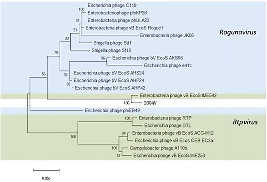
Pacífico et al.                                                                                                                    Natural Coliphages in Clinical Samples

TABLE 1 | Sampling scheme containing the type of biological fluid samples (urine, blood, respiratory tract fluids, and others) obtained during each of the four sampling
rounds (I–IV), in a total of 111 samples analyzed.

Sampling round                Collection date                                                        Type of sample (n)

                                                         Urine           Blood             Respiratory               Others (e.g., abdominal, lung,                 Total
                                                                                           tract fluids             pleural, shoulder and knee fluid,
                                                                                                                       nasal swabs, IV catheter)

Ia                                14/11/17                 12               2                   4                                    5                                23
II                                28/01/18                 12               6                   9                                    0                                27
III                               26/03/18                 11               6                   7                                    8                                32
IV                                22/08/18                 9                7                  13                                    0                                29
Total                                                      44              21                  33                                   13                               111
a The   samples collected during this sampling round were not tested for bacteria.

samples were centrifuged at 10,000 rpm during 5 min and the                              spectra of the phages. Clinical isolates from our own bacterial
upper layer was set aside, followed by a second centrifugation at                        collection and from the clinical samples analyzed in this study
the same conditions. The resulting supernatant was then filtered                         were included in the screening (Table 2). Ten microliters of
and used as described above.                                                             concentrated phage lysates (108 PFU/ml) were spotted onto MSA
                                                                                         plates previously overlaid with 100 µl of stationary phase cell
Statistical Analysis                                                                     suspensions mixed with 3 ml of Molten-soft agar supplemented
Statistical analysis was performed using Fisher’s exact test to                          with 10 mM CaCl2 . The plates were incubated overnight at
investigate the association between the absence/presence of E. coli                      37◦ C. Whenever lysis was observed, the efficiency of plating
in the body fluids analyzed and the absence/presence of lytic                            (EOP) was calculated. EOP was defined as the ratio between
coliphages. The significance level was set to a level of p < 0.05.                       PFU/ml on the sensitive bacteria and the PFU/ml on the
                                                                                         indicator strain.
Preparation of Concentrated Lysates
Bacteriophage suspensions were propagated by re-infection the
                                                                                         Tolerance to Disinfectants
indicator host in triplicate using the purified lysate and the soft                      Phage stability was determined in the presence of 0.25%
agar overlay method. After overnight incubation at 37◦ C, the                            commonly used hospital disinfectants TPH Protect and
soft agar was shredded and 3 ml of media were added. The                                 Hexaquart plus. Phage preparations of 107 PFU/ml were
                                                                                                     R

overlay was collected and centrifuged at 8,000 g during 5 min. The                       incubated at RT and phage titer was determined at 0, 15,
clear supernatant was filtered through a 0.22 µm-pore-size filter                        30, and 60 min by serial dilutions tested on the indicator
and kept at 4◦ C. Ten-fold serial dilutions (10−1 to 10−6 ) were                         strain by means of the soft agar overlay method, as before
prepared by plating 10 µl of the diluted lysate with the indicator                       (Salem and Skurnik, 2018). The phage particles were counted
bacteria. After overnight incubation at 37◦ C, the plaques were                          after overnight incubation at 37◦ C. Assays were performed
counted and the titer was expressed as PFU/ml.                                           in duplicate. All the tested conditions allowed the survival
                                                                                         and normal growth of the host strain, not hindering the
                                                                                         phage infection.
Transmission Electron Microscopy (TEM)
A droplet of the purified phage suspension (107 –108
                                                                                         Bacteriophage DNA Extraction
PFU/ml) was deposited on a copper grid (Science Services,
                                                                                         DNA was isolated using a phenol-chloroform extraction method
Munich, Germany) with carbon-coated Formvar film for
                                                                                         with some modifications. Concentrated phage suspensions
10 min at room temperature and stained with 4% aqueous
                                                                                         were pretreated with DNase I (Quantabio, Beverly, MA,
phosphotungstic acid (Merck, Darmstadt, Germany) at
                                                                                         United States) and RNase A (Sigma-Aldrich, St. Louis,
pH 7. The sample was air-dried overnight and analyzed
                                                                                         MO, United States) in 10 µg/ml final concentration and
with a Zeiss TEM 900 electron microscope (Carl Zeiss,
                                                                                         the mixture was inverted several times and incubated at
Oberkochen, Germany) operated at 50 kV. The phage
                                                                                         37◦ C during 30 min. Furthermore, 20 mg/ml proteinase K
particles were visualized using the Image SP software and
                                                                                         (40 µg) and a final concentration of 0.5% sodium dodecyl
a CCD camera (TRS, Tröndle Restlichtverstärkersysteme,
                                                                                         sulfate (SDS) were added and incubated at 65◦ C for 1 h.
Moorenweis, Germany).
                                                                                         The suspension was cooled to RT and phenol:chloroform
                                                                                         extraction was performed, followed by an ethanol precipitation.
Host Range Analysis and Efficiency of                                                    The precipitated DNA was dissolved in 200 µl ddH2 O.
Plating                                                                                  The DNA concentration was estimated based on the band
The laboratory host bacteria E. coli DSM 12242, ATCC 11303,                              intensity comparison to the 1 kb marker (peqGOLD 1 kb
JM109, W3110, MC1061, and DH5α and other members of                                      DNA-Ladder, PEQLAB Biotechnologie GmbH, Austria).
the Enterobacteriaceae family were used to assess the lytic                              Whenever the DNA concentration proved to be insufficient,

Frontiers in Microbiology | www.frontiersin.org                                      3                                           October 2019 | Volume 10 | Article 2484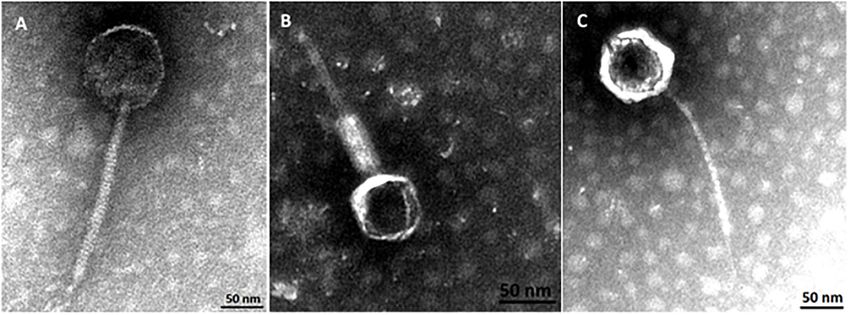
Pacífico et al.                                                                                                                  Natural Coliphages in Clinical Samples

TABLE 2 | Bacterial strains used to characterize the lysis spectra of the phages and respective EOP.

Bacteria                                                                                      Phage group                                           References

                                                                Peduovirus                   Tequintavirus                Tunavirinae
                                                              vB_EcoM-12474V                vB_EcoS-26175V              vB_EcoS-2004IV

Escherichia coli DSM 12242                                       1.000 ± 0.000                1.000 ± 0.000               1.000 ± 0.000             DSMZ
Escherichia coli ATCC 11303                                             –                     0.228 ± 0.048                      –                  ATCC
Escherichia coli W3110 (ATCC 27325)                                     –                     0.840 ± 0.260               0.812 ± 0.080             ATCC
Escherichia coli JM109 (DSM 3423)                                       –                     0.011 ± 0.004               0.145 ± 0.030             DSMZ
Escherichia coli DH5α (DSM 6897)                                        –                     0.195 ± 0.055                      –                  DSMZ
Escherichia coli MC1061 (ATCC 53338)                             0.283 ± 0.150                0.541 ± 0.287               0.509 ± 0.034             ATCC
Klebsiella pneumoniae sub. pneumoniae ATCC 13883                        –                              –                         –                  ATCC
Yersinia enterocolitica sub. palearctica DSM 11502                      –                              –                         –                  DSMZ
Salmonella enterica sub. enterica ATCC 14028                            –                              –                         –                  ATCC
Salmonella typhimurium DT104 isolate H3380                              –                              –                         –                  Clinical (Briggs and
                                                                                                                                                    Fratamico, 1999)
Escherichia coli isolated from samples (n = 18)                         –                              –                         –                  This study
Escherichia coli isolates (n = 20)                                      –                              –                         –                  Clinical
Escherichia coli isolate P3                                             –                              –                         –                  Clinical
Escherichia coli isolate P4                                             –                              –                         –                  Clinical
Escherichia coli isolate P5                                             –                              –                         –                  Clinical
Klebsiella pneumoniae isolate P6                                        –                              –                         –                  Clinical
Klebsiella pneumoniae isolate P7                                        –                              –                         –                  Clinical
Klebsiella pneumoniae isolate P8                                        –                              –                         –                  Clinical
Klebsiella oxytoca isolate P10                                          –                              –                         –                  Clinical

These consisted in E. coli laboratory strains used in phage enumeration and isolation, additional Enterobacteriaceae species, E. coli isolated from the biological fluid
samples and other bacterial isolates collected from patients and outpatients in this clinical setting.

ethanol precipitation was repeated using ammonium acetate                              Bioinformatics and Comparative
at a final concentration of 2.0–2.5 M. The DNA was restricted                          Genome Analysis
with enzyme PvuII (Roche Diagnostics GmbH, Vienna,
                                                                                       The taxonomic distribution was assessed using Kraken v1
Austria) using manufacturer’s protocol and separated on
                                                                                       (Wood and Salzberg, 2014). Phylogenetic analysis of the 43
a 1% agarose gel as an attempt to distinguish different
                                                                                       bacteriophages was re-constructed using VICTOR1 (Meier-
restriction profiles.
                                                                                       Kolthoff et al., 2013) with recommended settings for prokaryotic
                                                                                       viruses (Meier-Kolthoff and Göker, 2017). The presence
Whole Genome Sequencing (WGS)                                                          of antimicrobial resistance determinants such as antibiotic
All library preparation, DNA sequencing and sequence                                   resistance and virulence genes was investigated using the
trimming were performed at MicrobesNG (Birmingham,                                     command-line tool ABRicate v0.8.102 . Genome maps were
United Kingdom). Genomic libraries were prepared using                                 constructed using the web-based tool GenomeVx (Conant and
the Nextera XT Library Prep Kit (Illumina, San Diego, CA,                              Wolfe, 2008). Average nucleotide identity (ANI) was calculated
United States) according to manufacturer’s instructions with                           using OrthoANI3 (Ha et al., 2017). The amino acid sequences
slight modifications. Libraries were sequenced on the Illumina                         of the integrase (Peduovirus), the large subunit terminase
HiSeq using 250 bp paired-ends approach. The trimmed reads                             protein (Tunavirinae) and the DNA polymerase (Tequintavirus)
were analyzed using the INNUca pipeline (Machado et al.,                               were used for phylogenetic analysis. Sequence alignment
2016). Briefly, reads quality were improved using Trimmomatic                          was determined using Clustal Omega (Sievers et al., 2011;
v0.38 (Bolger et al., 2014) and assembly was performed                                 Chojnacki et al., 2017) and trees were reconstructed applying
using Pear v0.9.10 and SPAdes version 3.13 (Bankevich                                  a bootstrapped (500 replications) Maximum Likelihood (ML)
et al., 2012). Resulting assembly was corrected by Pilon v1.18                         analysis. Tree inference used a Nearest-Neighbor-Interchange
and contigs were annotated using Prokka 1.13.3 (Seemann,                               (NNI) as a heuristic method and a Jones-Thaylor-Thornton
2014). Hypothetical proteins were manually annotated by                                (JTT) as substitution model. Bacteriophages were compared with
using blastp against viral databases (Supplementary Data                               complete phage genomes deposited in the NCBI Viruses database
S2–S4). Contigs having a coverage lower than 1/3 of the                                for Peduovirus, Tequintavirus, and subfamily Tunavirinae (April
assembly and smaller than 200 bp were considered as putative
artifacts/contaminations and therefore removed from the                                1
                                                                                         https://ggdc.dsmz.de/victor.php
downstream analysis. Genomes yielding a coverage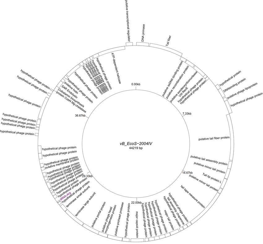
Pacífico et al. Natural Coliphages in Clinical Samples
2019). Accession numbers and ANI% of the closest homologs shared an ANI of 100%. From the 44 proteins predicted, 28
according to MASH4 are given in the Supplementary Data S5. were initially deemed as hypothetical. After carefully inspecting
Evolutionary analysis were carried out using MEGA X v10.0.5 each protein and using blastp searches, only two proteins were
(Kumar et al., 2018) and trees were visualized and edited with unique in our phages and had no homology in the NCBI viral
FigTree v1.4.45 . database. The remaining proteins were annotated or designated
as “hypothetical phage proteins” whenever homology in other
Sequence Accession Numbers phage sequences was found (Figure 2). Phage attachment
Raw sequence data were deposited in the ENA under project sites, attL (CCCGCCCGCTTCATGGGTCGGTTTTAATG) and
accession number PRJEB32459. Assembled genomes and attR (CCCGCCCGCTTCATGGGTCGGTTTTAATG) were also
annotations were directly submitted to NCBI (PRJNA541793). detected in the sequence.
Accession numbers are given in the Supplementary Data S1. Five “T5likevirus” phages were isolated during this study
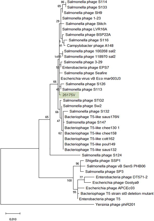
from a urine sample, sharing a 99.93–100% nucleotide sequence
homology among themselves. Phages vB_EcoS-26175 I, II, III, IV,
RESULTS and V have a genome length of 114,646–114,649 bp and between
39.9 and 40.0% GC content. These phages have 158 putative CDS
Phage, Bacteria, and Prevalence in and 31 tRNA genes. Eighty-seven phage-related hypothetical
proteins were found and two have not been described in the
Clinical Samples databases used (Figure 3). The remaining 34 phages were
A total of 111 samples were screened for the presence of lytic
classified as “Tunalikevirus” and had a genome of 44,219 bp, 64–
bacteriophages infecting E. coli DSM 12242 (Table 1). Coliphages
68 CDS and 1 tRNA gene. From these, 38 were phage-related
were detected in 16 samples, representing a prevalence of 14.4%
hypothetical proteins with homologs in other virus genomes in
in the body fluids analyzed. Phages were more commonly
the database and two are unique to these sequences (Figure 4).
found in blood (23.8%), followed by urine (20.5%) and tracheal
The GC content was between 44.4 and 44.5%.
aspirates (6.1%). No coliphages were found in other samples
No bacterial antimicrobial resistance gene, toxin or virulence
analyzed (abdominal, lung, pleural, shoulder and knee fluid, nasal
determinant were found in the phage genomes.
swabs, IV catheters). From 88 samples, 16 were positive for
the presence of E. coli, with bacterial counts ranging between
Peduovirus (“P2likevirus”)
1,65 × 10⁷ and 3,45 × 10¹² CFU/ml. From these 16 samples,
The greatest nucleotide similarity level identified with vB_EcoM-
in 5 of them lytic phages were concomitantly isolated. In fact, a
12474V were phages fiAA91-ss (NC_022750) and pro483
statistically significant association between the absence/presence
(NC_028943), both members of the Peduovirus genus. Similarity
of E. coli and the presence/absence of coliphages in the sample
to bacteriophage P2 (NC_001895), representative of this genus,
was determined (p = 0.016).
was 96.59%. The phylogeny of these phages was further
investigated by comparison of the amino acid sequence of
Bacteriophage Diversity the integrase (367 amino acids) (Figure 5). The Peduovirus
A total of 43 coliphages isolated from 16 human body fluid isolated in this study shares a 97.30% whole genome ANI with
samples were purified, sequenced and analyzed (Table 3). fiAA91-ss (NC_022750). These phages are closely associated with
Three distinct genetic groups of linear double-stranded DNA bacteriophage WPhi (NC_005056), L-413C (NC_004745), and
viruses were identified using both a Genome-BLAST Distance P2 (NC_001895), all officially classified as Peduovirus by the
Phylogeny (GBDP) method and a MASH all-versus-all approach, ICTV. Whole-genome based phylogeny of all Peduovirus clearly
where phage genomes were compared against each other and corroborates this data (Supplementary Figure S1) and adds
grouped based on sequence similarity (Figure 1). This genomic pro147 (NC_028896) and pro483 (NC_028943), also classified
diversity was also in accordance with the output of Kraken, Peduovirus, as closest homologs. In fact, the ANI% of vB_EcoM-
which evidenced the presence of three phage groups belonging 12474V is ≥95% with these seven phages, suggesting they
to the “P2likevirus,” “T5likevirus,” and “Tunalikevirus.” One are representatives of one of these species. An alignment of
representative phage of each genetic group was further selected the Peduovirus genomes shows non-collinearization of phage
for more in-depth analysis. genomes and almost no order conservation of the genetic
blocks. Nonetheless the synteny seems to be maintained
Basic Genome Characteristics and (Supplementary Figure S2).
Phylogenetic Analysis
All assemblies generated one single contig, with the exception of Tequintavirus (“T5likevirus”)
vB_EcoS-26174I (Table 3). The genome of the four “P2likevirus” The Tequintavirus genus belongs to the Siphoviridae family.
phages (vB_EcoM-12474II, III, IV, and V) is between 33,688 All the phages currently listed as part of this genus were
and 33,807 bp long, contains 44 protein-encoding CDS and no extracted from Genbank. Greatest similarity to vB_EcoS-
tRNA genes. The GC content is 50.9%. All four “P2likevirus” 26175V was found to phage vB_Eco_mar003J3 (LR027389),
bacteriophages were isolated from the same blood sample and S126 (MH370376), 3-29 (MK393882), A148 (MG065642), and
118970_sal2 (NC_031933), and Stitch (NC_027297) based on
4
https://github.com/marbl/Mash MASH identity. MASH identified at least 34 phages as closest
5
http://tree.bio.ed.ac.uk/software/figtree/ homologs and from these, 27 having at least 93.11% ANI to
Frontiers in Microbiology | www.frontiersin.org 5 October 2019 | Volume 10 | Article 2484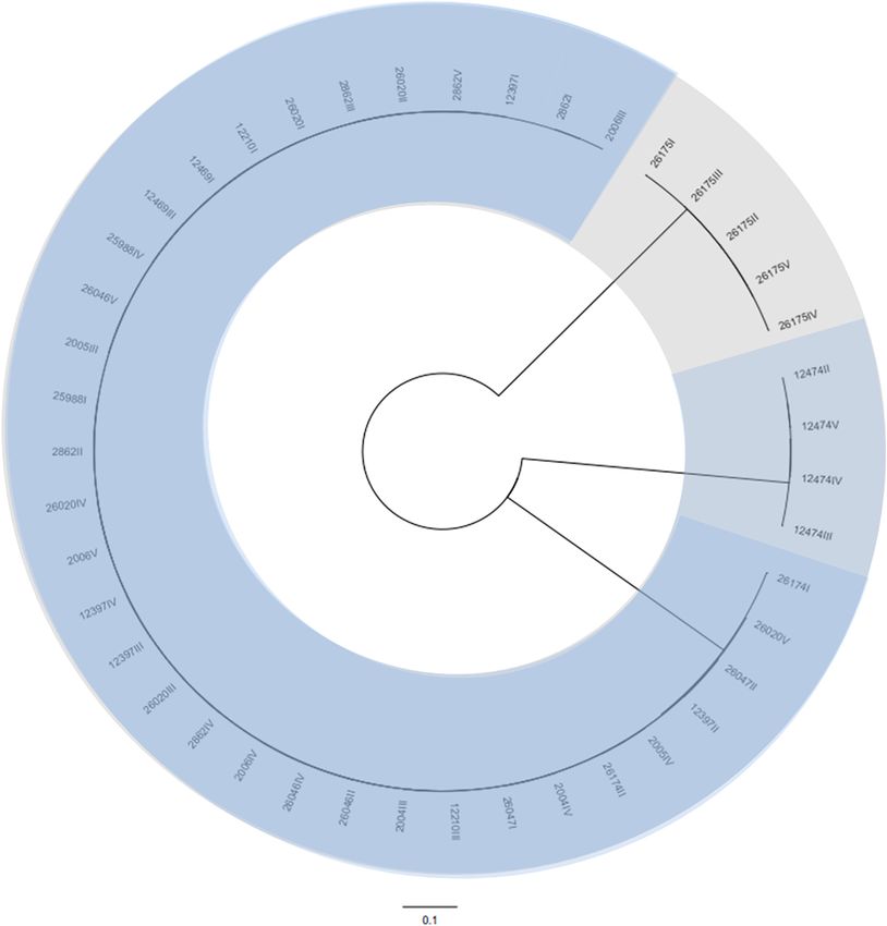
Pacífico et al.                                                                                                                                 Natural Coliphages in Clinical Samples

TABLE 3 | Description of the isolation source, morphology, and sequence characteristics of the 43 bacteriophages sequenced in the course of this study.

Phages analyzed          Sampling round         Isolation        Clinical history and               Morphology                              Genome characteristics
                                                source           microbiological findings

                                                                                                                         Size (Kb)        GC content (%)        CDS (n)     tRNAs (n)

vB_EcoS-2862I,                    II            Tracheal         Klebsiella variicola∗              Siphoviridae                44.2          44.4–44.5          65–66           1
vB_EcoS-2862II,                                 aspirate
vB_EcoS-2862III,
vB_EcoS-2862IV,
vB_EcoS-2862V
vB_EcoS-26020I,                    I            Urine            ∗∗                                 Siphoviridae                44.2          44.4–44.5          65–67           1
vB_EcoS-26020II,
vB_EcoS-26020III,
vB_EcoS-26020IV,
vB_EcoS-26020V
vB_EcoS-12469II,                  III           Blood            Urinary tract infection            Siphoviridae                44.2             44.4             66             1
vB_EcoS-12469III                                                 (E. coli, Bacteroides fragilis)
vB_EcoS-12210I,                   III           Blood            Status febrilis (S. aureus)        Siphoviridae                44.2             44.4            64–65           1
vB_EcoS-12210III
vB_EcoS-2005III,                  II            Urine            ∗∗                                 Siphoviridae                44.2             44.4             66             1
vB_EcoS-2005IV
vB_EcoS-26046II,                   I            Urine            ∗∗                                 Siphoviridae                44.2             44.4             66             1
vB_EcoS-26046III,
vB_EcoS-26046IV,
vB_EcoS-26046V
vB_EcoS-2006III,                  II            Urine            ∗∗                                 Siphoviridae                44.2             44.4             66             1
vB_EcoS-2006IV,
vB_EcoS-2006V
vB_EcoM-12474II,                  III           Blood            Urinary tract infection            Myoviridae           33.7–33.8               50.9             44             0
vB_EcoM-12474III,                                                (E. coli)
vB_EcoM-12474IV,
vB_EcoM-12474V
vB_EcoS-2004III,                  II            Urine            ∗∗                                 Siphoviridae                44.2             44.4             66             1
vB_EcoS-2004IV
vB_EcoS-25988I,                    I            Urine            ∗∗                                 Siphoviridae                44.2             44.4            65–66           1
vB_EcoS-25988IV
vB_EcoS-26174I,                    I            Urine            ∗∗                                 Siphoviridae                44.2             44.4            65–66           1
vB_EcoS-26174II
vB_EcoS-26175I,                    I            Urine            ∗∗                                 Siphoviridae            114.6             39.9–40.0           158           31
vB_EcoS-26175II,
vB_EcoS-26175III,
vB_EcoS-26175IV,
vB_EcoS-26175V
vB_EcoS-26047I,                    I            Urine            ∗∗                                 Siphoviridae                44.2             44.4            65–66           1
vB_EcoS-26047II
vB_EcoS-12397I,                   III           Tracheal         Oropharyngeal flora and            Siphoviridae                44.2             44.4            65–66           1
vB_EcoS-12397II,                                aspirate         Candida tropicalis ∗
vB_EcoS-12397III,
vB_EcoS-12397IV
∗ routine investigation of respiratory tract fluids, clinical history most likely not because of infectious disease;   ∗∗ all   urine samples were not tested for bacterial culture but
for Legionella or pneumococci-antigen only.

our phages (Supplementary Figure S3). Nucleotide similarity                                    the name Escherichia phage vB_EcoS-26175I, vB_EcoS-26175II,
to phage T5 (NC_005859) was only 82.79% and therefore it                                       vB_EcoS-26175III, vB_EcoS-26175IV, and vB_EcoS-26175V and
is not the same species (%ANI ≥ 95). ANI with 118970_sal2                                      their gene content was compared with the closest homologs
and Stitch was 95.08 and 95.45% respectively, but the closest                                  (Supplementary Figure S4).
nucleotide identity was 95.90% with phage 3–29. The phylogeny
of these phages was further investigated by comparison of                                      Tunavirinae
the amino acid sequence of the DNA polymerase (1216                                            Thirty-four identical phages from 14 samples were identified
amino acids), as previously (Sváb et al., 2018; Michniewski                                    in this study as putative “Tunalikevirus” according to Kraken,
et al., 2019; Figure 6). Phages S126 and S113 (MH370366)                                       but whole-genome sequence analysis puts them closer to the
appear closer to 26175V. The phages were deposited under                                       members of the Rtpvirus genus (Supplementary Figure S5).

Frontiers in Microbiology | www.frontiersin.org                                            6                                                  October 2019 | Volume 10 | Article 2484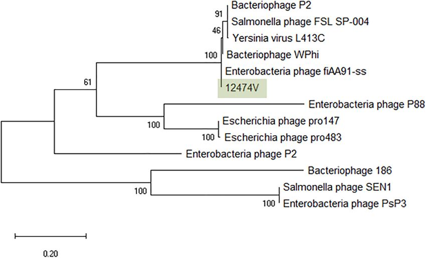
Pacífico et al.                                                                                                           Natural Coliphages in Clinical Samples

  FIGURE 1 | Phylogenetic tree of the 43 bacteriophages isolated during this study using VICTOR (Supplementary Data S1). The scale represents homology % and
  yields an average support of 33%. The number of clusters determined were three, representing the genetic groups here described – Peduovirus (mild blue),
  Tequintavirus (gray) and Tunavirinae (blue).

To classify the phages identified in this study, the amino                        study form a clade closer to the Rogunavirus genus. Phage
acid sequence of the large subunit terminase protein (534                         vB_EcoS_IME542 is not classified by the ICTV but rather
amino acids) was chosen (Kropinski et al., 2015; Michniewski                      shows up in the NCBI database as an Rtpvirus member.
et al., 2019) and compared with the same sequence of 20                           In regards to ANI, the most similar phage genomes at the
Tunavirinae members of both Rtpvirus and Rogunavirus genera                       nucleotide level are the Rtpvirus phages A110b (MG065688),
identified as closest homologs using MASH, all having an                          followed by the vB_Ecos_CEB_EC3a (KY398841), vB_EcoS-
ANI% ≥ 66.94 (Figure 7). According to the amino acid sequence                     IME253 (KX130960), vB_EcoS_ACG-M12 (NC_019404), DTL
of the terminase large subunit protein, both vB_EcoS_IME542                       (MG050172), RTP (NC_007603), and vB_EcoS_IME542. When
(MK372342) and the new Tunavirinae phages isolated in this                        looking at the amino acid sequence of the large subunit terminase,

Frontiers in Microbiology | www.frontiersin.org                               7                                         October 2019 | Volume 10 | Article 2484
Pacífico et al. Natural Coliphages in Clinical Samples
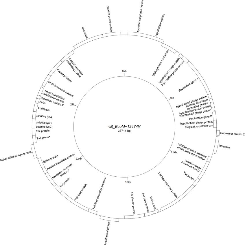
FIGURE 2 | Genome map of the representative Peduovirus phage vB_EcoM-12474V (MK907239) isolated during this study.
both vB_EcoS_IME542 and the newly isolated phage vB_EcoS- and vB_EcoS-2004IV show phages with icosaheadral heads and
2004IV are closer to Rogunavirus. Both share a 96.37% amino acid long tails (Figure 8). Phage vB_EcoM-12474V belongs to the
identity at the level of the large subunit terminase. Whole genome Myoviridae family and has a head of about 62 nm and a 155 nm
analysis and ANI% places them both closer to the Rtpvirus. The contractile tail. Phages vB_EcoS-2004IV and vB_EcoS-26175V
most similar phage is A110b, a non-classified phage also closer are both siphoviruses and have long non-contractile tails of 232
to the Rtpvirus genus according to whole-genome phylogeny and 186 nm, respectively. Phage heads have an approximate
reconstruction, with an 87.69% ANI in common with the new diameter of 100 and 85 nm.
phages, the highest value obtained when scanning the database The lytic capacity of phages from the different groups was
(Supplementary Figure S6). investigated using enterobacterial collection strains, clinical
isolates and E. coli isolated from the samples (Table 2).
Bacteriophage Characterization Peduovirus vB_EcoM-12474V, Tequintavirus vB_EcoS-26175V,
Phylogenetic analysis corroborates that three groups of and Tunavirinae vB_EcoS-2004IV showed different lysis
phages where identified in this study. Transmission electron patterns. None of the isolated phages lysed the bacteria isolated
micrographs from phages vB_EcoM-12474V, vB_EcoS-26175V, from the blood, urine and tracheal aspirates samples or other
Frontiers in Microbiology | www.frontiersin.org 8 October 2019 | Volume 10 | Article 2484Pacífico et al. Natural Coliphages in Clinical Samples FIGURE 3 | Genome map of the representative Tequintavirus phage vB_EcoS-26175V (MK907271) isolated during this study. clinical isolates previously collected in the hospital setting. the bacteria already acquired resistance to infection (in the case Whilst vB_EcoS-26175V could infect the six laboratory strains of the bacteria from the native sample). Bioinformatic searches tested, vB_EcoS-2004IV could only infect DSM 12242, JM109, in Enterobacteriaceae genomes deposited in the NCBI database W3110, and MC1061. The narrower host range was obtained revealed a 98–99% sequence identity of the Peduovirus isolated for vB_EcoM-12474V, which only infected two lab strains. This during this study with six E. coli isolates: STEC O145:H28 strain might indicate that the phages are either strain-specific or that 95-3192 (CP027362), non-O157 STEC FHI82 (LM996779), Frontiers in Microbiology | www.frontiersin.org 9 October 2019 | Volume 10 | Article 2484
Pacífico et al.                                                                                                          Natural Coliphages in Clinical Samples

  FIGURE 4 | Genome map of the representative Tunavirinae phage vB_EcoS-2004IV (MK907241) isolated during this study.

STEC O145:H28 strain RM12581 (CP007136), STEC O145:H28                            DISCUSSION
strain RM13514 (CP006027), STEC O145:H28 strain 2015C-
3125 (CP027763), and STEC O145 strain RM9872 (CP028379).                          In this study, we were able to successfully isolate natural-
Tequintavirus vB_EcoS-26175V and Tunavirinae phage                                occurring phages from different human sources and further
vB_EcoS-2004IV were not detected in previously sequenced                          characterize the phage community found not only at the
bacteria deposited in the database. The phage stability in the                    morphological level, but also at the nucleotide level. Coliphages
presence of the disinfectants TPH Protect and Hexaquart plus         R
                                                                                  that were able to infect the host strain E. coli DSM 12242 were
was investigated (Figure 9). A concentration as low as 0.25%                      present in 14.4% of the samples analyzed in this study. This
TPH Protect was sufficient to inactivate all phage particles of                   prevalence is below what was reported in a previous study that
the three phage groups described, after 15 min. After incubation                  surveyed the presence of phages, mainly siphoviruses in human
with Hexaquart plus for 60 min Peduovirus vB_EcoM-12474V
                     R
                                                                                  blood, ascitic fluid, urine, cerebrospinal fluid, and serum (>45%
and Tequintavirus vB_EcoS-26175V virions could still be                           of abdominal fluid and urine) (Brown-Jaque et al., 2016). The
isolated. Tunavirinae vB_EcoS-2004IV could no longer be                           successful isolation of virions from clinical samples mostly relies
detected after 30 min.                                                            on the use of large volumes of human body fluid samples. Despite

Frontiers in Microbiology | www.frontiersin.org                              10                                         October 2019 | Volume 10 | Article 2484
Pacífico et al. Natural Coliphages in Clinical Samples FIGURE 5 | Phylogenetic tree of Peduovirus phage based on the Clustal Omega alignment of the integrase protein sequence of the representative phage vB_EcoM-12474V (MK907239). The percentage of trees in which the associated taxa clustered together is shown next to the branches. The list of genomes included and GenBank accession numbers are described in Supplementary Data S5. the high level of sensitivity of E. coli DSM 12242 to detect Studies on the phage distribution in the human body are bacteriophages, as previously shown (International Organization quite scarce, especially in a clinical context (Brown-Jaque for Standardization, 2000; Shousha et al., 2015; Hilbert et al., et al., 2016; Navarro and Muniesa, 2017). The vast majority of 2017), we assume that some of the coliphages present in the studies available are mainly focused on the human gut (Górski samples analyzed might have been missed due to a narrow and Weber-Dabrowska, 2005; Górski et al., 2006; Manrique host range (e.g., strain specific) and to the low phage densities et al., 2017; Ma et al., 2018) and feces (Havelaar et al., 1986; despite the use of liquid cultures as an enrichment method. Cornax et al., 1994; Furuse et al., 2019), with other studies Additionally, some of the phages that are able to infect the strain describing phage colonization of the mouth (Ly et al., 2014; used may have reduced plating efficiency during the first passage Naidu et al., 2014) and respiratory tract (Willner et al., 2009; (Weinbauer, 2004). Willner and Furlan, 2010). Human body sites are not as sterile as previously thought Phages were previously expected to be found everywhere their and data from phage diversity studies showed that this diversity bacterial host is present, but recent studies suggest that bacterial tends to be very different between ecological niches. The amount abundance does not necessarily predict the relative abundance of uncharacterized, unknown, and abundant phage populations of their phages (Ly et al., 2014; Naidu et al., 2014; Brown-Jaque described in the late years in metagenomics studies highlighted et al., 2016). The observation of phages at body sites where the the emergent need to characterize bacteriophages and disclose bacterial host was not detected gives strength to the possibility their role in human health and disease (Hyman and Abedon, of phage translocation through the human body (Górski et al., 2012). Previous work conducted in our lab highlighted the 2006; Brown-Jaque et al., 2016; Tetz and Tetz, 2018). As a matter important role of phages in the transfer of antimicrobial of fact, in this study coliphages and E. coli were only found resistance genes through transduction mechanisms in other concomitantly in five samples. In 11 other samples, phages were environments (Shousha et al., 2015; Hilbert et al., 2017). We found despite the non-detection of the bacterial host. chose a culture-based method in order to be able to study Phage genomes are characterized by a rapid evolutionary transduction instead of a metagenomics approach that would rate, horizontal gene transfer events and recombination between only provide information about genes but not the mechanism and different viral genes, which makes the inference of phylogenetic ability of transfer. Moreover, the majority of the metagenomics relationships more reliable when considering individual genes studies cannot classify phages due to lack of homolog sequences or modules instead of whole genomes (Zois et al., 2011). This in the databases and the so-called “viral dark matter” remains in work relied on whole-genome analysis (VICTOR), gene content obscure (Breitbart et al., 2003; Willner et al., 2009; Pride et al., (multi-sequence alignment) and single-gene analysis (signature 2012; Dinakaran et al., 2014; Dutilh et al., 2014; Ma et al., 2018). genes with no signs of recombination) to better disclose the Frontiers in Microbiology | www.frontiersin.org 11 October 2019 | Volume 10 | Article 2484
Pacífico et al.                                                                                                              Natural Coliphages in Clinical Samples

  FIGURE 6 | Phylogenetic tree of Tequintavirus phage based on the Clustal Omega alignment of the DNA polymerase I protein sequence of the representative phage
  vB_EcoS-26175V (MK907271). The percentage of trees in which the associated taxa clustered together is shown next to the branches. The list of genomes included
  and GenBank accession numbers are described in Supplementary Data S5.

Frontiers in Microbiology | www.frontiersin.org                                12                                          October 2019 | Volume 10 | Article 2484
Pacífico et al.                                                                                                               Natural Coliphages in Clinical Samples

  FIGURE 7 | Phylogenetic tree of Tunavirinae phage based on the Clustal Omega alignment of the terminase large subunit protein sequence of the representative
  phage vB_EcoS-2004IV (MK907241). Phages currently classified as Rogunavirus and as Rtpvirus are highlighted in blue and green, respectively. The percentage of
  trees in which the associated taxa clustered together is shown next to the branches. The list of genomes included and GenBank accession numbers are described
  in Supplementary Data S5.

  FIGURE 8 | Virion morphology of phages, (A) vB_EcoS-2004IV (Siphoviridae), (B) vB_EcoM-12474V (Myoviridae), and (C) vB_EcoS-26175V (Siphoviridae).

phylogenetic relationship between these phages and other phages                      study. Phages belonging to the Peduovirus, Tequintavirus, and
from the viral databases. We were able to successfully isolate                       Tunavirinae subfamily were present in 16 samples. Interestingly,
and characterize 43 phages belonging to three different species                      Peduovirus and Tequintavirus were isolated from a single sample,
(Figure 2). There was a surprising low level of diversity observed,                  whilst the Tunavirinae were found across several samples. All of
regarding the taxonomical distribution of phages isolated in our                     the phages were very similar, reflecting a low level of intra-sample

Frontiers in Microbiology | www.frontiersin.org                                 13                                          October 2019 | Volume 10 | Article 2484
Pacífico et al.                                                                                                      Natural Coliphages in Clinical Samples

  FIGURE 9 | Disinfectant stability of the phages vB_EcoM-12474V (MK907239), vB_EcoS-26175V (MK907271) and vB_EcoS-2004IV (MK907241) in the presence
  of 0.25% (A) TPH Protect and (B) Hexaquart R plus.

diversity. The same can be said about the inter-sample diversity               the ICTV. Peduovirus vB_EcoM-12474V shared a 97.30% ANI
observed for the Tunavirinae, which was in fact unexpected.                    with phage fiAA91-ss, a recognized member of the Peduovirus
This low level of diversity might be mainly due to the ability                 genus previously isolated from an E. coli O157:H7 strain
of specific phages to survive and multiply in the biological                   from urban raw wastewater (Allué-Guardia et al., 2013). The
materials tested given the challenges that they present to the                 sequence characteristics are also in accordance with the phage
phage stability, such as e.g., high and/or low pH conditions                   genomes already described for members of this genus, which
(Weinbauer, 2004). Moreover, enteric bacteria and coliphage in                 range between 29.5 and 40.6 Kb and have 40–60 CDS. The
pure urine were found to be rapidly inactivated in stored urine,               classification of these phages as members of the Peduovirus
being highly dependent on its pH (Chandran and Pradhan, 2009).                 genus is also supported by the presence of signature genes as
The most similar phage genomes in the NCBI Viruses database                    the regulatory region containing the integrase gene int, the
was compared with the representative of each phage genome                      lytic-lysogenic transcriptional switch genes cox (lytic repressor
sequenced in this study (Supplementary Figures S2, S4, S6).                    gene) and C (immunity repressor gene), and the P2 type of late
Annotation of the features of the phage nucleotide sequences                   genes – capsid scaffold gene O, major capsid precursor gene N,
showed that these are conserved throughout the genomes, with                   small terminase subunit gene M and capsid completion gene
only some genomic rearrangements occurring, according to                       L (Supplementary Data S2). These genes control the central
the different modules that characterize each species analyzed.                 mechanism of integration/excision in the host genome and
The vast majority of the phage sequences were structural                       the life cycle after infection. The major difference between the
proteins, with a part of hypothetical proteins, which were                     vB_EcoM-12474 phages and fiAA91-ss relies on the absence of
further investigated using blastp searches in the viral database               the cytolethal toxin genes (Allué-Guardia et al., 2013). Altogether,
for homology in other phage species. This strategy allowed                     this suggests that the four phages are indeed part of the
the association of the majority of the hypothetical proteins as                Peduovirus genus, within a previously existing species. Previous
phage-related. Moreover, no features associated with antibiotic                works attempted to detect the presence of natural phages in
resistance or virulence were identified in any of the genomes.                 human or animal blood without success (Gaidelyte et al., 2007;
   When testing these phages for the host range, they could                    Brown-Jaque et al., 2016; Navarro and Muniesa, 2017). They
only infect E. coli laboratory strains DSM 12242, ATCC 11303,                  are thought to be inherently absent in the blood of healthy
JM109, W3110, MC1061, and DH5α, having no activity against                     individuals and are not expected to occur naturally. In fact, to
other Enterobacteriaceae or other E. coli clinical isolates tested             the best of our knowledge, the only report describing virulent
(Table 2). These observations highlight the importance of using                phage isolation from native blood was in septicemic patients
an adequate host strain for phage isolation, as the use of                     (Gaidelyte et al., 2007). The associated bacterial host DNA is
different E. coli strains to analyze the same set of samples                   usually co-detected, which may suggest the translocation of
has previously shown to yield different Siphoviridae coliphages                bacteria harboring phage particles or lysogenized prophages
(Chibani-Chennoufi et al., 2004).                                              into the bloodstream (Huh et al., 2019). Phage titers were also
   The genus Peduovirus, together with Hpunavirus, compose                     previously shown to increase with the severity of the symptoms.
the Peduovirinae subfamily, family Myoviridae according to                     In patients with leukemic diseases, an increase in coliphage titer

Frontiers in Microbiology | www.frontiersin.org                           14                                       October 2019 | Volume 10 | Article 2484
Pacífico et al.                                                                                              Natural Coliphages in Clinical Samples

and virulent phages (including T-phages) was previously reported          Tlsvirus, Tunavirus, and Webervirus [plus a non-classified species
(Furuse et al., 2019). In the case of the Peduoviruses isolated           Cronobacter virus Esp2949-1 (NC_019509)]. Members of this
in this study, the temperate lifestyle and the co-detection of            subfamily are between 45 and 52 kb and have between 45
bacteria suggests a case of lysogenized prophages released by the         and 94 CDS. The highly abundant Tunavirinae isolated across
E. coli found in the bloodstream. Moreover, these phages can be           our samples are confidently part of this subfamily, and highest
found as prophage sequences of Shiga toxin-producing E. coli              homology was found within the Rtpvirus. However, analysis
(STEC) strains when searching the NCBI bacterial database.                of the large subunit terminase suggests that they are closer to
Typically, Peduovirus are temperate phage-infecting members               the Rogunavirus. The closest related phages were all phages
of the γ-proteobacteria and tend to be similar when infecting             isolated in other Enterobacteriaceae such as Yersinia sp. and
the same host (Zois et al., 2011). The absence of bacteriophages          Salmonella sp. However, the Tunavirinae phage vB_EcoS-2004IV
in the blood of healthy individuals was also observed when                appears to be very similar to a campylobacter phage, which
compared with the virome of patients with HIV/AIDS (Li                    unfortunately is still deemed as unverified, which would suggest
et al., 2012). Other two blood samples contained siphoviruses,            an unusual level of promiscuity between phages that infect
with an apparent lytic lifestyle, which might have reached the            different host families. The closest homolog shares an 87.69%
bloodstream through translocation from other body sites. No               ANI, which is not enough to place the phages within a pre-
evidence of lysogeny was found in the phage genomes, suggesting           existent genus, but enough to classify it as a member of the
that these are lytic phages.                                              Tunavirinae family. Currently the ANI for the demarcation
    Data on the presence of bacteriophages in human urine is              of a new species is at 95% according to the ICTV criteria.
scarce (Caldwell, 1928; Thannesberger et al., 2017; Santiago-             Nonetheless, recent studies suggest that a 97% would provide
Rodriguez, 2018). It was for long assumed as sterile until the            better accuracy for Tequintavirus, Rb69virus and Seuratvirus
urethra level and the presence of microorganisms was normally             (Sazinas et al., 2018; Michniewski et al., 2019). In this case, the
associated with disease. This idea changed with the use of NGS            ANI criteria set to 97% would mean that also the Tequintavirus
that strongly support the presence of bacterial communities               phage isolated in our study would also be a new species
also in healthy individuals. The urine viral community was                within this genus. Phage vB_EcoS-26175V, despite containing a
previously shown to be quite robust, comprising of about 107              vast abundance of structural proteins, also contained proteins
virus-like particles (VLPs), both in healthy subjects and patients        associated with RNA metabolism (JOHFDMOO_00089) and
harboring an urinary tract infection (Santiago-Rodriguez et al.,          protein degradation (JOHFDMOO_00009).
2015). These urinary viral communities are mainly composed by                By surviving disinfection, these phages could be widespread
phages and Human Papillomaviruses (HPV). Phages in human                  around the clinical setting and in close proximity with the
urine are thought to be mostly lysogenic, infecting bacteria from         environment of the patients. The survival of phages in this
a wide range of phyla, such as Proteobacteria, Bacteroidetes,             environment might have deleterious consequences on the
Firmicutes and Verrucomicrobia (Santiago-Rodriguez et al.,                outcome of infectious diseases, by mediating the acquisition
2015; Santiago-Rodriguez, 2018). This same idea of sterility              and dissemination of antimicrobial resistance, as observed
was assumed for the respiratory tract. The respiratory tract              in other settings (Colomer-Lluch et al., 2011, 2014; Fard
of healthy people is believed to be poorly colonized by                   et al., 2011; Shousha et al., 2015; Hilbert et al., 2017). The
microorganisms, thus suggesting that no significant phage                 phage stability in the presence of the disinfectants TPH
population is established. However, samples from the sputum               Protect and Hexaquart plus was investigated, given their
                                                                                                    R

of cystic fibrosis patients revealed a core of cystic fibrosis-           common use as hospital disinfectants. In particular, TPH
associated phage, encoding a plethora of virulence factors                Protect is the disinfectant used in the hospital where the
as adhesins, biofilm-formation and quorum-sensing genes. In               samples were collected, and is used in a final concentration
contrast, phages collected from the sputum of healthy individuals         of 0.5% (v/v). None of the phages survived contact with
were highly diverse and most likely resembled environmental               half that concentration. However, upon exposure to 0.25%
communities (Willner et al., 2009; Willner and Furlan, 2010).             (v/v) Hexaquart plus, Peduovirus vB_EcoM-12474V and
                                                                                            R

All the phages detected in urine and sputum during this                   Tequintavirus vB_EcoS-26175V could still be detected after
study show a lack of integrative elements and seem to have a              60 min (Figure 9).
lytic lifestyle.                                                             The phages isolated during the course of this study
    Dominant phages that persist at an increased number in                and genome comparison analysis provided new insights
individuals through an extended period of time showed no                  regarding the phages that inhabit and colonize human
significant genome divergence or mutation, suggesting indeed              body surfaces and revealed the presence of at least one
a high genome stability of phage genomes, maybe due to                    new taxonomic group. Peduovirus vB_EcoM-12474V is
the absence of substantial selective pressure acting on either            a pre-existent species, Tequintavirus vB_EcoS-26175V
bacteria or phages (Reyes et al., 2010). Possibly, this might             might be a new species (depending on the ANI cut-off
explain why almost no inter-sample variability was found, and             considered) and Tunavirinae vB_EcoS-2004IV represents
why the Tunavirinae group described here was widespread                   a novel species. TEM also provided morphological
in almost all patients. The subfamily Tunavirinae belongs to              evidence that supports the inclusion of the phages of
the Siphoviridae family and has currently 8 genera classified:            this study in the above mentioned taxonomic groups
Eclunavirus, Hanrivervirus, Rogunavirus, Rtpvirus, Sertoctavirus,         (Figure 9). Instead of characterizing the whole population

Frontiers in Microbiology | www.frontiersin.org                      15                                     October 2019 | Volume 10 | Article 2484
Pacífico et al. Natural Coliphages in Clinical Samples
through metagenomics, this approach allowed the isolation and FUNDING
purification of bacteriophages. This work is a very important
contribution and a meaningful attempt in the disclosure of This work was in part supported by the NFB
the key players behind the viral dark matter observed, their (Niederoesterreichische Forschungs- und Bildungsgesellschaft)
interaction with bacteria found on site and their possible Grant No. LS14-006.
involvement in the health status of the individuals.
ACKNOWLEDGMENTS
DATA AVAILABILITY STATEMENT
We are thankful to the bioinformatics staff of the Molecular
The datasets generated for this study were deposited under the Microbiology and Infection Unit at the Instituto de Medicina
accession numbers PRJEB32459 and PRJNA541793. Molecular of the University of Lisbon for the assistance provided
and to João Costa-Nunes (Sechenova University, Moscow) and
João Viana da Silva for critically reviewing and correcting the
AUTHOR CONTRIBUTIONS manuscript. We are very grateful to Franz Trautinger for his
expert opinion as chair of the ethics commission of the Karl
FH acquired the funding and administered the project. FH and Landsteiner University on the sampling procedure.
CP conceived and designed the experiments. I-JP, CA, and CP
provided the data and collected the samples. CP, MH, and DS
performed the experiments. FH, CP, MH, ND, I-JP, CA, JC, and SUPPLEMENTARY MATERIAL
DS contributed with reagents, materials, and analysis tools. ND
and MH performed transmission electronic microscopy analysis. The Supplementary Material for this article can be found
CP prepared the original draft and wrote the manuscript. FH, CA, online at: https://www.frontiersin.org/articles/10.3389/fmicb.
I-JP, JC, and CP reviewed and edited the manuscript. 2019.02484/full#supplementary-material
REFERENCES Chibani-Chennoufi, S., Sidoti, J., Bruttin, A., Dillmann, M. L., Kutter, E., Qadri,
F., et al. (2004). Isolation of Escherichia coli bacteriophages from the stool of
Allocati, N., Masulli, M., Alexeyev, M. F., and Di Ilio, C. (2013). Escherichia coli pediatric diarrhea patients in Bangladesh. J. Bacteriol. 186, 8287–8294. doi:
in Europe: an overview. Int. J. Environ. Res. Public Health 10, 6235–6254. 10.1128/JB.186.24.8287-8294.2004
doi: 10.3390/ijerph10126235 Chojnacki, S., Cowley, A., Lee, J., Foix, A., and Lopez, R. (2017). Programmatic
Allué-Guardia, A., Imamovic, L., and Muniesa, M. (2013). Evolution of a self- access to bioinformatics tools from EMBL-EBI update: 2017. Nucleic Acids Res.
inducible cytolethal distending toxin type V- encoding bacteriophage from 45, 550–553. doi: 10.1093/nar/gkx273
Escherichia coli O157:H7 to Shigella sonnei. J. Virol. 87, 13665–13675. doi: Colomer-Lluch, M., Jofre, J., and Muniesa, M. (2011). Antibiotic resistance genes in
10.1128/JVI.02860-2813 the bacteriophage DNA fraction of environmental samples. PLoS One 6:e17549.
Bankevich, A., Nurk, S., Antipov, D., Gurevich, A. A., Dvorkin, M., Kulikov, A. S., doi: 10.1371/journal.pone.0017549
et al. (2012). SPAdes: a new genome assembly algorithm and its applications Colomer-Lluch, M., Jofre, J., and Muniesa, M. (2014). Quinolone resistance genes
to single-cell sequencing. J. Comput. Biol. 19, 455–477. doi: 10.1089/cmb.2012. (qnrA and qnrS) in bacteriophage particles from wastewater samples and the
0021 effect of inducing agents on packaged antibiotic resistance genes. J. Antimicrob.
Bibi, Z., Abbas, Z., and Rehman, S. (2016). The phage P. E1 isolated from hospital Chemother. 69, 1265–1274. doi: 10.1093/jac/dkt528
sewage reduces the growth of Escherichia coli. Biocontrol Sci. Technol. 26, Conant, G. C., and Wolfe, K. H. (2008). GenomeVx: simple web-based creation of
181–188. doi: 10.1080/09583157.2015.1086311 editable circular chromosome maps. Bioinformatics 24, 861–862. doi: 10.1093/
Bolger, A. M., Lohse, M., and Usadel, B. (2014). Trimmomatic: a flexible bioinformatics/btm598
trimmer for illumina sequence data. Bioinformatics 30, 2114–2120. doi: 10.1093/ Cornax, R., Morinigo, M. A., Gonzalez-jaen, F., Alonso, M. C., and Borrego,
bioinformatics/btu170 J. J. (1994). Bacteriophages presence in human faeces of healthy subjects and
Breitbart, M., Hewson, I., Felts, B., Mahaffy, J. M., Nulton, J., Salamon, P., et al. patients with gastrointestinal disturbances. Zentralbl Bakteriol 224, 214–224.
(2003). Metagenomic analyses of an uncultured viral community from human doi: 10.1016/S0934-8840(11)80572-80574
feces. J. Bacteriol. 185, 6220–6223. doi: 10.1128/JB.185.20.6220 Desiere, F., Mcshan, W. M., Sinderen, D., Van, Ferretti, J. J., and Brüssow,
Briggs, C. E., and Fratamico, P. M. (1999). Molecular characterization of H. (2001). Comparative genomics reveals close genetic relationships between
an antibiotic resistance gene cluster of Salmonella typhimurium DT104. phages from dairy bacteria and pathogenic streptococci: evolutionary
Antimicrob. Agents Chemother. 43, 846–849. implications for prophage-host interactions. Virology 341, 325–341. doi: 10.
Brown-Jaque, M., Muniesa, M., and Navarro, F. (2016). Bacteriophages in clinical 1006/viro.2001.1085
samples can interfere with microbiological diagnostic tools. Sci. Rep. 6:33000. Dinakaran, V., Rathinavel, A., and Pushpanathan, M. (2014). Elevated levels of
doi: 10.1038/srep33000 circulating DNA in cardiovascular disease patients: metagenomic profiling of
Brussow, H., Canchaya, C., and Hardt, W.-D. (2004). Phages and the evolution microbiome in the circulation. PLoS One 9:e0105221. doi: 10.1371/journal.
of bacterial pathogens: from genomic rearrangements to lysogenic conversion. pone.0105221
Microbiol. Mol. Biol. Rev. 68, 560–602. doi: 10.1128/MMBR.68.3.560 Dutilh, B. E., Cassman, N., Mcnair, K., Sanchez, S. E., Silva, G. G. Z., Boling, L.,
Caldwell, J. A. (1928). Bacteriologic and bacteriophagic infected urines. J. Infect. et al. (2014). A highly abundant bacteriophage discovered in the unknown
Dis. 43, 353–362. doi: 10.1093/infdis/43.4.353 sequences of human faecal metagenomes. Nat. Commun. 5, 1–11. doi: 10.1038/
Chandran, A., and Pradhan, S. K. (2009). Survival of enteric bacteria and coliphage ncomms5498
MS2 in pure human urine. J. Appl. Microbiol. 107, 1651–1657. doi: 10.1111/j. European Centre for Disease Prevention and Control (2016). Surveillance of
1365-2672.2009.04353.x antimicrobial resistance in Europe 2016. Annual Report of the European
Frontiers in Microbiology | www.frontiersin.org 16 October 2019 | Volume 10 | Article 2484You can also read