Proliferation zones in the axolotl brain and regeneration of the telencephalon
←
→
Page content transcription
If your browser does not render page correctly, please read the page content below
Maden et al. Neural Development 2013, 8:1
http://www.neuraldevelopment.com/content/8/1/1

 RESEARCH ARTICLE                                                                                                                                Open Access

Proliferation zones in the axolotl brain and
regeneration of the telencephalon
Malcolm Maden1*, Laurie A Manwell2,3 and Brandi K Ormerod2

  Abstract
  Background: Although the brains of lower vertebrates are known to exhibit somewhat limited regeneration after
  incisional or stab wounds, the Urodele brain exhibits extensive regeneration after massive tissue removal.
  Discovering whether and how neural progenitor cells that reside in the ventricular zones of Urodeles proliferate to
  mediate tissue repair in response to injury may produce novel leads for regenerative strategies. Here we show that
  endogenous neural progenitor cells resident to the ventricular zone of Urodeles spontaneously proliferate,
  producing progeny that migrate throughout the telencephalon before terminally differentiating into neurons. These
  progenitor cells appear to be responsible for telencephalon regeneration after tissue removal and their activity may
  be up-regulated by injury through an olfactory cue.
  Results: There is extensive proliferation of endogenous neural progenitor cells throughout the ventricular zone of
  the adult axolotl brain. The highest levels are observed in the telencephalon, especially the dorsolateral aspect, and
  cerebellum. Lower levels are observed in the mesencephalon and rhombencephalon. New cells produced in the
  ventricular zone migrate laterally, dorsally and ventrally into the surrounding neuronal layer. After migrating from
  the ventricular zone, the new cells primarily express markers of neuronal differentiative fates. Large-scale
  telencephalic tissue removal stimulates progenitor cell proliferation in the ventricular zone of the damaged region,
  followed by proliferation in the tissue that surrounds the healing edges of the wound until the telencephalon has
  completed regeneration. The proliferative stimulus appears to reside in the olfactory system, because telencephalic
  regeneration does not occur in the brains of olfactory bulbectomized animals in which the damaged neural tissue
  simply heals over.
  Conclusion: There is a continual generation of neuronal cells from neural progenitor cells located within the
  ventricular zone of the axolotl brain. Variable rates of proliferation were detected across brain regions. These neural
  progenitor cells appear to mediate telencephalic tissue regeneration through an injury-induced olfactory cue.
  Identification of this cue is our future goal.
  Keywords: Axolotl, Brain regeneration, DCX, GFAP, NeuN, Neural precursor cells, Telencephalon, Ventricular zone

Background                                                                             In mammals, this VZ separates into two layers: the epen-
During vertebrate central nervous system (CNS) devel-                                  dymal cells forming a single layer in contact with the
opment, cells of the pseudostratified epithelium undergo                               lumen, and the subventricular zone.
cytokinesis at the ventricular surface. One daughter cell                                Although cytokinesis persists throughout the adult
migrates peripherally along radial glial cells, whose pro-                             mammalian neuraxis [1,2], neuronal differentiation occurs
cesses extend from the ventricular to the pial surface, to                             only in two restricted regions of the telencephalon. Neural
differentiate in the mantle layer while the other daughter                             progenitor cells (NPCs) resident in the subventricular
cell is retained to continue proliferating at the ventricu-                            zone chain migrate as neuroblasts over long distances
lar surface, eventually forming the ventricular zone (VZ).                             along the rostral migratory stream to the olfactory bulbs,
                                                                                       where they terminally differentiate into GABAergic gra-
* Correspondence: malcmaden@ufl.edu                                                    nule neurons and dopaminergic periglomerular neurons
1
 Department of Biology & UF Genetics Institute, University of Florida, PO Box          [3]. NPCs resident in the hippocampal subgranular zone
118525, Gainesville, FL 32611, USA
Full list of author information is available at the end of the article                 differentiate into glutamatergic granule neurons of the

                                         © 2013 Maden et al.; licensee BioMed Central Ltd. This is an Open Access article distributed under the terms of the Creative
                                         Commons Attribution License (http://creativecommons.org/licenses/by/2.0), which permits unrestricted use, distribution, and
                                         reproduction in any medium, provided the original work is properly cited.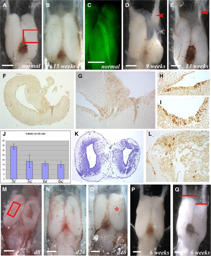
Maden et al. Neural Development 2013, 8:1                                                                          Page 2 of 15
http://www.neuraldevelopment.com/content/8/1/1

granule cell layer and a few hilar astrocytes [4]. Although      animals induces rapid tissue regeneration followed by more
NPCs capable of generating neurons in culture can be             prolonged retinotectal projection regeneration [33]. In
harvested from the entire neuraxis of living (throughout         addition, their mesencephalic dopaminergic neurons are
life) and cadaveric mammalian CNS tissue [4-8], they only        completely regenerated after 6-hydroxydopamine-induced
spontaneously generate significant numbers of neurons            ablation stimulates proliferation among normally quiescent
in vivo in the hippocampus and olfactory bulbs. In lower         mesencephalic ependymoglia [31,32]. The remarkable re-
vertebrates such as reptiles, amphibians and fish, VZ NPCs       generative ability of the axolotl telencephalon has been
are thought to proliferate throughout adulthood [9-12]. A        characterized most extensively by Kirsche [9,34-36] and
comparative approach that examines the distribution of           others [37,38]. They found that telencephalic segments can
proliferating NPCs and the differentiation and migration of      regenerate providing the olfactory nerves are intact, and
their progeny in lower vertebrates may reveal mechanisms         concluded that the stimulatory cue to regeneration pro-
that could be harnessed to stimulate neuronal regeneration       vided by the olfactory system is the cellular contribution
in the adult mammalian CNS.                                      provided by olfactory neuroblasts. Although the olfactory
   Precise localization labeling experiments in zebrafish        epithelium replaces lost neurons throughout life [39], and
and other teleosts [13] have revealed proliferating NPCs         transplanted olfactory epithelial ensheathing cells can
in VZs throughout the major subdivisions of the adult            minimize CNS lesions [40], there is little evidence in tele-
brain with the exception of the hypothalamus and cere-           osts, reptiles or mammals that olfactory epithelial cells mi-
bellum, where proliferative zones are located deeper in          grate into brain parenchyma. Rather, telencephalon VZ
the parenchyma [14,15]. Proliferation rates are faster in        NPCs produce neuroblasts that migrate toward the olfac-
ventral versus dorsal regions of the teleost telenceph-          tory bulbs in these species. Therefore, the olfactory nerve
alon, where cells migrate centrifugally away from the VZ         or bulb more likely provides an injury-induced cue that up-
as they differentiate into neurons that integrate into           regulates spontaneous VZ NPC proliferation to induce
neuronal circuits [15-19]. Ventral telencephalic neuro-          plasticity and regeneration in the Urodele brain.
blasts migrate through a rostral migratory stream that              Here, we conduct a detailed study of the distribution
resembles the mammalian rostral migratory stream, be-            of NPCs that divide spontaneously and in response to
fore terminally differentiating into neurons in the olfac-       injury in the axolotl brain. The axolotl is a champion
tory bulbs [16,20]. Although teleost brain regeneration is       of regeneration with the exceptional ability to regrow
far less studied than optic nerve or retinal regeneration,       limbs, heart, spinal cord, tail and large portions of brain
a stab wound can up-regulate VZ NPC proliferation,               tissue. Axolotl brain regeneration thus provides us with
which is followed by the migration of new cells into             an excellent model for examining the relationship be-
damaged telencephalic regions [21-25]. In the knifefish,         tween spontaneous and injury-induced NPC behavior
an incisional cerebellar wound stimulates proliferation at       and its relationship to olfactory stimuli. Our detailed
the site of injury that is followed by the migration of          analysis builds upon previous work that detected divid-
new cells along radial glial fibers into the wound [13].         ing NPCs in the axolotl Ambystoma mexicanum CNS
   In reptiles, spontaneous but variable rate NPC prolif-        and showed that the number of dividing NPCs declines
eration has been noted in the VZs of the telencephalon           with age [30]. We found proliferating NPCs in the VZs
and cerebellum [26,27]. In a manner similar to that              of the telencephalon, mesencephalon and rhombenceph-
noted in teleosts, neuroblasts (but not glioblasts) migrate      alon with activity that was variable along the rostrocau-
centrifugally away from the VZ and are thought to mi-            dal and dorsoventral neuraxes between these regions. In
grate through a rostral migratory stream into the olfac-         the VZ, dividing cells exhibited radial glial-like glial fi-
tory bulbs before terminally differentiating into neurons.       brillary acidic protein (GFAP)-positive phenotypes. Cells
In reptiles, incisional wounds can stimulate the prolifera-      positive for the cell synthesis marker bromodeoxyuridine
tion of VZ NPCs that appear to induce relatively slow            (BrdU) migrated laterally, dorsally and ventrally from the
and incomplete wound repair [28]. In the lizard, with            VZs and differentiated into either almost mature neurons
more drastic dorsal telencephalic segment removal some           positive for doublecortin (DCX) and/or neuronal nuclei
tissue regeneration is noted, but with limited cell layer-       (NeuN) or mature NeuN+ neurons by 2 weeks. There
ing, even after 260 days [29].                                   appears to be a relative reduction in the neuronal popula-
   The amphibian telencephalon contains a dorsal and             tion, suggesting a degree of selective cell death. Large-scale
thicker ventral matrix (ventricular) zone [9,30] that exhibits   telencephalon tissue removal stimulated VZ proliferation
higher proliferative and regenerative capacity than the tele-    and tissue regeneration. We only detected this injury-
ost and reptile telencephalon VZ. In the adult newt, NPCs        induced proliferative and regenerative response in animals
proliferate in the telencephalon VZ anterior to the quies-       with an intact olfactory nerve, suggesting that an olfactory
cent mesencephalon, hindbrain and cerebellum regions             cue could stimulate the response. We present this data as
[31,32]. Removal of 70% of the optic tectum in these             the beginning of a thorough cellular and molecular analysis
Maden et al. Neural Development 2013, 8:1 Page 3 of 15
http://www.neuraldevelopment.com/content/8/1/1
of the phenomenon of brain regeneration in axolotls and containing the cell synthesis marker BrdU. The brains
its relation to proliferation of stem cells within the CNS. were fixed, serially sectioned and BrdU+ nuclei identified
on immunohistochemistry. The following regions were
Results examined in transverse sections: anterior telencephalon
Proliferation zones in the axolotl brain and olfactory nerve (Figure 1A, level B), anterior telen-
To locate proliferating neural progenitor cells and their cephalon and olfactory bulb (Figure 1A, level C), posterior
progeny, juvenile axolotls (4 to 5 inches long; 3 to 4 telencephalon and choroid plexus (Figure 1A, level D),
months of age) were used. These animals do not meta- mesencephalon (Figure 1A, level E), anterior rhomben-
morphose, become sexually mature in the larval form cephalon and cerebellum (Figure 1A, level F), and the mid
and retain exceptional regenerative ability throughout rhombencephalon (Figure 1A, level G). To provide
their lives. They were placed overnight (18 h) in water comparative data, the location of every BrdU+ cell in five
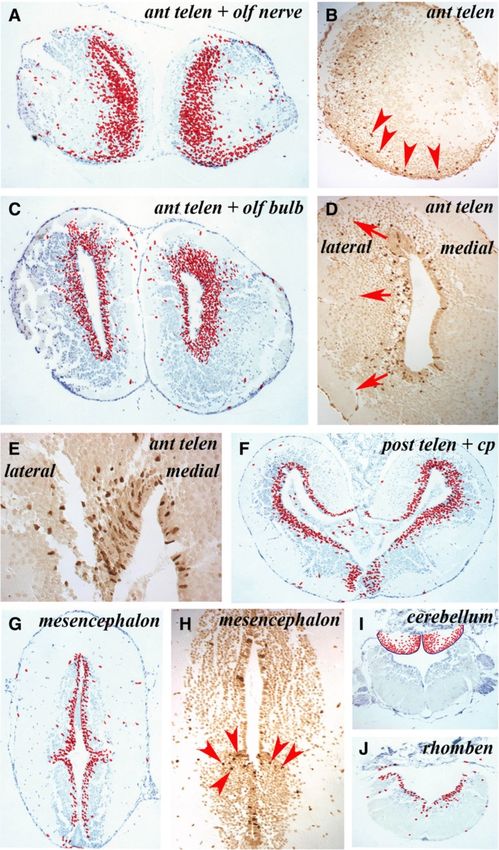
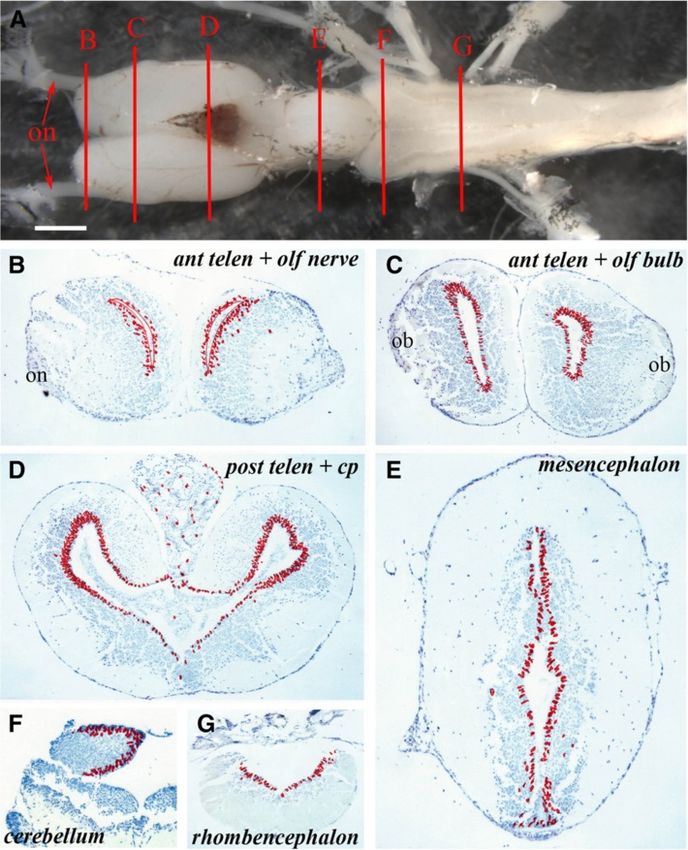
Figure 1 Ventricular zones with variable proliferative activity can be detected throughout the axolotl central nervous system.
(A) Structure of the axolotl brain showing the levels at which sections ventricular zone proliferation was analyzed. B = anterior telencephalon and
olfactory nerve, C = anterior telencephalon and olfactory bulb, D = posterior telencephalon, E = mesencephalon, F = cerebellum,
G = rhombencephalon; on: olfactory nerve. Scale bar = 1 mm. (B – G) Cumulative BrdU+ cell numbers on five sections from each level depicted
in A on which individual labeled cells are marked with a red dot. Regions of the brain are marked.; on: olfactory nerve; ob: olfactory bulb;
cp: choroid plexus.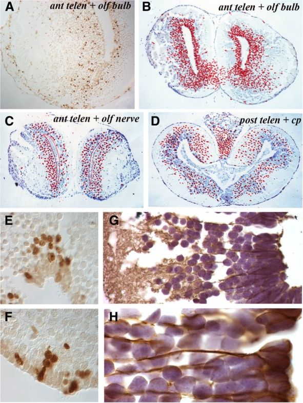
Maden et al. Neural Development 2013, 8:1                                                                        Page 4 of 15
http://www.neuraldevelopment.com/content/8/1/1

adjacent sections was recorded at each level and repre-         with the rhombencephalon revealing the lowest levels of
sented as a summary section in the figures.                     proliferation along with the ventral posterior telencephalon.
   The axolotl brain is typified by the presence of a narrow,     These data show that NPCs proliferate throughout the
one- to three-cell layered VZ (matrix zone) adjacent to         uninjured axolotl brain, with the highest levels detected in
the ventricle. The VZ is surrounded by a wide region            the dorsolateral telencephalon followed by the mediolat-
of uniformly spherical neurons surrounded by an axonal          eral telencephalon and cerebellum. There was an obvious
layer of varying thickness (Figure 1B-F). In axolotls sacri-    dorsoventral difference in VZ proliferation in the posterior
ficed the morning after overnight BrdU incorporation,           telencephalon that disappeared in more caudal regions.
more than 99% of the BrdU-labeled cells were located in         Occasional labeled cells were detected in parenchymal
the VZ. Only occasional labeled cells with a neuronal           regions of animals sacrificed immediately after overnight
phenotype were detected within the grey matter, or with a       BrdU labeling. The spherical morphology of these cells
meningeal phenotype at the periphery or within the cho-         suggests that they represent either a small population of
roid plexus. At the level of the anterior telencephalon and     the migrating neuroblast progeny of NPCs that had com-
olfactory nerve, BrdU+ cells were distributed uniformly in      pleted their synthesis phase at the beginning of the label-
the dorsoventral axis of the VZ and no labeled cells were       ing period or a small population of neurons undergoing
detected in the olfactory nerve (Figure 1B). At the olfac-      DNA repair. The former hypothesis would suggest that
tory bulb level (Figure 1C), BrdU+ cells were restricted to     neuroblasts migrate fairly quickly after NPCs complete
the VZ, but were apparently more abundant in dorsal and         division.
ventral regions than in the central VZ. Many BrdU+ cells
were detected in the VZ of the posterior telencephalon          Migration of labeled cells
(Figure 1D), with far more in the dorsal versus ventral VZ      To characterize the migration pattern of the VZ progeny
and more in the lateral than the medial VZ. Examination of      into the surrounding differentiated neuronal layer, ani-
cumulative cells labeled in the mesencephalon (Figure 1E)       mals were placed overnight (18 h) in water containing
showed that considerably fewer BrdU+ cells were present in      BrdU and then kept in fresh water for 1, 2, 3 or 4 weeks
this region of the brain than in the dorsal telencephalon       before being sacrificed.
and that the number of labeled cells was consistent between       In animals sacrificed 1 week after BrdU labeling, 1-
the dorsal and ventral mesencephalic VZ. In the anterior        week-old BrdU+ cells had migrated out of the VZ into
rhombencephalon, at the level of the cerebellum, labeled        the adjacent neuronal layer, most prominently in the tel-
cells were present both in the VZ and in the underlying         encephalon followed by the cerebellum (Figure 3A,I). At
parenchyma (Figure 1F) and in the mid-rhombencephalon           the level of the anterior telencephalon and olfactory
(Figure 1G) there was a fairly uniform, but lower number        nerve, there was an expanded VZ zone of labeled cells
of BrdU+cells along the VZ.                                     and apparent streams of labeled cells migrating laterally
   Figure 2 shows individual sections of the brain regions      and ventrally toward the olfactory bulb and nerve
to highlight these differences in the abundance of BrdU+        (Figure 3A) that could be readily detected in a single
VZ cells. The percentage of BrdU+ VZ cells in the dif-          section (Figure 3B). In the anterior telencephalon and ol-
ferent regions of the brain are show in Figure 2G.              factory bulb, the expanded zone of labeled cells was
Figure 2A,B revealed similar proliferation levels in the        more prominent on the lateral side of the VZ than the
dorsal region of the anterior versus the posterior telen-       medial side (Figure 3C,D). This difference can be clearly
cephalon. But within this region, especially notable in         seen in single sections such as the highlight of the dorsal
Figure 2B, the lateral side of the VZ (left side of             part of the telencephalic VZ (Figure 3E). In the posterior
Figure 2B) had more proliferating cells than the medial         telencephalon there was also a lateral expansion of the
side (right side of Figure 2B). This impression was con-        zone of labeled cells that was more prominent dorsally
firmed by cell counts, where the dorsolateral posterior         than ventrally (Figure 3F). In the mesencephalon, few la-
telencephalon had almost twice as many proliferating            beled cells appeared to have migrated away from the VZ
cells as the dorsomedial posterior telencephalon (24.2%         (Figure 3G) except at the junction between the thalamus
versus 14.9%; Figure 2G). The ventral posterior telen-          and the hypothalamus where labeled cells spreading
cephalon (Figure 2D) had low levels of proliferation            laterally could be observed (Figure 3H, red arrowheads).
(6.7%; Figure 2G), demonstrating how variable prolifera-        There was a high degree of movement among BrdU+
tion is within this one region of the brain. The mesen-         cells from the VZ to the parenchyma of the cerebellum
cephalon (Figure 2C) had similar levels of proliferation        (Figure 3I). Similar to the mesencephalon, very few
to the anterior telencephalon (13.2% versus 10.2%, non-         BrdU+ cells had migrated from the VZ into the paren-
significant; Figure 2G). The cerebellum (Figure 2E) had         chyma of the rhombencephalon (Figure 3J). In this and
far higher proliferative counts than its adjacent rhomb-        other sections, some BrdU+ cells could be seen far from
encephalon (Figure 2F) (15.2% versus 7.2%; Figure 2G)           the VZ, sometimes at the edge of the brain. We have not
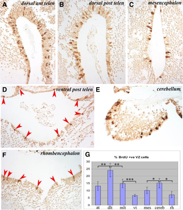
Maden et al. Neural Development 2013, 8:1                                                                                              Page 5 of 15
http://www.neuraldevelopment.com/content/8/1/1

 Figure 2 Cells labeled with BrdU after an overnight incubation are largely retained in the ventricular zone throughout the neuraxis,
 which shows regional variation in proliferative activity. (A-F) Examples of BrdU+ cells in the VZ of different brain regions show variation in
 numbers of labeled cells. Regions of the brain are marked. In D and F, BrdU+ cells are marked with red arrowheads. (G) Counts of proliferating VZ
 cells in different regions of the brain expressed as a percentage of total VZ cells. at: anterior telencephalon; BrdU: bromodeoxyuridine; cereb:
 cerebellum; dlt: dorsolateral telencephalon; mes: mesencephalon; mlt: mediolateral telencephalon; rh: rhombencephalon; vt: ventral telencephalon;
 VZ: ventricular zone. * significant (P = 0.025), ** very significant (P = 0.006), *** extremely significant (P = 0.0004) by Student’s T-test.

studied these cells in detail but suggest they are prolifer-               olfactory nerve, there was a much reduced and more gen-
ating in situ; for example BrdU+ meningeal cells could                     eralized migration of cells (Figure 4C). This was the most
occasionally be observed. These zones of higher migra-                     common response, also apparent at the posterior telen-
tion rates correlated with the zones of high proliferation                 cephalon level (Figure 4D) where BrdU+ cells were distrib-
(Figure 2G).                                                               uted throughout the neuronal layer with a reduced
  Over the ensuing weeks the patterns of movement                          number of labeled cells remaining in the VZ.
within the parenchyma appeared to continue, with BrdU+                        In less active regions, intensely labeled BrdU+ cells were
cells moving more laterally and ventrally away from the                    more frequently observed within the VZ at later time peri-
VZs of the telencephalon and cerebellum and, to a lesser                   ods. By contrast, in more active regions, paler stained
degree, in the less active mesencephalon and rhomben-                      nuclei were seen within the VZ, suggesting these were the
cephalon. At the anterior telencephalon and olfactory bulb                 progeny of cell divisions. Within the parenchyma, BrdU+
level (Figure 4A,B), for example, there was a widespread                   cells were often seen closely adjacent to labeled VZ cells
ventral migration both towards the olfactory bulb and                      as clumps (Figure 4E) or as lines (Figure 4F), suggesting
medially whereas, more anteriorly at the level of the                      lineage relationships.

Maden et al. Neural Development 2013, 8:1                                                                                               Page 6 of 15
http://www.neuraldevelopment.com/content/8/1/1

 Figure 3 Sections taken one week after overnight BrdU labeling show the migration of BrdU+ cells from the ventricular zone into the
 mantle zone at different rates at different levels of the central nervous system. (A, C, F, G, I, J) Positions of labeled cells on five cumulative
 sections at the levels marked on the figure. The migration of BrdU+ cells outwards at different rates can be appreciated by comparing A and C
 with G. (B, D, E, H) Labeled cells on individual sections.
Maden et al. Neural Development 2013, 8:1                                                                                              Page 7 of 15
http://www.neuraldevelopment.com/content/8/1/1

 Figure 4 Migration patterns and glial fibrillary acidic protein expression in the telencephalon. (A) Single section through the anterior
 telencephalon and olfactory bulb taken 4 weeks after overnight BrdU labeling shows a stream of cells moving laterally into the olfactory bulb.
 (B) Cumulative positions of BrdU+ cells in the anterior telencephalon and olfactory bulb level 4 weeks after overnight labeling. (C) Cumulative
 positions of BrdU+ cells in the anterior telencephalon and olfactory nerve level 4 weeks after overnight labeling showing a decreased number of
 BrdU+ cells in the VZ. (D) Cumulative positions of BrdU+ cells in the posterior telencephalon 4 weeks after overnight labeling showing a
 decreased number of BrdU+ cells in the VZ. (E,F) Patterns of BrdU+ cells in and adjacent to the VZ 3 weeks after overnight labeling. (G,H) Glial
 fibrillary acidic protein immunocytochemistry of the normal telencephalon at low power (G) and high power (H) to show the cytoplasmic
 extensions of radial glial cells. BrdU: bromodeoxyuridine; VZ: ventricular zone.

Phenotype of labeled cells                                                    To examine the differentiative properties of prolifera-
Most VZ cells contain flat apical surfaces lining the ven-                 tive VZ cells, sections were immunolabeled to detect the
tricle and pointed basal surfaces with elongated cytoplas-                 glial markers GFAP (astrocytes) and NG2 (early oligo-
mic extensions extending to the pial surface of the brain.                 dendroctyes) or the neuronal markers DCX (immature
These morphologies are consistent with the classical ra-                   neurons) and/or NeuN (mature neurons) expressed by
dial glial cell morphologies of lower vertebrates [41-46]                  BrdU+ cells (Figure 5A-C). The densities of BrdU+ cells
and the VZ cells all stain with GFAP (Figure 4G,H).                        located in the VZ (Figure 5D) and neuronal layers
Their glial fibers form columns with spherical NeuN+                       (Figure 5G) and their phenotypes (Figure 5B,C,E,G) in
neuronal cells arranged in columns between the radial                      uninjured animals that were sacrificed 2, 3 and 4 weeks
glial fibers (Figures 4H and 5C). A minority of GFAP+                      after BrdU labeling were then quantified under confocal
VZ cells had smaller nuclei and appeared not to stain                      microscopy. We have not been able to detect NG2+ cells
with GFAP (Figure 5B, yellow arrows).                                      with commercially available antibodies, previously and in
Maden et al. Neural Development 2013, 8:1 Page 8 of 15
http://www.neuraldevelopment.com/content/8/1/1
B1
A B *
DAPI
B2
*
Ventricle BrdU
ye
r
e
*
L a Zon B3
a l e u lar DAPI *
on on tric BrdU
ur rZ Ve
n
Ne la GFAP GFAP
tr icu
n DAPI DAPI
Ve BrdU *
NeuN C1
Ventricle DCX BrdU
* C
2
DCX
* * C3
DAPI
NeuN
BrdU
C Neuronal Layer * C
4
D E F G
% BrdU+ Cells Expressing Neuronal Markers
100%
% of VZ BrdU+ Cells Expressing GFAP
1000 100% Transition 20000
Density of BrdU/GFAP+ cels (/mm2)
90%
Mature
Density of New Neurons (/mm2)
900 90% 18000
800 80% 80% 16000
700 70% 70% 14000 **
600 60% 60% 12000
500 50% 50% 10000
400 40% 40% 8000
300 30% 30% 6000
200 20% 20% 4000
100 10% 10% 2000
0 0% 0% 0
2 3 4 2 3 4 2 3 4 2 3 4
Weeks Weeks Weeks Weeks
Figure 5 Two weeks after BrdU labeling, new cells have migrated and differentiated into neurons and a few new cells are retained in
the ventricular zone and express GFAP. (A) Confocal image at 10× magnification shows that, in the uninjured axolotl telencephalon, most
BrdU+ cells have migrated into the mantle zone 2 weeks after labeling. A few BrdU+ cells are retained in the VZ. (B,C) Confocal images taken at
40× objective (2.3× digital zoom) of sections stained with 40-6-diamidino-2-phenylindole (in white; B1 and C1) and antibodies against (B) BrdU
(in red; B2) and GFAP (in green; B3) or (C) BrdU (in red; C2), DCX (in blue; C3), NeuN (in green; C3). The image in (B) shows that very few BrdU+
cells are retained in the VZ by 2 weeks after overnight BrdU labeling, but those that do remain and express GFAP may represent new radial
glia-like cells. (C) by 2 weeks, all BrdU+ cells in the mantle zone express the mature neuronal marker NeuN (white and yellow arrows) and a small
percentage of the NeuN+ neurons retain DCX expression (yellow arrows) and are likely transitioning into mature neurons. (D) The relatively small
densities (versus F) of BrdU/GFAP+ cells retained in the VZ are resilient across weeks 2 to 4 after BrdU labeling. (E) Most BrdU+ VZ cells express
GFAP. (F) All BrdU+ cells in the neuronal layer express NeuN and a few NeuN+ neurons retain DCX expression. These percentages are consistent
across weeks 2 to 4 after BrdU labeling. (G) New neurons are either vulnerable to cell death or are still migrating because their densities
significantly decrease between weeks 2 and 3 (**PMaden et al. Neural Development 2013, 8:1 Page 9 of 15
http://www.neuraldevelopment.com/content/8/1/1
the current study, which could reflect that these anti- role in regeneration that were described over 50 years
bodies were raised against a non-homologous region of ago by Kirsche [9,34-36] and others [37,38]. Although
the mammalian protein or that NG2+ oligodendrocyte both telencephalons can, remarkably, regenerate in their
precursor cells are seldom found in the uninjured axo- entirety after simultaneous removal [48], we have found
lotl CNS. that removing tissue segments from one telencephalon
In the telencephalon, the migration of BrdU+ cells out while leaving the other intact as a control or removing
of the VZ appeared complete by 2 weeks because the tissue segments as well as severing the olfactory nerve
total percentage of BrdU+ cells that were retained in the provides better insights into the underlying mechanisms
VZ (2.50 ±0.53%, 2.0 ±0.1% and 1.1 ±0.3%, respectively; of regeneration.
F(2,10) = 1.14; P = 0.36.) versus those that had migrated Removed telencephalon segments, such as the middle
into the parenchymal neuronal layer (97.50 ±0.53%; third segment, regenerated within 12 to 15 weeks in
98.0 ±0.4% and 98.9 ±3.6%, respectively; (F(2,10) = 1.14; 4- to 5-inch-long animals (compare right telencephalon
P = 0.36) remained consistent across weeks 2 to 4. in Figure 6A with Figure 6B). Regenerated segments are
Figure 5D shows that the density of BrdU+ cells externally indistinguishable from undamaged segments
retained in the VZ is resilient, because although the at this time frame although, histologically, the full thick-
densities are relatively small, they did not change across ness of the telencephalon has not always recovered.
weeks 2 to 4 after labeling (F(2,10) = 1.31; P = 0.31). The Although the tissue removals described here sever the
majority (84.0 ±4.5%) of these BrdU+ VZ cells express median olfactory tract (Figure 6C), we have not tested
GFAP (F(2,10) = 1.56; P = 0.86; Figure 5E), suggesting that these animals to determine whether behavioral function
some progeny either retain a radial-glial like progenitor has returned nor have we performed electrophysiological
cell phenotype or differentiate into ventricular zone astro- assessment of connectivity. We have only assessed the
cytes. If these cells do represent a population of new radial regeneration of the damaged site morphologically. Over-
glia-like progenitors, their retention of BrdU suggests that night BrdU labeling experiments during the process of
they either remain quiescent or do not undergo multiple regeneration revealed that NPC proliferation in the VZ
divisions within the VZ. These speculations could be of the damaged telencephalon, which often exhibited a
tested in future experiments. All spherical BrdU+ cells that ventral thickening (Figure 6F,G), is up-regulated relative
had migrated out of the VZ into the surrounding neuronal to NPC proliferation in the VZ of the undamaged contra-
layer expressed the mature neuronal protein NeuN. A lateral telencephalon by approximately 3 weeks after the
small fraction (approximately 14.2 ±1.6%) of these BrdU/ damage (compare Figure 6H and I, counts in J). After 6
NeuN+ cells retained expression of the immature neuronal weeks, the damaged area had healed enough to restore the
marker DCX, suggesting that they were transitioning to a appearance of a well-defined ventricle (Figure 6K) and
mature state (Figure 5F). These data suggest that new NPC proliferation in the VZ of the damaged telenceph-
neurons mature along similar timelines in the uninjured alon had subsided to resemble that in the VZ of the con-
axolotl telencephalon and rodent hippocampus [47]. In trol telencephalon (Figure 6J). However, at this later stage,
contrast to the BrdU/GFAP+ cells retained in the VZ, dividing NPCs were additionally detected in the cells sur-
BrdU+ neurons (DCX and/or NeuN+) that had migrated rounding the wound margins immediately after a BrdU
into the parenchyma appeared to either be vulnerable to pulse (Figure 6L). By contrast, BrdU+ cells are never
cell death or continue to migrate because their densities detected in the undamaged parenchyma immediately after
decreased across weeks 2 to 4 (F(2,10) = 14.66; P = 0.001). a short BrdU exposure. These data suggest that injury-
Specifically, relative to 2-week-old neurons, fewer 3-week- induced regeneration of the telencephalon may be accom-
old (P = 0.005) and 4-week-old (P = 0.001) neurons were plished initially by a cue that stimulates the proliferation
detected (Figure 5G). If this phenomenon reflects the of NPCs in the VZ and then latently by a cue that stimu-
attrition that has been well documented in the rodent lates a more local proliferative response. One possibility is
hippocampus [47] then it may be occurring somewhat that the first response may recruit a large number of cells
more latently in the axolotl CNS, although we have not to replace missing tissue whereas the second response
investigated other possibilities and we did not directly may harmonize the cut surfaces. These conjectures are
measure cell death in either the VZ or parenchyma. based on the assumption that BrdU uptake indicates cell
cycling but other explanations are equally valid, such as
Characteristics of telencephalic regeneration that the localized BrdU+ cells in Figure 6J could be simply
To begin an analysis of the relationship between VZ pro- repairing their DNA.
liferation in response to damage, the olfactory nerve and We found several lines of evidence consistent with the
regenerative ability we have investigated various regen- hypothesis that telencephalic regeneration may be stimu-
erative regimes in the telencephalon. We have replicated lated by a cue provided by the olfactory nerve. In cases
the striking findings that the olfactory system plays a where the olfactory nerve is severed along with removalMaden et al. Neural Development 2013, 8:1 Page 10 of 15 http://www.neuraldevelopment.com/content/8/1/1 Figure 6 (See legend on next page.)
Maden et al. Neural Development 2013, 8:1                                                                                               Page 11 of 15
http://www.neuraldevelopment.com/content/8/1/1

 (See figure on previous page.)
 Figure 6 Regeneration of the axolotl telencephalon. (A) Normal forebrain (ablated region marked with red box). (B) 15 weeks later showing
 complete regeneration. (C) RT97 wholemount showing the medial olfactory tract. (D) 9 weeks after removal of the right anterior third and
 olfactory bulb showing failure of regeneration and swelling of the regenerating olfactory nerve (arrowhead). (E) After removal of the olfactory
 bulb (right) the telencephalon heals but does not regenerate until the olfactory nerve regenerates (arrowhead = olfactory nerve swelling). (F-L)
 Regeneration after removal of the middle third of the telencephalon (as in A). After 3 weeks (F, G) there is no regeneration but an increase in
 BrdU+ cells in the damaged VZ (right). Close-up of the undamaged (H) and damaged (I) VZ shows more BrdU+ cells in I. J, BrdU+ counts in the
 VZ from 3-week regenerating (3r), 3-week undamaged (3c), 6-week regenerating (6r), 6-week undamaged (6c) with standard deviations. K, after 6
 weeks there is restoration of the damaged side (right) but not full tissue replacement. After overnight BrdU labeling at 6-weeks there is local
 proliferation at the cut site (L). (M-Q) Regeneration after dorsal pallium removal (red box in M). M, damage site (right) after 8 days. N, after 24
 days there is near complete regeneration. O, 40 days after removal the left and right telencephalons are indistinguishable (damage site marked
 with red star). P, regenerated brain 6 weeks after removal of the right dorsal pallium (as in M) shows complete regeneration. Q, 6 weeks after
 right dorsal pallium removal and olfactory nerve severing there is wound repair but shrinkage of the right telencephalon while the olfactory
 nerve regenerates. Red bars show the left/right difference in telencephalon length. Scale bars = 1 mm.

of the anterior third of the telencephalon, regeneration                     size of the right telencephalon. While Kirsche [9,34-36]
only begins once the olfactory nerve regrows enough to                       and others [37,38] suggested that the olfactory nerve may
contact the residual portion of the telencephalon. During                    be a source for cells that regenerate the telencephalon,
this period the remaining part of the telencephalon has                      data from the current study and work in mammals, tele-
undergone wound healing but not regeneration, and re-                        osts and reptiles suggests that the more likely explanation
growth is suspended until contact is made. For example,                      is that the olfactory nerve provides a trophic stimulus to
by approximately 9 weeks after anterior telencephalon                        VZ and perhaps parenchymal NPCs. Future experiments
damage, which is coincident with the time frame that a                       will attempt to determine the nature of this trophic regen-
removed middle third segment of the telencephalon                            erative stimulus.
would have nearly completed regeneration (for example,
Figure 6B), the re-growing olfactory nerve exhibits a dis-                   Discussion
tal swelling as contact is made (Figure 6D, red arrow-                       The regenerative ability of the axolotl is unparalleled
head). This swelling was described as containing                             among vertebrates and includes the ability to regenerate
olfactory neuroblasts [9,34-38]. The same phenomena of                       large parts of the brain. By identifying the mechanisms
wound healing, delayed regeneration and olfactory nerve                      that control this process in the axolotl we hope to gain
swelling can also be seen in more precise tissue removal,                    insight into why this process does not occur spontaneously
for example of only the olfactory bulb (Figure 6E, red                       in higher vertebrates, including man, despite the presence
arrowhead).                                                                  of forms of CNS plasticity and NPCs capable of proliferat-
  A less invasive procedure that does not remove the                         ing and generating neurons that incorporate into func-
olfactory bulb and where the effect of the olfactory nerve                   tional neural circuits [1,4-8]. Brain regeneration, at least in
can readily be seen is after the removal of a rectangular                    response to incisional or stab wounds, occurs in fish,
segment of the dorsal pallium (Figure 6M). In one group                      amphibians and reptiles throughout life [10,13,21,23,26].
of animals where the dorsal pallium was removed with-                        Although NPCs are distributed throughout the neuraxis of
out damage to the olfactory nerve or bulb, the damage                        adult mammals [1,5,8], neurons are only spontaneously
heals and the missing tissue is rapidly replaced. After 24                   generated in highly restricted areas [3,4] and only ex-
days the wound size has vastly reduced (Figure 6N) and                       tremely limited abortive neurogenesis occurs in response
by 40 days looks to have completely regenerated                              to injury in extra-neurogenic regions (for review see [49]).
(Figure 6O). Sections reveal, however, that the full thick-                  Here we characterize the effects of removing variably-sized
ness of the dorsal pallium has not yet regenerated by this                   telencephalon sections on NPC proliferation and new cell
time after damage. In the second group of animals in                         migration in the axolotl to establish a model with which to
which the olfactory nerve is also severed at the time of                     investigate the mechanisms of brain regeneration.
dorsal pallium removal, the damage heals successfully,                          The axolotl VZ, like other Urodeles, retains embryonic
but the telencephalon shrinks in length and does not                         characteristics that include the presence of abundant
begin to regenerate its size until the olfactory nerve                       GFAP+ radial glia with nuclei that line the ventricle and
returns. This can be seen by comparing Figure 6P, taken 6                    long cytoplasmic processes that extend to the pial sur-
weeks after dorsal pallium removal on the right side show-                   face [41-46]. New cells appear to migrate away from the
ing good regeneration, with Figure 6Q, taken at the same                     VZ along these extensions to terminally differentiate
time after dorsal pallium removal plus olfactory nerve                       into neurons once they reach the mantle zone. This
severing. The latter shows repair of the wound site but no                   process appears to be complete within 2 weeks, at which
regeneration at this time as there is a clear reduction in                   point all cells that have entered the mantle zone express
Maden et al. Neural Development 2013, 8:1                                                                         Page 12 of 15
http://www.neuraldevelopment.com/content/8/1/1

the mature neuronal marker NeuN. A few of these cells re-        higher vocal center of birds [50] and the hippocampal
tain expression of the immature neuronal marker DCX              dentate gyrus, olfactory epithelium and olfactory bulbs of
even 4 weeks after their birth as they transition from an        mammals [51]. Considering the latter possibility, Urodele
immature to mature status. Why a small population of             cells or their niches may be unique. Although spontan-
new neurons exhibits delayed maturation is unclear. Simi-        eous NPC proliferation is detected throughout the VZs of
lar to new mammalian neurons, these new axolotl neurons          the axolotl brain, it is only detected in the VZ of the telen-
appear vulnerable to cell death because their densities          cephalon in adult newts [31]. Yet 70% of the optic tectum
decrease in the month after their birth. Whether these           [33] and the entire set of dopaminergic neurons in the
dying cells supply a stimulus to VZ NPCs to maintain             ventral midbrain [31,32] can be regenerated in response to
spontaneous neuronal turnover would be interesting to test       tissue removal or chemical injury in the adult newt. In
in future experiments. A small population of the new cells       these circumstances, injury induces normally quiescent
remains in the VZ and expresses GFAP. These cells likely         NPCs to proliferate until the lost cells are regenerated.
replenish the VZ radial glia population as we did not detect     These data suggest that the ability to regenerate neurons
any GFAP+ cells in the mantle zone, which is consistent          is not simply a by-product of continual growth, and gives
with previous data collected in the axolotl [44,45], and in      us a greater opportunity for investigating the nature of the
fish and reptiles [17,27].                                       mechanisms behind spontaneous and injury-induced NPC
   Spontaneous NPC proliferation and neurogenesis oc-            proliferation and neuronal regeneration. As a first step, we
curred in the VZs throughout the axolotl brain but some          have confirmed the importance of the olfactory nerve as a
zones, such as the dorsal and ventral telencephalon, exhi-       source of the injury-induced stimulus for telencephalic
bited elevated activity levels. Whether neuronal turnover        regeneration in the axolotl. Discovering the nature of this
is higher in these highly active regions is currently unclear.   stimulus will be the subject of our future investigations.
The axolotl does continue to grow in adulthood so these
new neurons could also contribute to areas of elevated           Conclusion
growth. Indeed, spontaneous VZ NPC proliferation varies          There is a continual generation of neuronal cells from
across 16 zones in the adult zebrafish [15,16]. This situ-       neural progenitor cells located within the ventricular
ation in axolotls is likely related to their pedamorphic         zone of the axolotl brain. Variable rates of proliferation
nature, as adult newts show VZ proliferation only in the         were detected across brain regions and variable migra-
telencephalon [31] as do reptiles [26]. Future work testing      tion routes observed over time. The telencephalon and
how widespread spontaneous and injury-induced NPC                the cerebellum were the most active brain regions and
proliferation and neurogenesis is in the CNS of metamor-         ventricular zone cells either generated more radial glial
phosed axolotls would be an interesting test of their re-        cells, which remained in the VZ, or new neurons. New
generative potential. When studied over time, BrdU+ cells        neurons appeared to undergo a similar attrition to that
appeared to migrate from these regions of high activity in       seen in mammals as their numbers declined over time.
a lateral and dorsoventral rather than medial direction          These neural progenitor cells in the VZ appear to medi-
into the mantle zone. A ventral stream of BrdU+ cells ori-       ate complete telencephalic tissue regeneration through
ginating from the ventromedial surface of the anterior           an injury-induced olfactory cue. Identification of this cue
telencephalon toward the olfactory bulbs resembled the           is our future goal.
rostral migratory stream in the mammalian brain and was
consistent with reports in other vertebrate brains. Similar      Methods
to differential rates of NPC proliferation throughout the        Animals
brain, migration of new cells into the mantle varied across      Axolotls (Ambystoma mexicanum) of 4 to 6 inches in
brain regions with the most active areas occurring in the        length were purchased from the Ambystoma Genetic
telencephalon and the cerebellum with far less activity in       Stock Centre (University of Kentucky, Lexington, KY,
the mesencephalon and rhombencephalon.                           USA). They were maintained in 40% Holtfreter’s solution
   It is important to consider whether the ability to prolif-    and treated in accordance with the University of Florida’s
erate and generate new neurons per se endows the brain           Institutional Animal Care and Use Committee’s regula-
with regenerative capacity or whether regenerative capa-         tions. For all the experiments, group sizes were between
city is a unique property of particular taxa [11]. Consider-     two and four animals for each data point.
ing the former possibility, brain regeneration, at least in
response to incisional or stab wounds, occurs in fish and        BrdU labeling and histology
reptiles that show extensive spontaneous NPC prolifera-          Animals were placed in 1 L of Holtfreter’s solution con-
tion throughout the adult VZ. In birds and mammals, pro-         taining 50 mg of the cell synthesis marker BrdU (Sigma
liferation occurs in more restricted proliferative zones [21]    Aldrich, St. Louis, MO, USA) overnight (18 hrs) and
and neurogenesis is restricted to regions such as the            then either sacrificed or placed into fresh Holtfreter’s
Maden et al. Neural Development 2013, 8:1                                                                   Page 13 of 15
http://www.neuraldevelopment.com/content/8/1/1

solution for another 1, 2, 3 or 4 weeks before being          that we did not detect NG2+ cells in the axolotl brain
sacrificed. At each time point, the animals were anaes-       with the antibody used.
thetized in 1:5,000 tricaine methanesulfonate (MS222;           For whole-mount immunocytochemistry, brains were
Argent Chemical Laboratories, Redmond, Washington,            dissected from the animal and fixed overnight in 4% par-
USA) before their brains were dissected out of the cra-       aformaldehyde. They were washed several times in PBS-
nium and post-fixed overnight in 4% paraformaldehyde at       Tween (PBST) and then placed in RT97 antibody (1:50;
4°C. The brains were dehydrated, embedded in paraffin         Developmental Studies Hybridoma Bank, University of
wax and sectioned at 10 μm.                                   Iowa, Iowa City, IA, USA) made up in PBST at 4°C for 6
                                                              days. After washing for 1 day in PBST, immunoreactivity
Immunocytochemistry                                           was visualized with a fluorescent donkey anti-mouse im-
To localize dividing NPCs in brains fixed after overnight     munoglobulin G (1:200; Alexa Fluor, Life Technologies,
exposure to BrdU and surviving new cells in brains fixed      Grand island, NY, USA) made up in PBST for 2 days,
1, 2, 3 or 4 weeks after BrdU exposure, BrdU+ cells were      washed extensively and then cleared with 80% glycerol.
revealed enzymatically with 3,3’-diaminobenzidine tetra-
hydrochloride and imaged under light microscopy. Sec-         New cell densities and phenotyping
tions were dewaxed, incubated in 0.3% H2O2 in 0.1 M           New cell densities were confirmed in the neuronal
Tris-buffered saline (pH 7.4) for 10 min to quench en-        mantle by counting the total number of BrdU+ cells in
dogenous peroxidase, and then in 2 M HCl for 20 min           images of non-overlapping telencephalon sections. New
at 37°C to denature DNA. They were then blocked in            cell densities in the VZ were confirmed by counting
normal donkey serum (Tris-buffered saline, 3% normal          BrdU+ cells on VZ areas measured using Zeiss Zen soft-
donkey serum and 0.1% triton-X) and incubated over-           ware, because the VZ only comprised a portion of an
night at 4°C with rat anti-BrdU (OTB0030CX, 1:500;            imaged area. We examined more than 100 BrdU+ cells
AbD Serotec, Raleigh, NC, USA). The next day, the             on triple fluorescent-stained sections (two to four sec-
sections were incubated in biotinylated secondary anti-       tions per axolotl) for the co-expression of neuronal or
rat IgG (Jackson ImmunoResearch, West Grove, PA;              glial proteins using a Zeiss meta LSM 710 fully spectral
USA, 712-065-150, 1:500) for 4 h at room temperature          laser scanning confocal microscope (with 405, 488, 510,
and then in avidin-conjugated horseradish peroxidase          543 and 633 laser lines, Carl Zeiss, Jena, Germany).
(PK6100; Vector Laboratories, Burlingame, CA, USA)            BrdU+ cells were considered co-labeled when a full
for 2 h before being reacted in a solution of 0.02%           ‘z-dimension’ scan revealed its BrdU/40-6-diamidino-2-
3,3’-diaminobenzidine tetrahydrochloride (D9015; Sigma        phenylindole+ nucleus was unambiguously associated with
Aldrich) and 0.5% H2O2.                                       a lineage marker such as the immature neuronal protein
  After BrdU immunocytochemistry, the numbers of              DCX [52-54], the mature neuronal protein NeuN [55], the
positive cells were counted throughout the brain as the       oligodendrocyte precursor protein NG2 [56] or the astro-
percentage positive cells in the VZ. To visualize their       cyte protein GFAP [57]. The percentage of BrdU+ cells
localization and their spread from the VZ over time,          expressing each phenotype was calculated.
BrdU+ cells were also localized on photographs of the
relevant sections by marking their position with a red        Brain regeneration experiments
dot. The position of every positive cell on five adjacent     Operations were performed by anaesthetizing animals in
sections was marked on the photographs.                       1:5,000 MS222. The skin was removed from above the
  To identify the phenotype of surviving new cells, the       cranium on three sides and flapped back caudally to
expression of neuronal or glial proteins by BrdU+ cells was   expose the cartilaginous cranium roof. This was also cut
quantified using fluorophore-conjugated secondary anti-       on three sides and bent back caudally to reveal the telen-
bodies. After overnight incubation in rat anti-BrdU, these    cephalon and mesencephalon. Various operations were
sections were incubated for 4 h at room temperature in        performed on one telencephalon using fine iridectomy
Cy3-conjugated anti-rat immunoglobulin G and then             scissors. These operations included, first, removal of the
mouse anti-NeuN (1:500; Chemicon, Temecula, CA, USA)          middle third of the telencephalon while leaving the ol-
and goat anti-DCX (1:500; Santa Cruz Biotechnology, Santa     factory bulb and olfactory nerve intact; second, removal
Cruz, CA, USA) to detect neuronal phenotypes or               of the anterior third including severing the olfactory
rabbit anti-chondroitin sulfate proteoglycan (NG2;            nerve and removal of the olfactory bulb; and third, re-
1:500; Chemicon) and chicken anti-GFAP (EnCor                 moval of a rectangular piece of the dorsal pallium with
Biotech, Gainesville, FL, USA) to detect glia and then        or without severing the olfactory nerve. All operations
the appropriate maximally cross-adsorbed cyanine 5-           were carefully performed to prevent damage to the cho-
and fluorescein isothiocyanate-conjugated secondary           roid plexus, and to ensure minimal blood loss. Following
antibodies (1:500; Jackson ImmunoResearch). Note              the relevant operation, the cartilage flap of the cranium
Maden et al. Neural Development 2013, 8:1 Page 14 of 15
http://www.neuraldevelopment.com/content/8/1/1
was replaced followed by the skin. The skin flap was kept 4. Zhou C, Deng W, Gage FH: Mechanisms and functional implications of
in place with superglue. The animals were then replaced adult neurogenesis. Cell 2008, 132:645–660.
5. Palmer TD, Ray J, Gage FH: FGF-2-responsive neuronal progenitors reside
in Holtfreter’s solution. All operated animals survived the in proliferative and quiescent regions of the adult rodent brain. Mol Cell
surgery and showed apparently normal behavior in the Neurosci 1995, 6:474–486.
ensuing weeks. At weekly intervals, operated brains were 6. Palmer TD, Schwartz PH, Taupin P, Kaspar B, Stein SA, Gage FH: Cell culture.
Progenitor cells from human brain after death. Nature 2001, 411:42–43.
sampled for histology and cell proliferation after overnight 7. Arsenijevic Y, Villemure JG, Brunet JF, Bloch JJ, Déglon N, Kostic C, Zurn A,
BrdU incorporation. Nissl staining was performed for Aebischer P: Isolation of multipotent neural precursors residing in the
structural analysis and BrdU immunocytochemistry per- cortex of the adult human brain. Exp Neurol 2001, 170:48–62.
8. Laywell ED, Kukekov VG, Suslov O, Zheng T, Steindler DA: Production and
formed as described above. analysis of neurospheres from acutely dissociated and postmortem CNS
specimens. Methods Mol Biol 2002, 198:15–27.
Statistical analyses 9. Kirsche W: The significance of matrix zones for brain regeneration and
brain transplantation with special consideration of lower vertebrates. In
Analyses were performed using STATISTICA software Neural Tissue Transplantation Research. Edited by Wallace R, Das GD. New
(StatSoft; Tulsa, OK, USA). Analyses of variance were York: Springer; 1983:65–104.
used to explore the effects of the independent variables 10. Chapouton P, Jagasia R, Bally-Cuif L: Adult neurogenesis in non-
mammalian vertebrates. Bioessays 2007, 29:745–757.
of survival time on measures of neurogenesis (new cell 11. Ferretti P: Is there a relationship between adult neurogenesis and neuron
densities and new cell phenotypes). Newman Keuls post generation following injury across evolution? Eur J Neurosci 2011,
hoc tests were used to reveal group differences. All data 34:951–962.
12. Kizil C, Kaslin J, Kroehne V, Brand M: Adult neurogenesis and brain
is represented in figures as the group average ± standard regeneration in zebrafish. Dev Neurobiol 2012, 72:429–461.
error of the mean and significance for all statistical ana- 13. Zupanc GHK: Adult neurogenesis and neural regeneration in the brain of
lyses was set at α = 0.05. teleost fish. J Physiol Paris 2008, 102:357–373.
14. Zupanc GHK, Hinsch K, Gage FH: Proliferation, migration, neuronal
Abbreviations differentiation, and long-term survival of new cells in the adult zebrafish
BrdU: 5-bromo-2’-deoxyuridine; CNS: central nervous system; brain. J Comp Neurol 2005, 488:290–319.
DCX: doublecortin; GFAP: glial fibrillary acidic protein; NeuN: neuronal nuclei; 15. Grandel H, Kaslin J, Ganz J, Wenzel I, Brand M: Neural stem cells and
NG2: chondroitin sulfate proteoglycan; NPC: neural progenitor cells; neurogenesis in the adult zebrafish brain: origin, proliferation dynamics,
PBST: phosphate-buffered saline-Tween; VZ: ventricular zone. migration and cell fate. Dev Biol 2006, 295:263–277.
16. Adolf B, Chapouton P, Lam CS, Topp S, Tannhauser B, Strahle U, Gotz M,
Competing interests Bally-Cuif L: Conserved and acquired features of adult neurogenesis in
The authors declare that they have no competing interests. the zebrafish telencephalon. Dev Biol 2006, 295:278–293.
17. Rothenaigner I, Krecsmarik M, Hayes JA, Bahn B, Lepier A, Fortin G, Gotz M,
Authors’ contributions Jagasia R, Bally-Cuif L: Clonal analysis by distinct viral vectors identifies
MM designed and performed the experimental analyses and wrote the bona fide neural stem cells in the adult zebrafish telencephalon and
manuscript; LM performed the immunohistochemical analyses, confocal characterizes their division properties and fate. Development 2011,
microscopy and cell counting; BO designed the experiments, analyzed the 138:1459–1468.
data and co-wrote the manuscript. All authors read and approved the final 18. März M, Chapouton P, Diotel N, Vaillant C, Hesl B, Takamiya M, Lam CS, Kah
manuscript. O, Bally-Cuif L, Strähle U: Heterogeneity in progenitor cell subtypes in the
ventricular zone of the zebrafish adult telencephalon. Glia 2010,
Acknowledgements 58:870–888.
This work was performed with funds from ‘The Regeneration Project’, 19. Ganz J, Kaslin J, Hochmann S, Freudenreich D, Brand M: Heterogeneity and
University of Florida (MM), a NIH GO grant RC2 NS069480 (MM), J. Crayton Fgf dependence of adult neural progenitors in the zebrafish
Pruitt Family Department of Biomedical Engineering startup funds (BKO) and telencephalon. Glia 2010, 15:1345–1363.
a Natural Sciences and Engineering Research Council of Canada - Michael 20. Kishimoto N, Alfaro-Cervello C, Shimizu K, Asakawa K, Urasaki A, Nonaka S,
Smith Foreign Student Supplement (LM). Publication of this article was Kawakami K, Garcia-Verdugo JM, Sawamoto K: Migration of neuronal
funded in part by the University of Florida Open-Access Publishing Fund. precursors from the telencephalic ventricular zone into the olfactory
bulb in adult zebrafish. J Comp Neurol 2011, 519:3549–3565.
Author details 21. Kaslin J, Ganz J, Brand M: Proliferation, neurogenesis and regeneration in
1 the non-mammalian vertebrate brain. Phil Trans R Soc B 2008,
Department of Biology & UF Genetics Institute, University of Florida, PO Box
118525, Gainesville, FL 32611, USA. 2J. Crayton Pruitt Family Department of 363:101–122.
Biomedical Engineering, Department of Neuroscience and McKnight Brain 22. Ayari B, El Hachimi KH, Yanicostas C, Landoulsi A, Soussi-Yanicostas N:
Institute, University of Florida, Gainesville, FL, USA. 3Department of Prokineticin 2 expression is associated with neural repair of injured adult
Psychology, Sir Wilfrid Laurier University, Waterloo, ON, Canada. zebrafish telencephalon. J Neurotrauma 2010, 2010(27):959–972.
23. Kroehne V, Freudenreich D, Hans S, Kaslin J, Brand M: Regeneration of the
Received: 2 October 2012 Accepted: 14 December 2012 adult zebrafish brain from neurogenic radial glia-type progenitors.
Published: 17 January 2013 Development 2011, 138:4831–4841.
24. Kishimoto N, Shimizu K, Sawamoto K: Neuronal regeneration in a zebrafish
References model of adult brain injury. Dis Model Mech 2012, 5:200–209.
1. Palmer TD, Markakis EA, Willhoite AR, Safar F, Gage FH: Fibroblast growth 25. März M, Schmidt R, Rastegar S, Strähle U: Regenerative response following
factor-2 activates a latent neurogenic program in neural stem cells from stab injury in the adult zebrafish telencephalon. Dev Dyn 2011,
diverse regions of the adult CNS. J Neurosci 1999, 18:8487–8497. 240:2221–2231.
2. Horner PJ, Power AE, Kempermann G, Kuhn HG, Palmer TD, Winkler J, 26. Font E, Desfilis E, Perez-Canellas MM, Garcia-Verdugo JM: Neurogenesis and
Thal LJ, Gage FH: Proliferation and differentiation of progenitor cells neuronal regeneration in the adult reptilian brain. Brain Behav Evol 2001,
throughout the intact adult rat spinal cord. J Neurosci 2000, 20:2218–2228. 58:276–295.
3. Doetsch F, García-Verdugo JM, Alvarez-Buylla A: Cellular composition and 27. Garcia-Verdugo JM, Ferron S, Flames N, Collado L, Desfilis E, Font E: The
three-dimensional organization of the subventricular germinal zone in proliferative ventricular zone in adult vertebrates: a comparative study
the adult mammalian brain. J Neurosci 1997, 17:5046–5061. using reptiles, birds and mammals. Brain Res Bull 2002, 57:765–775.You can also read


