Differential Uptake and Processing of a Haemophilus influenzae P5-Derived Immunogen by Chinchilla Dendritic Cells䌤
←
→
Page content transcription
If your browser does not render page correctly, please read the page content below
INFECTION AND IMMUNITY, Mar. 2008, p. 967–977                                                                                    Vol. 76, No. 3
0019-9567/08/$08.00⫹0 doi:10.1128/IAI.01395-07
Copyright © 2008, American Society for Microbiology. All Rights Reserved.

       Differential Uptake and Processing of a Haemophilus influenzae
            P5-Derived Immunogen by Chinchilla Dendritic Cells䌤
      Laura A. Novotny, Santiago Partida-Sánchez, Robert S. Munson, Jr., and Lauren O. Bakaletz*
                   Center for Microbial Pathogenesis, The Research Institute at Nationwide Children’s Hospital and
                                The Ohio State University College of Medicine, Columbus, Ohio 43205-2696
                    Received 17 October 2007/Returned for modification 29 November 2007/Accepted 17 December 2007

             Dendritic cells (DCs) are potent antigen-presenting cells involved in the initiation and modulation of
          immune responses after immunization via their ability to process and present antigen to naive T cells. We

                                                                                                                                                  Downloaded from http://iai.asm.org/ on February 17, 2021 by guest
          wanted to examine the role of DCs in the development of protective immunity against nontypeable Haemophilus
          influenzae (NTHI)-induced experimental otitis media (OM) after intranasal immunization of chinchillas with
          an NTHI P5-derived synthetic peptide immunogen called LB1. As chinchilla DCs have not been described, we
          adapted well-established protocols to induce the differentiation of chinchilla bone marrow precursor cells into
          DCs, which resulted in cells that were morphologically and phenotypically similar to DCs of other species. In
          vitro, chinchilla DCs readily internalized LB1, upregulated expression of the maturation markers CD80 and
          major histocompatibility complex class II, and presented processed LB1 to primed CD3ⴙ T cells, which
          resulted in antigen-specific T-cell proliferation. In vivo, LB1-activated DCs trafficked from the chinchilla nasal
          cavity primarily to the nasal-associated lymphoid tissues and were detected in close proximity to CD3ⴙ T cells
          within this lymphoid aggregate. These data are the first to characterize chinchilla DCs and their functional
          properties. Furthermore, they suggest an important role for chinchilla DCs in the development of protective
          immunity against experimental NTHI-induced OM after intranasal immunization.

   Nontypeable Haemophilus influenzae (NTHI) is a commen-                     by dendritic cells (DCs) can influence vaccine efficacy (14, 16),
sal of the human nasopharyngeal flora yet is also responsible                 as upon migration to lymphoid tissues, DCs present antigen in
for diseases of the upper and lower airway, including otitis                  the context of major histocompatibility complex (MHC) mol-
media (OM), sinusitis, bronchitis, and exacerbations of chronic               ecules to naive T cells, serving to either initiate an immune
obstructive pulmonary disease (17, 50). Adherence of the bac-                 response or induce T-cell tolerance (10, 59, 67, 73). Therefore,
terium at mucosal sites is a critical first step toward NTHI                  characteristics of the antigen itself and its processing by the
pathogenesis, and as a result, there is great interest in the                 DCs could influence the outcome of this interaction (immune
development of vaccines that target NTHI surface-exposed                      induction versus tolerance).
adhesins (13, 21, 51, 56). The outer membrane protein (OMP)                      Recently, it was reported that recombinant OmpA proteins
P5-homologous adhesin (OMP P5) is one of several adhesins                     from Escherichia coli O157:H7, Klebsiella pneumoniae, and
expressed by NTHI and has been shown to facilitate the ad-                    Acinetobacter baumannii interact with and induce maturation
herence of the bacterium to human oropharyngeal cells (9) and                 of DCs, an observation that resulted in the designation of the
nasopharyngeal mucin (57), Chinchilla Eustachian tube mucin                   OmpA family of bacterial proteins as a new pathogen-associ-
(47), respiratory syncytial virus-infected A549 cells (35), carci-            ated molecular pattern (PAMP) (32, 33, 43, 71). As OMP P5 is
noembryonic antigen-related cell adhesion molecule 1-trans-                   a member of the OmpA family of proteins (64), we also wanted
fected HeLa (11) or CHO (26) cells, and human intercellular                   to determine whether LB1, which is derived from OMP P5,
adhesion molecule 1 (4).
                                                                              showed properties similar to those of a PAMP (i.e., the ability
   A synthetic chimeric peptide immunogen, called LB1, was
                                                                              to interact with and induce the maturation of DCs), among
derived from a surface-exposed region of OMP P5 (8). In rat
                                                                              others.
and chinchilla models of OM, LB1 has demonstrated signifi-
                                                                                 Here, we have identified the optimal conditions under which
cant protective efficacy against both homologous and heterol-
                                                                              to culture chinchilla bone marrow-derived precursor cells to
ogous NTHI challenge when delivered parenterally (39, 42).
                                                                              promote their differentiation into DCs and have characterized
Furthermore, significant efficacy was observed when LB1 was
                                                                              the differential uptake and presentation of LB1 by these cells
administered intranasally (i.n.) to chinchillas, despite the in-
duction of relatively low levels of serum and mucosal antibod-                in vitro. Additionally, we began to identify sites of immune
ies compared to those induced by immunization via a paren-                    induction by tracking the migration of DCs to lymphoid tissues
teral route (28, 53). It has been shown that antigen processing               in vivo. To do so, we labeled DCs resident within the chinchilla
                                                                              nasal cavity by i.n. delivery of the dye carboxyfluoroscein suc-
                                                                              cinimidyl ester (CFSE), followed by i.n. administration of LB1.
   * Corresponding author. Mailing address: Center for Microbial              We subsequently confirmed our observations by specifically
Pathogenesis, The Research Institute at Nationwide Children’s Hos-            tracking LB1-activated, bone marrow-derived DCs that had
pital, Rm. W591, The Ohio State University College of Medicine, 700
                                                                              been labeled with CFSE in vitro prior to their i.n. administra-
Children’s Drive, Columbus, OH 43205-2696. Phone: (614) 722-2915.
Fax: (614) 722-2818. E-mail: lauren.bakaletz@nationwidechildrens.org.         tion. These data are the first to characterize chinchilla DCs.
   䌤
     Published ahead of print on 26 December 2007.                            Moreover, our data demonstrated a role for DCs in the pro-

                                                                        967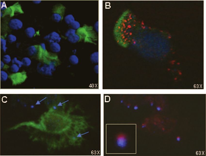
968 NOVOTNY ET AL. INFECT. IMMUN.
cessing and presentation of the peptide immunogen LB1 when Antigen endocytosis. Immature DCs were mixed with 1 g FITC-labeled
delivered to the chinchilla nasal cavity, which likely underlies peptide or protein/ml medium and allowed to adhere to poly-L-lysine (Sigma)-
coated coverslips for 30 min at 37°C. Alternatively, to block endocytosis, imma-
the observed preclinical vaccine efficacy obtained upon i.n. ture DCs were preincubated with 10 nM cytochalasin D (Sigma) for 20 min prior
immunization. to and during incubation with FITC-labeled peptides. The cells were then fixed,
and nonspecific binding sites were blocked with either 2% bovine serum albumin
in Dulbecco’s phosphate-buffered saline (DPBS) (Mediatech) or 10% normal
MATERIALS AND METHODS goat serum in DPBS (Zymed). To label F-actin, cells were incubated with
rhodamine-phalloidin (Molecular Probes). To label MHC class II molecules,
Animals. Adult chinchillas (Chinchilla lanigera) were obtained from Raus-
DCs were incubated with mouse anti-guinea pig MHC class II (AbD Serotec)
cher’s Chinchilla Ranch (LaRue, OH). Animal care and all related procedures
and detected with goat anti-mouse IgG conjugated to Texas Red (Invitrogen). To
were performed in accordance with institutional and federal guidelines and were
inhibit endosome acidification and MHC class II antigen presentation, cells were
conducted under an IACUC-approved protocol.
treated with 10 nM choloroquine (MP Biologicals) for 20 min prior to and during
Generation of chinchilla DCs. Bone marrow cells were isolated from the
incubation with FITC-labeled peptides. An appropriate isotype-matched anti-
femurs of adult chinchillas and cultured in RPMI 1640 medium containing 2 mM
body control was included in each assay. To inhibit fluorescence quenching,
L-glutamine, 100 U penicillin/ml, 100 g streptomycin/ml, 1 mM pyruvate
Prolong AntiFade Gold (Invitrogen) was added to each slide prior to sealing it.
(Mediatech), 5 mM HEPES (Acros), 0.75 g NaHCO3/ml (Fisher Scientific), and
Downloaded from http://iai.asm.org/ on February 17, 2021 by guest
The slides were examined with a Zeiss LSM 510 Meta confocal system attached
0.05 mM -mercaptoethanol (Sigma). The DCs were induced by culturing 2 ⫻
to a Zeiss Axiovert 200 inverted microscope (Carl Zeiss, Inc.).
106 bone marrow cells in 8 ml medium supplemented with 40 ng recombinant
Antigen presentation by chinchilla DCs. For immunization, three doses of 10
human granulocyte-macrophage colony-stimulating factor (GM-CSF)/ml and 40
g LB1 plus 10 g monophosphoryl lipid A (MPL) (Corixa) or 10 g MPL alone
ng recombinant human interleukin 4 (IL-4)/ml (R&D Systems) in 100-mm tissue
were administered to alert chinchillas in a volume of 100 l via subcutaneous
culture dishes. The medium was also supplemented with either 5% fetal bovine injection at weekly intervals. One week after receipt of the third dose, chinchilla
serum (FBS) (Mediatech) or 5% naive chinchilla serum. On day 4 of culture, an splenocytes were isolated by crushing the spleen and passing the cells through a
additional 4 ml of fresh medium was added to the cultures, and on day 6, 40-m cell strainer. Erythrocytes were removed, and the suspensions were en-
nonadherent cells were collected for assay as immature cells. riched for viable cells by centrifugation with Ficoll Paque (GE Healthcare). The
Antibodies and antigens. We used the following fluorochrome-conjugated, resultant cells were labeled with mouse anti-rat CD3 conjugated to biotin (AbD
commercially available antisera to detect cell surface molecules expressed by Serotec) for enrichment of CD3-positive (CD3⫹) T cells by streptavidin-conju-
chinchilla bone marrow-derived DCs: mouse anti-rat CD11c-fluorescein isothio- gated magnetic-bead separation (Miltenyi). CD3⫹ T cells were then cultured in
cyanate (FITC), mouse anti-rat CD80-phycoerythrin, and mouse anti-guinea pig 100-mm2 petri dishes in culture medium supplemented with 5% naive chinchilla
MHC class II revealed with goat anti-mouse immunoglobulin G (IgG)-phyco- serum, but without cytokines. As chinchillas are outbred and thus are not ge-
erythrin (AbD Serotec). Appropriate isotype-matched antibody controls were netically identical animals, we also cultured bone marrow cells from the same
included in all assays. To induce the maturation of chinchilla DC-like cells in animal for 3 days to allow differentiation of precursor cells into DCs in order to
vitro, the cells were stimulated with either 10 ng E. coli lipopolysaccharide demonstrate that the observed T-cell proliferation was due to the presentation of
(LPS)/ml (Sigma) or 10 ng recombinant human tumor necrosis factor alpha antigen by DCs and not to an allogeneic MHC molecule response. After 3 days
(TNF-␣)/ml (Sigma) for 24 h prior to labeling the cells for flow cytometry. in culture, the CD3⫹ T cells were seeded into wells of 96-well round-bottom
Native P5 protein is an approximately 36-kDa OMP and was isolated and plates at a density of 2 ⫻ 105 cells per well in triplicate. The autologous bone
purified from NTHI strain 86-028NP, as previously described (64). LB1 is a marrow-derived DCs were incubated with 1 g or 10 g LB1 or control pep-
4.6-kDa 40-mer synthetic chimeric peptide vaccinogen derived from a surface- tide/ml for 2 h to allow the uptake of antigen, washed, and then added to wells
exposed loop of OMP P5 and has also been described (8). To manage potential that contained the CD3⫹ T cells at a density of 104 cells per well in medium
differences in the binding and uptake of antigens by the chinchilla DCs due to the without cytokines. Controls consisted of wells that contained 1 g or 10 g
substantial difference in the relative molecular masses of OMP P5 and LB1, a concanavalin A/ml (Sigma) plus CD3⫹ T cells and DCs, wells without an antigen
26-kDa NTHI protein (41) called “control protein” and a 3.5-kDa 34-mer syn- (unstimulated), or wells with CD3⫹ T cells only. After 72 h, 20 l of Alamar blue
thetic peptide called “TFPQ3” (1, 2, 5) were used as size control antigens. These (AbD Serotec) was added to each well and the cultures were incubated for an
“control” antigens were selected based exclusively on molecular mass and not on additional 24 h. Proliferation was assessed as the chemical reduction of Alamar
potential known immunoreactivity or lack thereof. Endotoxin contamination of blue by the cells in culture and was detected as the fluorescence of each well at
the proteins and synthetic peptides was determined to be ⱕ0.25 endotoxin 530-nm excitation and 590-nm emission wavelengths with a Bio-Tek Synergy HT
units/ml by Pyrosate agglutination assay (Associates of Cape Cod, Inc.). Peptides microplate reader. The statistical significance of three independent assays was
and proteins were coupled to FITC with a fluorescein protein-labeling kit ac- determined by Tukey’s honestly significant difference test for analysis of variance
cording to the manufacturer’s instructions (Pierce Biotechnology). Based on the pairwise comparisons.
coupling chemistries and amino acid compositions of LB1 and TFPQ3, the In vivo migration of chinchilla respiratory DCs following i.n. immunization.
peptides were estimated to be comparably labeled with FITC. To track the migration of chinchilla nasal mucosa DCs, we adapted the technique
Chemotaxis. To demonstrate the ability of immature LB1-activated or described by Legge and Braciale (44) for use in the chinchilla host. CFSE
TFPQ3-activated chinchilla DCs to sense and respond to a stimulus, a chemo- (Invitrogen) was dissolved to 100 M in dimethyl sulfoxide and further diluted in
taxis assay was performed. Immature DCs were induced to mature by incubation pyrogen-free saline to 8 M prior to administration via microaerosol sprayer
overnight with 1 g of either TFPQ3 or LB1/ml culture medium supplemented (Wolfe Tory Medical, Inc.) in a volume of 40 l/naris to alert, prone chinchillas.
with 5% naive chinchilla serum, but without cytokines. The cells were washed Fluid administered via this regimen is retained exclusively within the chinchilla
and adjusted to a concentration of 106 DCs/ml, and 12 l of unstimulated or nasal cavity, with no dose loss to the respiratory or gastrointestinal tract (72). Six
stimulated DCs was added to one of two horizontally positioned compartments hours later, the chinchillas received either 10 g LB1 plus 10 g MPL or 10 g
that were separated by a 260-m gradient channel in a TAXIScan instrument MPL alone via microaerosol spray as before. One, 3, and 12 h later, animals were
(Effector Cell Institute, Inc.) (38, 52). To align the DCs with the edge of the sacrificed and the cervical, brachial, axillary, and mediastinal lymph nodes were
channel, buffer was aspirated from the second compartment. A volume of 15 l removed, in addition to the spleen. The nasal-associated lymphoid tissue
of 1 M recombinant human C5a (Calbiochem) or buffer was then added to the (NALT) was also removed (37). Tissues were crushed into DPBS, and the cells
second compartment. After 60 min, the cumulative number of DCs that had were passed through a 40-m cell strainer. The spleen was also crushed and
entered the chemotactic gradient in the channel toward C5a or buffer was strained, and the cells were centrifuged with Ficoll-Paque. DCs were labeled with
calculated for each of three independent experiments. mouse anti-rat CD11c-Alexa Fluor 647 (AbD Serotec), and CD11c⫹ CFSE⫹
Antigen binding and internalization by chinchilla DCs. Immature DCs (1 ⫻ cells were detected by flow cytometry. A total of 5,000 viable events were
105) were incubated with medium containing 1 g FITC-labeled peptide or collected for each of three independent assays.
protein/ml for 1 h at 4°C (to assay surface binding of antigen) prior to shifting the To investigate whether LB1-activated, bone marrow-derived DCs would sim-
cultures to 37°C (to assay internalization of the bound antigen). The cells were ilarly follow homing signals within the nasal cavity to the NALT, in vitro-derived
washed, fixed with 2% paraformaldehyde in 0.1 M phosphate buffer, and assayed DCs were labeled with 10 M CFSE prior to incubation with either 10 g LB1
immediately by flow cytometry with a BD FACSCalibur. A total of 10,000 viable plus 10 g MPL or 10 g MPL alone for 15 min. These treated DCs were washed
events were collected for three independent assays, and the data were analyzed to remove unbound peptide, adjusted to 106 cells/80 l, and instilled dropwise by
with FloJo software (Tree Star, Inc.). micropipette to alert, prone chinchillas in a volume of 40 l per naris. The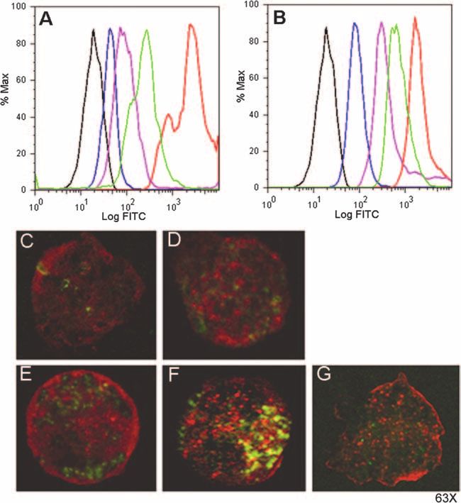
VOL. 76, 2008                                                              ANTIGEN PROCESSING BY CHINCHILLA DENDRITIC CELLS                             969

animals were sacrificed 1 h later, and the lymphoid tissues were processed as
described above. The DCs were labeled with mouse anti-rat CD11c-Alexa Fluor
647, and CD11c⫹ CFSE⫹ cells were detected by flow cytometry. A total of 5,000
viable events were collected for each of three independent assays.
   Aliquots of the NALT cell suspension retrieved 1 h after i.n. administration of
LB1 plus MPL were incubated for 1 hour at 37°C and 5% CO2 on poly-L-lysine-
coated coverslips. The cells were fixed, blocked with 10% normal goat serum, and
incubated with mouse anti-rat CD11c conjugated to Alexa Fluor 647, mouse
anti-guinea pig MHC class II revealed with goat anti-mouse IgG-conjugated to
Texas Red, and mouse anti-rat CD3 conjugated to biotin revealed with strepta-
vidin conjugated to Alexa Fluor 405 (Invitrogen). The slides were sealed after the
addition of Prolong AntiFade Gold, with DAPI (4⬘,6⬘-diamidino-2-phenylindole)
or without DAPI, prior to being viewed via confocal microscopy.

                                  RESULTS

                                                                                                                                                               Downloaded from http://iai.asm.org/ on February 17, 2021 by guest
   The cytokine and serum source influenced the differentia-
tion of chinchilla bone marrow-derived cells in culture. To
establish in vitro cultures of chinchilla DCs, we isolated bone
marrow cells from the femurs of chinchillas and cultured these
cells in medium supplemented with recombinant human cyto-
kines (40 ng GM-CSF/ml and 40 ng IL-4/ml), which resulted in
viable cultures. We further assessed whether the serum source
in the medium influenced the development of these cells. After
1 day, cells in the medium supplemented with a homologous
serum source (naive chinchilla serum) remained in suspension
as individual cells (Fig. 1A) that, in time, developed into a
mixture of floating and adherent cells (Fig. 1C). After 6 days,
we observed numerous floating aggregates of cells (Fig. 1E)
within this culture. Chinchilla bone marrow cells cultured in
medium supplemented with FBS, a serum source widely used
in cell culture systems, formed aggregates suggestive of rapidly
proliferating cells that had attached to the bottom of the cul-
ture dish (Fig. 1B), which appeared larger after 4 days in
culture (Fig. 1D) and finally dispersed after 6 days (Fig. 1F).
   By flow cytometry, the viable cells cultured in medium sup-
plemented with naive chinchilla serum were comparable in size
to those within the medium containing FBS, as demonstrated
by the forward scatter profile (Fig. 1G and H, respectively).
However, the cells cultured in medium supplemented with
chinchilla serum appeared more complex (as demonstrated by
the side scatter profile) than cells within the medium contain-
ing FBS. This observation suggested differences in the inner
complexity of the cells (i.e., the shape of the nucleus, the                            FIG. 1. Influence of serum source on the survival and differentia-
amount and type of cytoplasmic granules, or the membrane                             tion of chinchilla bone marrow-derived cells in culture. (A to F) Light
                                                                                     microscopy images of chinchilla bone marrow-derived cells cultured in
roughness). As the development of cells within the medium                            medium supplemented with 5% naive chinchilla serum (A, C, and E)
supplemented with naive chinchilla serum closely followed that                       or fetal bovine serum (B, D, and F). The arrows in panels E and F
reported for murine and rat bone marrow cultures (31, 70), we                        indicate floating clusters of cells. (G and H) Representative dot plots
concluded that optimal medium supplements required to cul-                           demonstrating forward scatter and side scatter profiles of bone mar-
ture chinchilla bone marrow cells included the use of recom-                         row-derived cells cultured for 6 days in medium supplemented with
                                                                                     either naive chinchilla serum (G) or FBS (H).
binant human cytokines and a homologous serum source.
   Chinchilla bone marrow-derived cells exhibited classic DC
morphology and phenotype after culture with GM-CSF and
IL-4. In order to examine the expression of classical DC sur-                        routinely use rat-specific (and human-specific) reagents for
face markers by the chinchilla DC-like cells, we tested anti-                        immunodetection in the chinchilla model (18, 29, 40, 53, 62).
bodies raised against molecules expressed by other species,                          Greater than 75% of cells within each culture were CD11c⫹, as
including human, mouse, and rat, for reactivity with the chin-                       determined by flow cytometry, also comparable to reports of
chilla bone marrow-derived cells, as no chinchilla-specific re-                      cells cultured from other species. We further examined the
agents are available commercially. Of the panel tested, we                           morphology and phenotype of the DC-like cells by inducing
observed the greatest positive result with mouse anti-rat                            the maturation of the bone marrow-derived cells with 10 ng E.
CD11c (Fig. 2A). It was not unprecedented that a rat-specific                        coli LPS/ml. Immature (unstimulated) cells in culture ex-
reagent recognized a chinchilla homologue, as we and others                          pressed fine processes that extended from the cell surface (Fig.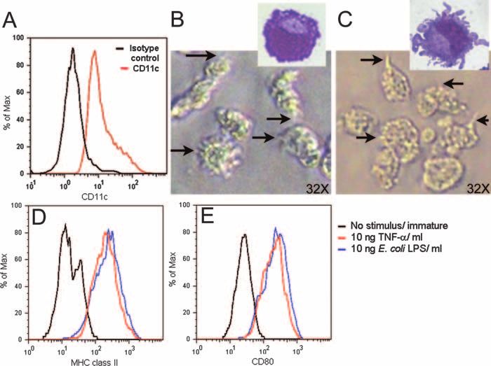
970 NOVOTNY ET AL. INFECT. IMMUN.
Downloaded from http://iai.asm.org/ on February 17, 2021 by guest
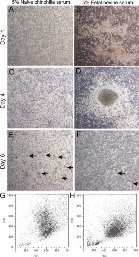
FIG. 2. Chinchilla bone marrow-derived cells exhibited DC-like phenotype and morphology. (A) Representative histogram demonstrating
expression of CD11c by chinchilla bone marrow-derived cells as determined by flow cytometry. Black histogram, isotype control; red histogram,
anti-CD11c. (B and C) Light microscopic images of immature (B) or LPS-matured (C) chinchilla bone marrow-derived cells in culture. The arrows
indicate surface expression of fine processes by immature cells or of larger dendrites by LPS-matured DCs. (Insets) Morphology of immature (B) or
LPS-matured (C) cells after being stained with Giemsa. (D and E) Histograms demonstrating expression of MHC class II (D) and CD80 (E) by
immature chinchilla bone marrow-derived cells (black histograms), TNF-␣-matured cells (red histograms), and LPS-matured cells (blue histo-
grams) as determined by flow cytometry.
2B) and had a large, indented nucleus, which is commonly activated DCs was marginally greater than that of immature
described for DCs (Fig. 2B, inset), whereas mature or LPS- DCs. In contrast, LB1-stimulated DCs continually entered the
stimulated DC-like cells expressed much larger dendrites (Fig. chamber when C5a was present. These data demonstrated that
2C and inset). CD11c⫹ DC-like cells stimulated with either DCs stimulated with LB1 were induced to mature and re-
LPS or recombinant human TNF-␣ upregulated expression of sponded to the presence of a chemoattractant.
both MHC class II molecules (Fig. 2D) and CD80 (Fig. 2E)
compared to immature cells. Therefore, as the chinchilla DC-
like cells were both morphologically and phenotypically com-
parable to described murine and rat bone marrow-derived
DCs, we were confident we had identified conditions to pro-
mote the differentiation of precursor cells within the chinchilla
bone marrow into DCs.
Chinchilla DCs exhibited chemotaxis. One of many func-
tions of DCs is the ability to sense a chemokine gradient and to
respond by directional movement. We therefore assessed chin-
chilla DC chemotaxis toward a classic chemotactic agonist,
C5a, in vitro (48). As demonstrated by the pseudocolored
images captured with the TAXIScan system after 30 min, a
greater number of LB1-activated DCs were present within the
channel compared to the DCs that had been activated with
another synthetic peptide of similar mass (TFPQ3) (Fig. 3A
FIG. 3. Chemotaxis of chinchilla DCs toward C5a. (A and B) Rep-
and B). Furthermore, the chemotactic response was specifi- resentative images captured with the TAXIScan instrument after 30
cally directed toward compartments that contained C5a, as min of chemotaxis toward C5a. The pseudocolored images show
minimal spontaneous migration of cells was observed toward TFPQ3-activated DCs (A) and LB1-activated DCs (B). (C) Plot dem-
compartments that contained only buffer (Fig. 3C), regardless onstrating the cumulative number of DCs that entered the channel
over time. The solid lines depict DC chemotaxis toward C5a, while the
of the maturation status. When plotted as the cumulative num- dashed lines demonstrate spontaneous migration of DCs in buffer. Red
ber of DCs that entered the chemotactic gradient in the chan- lines, LB1-activated DCs; green lines, TFPQ3-activated DCs; gray
nel and were thus moving toward C5a, the number of TFPQ3- lines, immature/unstimulated DCs.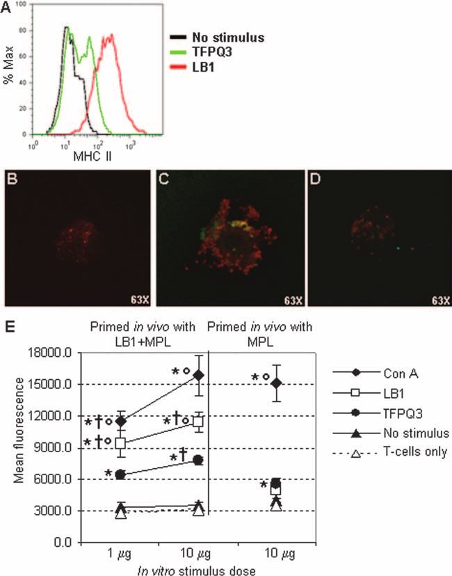
VOL. 76, 2008                                                   ANTIGEN PROCESSING BY CHINCHILLA DENDRITIC CELLS                             971

                                                                                                                                                    Downloaded from http://iai.asm.org/ on February 17, 2021 by guest
  FIG. 4. Chinchilla DCs differentially bound and internalized FITC-labeled proteins and peptides. (A and B) Representative fluorescence-
activated cell sorter histograms demonstrating the mean fluorescence of chinchilla DC cultures incubated at 4°C to assess cell surface binding of
antigens (A), followed by a temperature shift to 37°C to assess internalization of the bound antigens (B). Representative histograms demonstrate
the relative fluorescence of immature DCs (black histograms) and DCs incubated with FITC-labeled antigens: control protein (blue histograms),
OMP P5 (purple histograms), TFPQ3 (green histograms), or LB1 (red histograms). (C to G) Confocal microscopy images demonstrate the
localization of FITC-labeled proteins and peptides within chinchilla DCs (green fluorescence) after the temperature shift to 37°C. DCs were
incubated with either control protein (C), OMP P5 (D), TFPQ3 (E), or LB1 (F). F-actin was labeled with rhodamine-phalloidin (red fluorescence).
Note the yellow fluorescence in panel F, which demonstrated colocalization of FITC-labeled LB1 with actin, which was abrogated upon treatment
with cytochalasin D (G).

   Chinchilla DCs preferentially bound and internalized LB1.               were more fluorescent than cultures incubated with either pro-
To examine the relative abilities of immature chinchilla DCs to            tein, which suggested that the smaller molecular size of the
bind and internalize NTHI OMP P5 and LB1, we utilized a                    peptides than of the native proteins may facilitate antigen
panel of both native proteins and synthetic peptides. FITC-                binding. Furthermore, the greater binding and uptake of LB1
labeled proteins or peptides were incubated with immature                  than of the peptide of comparable size suggested a unique
chinchilla DCs at 4°C to examine antigen surface binding, and              characteristic specific to that immunogen, for example, the
then the cultures were shifted to 37°C to promote internaliza-             presence of a PAMP-like motif.
tion of the bound antigen. At 4°C, all cultures demonstrated                  To further investigate this theory and to assess whether the
binding of the protein or peptide, although at various magni-              preferential binding of LB1 by the chinchilla DCs was also
tudes (Fig. 4A). Whereas DCs incubated with FITC-labeled                   receptor mediated, we examined the localization of the FITC-
OMP P5 demonstrated greater binding than the 26-kDa con-                   labeled proteins and peptides within DCs by confocal micros-
trol peptide, the culture of DCs incubated with FITC-labeled               copy after the temperature shift to 37°C. In agreement with the
LB1 was the most fluorescent of all cultures, with approxi-                flow cytometry data, immature chinchilla DCs internalized the
mately eightfold-greater relative fluorescence than DCs incu-              FITC-labeled proteins and peptides, although to differing mag-
bated with a peptide of similar molecular mass (TFPQ3).                    nitudes. Whereas DCs incubated with the FITC-labeled 26-
   Upon temperature shift to 37°C, the trend of relative fluo-             kDa control protein (Fig. 4C) or FITC-labeled OMP P5 (Fig.
rescence remained the same as that observed at 4°C; however,               4D) contained a few areas of punctate green fluorescence,
the mean fluorescence of each culture increased (Fig. 4B). At              greater internal fluorescence was observed in DCs incubated
either temperature, DC cultures incubated with the peptides                with either FITC-labeled TFPQ3 (Fig. 4E) or FITC-labeled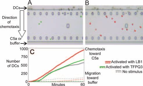
972     NOVOTNY ET AL.                                                                                                            INFECT. IMMUN.

                                                                                                                                                     Downloaded from http://iai.asm.org/ on February 17, 2021 by guest
   FIG. 5. Chinchilla DCs expressed MHC class II molecules and effectively presented antigen to CD3⫹ T cells in vitro. (A) Upregulation of MHC
class II expression as demonstrated by flow cytometry. Black histogram, no stimulus; green histogram, DCs incubated with TFPQ3; red histogram,
DCs incubated with LB1. (B and C) Confocal microscopy images demonstrating MHC class II molecules (red fluorescence) on the surfaces of
chinchilla DCs incubated with either TFPQ3 (B) or LB1 (C). (D) Chloroquine treatment of DCs incubated with LB1 abrogated the MHC class
II-specific fluorescent signal. (E) Chinchilla CD3⫹ T cells primed in vivo with LB1 proliferated upon DC presentation of LB1 in vitro. The results
are shown as the mean ⫾ standard deviation of three independent experiments. *, P ⱕ 0.05 compared to cells with no stimulus; †, P ⱕ 0.05
compared to CD3⫹ T cells isolated from chinchillas immunized with MPL only; °, P ⱕ 0.05 compared to cells stimulated with TFPQ3. Significance
was determined by the Tukey honestly significant difference test for analysis of variance pairwise comparisons.

LB1 (Fig. 4F). The greatest internal fluorescence was clearly              diated by receptor-mediated endocytosis and, further, that a
visible within the cells incubated with LB1, as numerous punc-             motif might be present within the peptide immunogen LB1
tate signals were observed (Fig. 4F). Furthermore, colocaliza-             itself which facilitated this interaction.
tion of actin and LB1 was observed (Fig. 4F, yellow signal),                  DCs upregulated MHC class II molecules and induced T-
which suggested that uptake of the FITC-labeled LB1 likely                 cell proliferation upon activation. To more closely examine the
occurred via endocytosis. To confirm this, DCs were treated                specific interaction between the chinchilla DCs and LB1, we
with cytochalasin D prior to and during incubation with FITC-              examined whether immature chinchilla DCs stimulated with
labeled LB1 to inhibit actin polymerization and formation of               either LB1 or a peptide of similar size subsequently upregu-
endocytic vesicles. Whereas cytochalasin D treatment did not               lated the expression of MHC class II molecules. As detected by
prevent binding of LB1 by the chinchilla DCs, as demonstrated              flow cytometry (Fig. 5A) and confocal microscopy (Fig. 5B),
by the punctate green signals in Fig. 4G, internalization of the           chinchilla DCs incubated with TFPQ3 were labeled with an
peptide was inhibited, as evidenced by the fact that no colo-              MHC class II-specific antibody, although the DCs exhibited
calization of actin and LB1 was observed. These data suggested             only relatively moderate upregulation of MHC class II mole-
that binding and internalization of LB1 were potentially me-               cules over unstimulated DCs (Fig. 5A). Upon incubation of
VOL. 76, 2008 ANTIGEN PROCESSING BY CHINCHILLA DENDRITIC CELLS 973
Downloaded from http://iai.asm.org/ on February 17, 2021 by guest
FIG. 6. DCs resident within the nasal mucosa migrated to the local lymphoid tissues after i.n. administration of LB1 plus MPL. The scatter plots
depict cells isolated 1 h after receipt of LB1 plus MPL (top plot) or MPL alone (bottom plot) for each lymphoid tissue examined. The shaded
region represents CD11c⫹ CFSE⫹ cells. The bar graphs demonstrate the corresponding mean CFSE⫹ fluorescence of the CD11c⫹ CFSE⫹ DCs
1, 3, or 12 h after i.n. administration of LB1 plus MPL or MPL alone. Note the difference in scale between NALT and other lymphoid tissues.
DCs with LB1, however, notably greater MHC class II-specific statistically significant compared to cultures with no stimu-
fluorescence was observed (Fig. 5A and C). Treatment of the lus (P ⱕ 0.05) and thus confirmed the utility of this assay
DCs with chloroquine to inhibit endosome acidification and system to measure chinchilla T-cell proliferation.
MHC class II antigen presentation abrogated the MHC class To examine the proliferation induced by a specific stimulus,
II-specific fluorescent signal (Fig. 5D). These data further DCs were incubated with LB1 prior to culture with CD3⫹ T
demonstrated that internalization of LB1 induced DC matu- cells isolated from chinchillas immunized with LB1 plus MPL.
ration, as evidenced here by upregulated expression of MHC Dose-dependent proliferation resulted, which was significantly
class II molecules. greater than the proliferation induced by DCs incubated with
We next examined the consequence of LB1 internaliza- cells with no stimulus or by CD3⫹ T cells isolated from animals
tion and upregulation of MHC class II molecules by mea- immunized with MPL only (P ⱕ 0.05). These data indicated
suring the induction of CD3⫹ T-cell proliferation in vitro. that upon uptake of LB1, chinchilla DCs effectively processed
We first confirmed that chinchilla T cells could be induced the immunogen and presented peptide fragments, likely in the
to proliferate, as there have been no reports, to our knowl- context of MHC class II and costimulatory molecules, to T
edge, of chinchilla T-cell proliferation assays. Using a non- cells, thereby inducing their proliferation. Moreover, our data
specific stimulus, we observed dose-dependent proliferation suggested that T cells recovered from chinchillas immunized
upon in vitro stimulation of chinchilla CD3⫹ T cells with 10 with LB1 demonstrated a memory response, as significantly
g or 1 g concanavalin A/ml in the presence of DCs (Fig. greater proliferation of T cells isolated from chinchillas immu-
5E). Furthermore, CD3⫹ T cells isolated from chinchillas nized with LB1 was induced by DCs presenting the same an-
immunized with MPL exhibited comparable proliferation at tigen than by DC presentation of an unrelated peptide of
the 10-g concanavalin A/ml dose. These responses were comparable size (P ⱕ 0.05).974 NOVOTNY ET AL. INFECT. IMMUN.
FIG. 7. CFSE-labeled, LB1-activated bone marrow-derived DCs
Downloaded from http://iai.asm.org/ on February 17, 2021 by guest
migrate to the NALT upon i.n. instillation. The scatter plots represent
cells isolated from the NALTs of animals that received DCs activated
in vitro with LB1 plus MPL (A) or MPL (B) 1 h after i.n. administra-
tion.
FIG. 8. Interaction of respiratory CFSE⫹ DCs with CD3⫹ T cells
upon migration to the NALT. (A) Confocal microscopy images of cells
isolated from the NALT 1 hour after i.n. administration of LB1 plus
Chinchilla respiratory DCs migrated to local lymphoid tis-
MPL, demonstrating the presence of CFSE⫹ cells (green) within the
sues following i.n. administration of LB1. We next examined cell suspension (nuclei stained blue). (B) CFSE⫹ cell expressing the
the migration of DCs resident within the chinchilla nasal cavity DC marker CD11c (red). (C) Detection of CD3⫹ fluorescent signals
following i.n. administration of LB1 plus the adjuvant MPL. To (blue; see the arrows) in close proximity to a CFSE⫹ DC (green).
identify potential sites of immune induction, we administered (D) Expression of MHC class II (red) by the CFSE⫹ cell in panel C is
localized directly under the CD3⫹ T cells (blue).
the dye CFSE via microaerosol spray to chinchillas prior to
administration of the immunogen. CFSE labeling allowed dis-
crimination between cells resident within lymphoid tissue and
DCs that had migrated to these tissues from the nasopharyn- from culture of bone marrow cells with CFSE and then acti-
geal mucosa and had been shown not to disturb DC homeosta- vated these cells with LB1 plus MPL or MPL alone in vitro.
sis (44). By this method, at the 1- and 3-hour time points after These CFSE-labeled and activated cells were then instilled into
i.n. administration, the greatest mean fluorescence of CD11c⫹ the chinchilla nasal cavity. One hour later, a distinct population
CFSE⫹ cells was detected within the NALTs of the animals of CD11c⫹ CFSE⫹ cells was detected within the NALT after
that received LB1 plus MPL (Fig. 6A) compared to all other receipt of DCs activated with LB1 plus MPL (Fig. 7A) but not
tissues examined (Fig. 6B to F), suggesting that the NALT when activated with MPL alone (Fig. 7B). Further, 11.5% of
could serve as a primary inductive site following i.n. immuni- the cells isolated from the NALT of the animal administered
zation of the chinchilla host. Moreover, in addition to the LB1-activated DCs were CD11c⫹ CFSE⫹, whereas popula-
NALT, greater mean CFSE⫹ fluorescence was detected from tions of ⱕ1% were detected within the brachial, axillary, cer-
CD11c⫹ cells within the brachial and axillary lymph nodes vical, and mediastinal lymph nodes and the spleen (data not
from the animals that received LB1 plus MPL than from those shown). Whereas homing of nasal mucosa DCs to the axillary
from animals that received only MPL (Fig. 6B and C). These and brachial lymph nodes is shown in Fig. 6B and C, few of the
data demonstrated that the LB1 delivered with MPL influ- instilled bone marrow-derived DCs were detected within these
enced greater DC activation and subsequent migration to these tissues, suggesting subtle differences between the DC types
lymphoid tissues than did the adjuvant alone. Over a 12-h (i.e., those derived from culture in vitro versus those cells
period, a decrease in CFSE⫹ fluorescence was observed within which develop in vivo). Nevertheless, the migration of the
the NALT and brachial and axillary lymph nodes, which indi- instilled DCs from the nasal cavity primarily to the NALT
cated that i.n. delivery of LB1 plus MPL triggered rapid and correlated well with the study in which CFSE had been deliv-
transient influx of DCs from the nasal cavity. In contrast, the ered directly to the nasal cavity (Fig. 6A). The similarity of the
CFSE⫹ signal detected in the cervical and mediastinal lymph results obtained using these two distinct but complementary in
nodes, as well as the spleens, from the animals that received vivo assays strongly indicated that local cytokine/chemokine
LB1 plus MPL was comparable to that from animals that signals likely guided the LB1-activated DCs from the chinchilla
received MPL alone (Fig. 6D to F), which suggested that these nasal cavity to the NALT.
lymphoid tissues likely were not primary inductive sites follow- By confocal microscopy, we further verified that DCs res-
ing i.n. immunization of the host. Thus, DCs within the chin- ident within the nasal cavity migrated to the NALT after i.n.
chilla nasal cavity were induced to mature following i.n. deliv- administration of LB1 plus MPL. CFSE⫹ cells were easily
ery of LB1 plus MPL, which resulted in migration of these distinguished from the other cells isolated from this lym-
activated cells, primarily to the NALT, but also to the axillary phoid aggregate due to their intense green fluorescence and
and brachial lymph nodes. unique morphology (i.e., expression of extensive dendrites)
To confirm that the positive signal described above was (Fig. 8A). Furthermore, the CFSE⫹ cells were labeled with
specifically due to an influx of DCs from the nasal cavity and an anti-CD11c antibody, thus confirming that these cells
not to diffusion of dye from that site, we labeled DCs derived were DCs (Fig. 8B, red fluorescence). We also observedVOL. 76, 2008 ANTIGEN PROCESSING BY CHINCHILLA DENDRITIC CELLS 975
CD3⫹ T cells in close proximity to the DCs within this ized OMP P5 in vitro. However, this result was overshadowed
sample (Fig. 8C, blue fluorescence), suggesting potential by far greater binding and internalization of LB1 by the DCs.
interaction between the two cell types. Upon closer exami- This may have been due to the smaller molecular mass of LB1
nation, MHC class II molecule expression on the DCs was than the native protein (4.6 kDa versus 36.4 kDa). Alterna-
localized directly under the CD3⫹ T-cell fluorescent signal tively, a motif from OMP P5 that is included in the sequence of
(Fig. 8D and inset; red and blue fluorescence, respectively). LB1 to which the DCs bind may be more optimally presented
These data suggested that delivery of LB1 to the nasal than in the native protein. It is interesting to speculate that this
cavities of chinchillas by microaerosol spray induced DCs 19-mer motif (known to be a B-cell epitope) is a PAMP, which
resident within the nasal cavity to take up the antigen, ma- would allow DC binding to the immunogen via a pattern rec-
ture, and migrate to local lymphoid tissues, where they ap- ognition receptor (3). We continue to investigate this hypoth-
peared to interact with T cells within the NALT. esis.
In vivo, by two techniques, we tracked the migration of DCs
from the chinchilla nasal cavity to the NALT. These data
DISCUSSION
demonstrated that LB1 induced DC maturation, which re-
Downloaded from http://iai.asm.org/ on February 17, 2021 by guest
Despite the great utility of the chinchilla for studies of viral sulted in the ability of these cells to sense and respond to local
and bacterial pathogenesis in OM (6, 12, 15, 19, 23, 30, 36, 46, homing signals within the nasal cavity, as similar migration was
49, 54, 69), as well as for preclinical vaccine efficacy studies (7, not detected when only MPL was administered. Our data fur-
20, 22, 39, 56, 68), the cellular mechanisms for the induction of ther demonstrated that a single dose of LB1 delivered i.n. was
a protective immune response in this rodent host have yet to be sufficient to induce the maturation and migration of the DCs.
fully characterized. One component of the innate immune Moreover, the NALT may serve as a primary inductive site
system is the DC, a potent antigen-presenting cell found in following i.n. immunization in this host. Rodent NALT has
many tissues of the body that is capable of initiation and been reported to have functional similarities to Waldeyer’s
modulation of primary immune responses (10, 55, 66). As we lymphoid ring, the lymphatic tissue of the pharynx, palatine
have observed significant efficacy against the development of tonsils, and lingual tonsils, as well as other lymphoid tissue in
NTHI-induced OM after i.n. immunization of the chinchilla the area that encircles the human nasopharynx and oropharynx
with LB1, we were interested to determine whether processing (25, 65, 74). As such, the ability to target the lymphoid tissues
and presentation of the peptide immunogen by nasal-cavity proximal to the anatomical site of infection should be an im-
DCs likely participated in the observed protective response. portant consideration when developing a vaccine and a vaccine
We therefore examined the capture and presentation of LB1 delivery system.
by chinchilla bone marrow-derived DCs in vitro and by DCs In summary, our data are the first to describe the in vitro
within the chinchilla nasal cavity in vivo. culture, functional characterization, and in vivo tracking of
It is well established that precursor cells within the bone chinchilla DCs. We continue to examine the role of DCs in the
marrow of mice and rats can be induced to differentiate into development of a protective immune response following i.n.
DCs by the inclusion of GM-CSF and IL-4 in the culture immunization with LB1 and several other NTHI adhesin-de-
medium (31, 45, 58, 60). We therefore sought to adapt this rived or surface-accessible antigens, for the prevention of
methodology to generate DCs from chinchilla bone marrow NTHI-induced OM. Moreover, the chinchilla serves as a rel-
cells. Two factors impacted our ability to culture the chinchilla- evant animal in which to model multiple viral and bacterial
derived cells: the cytokine specificity and the serum source. diseases of the human respiratory tract; thus, the potential to
First, human-specific recombinant GM-CSF and IL-4, but not apply the methods described here need not be limited to ex-
murine-specific cytokines, supported the viability of these cells amination of vaccine efficacy for NTHI-induced OM only.
in culture. This observation was not without precedent, as the Components of both innate and adaptive immunity can influ-
use of human-specific (and rat-specific) reagents for immuno- ence DC functions. Therefore, the data presented here will be
detection is more successful than when one tries to use mouse- useful as we continue to examine the roles of DCs in the
specific reagents (27, 29, 63), which indirectly suggests that the initiation and modulation of the immune response in the chin-
chinchilla is closer to the rat and human than to mice in terms chilla model.
of similarity of immune molecules. Second, the serum source
used to supplement the medium influenced the development ACKNOWLEDGMENTS
of the culture, an observation similarly reported for murine This work was supported by a grant from the NIH (NIDCD R01
DCs (24, 61). Therefore, whereas it is not surprising that chin- DC03915) to L.O.B.
chillas possess DCs, our data are the first to describe their We thank Joseph Jurcisek, Harivadan Bhagat, and Adrianna
culture. Sumoza-Toledo for expert technical assistance, John Hayes for statis-
tical analysis, and Jennifer Neelans for manuscript preparation. We
It has been reported that DCs bind to OmpA proteins from are also grateful to Kevin Mason for helpful comments and sugges-
K. pneumoniae, E. coli, and A. baumannii, inducing DC matu- tions.
ration and activation, including expression of costimulatory
REFERENCES
molecules, cytokine secretion, and stimulation of T-cell prolif-
1. Adams, L., L. A. Novotny, S. R. Hill, S. Sethi, T. F. Murphy, and L. O.
eration (34, 43, 71). These observations, among others, have Bakaletz. 2007. Epitope-mapping the immune response of children with
led to the designation of this highly conserved family of bac- otitis media and adults with chronic obstructive pulmonary disease to the
terial proteins as a new PAMP (33). NTHI OMP P5 is an PilA protein of the nontypeable Haemophilus influenzae type IV pilus, abstr.
D-58, p. 253. Abstr. 107th Gen. Meet. Am. Soc. Microbiol. American Society
OmpA protein homologue (64). As shown by flow cytometry for Microbiology, DC.
and confocal microscopy, chinchilla DCs bound and internal- 2. Adams, L., L. A. Novotny, S. Sethi, T. F. Murphy, and L. O. Bakaletz. 2007.976 NOVOTNY ET AL. INFECT. IMMUN.
Eptiope-mapping the immune response of children with otitis media and to circumvent nonspecific immunosuppressive activity in vivo. Blood 106:
adults with chronic obstructive pulmonary disease to the PilA protein of 4225–4233.
nontypeable Haemophilus influenzae type IV pilus, p. 130. Abstr. Ninth Int. 25. Heritage, P. L., B. J. Underdown, A. L. Arsenault, D. P. Snider, and M. R.
Symp. Recent Adv. Otitis Media. McDermott. 1997. Comparison of murine nasal-associated lymphoid tissue
3. Akira, S., S. Uematsu, and O. Takeuchi. 2006. Pathogen recognition and and Peyer’s patches. Am. J. Respir. Crit. Care Med. 156:1256–1262.
innate immunity. Cell 124:783–801. 26. Hill, D. J., M. A. Toleman, D. J. Evans, S. Villullas, L. Van Alphen, and M.
4. Avadhanula, V., C. A. Rodriguez, G. C. Ulett, L. O. Bakaletz, and E. E. Virji. 2001. The variable P5 proteins of typeable and non-typeable Hae-
Adderson. 2006. Nontypeable Haemophilus influenzae adheres to intercellu- mophilus influenzae target human CEACAM1. Mol. Microbiol. 39:850–862.
lar adhesion molecule 1 (ICAM-1) on respiratory epithelial cells and up- 27. Hill, S. R., L. A. Novotny, and L. O. Bakaletz. 2005. Characterization of the
regulates ICAM-1 expression. Infect. Immun. 74:830–838. immune response induced by a parenteral prime-mucosal boost immuniza-
5. Bakaletz, L. O., B. D. Baker, J. A. Jurcisek, A. Harrison, L. A. Novotny, J. E. tion strategy vs. a mucosal priming and boosting regimen using the vaccine
Bookwalter, R. Mungur, and R. S. Munson, Jr. 2005. Demonstration of type candidate LB1 against nontypeable Haemophilus influenzae-induced otitis
IV pilus expression and a twitching phenotype by Haemophilus influenzae. media, p. 148. Abstr. Fifth Extraordinary Int. Symp. Recent Adv. Otitis
Infect. Immun. 73:1635–1643. Media.
6. Bakaletz, L. O., and K. A. Holmes. 1997. Evidence for transudation of 28. Hill, S. R., L. A. Novotny, and L. O. Bakaletz. 2005. Characterization of the
specific antibody into the middle ears of parenterally immunized chinchillas protective response induced by a mucosal immunization strategy vs. one of
after an upper respiratory tract infection with adenovirus. Clin. Diagn. Lab. parenteral priming and mucosal boosting using an adhesin-based vaccine
Immunol. 4:223–225. candidate against otitis media caused by nontypeable Haemophilus influen-
Downloaded from http://iai.asm.org/ on February 17, 2021 by guest
7. Bakaletz, L. O., B. J. Kennedy, L. A. Novotny, G. Duquesne, J. Cohen, and zae, abstr. D-099, p. 214. Abstr. 105th Gen. Meet. Am. Soc. Microbiol.
Y. Lobet. 1999. Protection against development of otitis media induced by American Society for Microbiology, Washington, DC.
nontypeable Haemophilus influenzae by both active and passive immuniza- 29. Hong, W., K. Mason, J. Jurcisek, L. Novotny, L. O. Bakaletz, and W. E.
tion in a chinchilla model of virus-bacterium superinfection. Infect. Immun. Swords. 2007. Phosphorylcholine decreases early inflammation and pro-
67:2746–2762. motes the establishment of stable biofilm communities of nontypeable Hae-
8. Bakaletz, L. O., E. R. Leake, J. M. Billy, and P. T. Kaumaya. 1997. Relative mophilus influenzae strain 86-028NP in a chinchilla model of otitis media.
immunogenicity and efficacy of two synthetic chimeric peptides of fimbrin as Infect. Immun. 75:958–965.
vaccinogens against nasopharyngeal colonization by nontypeable Haemophi- 30. Hong, W., B. Pang, S. West-Barnette, and W. E. Swords. 2007. Phosphoryl-
lus influenzae in the chinchilla. Vaccine 15:955–961. choline expression by nontypeable Haemophilus influenzae correlates with
9. Bakaletz, L. O., B. M. Tallan, T. Hoepf, T. F. DeMaria, H. G. Birck, and D. J. maturation of biofilm communities in vitro and in vivo. J. Bacteriol. 189:
Lim. 1988. Frequency of fimbriation of nontypable Haemophilus influenzae 8300–8307.
and its ability to adhere to chinchilla and human respiratory epithelium. 31. Inaba, K., M. Inaba, N. Romani, H. Aya, M. Deguchi, S. Ikehara, S. Mura-
Infect. Immun. 56:331–335. matsu, and R. M. Steinman. 1992. Generation of large numbers of dendritic
10. Banchereau, J., and R. M. Steinman. 1998. Dendritic cells and the control of cells from mouse bone marrow cultures supplemented with granulocyte/
immunity. Nature 392:245–252. macrophage colony-stimulating factor. J. Exp. Med. 176:1693–1702.
11. Bookwalter, J. E., J. A. Jurcisek, S. D. Gray-Owen, S. Fernandez, G. McGil- 32. Jeannin, P., B. Bottazzi, M. Sironi, A. Doni, M. Rusnati, M. Presta, V.
livary, and L. O. Bakaletz. 2008. A carcinoembryonic antigen-related cell Maina, G. Magistrelli, J. F. Haeuw, G. Hoeffel, N. Thieblemont, N. Corvaia,
adhesion molecule 1 homologue plays a pivotal role in nontypeable Hae- C. Garlanda, Y. Delneste, and A. Mantovani. 2005. Complexity and comple-
mophilus influenzae colonization of the chinchilla nasopharynx via the outer mentarity of outer membrane protein A recognition by cellular and humoral
membrane protein P5-homologous adhesin. Infect. Immun. 76:48–55. innate immunity receptors. Immunity 22:551–560.
12. Bouchet, V., D. W. Hood, J. Li, J. R. Brisson, G. A. Randle, A. Martin, Z. Li, 33. Jeannin, P., G. Magistrelli, L. Goetsch, J. F. Haeuw, N. Thieblemont, J. Y.
R. Goldstein, E. K. Schweda, S. I. Pelton, J. C. Richards, and E. R. Moxon. Bonnefoy, and Y. Delneste. 2002. Outer membrane protein A (OmpA): a
2003. Host-derived sialic acid is incorporated into Haemophilus influenzae new pathogen-associated molecular pattern that interacts with antigen pre-
lipopolysaccharide and is a major virulence factor in experimental otitis senting cells—impact on vaccine strategies. Vaccine 20(Suppl. 4):A23–A27.
media. Proc. Natl. Acad. Sci. USA 100:8898–8903. 34. Jeannin, P., T. Renno, L. Goetsch, I. Miconnet, J. P. Aubry, Y. Delneste, N.
13. Cripps, A. W., and D. C. Otczyk. 2006. Prospects for a vaccine against otitis Herbault, T. Baussant, G. Magistrelli, C. Soulas, P. Romero, J. C. Cerottini,
media. Exp. Rev. Vaccines 5:517–534. and J. Y. Bonnefoy. 2000. OmpA targets dendritic cells, induces their mat-
14. Crowe, S. R., S. C. Miller, R. M. Shenyo, and D. L. Woodland. 2005. uration and delivers antigen into the MHC class I presentation pathway. Nat.
Vaccination with an acidic polymerase epitope of influenza virus elicits a Immunol. 1:502–509.
potent antiviral T cell response but delayed clearance of an influenza virus 35. Jiang, Z., N. Nagata, E. Molina, L. O. Bakaletz, H. Hawkins, and J. A. Patel.
challenge. J. Immunol. 174:696–701. 1999. Fimbria-mediated enhanced attachment of nontypeable Haemophilus
15. Daines, D. A., M. Bothwell, J. Furrer, W. Unrath, K. Nelson, J. Jarisch, N. influenzae to respiratory syncytial virus-infected respiratory epithelial cells.
Melrose, L. Greiner, M. Apicella, and A. L. Smith. 2005. Haemophilus Infect. Immun. 67:187–192.
influenzae luxS mutants form a biofilm and have increased virulence. Microb. 36. Jurcisek, J. A., and L. O. Bakaletz. 2007. Biofilms formed by nontypeable
Pathog. 39:87–96. Haemophilus influenzae in vivo contain both double-stranded DNA and type
16. Del Giudice, G. 2003. Vaccination strategies. An overview Vaccine 21(Suppl. IV pilin protein. J. Bacteriol. 189:3868–3875.
2):S83–S88. 37. Jurcisek, J. A., J. E. Durbin, D. F. Kusewitt, and L. O. Bakaletz. 2003.
17. Eldika, N., and S. Sethi. 2006. Role of nontypeable Haemophilus influenzae Anatomy of the nasal cavity in the chinchilla. Cells Tissues Organs 174:136–
in exacerbations and progression of chronic obstructive pulmonary disease. 152.
Curr. Opin. Pulm. Med. 12:118–124. 38. Kanegasaki, S., Y. Nomura, N. Nitta, S. Akiyama, T. Tamatani, Y. Gos-
18. Figueira, M. A., S. Ram, R. Goldstein, D. W. Hood, E. R. Moxon, and S. I. hoh, T. Yoshida, T. Sato, and Y. Kikuchi. 2003. A novel optical assay
Pelton. 2007. Role of complement in defense of the middle ear revealed by system for the quantitative measurement of chemotaxis. J. Immunol.
restoring the virulence of nontypeable Haemophilus influenzae siaB mutants. Methods 282:1–11.
Infect. Immun. 75:325–333. 39. Kennedy, B. J., L. A. Novotny, J. A. Jurcisek, Y. Lobet, and L. O. Bakaletz.
19. Giebink, G. S. 1999. Otitis media: the chinchilla model. Microb. Drug Resist. 2000. Passive transfer of antiserum specific for immunogens derived from a
5:57–72. nontypeable Haemophilus influenzae adhesin and lipoprotein D prevents
20. Giebink, G. S., M. Koskela, P. P. Vella, M. Harris, and C. T. Le. 1993. otitis media after heterologous challenge. Infect. Immun. 68:2756–2765.
Pneumococcal capsular polysaccharide-meningococcal outer membrane pro- 40. Kerschner, J. E., T. K. Meyer, A. Burrows, G. Ehrlich, and J. C. Post. 2006.
tein complex conjugate vaccines: immunogenicity and efficacy in experimen- Mucin gene cDNA sequence characterization in chinchilla middle ear epi-
tal pneumococcal otitis media. J. Infect. Dis. 167:347–355. thelium. Int. J. Pediatr. Otorhinolaryngol. 70:1449–1456.
21. Giebink, G. S., Y. Kurono, L. O. Bakaletz, J. M. Kyd, S. J. Barenkamp, T. F. 41. Kyd, J. M., and A. W. Cripps. 1998. Potential of a novel protein, OMP26,
Murphy, B. Green, P. L. Ogra, X. X. Gu, J. A. Patel, T. Heikkinen, S. I. from nontypeable Haemophilus influenzae to enhance pulmonary clearance
Pelton, M. Hotomi, and P. Karma. 2005. Recent advances in otitis media. 6. in a rat model. Infect. Immun. 66:2272–2278.
Vaccine. Ann. Otol. Rhinol. Laryngol. Suppl. 194:86–103. 42. Kyd, J. M., A. W. Cripps, L. A. Novotny, and L. O. Bakaletz. 2003. Efficacy
22. Giebink, G. S., J. D. Meier, M. K. Quartey, C. L. Liebeler, and C. T. Le. 1996. of the 26-kilodalton outer membrane protein and two P5 fimbrin-derived
Immunogenicity and efficacy of Streptococcus pneumoniae polysaccharide- immunogens to induce clearance of nontypeable Haemophilus influenzae
protein conjugate vaccines against homologous and heterologous serotypes from the rat middle ear and lungs as well as from the chinchilla middle ear
in the chinchilla otitis media model. J. Infect. Dis. 173:119–127. and nasopharynx. Infect. Immun. 71:4691–4699.
23. Gitiban, N., J. A. Jurcisek, R. H. Harris, S. E. Mertz, R. K. Durbin, L. O. 43. Lee, J. S., J. C. Lee, C. M. Lee, I. D. Jung, Y. I. Jeong, E. Y. Seong, H. Y.
Bakaletz, and J. E. Durbin. 2005. Chinchilla and murine models of upper Chung, and Y. M. Park. 2007. Outer membrane protein A of Acinetobacter
respiratory tract infections with respiratory syncytial virus. J. Virol. 79:6035– baumannii induces differentiation of CD4⫹ T cells toward a Th1 polarizing
6042. phenotype through the activation of dendritic cells. Biochem. Pharmacol.
24. Haase, C., M. Ejrnaes, A. E. Juedes, T. Wolfe, H. Markholst, and M. G. von 74:86–97.
Herrath. 2005. Immunomodulatory dendritic cells require autologous serum 44. Legge, K. L., and T. J. Braciale. 2003. Accelerated migration of respiratoryYou can also read