Escherichia coli's physiology can turn membrane voltage dyes into actuators
←
→
Page content transcription
If your browser does not render page correctly, please read the page content below
bioRxiv preprint first posted online Apr. 12, 2019; doi: http://dx.doi.org/10.1101/607838. The copyright holder for this preprint
(which was not peer-reviewed) is the author/funder, who has granted bioRxiv a license to display the preprint in perpetuity.
                               It is made available under a CC-BY-ND 4.0 International license.

                       Escherichia coli’s physiology can turn
                       membrane voltage dyes into actuators
                       L Mancini1 , G Terradot1,* , T Tian2,* , Y Pu2 , Y Li2 , CJ Lo3 , F Bai2 , and T
                       Pilizota1,+
                       * these   authors contributed equally to this work
                       + Corresponding    author:teuta.pilizota@ed.ac.uk

                       ABSTRACT

                        The electrical membrane potential (Vm ) is one of the components of the electrochemical potential of
                        protons across the biological membrane (proton motive force), which powers many vital cellular processes,
                        and Vm also plays a role in signal transduction. Therefore, measuring it is of great interest, and over
                        the years a variety of techniques has been developed for the purpose. In bacteria, given their small
                        size, Nernstian membrane voltage probes are arguably the favourite strategy, and their cytoplasmic
                        accumulation depends on Vm according to the Nernst equation. However, a careful calibration of Nernstian
                        probes that takes into account the trade-offs between the ease with which the signal from the dye is
                        observed, and the dyes’ interactions with cellular physiology, is rarely performed. Here we use a
                        mathematical model to understand such trade-offs and, based on the knowledge gained, propose a
                        general work-flow for the characterization of Nernstian dye candidates. We demonstrate the work-flow on
                        the Thioflavin T dye in Escherichia coli, and identify conditions in which the dye turns from a Vm probe
                        into an actuator.

                       Keywords:       Nernstian probes, single-cell microscopy, Escherichia coli

                       SIGNIFICANCE STATEMENT
                       The phospholipid bilayer of a biological membrane is virtually impermeable to charged molecules. Much
                       like in a rechargeable battery, cells harness this property to store an electrical potential that fuels life
                       reactions but also transduces signals. Measuring this electrical potential, also referred to as membrane
                       voltage, is therefore of great interest and a variety of techniques have been employed for the purpose,
                       starting as early as the 1930s. For the case of bacteria, which are smaller in size and possess a stiffer
                       cell wall, arguably the most popular approach to measuring membrane voltage are Nernstian probes
                       that accumulate across the bacterial membrane according to the Nernst potential. The present study
                       characterizes the undesired effects Nernstian probes can have on cell physiology, which can be crucial for
                       the accurate interpretation of experimental results. Using mathematical modelling and experiments, the
                       study provides a general, simple workflow to characterise and minimise these effects.

                       INTRODUCTION
                       Living cells maintain an electric potential difference (Vm ) across the plasma membrane that acts like a
                       capacitor, and is achieved by active transport of ions:
                                       Qin
                              Vm = F ·                                                                                       (1)
                                        C
                       where Qin is the intracellular charge (in mole), C the membrane capacitance and F the Faraday constant.
                       Membrane potential stands at the basis of fundamental biological processes, such as signal transduction
                       and energy production [Del Castillo and Katz, 1954, Mitchell, 1961]. For the latter, Vm adds up to the
                       chemical potential of protons, arising from their concentration difference across the membrane, to result
                       in the proton electrochemical gradient, so called proton motive force (PMF). The PMF drives numerous
                       cellular processes, most notably the production of ATP [Mitchell, 1961], import of nutrients or osmolites
                       [Bradbeer, 1993, Jahreis et al., 2008, Ramos and Kaback, 1977, Wood, 2015] rotation of the bacterial
                       flagellar motor [Sowa and Berry, 2008], and it is necessary for cell division [Strahl and Hamoen, 2010].

                                                                                   1
bioRxiv preprint first posted online Apr. 12, 2019; doi: http://dx.doi.org/10.1101/607838. The copyright holder for this preprint
(which was not peer-reviewed) is the author/funder, who has granted bioRxiv a license to display the preprint in perpetuity.
                               It is made available under a CC-BY-ND 4.0 International license.

                            The notion that Vm lies at the very basis of life motivated decades long efforts to measure it. The
                       first technique dates to 1939 and relies on the mechanical insertion of microelectrodes into squid giant
                       axons [Hodgkin and Huxley, 1939]. The method was later applied to neurons [Ling and Gerard, 1949],
                       and led to the development of the patch-clamp technique, which advanced the understanding of neuron
                       signal transduction [Neher and Sakmann, 1976, Sakmann and Neher, 1984]. However, applicability of
                       microelectrodes for the measurement of bacterial Vm is limited, owing to the small size of the organisms
                       and the presence of the cell wall [Martinac et al., 1987, 2013]. Some of the subsequently developed
                       methods overcome such limits with the use of molecular sensors [Felle et al., 1980], grouped in two
                       categories: conformational-change-based sensors and Nernstian sensors. The former are static molecules
                       or proteins that sit inside the membrane, or in its close proximity, and change conformation or electron
                       distribution in response to changes in Vm , which in turn affect the optical properties of the chromophores
                       thus transducing signal [Fluhler et al., 1985, Kralj et al., 2011, Tsutsui et al., 2008]. Here we focus on
                       the latter, the Nernstian sensors, and on the parameter range in which they serve as Vm indicators, using
                       Escherichia coli as the model organism.
                            Nernstian sensors are charged molecules that can diffuse across the biological membranes and
                       distribute according to the Nernst equation:


                                     RT      cout
                              Vm =      ln                                                                                           (2)
                                     zF      cin

                       where R, T , z, F, cout , cin denote respectively gas constant, temperature, valence of the charged molecule,
                       Faraday’s constant, external and internal concentration of the charged molecule. For a measurement to
                       be attained, these molecules need to emit a signal that is a proxy for their number. Therefore, Nernstian
                       Vm dyes are usually radio-labeled or fluorescent molecules [Felle et al., 1980, Sims et al., 1974], and Vm
                       is calculated from equation (2) by measuring the cytoplasmic (cin ) and the external dye concentration
                       (cout [Lo et al., 2007].
                            However, Nernstian dyes are used in complex biological systems and a number of factors can be
                       responsible for an incomplete adherence to a fully Nernstian behavior. In Fig. 1 we give a cartoon
                       representation of the trade-offs imposed on a Nernstian dye by plotting the dye intensity inside E. coli’s
                       cytoplasm against the time. The chosen dye concentration should be such that the signal is sufficiently
                       above the background (∆I is sufficiently large). Yet, with increasing dye concentration, cell’s Vm is more
                       likely to be affected by the dye. This caveat is inherent to positively charged dyes as these directly lower
                       Vm and more so at higher concentrations [Kashket, 1985]. The first requirement for a Nernstian dye is,
                       thus, existence of a range of concentrations that give sufficient signal without extensively affecting the Vm .
                       Likewise, cellular processes should not interfere with the Nernstian behavior of the dye, for example by
                       actively importing or exporting it. Instead, the dye should be able to diffuse across the membrane and
                       its diffusion constant will determine the time it takes for the dye to equilibrate across the membrane in
                       agreement with equation (2) (τeq in Fig. 1). All phenomena that occur quicker than τeq are beyond the
                       dye’s temporal resolution, and likewise, all the measurements taken before τeq do not faithfully report
                       Vm . Lastly, for quantitative measurements, Nernstian dyes should exhibit a well defined and constant
                       correlation between the concentration and the signal, e.g. the dye should not self-quench at any point
                       [te Winkel et al., 2016] or undergo signal enhancements.
                            To summarize, to be used as a Nernstian sensor, a cationic dye should: (i) give a sufficiently high
                       signal without affecting cell’s Vm ; (ii) diffuse through the membrane with τeq on the order of minutes; (iii)
                       stay inert, despite being charged, and not form bonds or in any way interact with cell; (iv) have constant
                       signal per molecule. Yet, when using such dyes these requirements are rarely assessed in a systematic
                       manner before measurements commence. In this work we identify a work-flow that should be adopted in
                       order to identify the parameter range in which Nernstain dyes act as sensors, rather than actuators. We
                       start with a mathematical model that helps us understand relationships and define trade-offs between dye
                       working concentration and signal intensity, equilibration time and Vm perturbation. We then show how
                       the identified work-flow can be used to benchmark new Nernstian dyes by characterizing the recently
                       reported dye Thioflavin T (ThT) used in Bacillus subtilis [Prindle et al., 2015], for use in E. coli. We
                       describe the physiological range in which ThT enables Vm sensing in E. coli, and, in the range where we
                       find it turns into an actuator, we investigate the mechanistic reasons. Our work-flow can be applied to the
                       characterization of other Nernstian dyes and provide novel insights for the established ones.

                                                                                                                                    2/15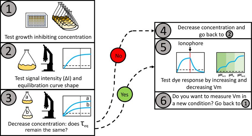
bioRxiv preprint first posted online Apr. 12, 2019; doi: http://dx.doi.org/10.1101/607838. The copyright holder for this preprint
(which was not peer-reviewed) is the author/funder, who has granted bioRxiv a license to display the preprint in perpetuity.
                               It is made available under a CC-BY-ND 4.0 International license.

                       Figure 1. A schematic plot of a Nernstian dye equilibration curve. Equilibration time, τeq , is defined
                       as the time at which the internalized dye Iin reaches the 90% of its final value. Vm,0 and Vm,dye indicate
                       the membrane potential before and after the addition of the dye, respectively. Inset: cartoon showing the
                       mechanism of accumulation of cationic dyes, which accumulate more in cells with a highly negative Vm .

                       METHODS
                       Bacterial strains
                       All experiments where no mutation is explicitly indicated were carried out in the MG1655 strain. For the
                       BFM speed assay we used MG1655 carrying the FliCsticky mutation from [Krasnopeeva et al., 2018].
                       ∆tolC mutants were obtained from the Keio collection [Baba et al., 2006]. Kanamycin resistance of the
                       Keio deletion strain was removed via one-step inactivation with the plasmid pCP20 [Datsenko and Wanner,
                       2000]. Kanamycin resistance inactivation and elimination of the pCP20 plasmid were confirmed via
                       Kanamycin, Chloramphenicol and Ampicillin sensitivity tests. Both the strain carrying the ∆tolC mutation
                       and MG1655 wild-type were transformed with plasmid pTP20-mKate2 (Fig. SI9) for cytoplasmic volume
                       measurements. pTP20-mKate2 contains the red fluorescent protein mKate2 and the ribosomal binding
                       site (RBS) of mCherry. The plasmid was constructed as follows: the backbone from pWR20 [Pilizota and
                       Shaevitz, 2012] and the sequence containing the RBS of mCherry and mKate2 were PCR amplified. The
                       products were purified, cleaved with the restriction enzymes AvrII and NotI (NEB, UK) and ligated using
                       T4 DNA ligase (Promega, UK). Chemically competent cells were transformed with the ligation mixes
                       and transformants were confirmed by colony PCR and subsequently sequenced. A map of the plasmid
                       and the primers are given in SI (Fig. SI9, and Table SI1). All the strains used in the study are summarized
                       in the Table SI2.

                       Bacterial growth conditions
                       Cells for fluorescence microscopy were grown from an overnight culture by diluting it 1:80 times in LB
                       (0.5% Yeast Extract, 1% Bacto tryptone, 0.5% NaCl). The culture was shaken at 220 rpm at 37◦ C and
                       harvested at OD600 =0.3-0.5. Upon harvest we washed the cells into fresh LB or MM9 + glucose medium
                       (50mM Na2 HPO4 , 25mM NaH2 PO4 , 8.5mM NaCl, 18.7mM NH4Cl, 0.1mM CaCl2 , 1mM KCl, 2mM
                       MgSO4 , 1x MEM essential amino acids (Gibco, UK) and 0.3% glucose). For the simultaneous BFM
                       speed and ThT fluorescence measurements cells were grown from an overnight culture by diluting it 1:80
                       times in TB (1% Bacto tryptone, 0.5% NaCl) at 200 rpm and 30◦ C. Cells were harvested at OD600 =0.8 as
                       before [Rosko et al., 2017] and washed to fresh MM9 via centrifugation. Growth curves were obtained
                       in a Spectrostar Omega microplate reader (BMG, Germany) using a flat-bottom 96-well plate that was
                       covered with a lid during the experiments (Costar, UK). Each well contained 200µl of growth media,
                       either MM9 + glucose or MM9 + glycerol (50mM Na2 HPO4 , 25mM NaH2 PO4 , 8.5mM NaCl, 18.7mM
                       NH4Cl, 0.1mM CaCl2 , 1mM KCl, 2mM MgSO4 , 1x MEM essential aminoacids (Gibco, UK) and 0.3%

                                                                                                                                    3/15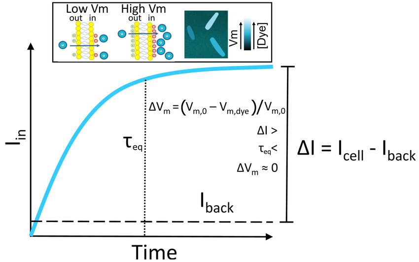
bioRxiv preprint first posted online Apr. 12, 2019; doi: http://dx.doi.org/10.1101/607838. The copyright holder for this preprint
(which was not peer-reviewed) is the author/funder, who has granted bioRxiv a license to display the preprint in perpetuity.
                               It is made available under a CC-BY-ND 4.0 International license.

                       glycerol), and was inoculated with 2µl (1:100 dilution) of an overnight culture. Plates were grown at
                       37◦ C with 300 rpm shaking (double orbital mode) . ThT (Acros organics, USA) solutions were prepared
                       from a 10 mM stock of ThT in water made at least monthly and stored at 4◦ C in the dark.

                       Fluorescence microscopy
                       Imaging was carried out in a custom-built microscope with a 100x oil immersion objective lens (Nikon,
                       Japan), Neutral White LED as a source of illumination (Cairn Research Ltd, UK) and images were
                       taken with an iXon Ultra 897 EMCCD camera (Andor, UK) [Krasnopeeva, 2018, Rosko, 2017]. ThT
                       fluorescence was measured with ZET436/20x and ET525/40m, and mKate2 and PI fluorescence with
                       ET577/25x and ET632/60m (Chroma Technology, USA) excitation and emission filters, respectively.
                       Exposure time was 50 ms and Andor camera gain 25. We note that ThT undergoes a spectral shift and
                       intensity increase when highly concentrated or when spatially constricted, either by binding to amyloid
                       fibrils or by viscosity [Maskevich et al., 2015, 2007, Sulatskaya et al., 2017]. Our choice of filters
                       aims at minimizing these effects and the damage that shorter wavelengths cause to E. coli [Vermeulen
                       et al., 2008]. Cells were imaged in a custom-built flow-cell (Fig. SI10, [Krasnopeeva et al., 2018]), and
                       attached to the coverslip surface as before [Krasnopeeva et al., 2018, Rosko et al., 2017]. Briefly, 1%
                       Poly-L-Lysine (Sigma, UK) is flushed through the flow cell and washed with 3-5 ml of growth media after
                       10 s. Polystyrene particles (beads) with a diameter of 1 µm (Bangs Laboratories, USA), were delivered
                       into the flow-cell and allowed to attach to the coverslip surface. After 10 min unattached beads were
                       flushed away with 1-2 ml of growth media. Next, 200 µl of cells were delivered to the flow-cell and
                       allowed to attach for 10-30 min, after which the unattached cells were removed with 1 ml of growth
                       medium. 10 µM ThT in growth media was delivered with a peristaltic pump (Fusion 400, Chemyx, USA)
                       using 50 µl/min flow rate while imaging. We deliver 5 µM of PI stain (MP Biomedicals, USA) in the
                       same way. 5mM PI stock solution (in water) was stored at 4◦ C in the dark. Images were stabilized in x, y
                       and z position using a bead attached to the cover-slip and back-focal-plane interferometry [Buda et al.,
                       2016, Pilizota and Shaevitz, 2012].

                       Motor speed measurements
                       Single motor speeds were measured as before [Krasnopeeva et al., 2018, Rosko et al., 2017]. Briefly,
                       we sheared flagellar filaments by passing them through two syringes with narrow-gauge needles (26
                       gauge) connected by plastic tubing. The cell attachment protocol was as above, except 0.5 µm beads
                       (Polysciences, USA) were delivered after cell attachment allowing them to attach to filament stubs. Motor
                       speed was measured during continuous flow that delivered MM9 + glucose medium supplemented with
                       10 µM ThT. Back-focal-plane interferometry setup and recording conditions are as before [Rosko et al.,
                       2017].

                       Data analysis
                       Motor speed traces
                       Raw traces of the position of the bead attached to the filament stub were analyzed by a moving-window
                       discrete Fourier transform as in [Rosko et al., 2017]. From the obtained motor speed traces DC frequency
                       (50 Hz) was removed, speeds lower then 5 Hz ignored, and subsequently a median filter (window size
                       11) was applied [Krasnopeeva et al., 2018]. We note that we use a wild type strain for which the BFM
                       can change rotational direction, which appears as a negative speed after application of the moving-
                       window Fourier transform. However, for the purpose of PMF measurements these short intervals can be
                       disregarded, and we only show the speed values above 0 Hz.
                       Fluorescence images
                       The image analysis was carried out with a custom written software. From fluorescence images, rectangles
                       containing ’flat’ cells, i.e. cells that are uniformly attached to the coverslip surface, as well as background
                       rectangles within each cell-containing rectangle, were manually selected [Buda et al., 2016, Pilizota and
                       Shaevitz, 2012]. The edge of the cell was identified within the cell-containing rectangle by applying a
                       global threshold via the Otsu’s method [Otsu, 1979]. To total cells’ intensity values were obtained by
                       summing up and averaging pixel belonging to the cells. Values obtained from the background rectangles
                       at the time points when ThT was loaded in the channel but cells had not taken it up yet, were subtracted
                       from the cell intensity values. The beads used for image stabilisation stain easily with ThT, and were
                       used as a point of reference for dye entry (which in our case occurred 7 to 10 min from the start of

                                                                                                                                    4/15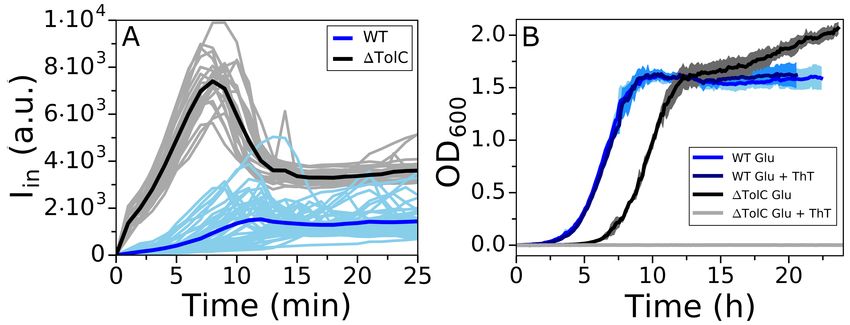
bioRxiv preprint first posted online Apr. 12, 2019; doi: http://dx.doi.org/10.1101/607838. The copyright holder for this preprint
(which was not peer-reviewed) is the author/funder, who has granted bioRxiv a license to display the preprint in perpetuity.
                               It is made available under a CC-BY-ND 4.0 International license.

                       imaging). We show fluorescence intensity traces that start at the point of ThT entry, but note that cells
                       were exposed to fluorescence illumination in the 7-10 min interval before. For the low fluorescence values
                       characteristic of the early stages of dye equilibration, our script fails to identify cells, in which case we
                       linearly interpolate values between two closest events of successful cell identification. Cell area was
                       measured from intensity profiles, by normalizing them and counting the pixels above 30% of maximum
                       intensity as described previously [Buda et al., 2016, Pilizota and Shaevitz, 2012]
                       Plate reader data
                       Individual growth curves were analysed with the software deODorizer from [Swain et al., 2016]. To
                       extract the maximum growth rate, 3 or more repeats in the same condition were aligned by the chosen
                       OD value (usually OD∼0.4 ) using the growth curve that reached it first (in the given condition). The
                       maximum growth rates given in Fig. 3C were normalized by the maximum growth rate in [Dye]out =0
                       condition.

                       RESULTS
                       Mathematical model of Nernstian dye’s behavior defines its working parameter range
                       To predict and understand the mutual effects of dye concentration and cell physiology we turn to a
                       mathematical model. We assume that the cytoplasmic and extracellular liquids are electrical conductors
                       separated by the membrane, which we treat as a parallel-plate capacitor (equation 1) [Grabe and Oster,
                       2001]. We account for four types of charge carriers and assume that all are monovalent to simplify the
                       model without altering the results with respect to Vm dye behavior: (i) negatively charged molecules to
                       which the membrane is close to non-permeable denoted Y , (ii) cationic species actively pumped outward
                       denoted C+ , (iii) anionic species, which equilibrates across the membrane A− and (iv) cationic species
                       that equilibrate across the membrane playing the part of a cationic dye. Thus Qin is:

                              Qin = VCell · [Dye]in + [C+ ]in − [A− ]in − [Y ]in

                                                                                                                           (3)

                       where Vcell is the intracellular volume. The extracellular concentrations and [Y ]in are constants set by the
                       initial conditions (we assume that the cell does not affect the ionic composition of its environment and we
                       treat [Y ]in as unable to cross the membrane). We also note that [Dye]in and [Dye]out are experimentally
                       determined from fluorescence intensity signal, thus when presenting experimental results we use Iin and
                       Iout instead.
                                                                                                                   +
                            The charge separation, and so Vm , is achieved in two ways. First, by pumping Cin         outwards and
                       thus creating an overall negatively charged intracellular environment, and second by maintaining [Y ]in
                       (an example of [Y ]in is glutamate). We set the free-energy for the outward pumping of C+ against its
                       electrochemical gradient to be a constant, and label it ∆GE (where ∆GE < 0). For example, in the case
                       of a proton:ion antiporter with 1:1 exchange stoichiometry the free energy is the PMF itself, and for a
                       similar antiporter with 2:1 proton:ion stoichiometry it is 2 × PMF.
                            The rate at which C+ is pumped out of the cell is jP = kP · 1 − e∆GP /(RT ) , where kP is a function
                       that describes the specifics of the transport mechanism by a given pump, here, we keep it a constant. ∆GP
                       depends on the electrochemical potential of the pumped cation (∆GC+ ) and ∆GE . The chosen functional
                       dependency of jP gives the simplest pump kinetics, sufficient for our purpose, which can be expanded
                       to include more complex pumping scenarios Keener and Sneyd [2009]. Therefore, the rate of pumping
                       (positive flux means C+ is extruded) depends on the intracellular ionic composition via Vm and [C+ ]in :

                               jP = kP · 1 − e∆GP /(RT )                                                                         (4)
                              ∆GP = ∆GE − ∆GC+                                                                                       (5)
                                                                [C+ ]in

                              ∆GC+ = F ·Vm + RT · ln                                                                                 (6)
                                                               [C+ ]out

                           Note that in order for the pump to move C+ outward jP > 0, and consequently ∆GE < ∆GC+ , i.e. the
                       free-energy providing reaction has to be able to overcome the electrochemical gradient of the C+ .

                                                                                                                                    5/15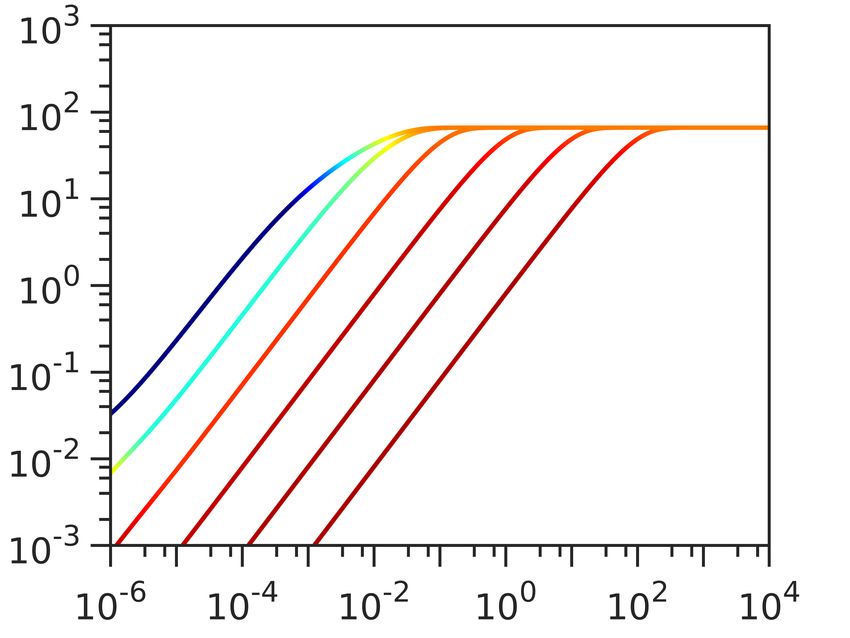
bioRxiv preprint first posted online Apr. 12, 2019; doi: http://dx.doi.org/10.1101/607838. The copyright holder for this preprint
(which was not peer-reviewed) is the author/funder, who has granted bioRxiv a license to display the preprint in perpetuity.
                               It is made available under a CC-BY-ND 4.0 International license.

                          Finally, the dye, the anion and the cation leak through the membrane (positive flux means x is moved
                       inward) at the rate:

                             jL,x = kL,x · 1 − e∆Gx /(RT ) , x ∈ {Dye,C+ , A− }                                             (7)

                                                         [x]in
                            ∆Gx = F ·Vm + RT · ln                                                                           (8)
                                                        [x]out

                       Similarly to kP , kL,x is a function whose shape depends on the mechanisms by which an ion leaks across
                       the E. coli membrane, which in turn depends on the electrostatic potential at a position z within the
                       membrane, V (z). To the best of our knowledge, V (z), and consequently dV (z)/dz, are not known for
                       E. coli. We chose Eyring’s model that has been verified for cationic leakage across the mitochondrial
                       membrane [Garlid and Paucek, 2003], and which assumes V (z) abruptly changes in the middle of the
                       lipid bilayer, such that dV (z)/dz = 0 everywhere but at the geometrical middle of the membrane where
                       dV (z)/dz = Vm [Garlid et al., 1989]. We then have:

                                                              F · zx ·Vm
                                     Scell                  −
                              kL,x =       · Px · [x]out · e 2 · RT , with x ∈ {Dye,C+ , A− }                                        (9)
                                     Vcell

                           where Scell denotes the cell’s surface area and Px the permeability of the membrane for x ∈ {Dye,C+ , A− }.
                       At steady-state Dye and A− equilibrate across the membrane according to Nernst equation (d[Dye]in /dt =
                       jL,Dye = 0 ⇔ ∆GDye = 0, leading to (2)), whereas for the monovalent cation d[C+ ]i /dt = 0 ⇔ jL,C+ = jP .
                       Next we introduce a new variable (“pump-leak ratio”) defined as:
                                                                                  F ·Vm
                                                                Vcell
                              ρ = kP /kL,C+ = kP ·                            · e 2 · RT                                            (10)
                                                       Scell · PC+ · [C+ ]out

                       and re-write the steady-state condition for C+ as:

                              1 − e∆GC+ /(RT ) = ρ · 1 − e(∆GE −∆GC+ )/(RT )                                                        (11)

                       Given a certain extracellular composition ([Dye]out , [C+ ]out and [A− ]out ), and taking into account that
                       [Dye]in and [A− ]in are defined by the Nernst equation at steady-state, equation (11) gives us a unique
                       solution for steady-state Vm for a set of {[Y ]in , ρ, ∆GE } values.
                           We note from Eq. (11) that changing the functional dependency of kL,x or kP does not affect how
                       the steady-state Vm depends on ρ. However, the steady-state potential after addition of the dye relative
                       to the steady-state potential in absence of the dye (∆Vm = (Vm,0 − Vm,Dye )/Vm,0 ) does. Similarly, the
                       dynamics of dye equilibration are dependent on the functional dependency of kL,x , kP , e.g. assuming
                       Goldman–Hodgkin–Katz flux equation Goldman [1943] instead of Erying’s model for kL,x will give
                       a slightly different dye equilibration profile (Fig. SI1). However, such changes will not influence the
                       conclusions we reach based on our model predictions, because we are interested in the changes of the
                       intracellular dye concentration dynamics at, e.g. different extracellular dye concentrations, Vm,0 or PDye .
                       These partial derivatives of the intracellular dye concentration are invariant to the choice of kL,x and kP .
                           Having constructed the model, we obtain the computational data in Fig. 2 in two steps. In the first
                       step we allow the ODE system described by Eqs. SI 20, 21 to reach the steady-state (Vm,0 ) for a 3-D grid
                       of {[Y ]in , ρ, ∆GE }. We note that in this step we do not need to specify ions’ permeabilities nor the rate
                       function for leakage kL,x , because we define the values of ρ, which is the ratio of the two. We then use the
                       obtained Vm,0 as the initial condition for the second step of the numerical experiment, which requires us
                       to specify: (i) the rate function for leakage (Eq. (9)), (ii) the permeability of the membrane to the dye
                       PDye and (iii) the concentration of the dye in the extracellular space [Dye]out .
                           As increasing the [Dye] gives better signal-to-noise ratio, we first look at the dye equilibration profile
                       ([Dye]in ) for fixed Vm,0 , [Y ]i = 150 mM, ∆GE = −210 mV and at different external dye concentrations
                       ([Dye]out ). As expected, Fig. 2A shows that increasing [Dye]out improves the signal-to-noise ratio and
                       shortens τeq , but at the same time increasingly depolarizes the membrane. The extent by which ∆Vm drops
                       does not solely depend on the [Dye]out , but also on the initial Vm,0 . Fig. 2B shows dye equilibration profile

                                                                                                                                    6/15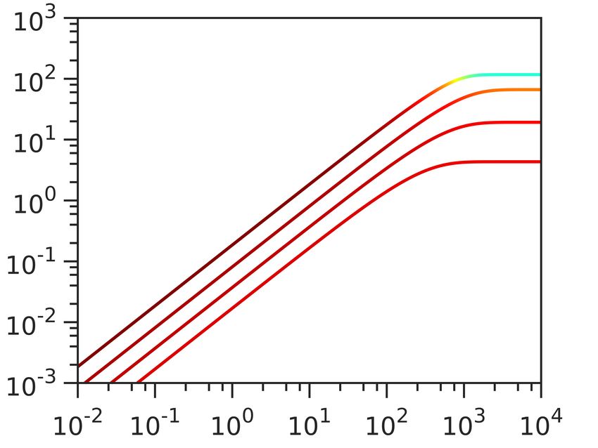
bioRxiv preprint first posted online Apr. 12, 2019; doi: http://dx.doi.org/10.1101/607838. The copyright holder for this preprint
(which was not peer-reviewed) is the author/funder, who has granted bioRxiv a license to display the preprint in perpetuity.
                               It is made available under a CC-BY-ND 4.0 International license.

                         for a fixed [Dye]out , but for different Vm,0 indicating that highly polarized cells are more susceptible to Vm
                         loss. Apart from the value of Vm,0 , ∆Vm will also depend on the charged permeable and non-permeable
                         species that are generating it, as shown in Fig. SI2. If a given Vm,0 value is generated in the presence of
                         a higher concentration of charged, impermeable intracellular species or at a higher energetic cost, ∆Vm
                         will increase for the same [Dye]out . Thus, the extent to which a given [Dye] becomes and actuator and
                         affects the ∆Vm is context dependant. Furthermore, while increasing [Dye]out shortens τeq this is only
                         the case when Vm,0 is affected, as seen in Fig. SI3. Lastly, we look at the dye equilibration profile for
                         different permeabilities of the membrane to the dye (PDye ) in Fig. 2C, and show that for higher PDye same
                         concentration of the dye lowers Vm,0 more. Fig. SI4 shows τeq as a function of PDye for different Vm,0 .
                       Intracellular Dye [mM]

                                                A                    B                                 C

                                                                                                                                           ΔVm [% of Vm,0]
                                                                             Time [min]
                         Figure 2. Computational data describing the parameter landscape associated with cationic dye usage as
                         Nernstian sensors. (A) Vm,0 = −140 mV, Yi = 150 mM, INa = 210 mV. Intracellular Dye concentration as
                         a function of time, for extracellular dye concentrations 10, 50, 100, 200, 400 and 1000 µM. The arrow
                         indicated increasing [Dye]out . (B) Extracellular dye concentration of 100 µM, Yi = 150 mM, ∆GE = −210
                         mV. Intracellular dye concentration as a function of time, for different Vm,0 : −220, −180, −140 and
                         −100 mV. The arrow indicates increasing absolute value of the Vm,0 . (C) Vm,0 = −180 mV, Yi = 150 mM,
                         ∆GE = −210 mV, Extracellular dye at 100 µM. Intracellular dye concentration as a function of time,
                         for different apparent permeabilities of the membrane to the dye: 10−12 , 10−10.8 , 10−9.6 , 10−8.4 , 10−7.2 ,
                         10−6 meter per second. The arrow indicates increasing permeability.

                         The working concentration of Nernstain dye Thioflavin T for E. coli is in µm range
                         Guided by the model predictions we devise an experimental work-flow for assessing the parametric range
                         in which a candidate cationic dye behaves like a Nernstian sensor, and choose Thioflavin T (ThT) for the
                         purpose. ThT has recently been used as a Vm dye in B. subtilis [Prindle et al., 2015], but has not been
                         characterized for use in E. coli. We start by identifying the working concentration that gives sufficiently
                         large signal, yet minimizes the membrane voltage perturbation, ∆Vm , and use the growth rate as a proxy
                         for affected ∆Vm . Fig. 3A and B show E. coli growth curves in MM9 media supplemented with glucose or
                         glycerol respectively (see Materials and Methods for detailed media composition), and in the presence of
                         a range of ThT concentrations. Fig. 3C shows growth rates plotted against the ThT concentration for both
                         media, demonstrating that 10 µM ThT or less does not significantly affect the growth rate in either media.
                         We call this concentration MNC (Maximum Non-inhibitory Concentration). However, the growth rate
                         reduction observed for higher ThT concentrations is media dependent (Fig. 3C). The result is consistent
                         with the finding of our model that the effect of the dye on cell’s physiology is environment dependent.
                             We next check that the highest ThT concentration, which does not affect the growth rate (MNC), 10
                         µM, gives sufficiently high signal-to-noise ratio by observing the dye equilibration in different media.
                         We note that if sufficient ∆I is achieved with 10 µM ThT we would further check that ∆Vm < 1% by
                         measuring the τeq with both 10 µM and a lower dye concentration. If ∆Vm < 1%, we expect τeq not to
                         change based on the results of our model (Fig. 2A). Fig. 3D shows Iin in time in LB and Fig. 3E the
                         same in MM9 media supplemented with glucose. In both cases fresh media with ThT is continuously
                         supplied using a customized flow-cell (see Materials and Methods), and in both cases ∆I is sufficiently
                         high. However, observed profiles are different from expected (Fig. 1), and show a characteristic initial
                         peak and a final plateau (SI Video 1). We reasoned that the peak could either be a real fluctuation in Vm or
                         it could indicate an unknown dye export mechanism.

                                                                                                                                    7/15
bioRxiv preprint first posted online Apr. 12, 2019; doi: http://dx.doi.org/10.1101/607838. The copyright holder for this preprint
(which was not peer-reviewed) is the author/funder, who has granted bioRxiv a license to display the preprint in perpetuity.
It is made available under a CC-BY-ND 4.0 International license.
Figure 3. E. coli growth in the presence of ThT. E. coli growing in MM9 media supplemented with
(A) glucose or (B) glycerol at increasing ThT concentration (colourmap). ThT concentrations in (A) are
10, 25, 50, 75, 100, 200, 350 µM and in (B) 10, 25, 50, 75, 100, 150, 175, 200 µM). The error bars are
standard deviations. (C) Maximum growth rates from (A) and (B) for each ThT concentration are given in
red and blue respectively. Each condition was done at least in triplicate and error bars are the standard
deviation. (D) Iin against time in LB and in (E) MM9 + glucose media. Individual cells are shown in cyan
(45 in (D) and 52 in (E) from at least 9 independent experiments), and the average trace is shown in blue.
The imaging conditions and Iex = 10µM are the same for (E) and (D).
Multi-drug efflux pumps influence ThT accumulation in E. coli
To determine whether the observed peak in Iin is due to active export of the dye we first check that in E. coli
ThT is not a multi-drug-efflux pump substrate. We are motivated by previous reports that show that dyes
such as ethidium bromide and Nile red are substrates of pumps belonging to the five bacterial structural
families: ABC (ATP-binding cassette), RND (resistance/nodulation/division), MATE (multidrug and
toxic compound extrusion), MFS (major facilitator superfamily) and SMR (small multidrug resistance)
[Alvarez-Ortega et al., 2013, Bay et al., 2008, Kuroda and Tsuchiya, 2009, Lubelski et al., 2007, Nikaido
and Takatsuka, 2009]. Fig. 4A shows dye equilibration curves in a wild type (WT) strain compared to the
strain bearing a deletion of TolC, which is a gene encoding for an outer membrane protein (OMP) that
is a ubiquitous component of multi-drug efflux pumps [Anes et al., 2015]. The Iin peak in the deletion
mutant did not disappear, instead the intensity level of the peak was even higher, suggesting the pump is
involved in the dye export, but also that the qualitative difference between the expected and the observed
equilibration curve shape is not due to dye export. Interestingly, in the mutant, the peak also occurred
earlier in time during the loading and with less cell-to-cell variability. We next tested the effect of the
ThT dye on the ∆TolC mutant growth rates, Fig. 4B. We found that at the MNC for the WT, the mutants’
growth was inhibited over the course of our experiment. Taken together the results indicate that ThT is
likely pumped out of the cells by one or more types of TolC-dependent efflux pumps, thereby preventing
its use for quantitative Vm measurements in WT E. coli.
Changing the membrane permeability during ThT loading can lead to loss of Vm
We next tested our second hypothesis that the Iin peak is due to a decrease in Vm . To this end, we
performed measurements of bacterial flagellar motor (BFM) speed [Krasnopeeva et al., 2018] during
ThT loading. BFM is a rotary molecular motor roughly 50 nm in size that enables bacterial swimming
[Sowa and Berry, 2008] via PMF driven rotation [Fung and Berg, 1995, Manson et al., 1980, Matsuura
8/15bioRxiv preprint first posted online Apr. 12, 2019; doi: http://dx.doi.org/10.1101/607838. The copyright holder for this preprint
(which was not peer-reviewed) is the author/funder, who has granted bioRxiv a license to display the preprint in perpetuity.
It is made available under a CC-BY-ND 4.0 International license.
Figure 4. Comparison of WT and ∆TolC mutant response to ThT. (A) Iin versus time for the WT (cyan)
and ∆TolC (gray) loaded in LB. WT traces are reproduced from Fig. 3D and ∆TolC traces were obtained
from 5 independent experiments to give 23 single cell traces. Averaged traces for the WT and ∆TolC are
given in blue and black respectively. (B) Growth curves of WT and ∆TolC in MM9 + glucose media are
given in blue (reproduced from Fig. 3A) and black, respectively. Growth curves in the same media, but in
the presence of 10 µM ThT, are given in cyan (WT) and gray (∆TolC). The shaded areas show standard
deviation and cyan and blue growth curves for the WT overlap.
et al., 1977, Meister and Berg, 1987]. The motor speed (ω) varies linearly with PMF [Fung and Berg,
1995, Gabel and Berg, 2003], which enables its use as a PMF, and when pHin = pHout , as a Vm indicator
as well [Krasnopeeva et al., 2018]. In our conditions, pHout is 7 and pHin is 7.86 (Fig. SI5) making the
contribution to the PMF from ∆pH ∼50 mV. Thus, even if during our experiment ∆pH goes to 0, we can
learn about the Vm behaviour from the PMF measurements via the motor speed. We measure ω as before,
using back-focal-plane interferometry [Svoboda et al., 1993] and a polystyrene bead attached to a short
filament stub (see Materials and Methods) [Bai et al., 2010, Krasnopeeva et al., 2018, Rosko et al., 2017,
Ryu et al., 2000].
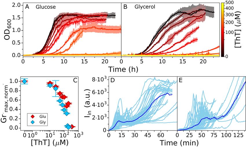
Fig. 5A shows simultaneous measurements of ThT intensity and normalized motor speed during dye
equilibration in MM9 + glucose. The motor speed decreases during ThT equilibration and BFM stops
at the point of Iin decrease. Furthermore, BFM does not resume spinning even as Iin further increases,
suggesting that the second ThT intensity increase that culminates in a plateau, is not driven by Vm . To
confirm the result, during ThT equilibration, we supplemented the medium with propidium iodide (PI). PI
permeates bacterial membrane that lost its integrity and significantly enhances its quantum yield upon
binding to DNA, which is commonly interpreted as an indication of cell death [Krämer et al., 2016, Lopez-
Amoros et al., 1995]. We found that the cells stained with PI although ThT intracellular concentration
remained high, Fig. 5B. In addition, at the time point of Iin decrease cellular volume suddenly increases,
and cytoplasmically expressed fluorescent protein mCherry-mKate2 hybrid (referred to as mKate2 for
brevity) [Lord, 2014] starts leaking out of the cell, Fig. 5C, SI Video 2.
These results are in contradiction with our estimate of dye working concentration, and we wondered,
based on Fig. 2C, if the changes in PDye could be the explanation. The cell culture in Fig. 3 was briefly
exposed to light at 600 nm every 7.5 min, whereas cells in our flow-cell were exposed to light of 435 nm
every minute for the purpose of imaging the ThT dye. We have previously reported loss of Vm and
PMF due to light-induced decrease of E. coli membrane’s resistance at effective powers higher than ∼
17 mW/cm2 , and for a combination of 395 and 475 nm wavelengths [Krasnopeeva et al., 2018]. Light
damage is wavelength dependent [Vermeulen et al., 2008], and we therefore characterized the light damage
caused by our imaging conditions, i.e. 435 nm wavelength and effective power of Peff ∼ 11 mW/cm2 .
Fig. SI7 shows a decrease in BFM motors’ speed, and thus cell’s PMF. However, the PMF is not fully lost,
indicating that the loss of PMF observed in Fig. 5A is likely caused by the combination of light induced
increase in PDye and exposure to 10 µM ThT.
To prove it, we exposed the bacteria to 10 µM ThT in LB as before, but this time we observe the
cells under bright-field illumination for 45 min, at which point we turn on the 435 nm light used for
epifluorescent imaging of ThT. Fig. 5E shows that after 45 min cells not exposed to 435 nm light did not
9/15bioRxiv preprint first posted online Apr. 12, 2019; doi: http://dx.doi.org/10.1101/607838. The copyright holder for this preprint
(which was not peer-reviewed) is the author/funder, who has granted bioRxiv a license to display the preprint in perpetuity.
It is made available under a CC-BY-ND 4.0 International license.
Figure 5. (A) Average traces of ThT fluorescence (in blue) and motor speed (in black) simultaneously
measured in 5 individual cells (individual cell traces are given in Fig. SI6). The shaded areas show the
standard deviation and the motor speed has been normalized to the initial value as described in Materials
and Methods. (B) ThT (y axes) and PI (colourmap) equilibration profile in LB. 25 individual traces are
given. (C) Average of ThT (in blue), mKate2 (in red) fluorescence and cell area (in green) simultaneously
measured in 12 individual cells from 3 independent experiments. The shaded areas show the standard
deviation. (D) Equilibration profile of 1 µM ThT in LB. 8 single cell traces and average trace are given in
cyan and blue, respectively. (E) Equilibration profile of 10 µM ThT in LB in the absence (gray shaded
area) and presence of epifluorescent illumination (light area). The dye was flown in the flow cell for the
whole length of the experiment, imaging conditions in the light area were the same as in Figure 3 and 5D.
44 cells from 8 independent experiments are given.
take up ThT, in contrast to Fig.3D where cells exposed to 435 nm light from the start, took up ThT in the
first 30 min.
Actively changing membrane permeability has been used to facilitate loading of Nernstian sensors
[Lo et al., 2007], and Fig. 5E shows that this can change the dye into an actuator because it can influence
Vm . Our mathematical model predicts that if a given concentration of the dye is lowering Vm , an even
lower concentration of the dye will result in a change of τeq (Fig. SI3). In agreement, Fig. 5D shows
that for 1 µM concentration of ThT τeq lasted longer then with 10 µM. Thus, 10 µM ThT in LB, under
435 nm light, affects Vm . Assessing the suitability of the dye working concentration by confirming that a
lower dye concentration does not alter τeq is a suitable additional control we propose, especially if PDye is
being altered as part of the experiments.
We note that in our plate reader experiments (Fig. 2) we observed the effect of the dye (above 10µM)
on cell growth, while in our microscopy experiments, in the absence of light damage, ThT does not
permeate WT cells. We thus wanted to confirm that at higher concentrations ThT permeates the cells and
for the purpose we imaged the cells from the wells at representative ThT concentrations in MM9+glucose
(10, 50 and 100 µM) and MM9+glycerol (10 µM). We found that in MM9 glucose cell brightness
increases with the extracellular dye concentration and that in MM9+glycerol, at 10µM, ThT signal from
the cells is overall greater that in glucose (Fig. SI8).
Having identified the mechanisms behind the shape of the ThT loading curve we observed in Fig. 3
we should now be able to reproduce it with our mathematical model. Based on Fig 5A we assume that Vm
decays exponentially immediately after addition of the dye [Krasnopeeva et al., 2018]: Vm (t) = Vm,0 ·2t/t1/2
where t1/2 is the time at which voltage is half that of Vm,0 . The dynamics of dye entry are then modelled
by Eyring’s rate law Eq. (9) taking into account Vm,0 , t1/2 and PDye (see Supplementary Information for
further details on the model). Fig. SI9 shows that the model reproduces the peak in [Dye]in observed in
Fig. 3. Immediately upon addition, the positively charged dye moves inwards because its extracellular
concentration is higher than the intracellular and the cell is negatively polarized. Thus, [Dye]in increases
and becomes greater than [Dye]out until the electrochemical potential reaches ∆GDye = 0 (at the peak).
10/15bioRxiv preprint first posted online Apr. 12, 2019; doi: http://dx.doi.org/10.1101/607838. The copyright holder for this preprint
(which was not peer-reviewed) is the author/funder, who has granted bioRxiv a license to display the preprint in perpetuity.
It is made available under a CC-BY-ND 4.0 International license.
As the Vm decays and since [Dye]in > [Dye]out , the dye now starts moving outwards and its intracellular
concentration decreases. The time at which the peak occurs as well as its intensity depend on PDye as
follows: (i) the time of the peak decreases with increasing PDye and increases with increasing t1/2 , and (ii)
the intensity of the peak increases with increasing PDye and t1/2 , Fig SI9 C and D.
The dye still equilibrates according to equation (2), but this is achieved transiently at the time of the
peak, which is the time point at which Vm can be calculated from equation (2). Since the Vm varies during
the course of the experiment, the Vm measured at the peak is not equal to the Vm,0 . Nonetheless, if we
measure Vm (t)/Vm,0 as well as calculate the Vm at the time of the peak using eq. (2), in principle we can
estimate Vm,0 as well. Thus, charged dyes can be used to estimate initial Vm even in conditions where they
act as actuators and collapse Vm , if the dynamical shape of the Vm loss is known.
DISCUSSION
Nernstian probes are a popular choice for estimating bacterial Vm , because their concentration directly
depends on Vm according to the Nernst equation. Despite the wide usage, the probes are often not
sufficiently calibrated before use in different conditions. Here we present a mathematical model that
shows trade-offs between requirements imposed on the dye: sufficient signal-to-noise ratio, sufficiently
short dye equilibration time and minimum effect on the cells’ physiology. Based on the model results we
propose a general work-flow for the characterization of Nernstian dye candidates (Fig. 6), and demonstrate
it on a newly suggested, and previously uncharacterized in E. coli, fluorescent dye Thioflavin T. We find
that the suitability of a candidate Nernstian dye is context dependent, and reveal that the candidate dyes
can be substrates for bacterial drug-export systems. We believe our work-flow is sufficiently simple and
general to provide a common standard for benchmarking the cationic dye behavior and thus improve the
robustness of Vm measurements.
We found as well that imaging conditions can affect how well the dye crosses bacterial membrane,
which suggest that on its own E. coli membrane exhibits low permeability to this Nernstian sensor, or that
its export by membrane efflux pumps is substantial. The observation is consistent with previous results
that needed to permeabilize the membrane by EDTA to achieve experimentally reasonable loading times
[Lo et al., 2007]. However, changing the membrane permeability is often not dye specific, and can lead to
changes in the overall Vm of the cell. Our model suggests that if the chosen dye working concentration is
not affecting Vm , lower dye concentration should leave τeq unchanged. The finding offers a simple test
to confirm the suitability of the chosen dye working concentration, which should be performed in each
environmental and physiological condition.
As a final remark, the fact that in the absence of illumination of shorter wavelengths, i.e. in our plate
reader experiments, ∆TolC mutant is affected significantly more by the dye than the wild type suggest that,
in the rich defined medium such as MM9 + glucose and amino acids, low cytoplasmic dye concentration
is not just a consequence of low membrane permeability to the dye, but also of the active efflux. Then,
in a condition where membrane permeability is affected, leading to the PMF loss, which is in turn the
energy source for efflux pumps, the cytoplasmic concentration of the dye is no longer in accordance with
equation (2) and could result in interesting dynamics such as in Fig. 5A, where we observe significant
accumulation of the dye only after the PMF drops to a certain threshold value.
11/15bioRxiv preprint first posted online Apr. 12, 2019; doi: http://dx.doi.org/10.1101/607838. The copyright holder for this preprint
(which was not peer-reviewed) is the author/funder, who has granted bioRxiv a license to display the preprint in perpetuity.
It is made available under a CC-BY-ND 4.0 International license.
Figure 6. Proposed work-flow for characterizing the Nernstian behavior of a candidate cationic mem-
brane voltage dye. The working concentration is estimated in steps 1 to 4, we define it as the maximum
dye concentration that does not affect membrane voltage and that yields sufficient amount of signal.
(1) The MNC is estimated. (2) The MNC is tested for sufficient signal intensity. (3) The effect of the
dye on Vm is determined by measuring τeq at different below-MNC concentrations. (4) Different τeq for
different dye concentrations indicate that the probe is altering Vm and the working concentration should
be reduced and the protocol resumed from (2). Equal τeq indicates that the probe is not altering Vm . (5)
Common procedures to test Nernstian dye responses can then be applied, such as the introduction of a
ionophore that neutralizes Vm , or changes in external pH that induce changes in Vm [Lo et al., 2007]. (6)
Because the effects depend on the physiological state of the cell, the procedure should be repeated for
every experimental condition.
AUTHOR CONTRIBUTIONS
LM, GT, CJL, BF and TP conceived the experiments and the computational work. LM, TT, YP and YL
performed experiments. GT performed computational work. LM analysed experimental data, and LM,
GT, CJL, BF and TP interpreted the results and wrote the manuscript.
ACKNOWLEDGEMENTS
We thank Dario Miroli for help with image analysis, Nathan Lord and Sebastian Jaramillo-Riveri for
donating us the construct containing the hybrid mCherry-mKate2 sequence and Angela Dawson for
retrieving the ∆TolC mutant from the Keio collection. This work was financially supported by the
Cunningham Trust scholarship ACC/KWF/PhD1 to TP and LM, the National Natural Science Foundation
of China under grants No. 31722003 and No. 31770925 to FB, the Ministry of Science and Technology,
Republic of China, under contract No. MOST-107-2112-M-008-025-MY3 to CJL and Human Frontiers
Program grant RGP0041/2015 to TP, FB and CJL. TP acknowledges the support of UK Research Councils
Synthetic Biology for Growth programme and is a member of the BBSRC/EPSRC/MRC funded Synthetic
Biology Research Centre (BB/M018040/1).
REFERENCES
Alvarez-Ortega, C., Olivares, J., and Martı́nez, J. L. (2013). Rnd multidrug efflux pumps: what are they
good for? Front Microbiol, 4:7–7. 23386844[pmid].
Anes, J., McCusker, M. P., Fanning, S., and Martins, M. (2015). The ins and outs of rnd efflux pumps in
escherichia coli. Front Microbiol, 6:587–587. 26113845[pmid].
12/15bioRxiv preprint first posted online Apr. 12, 2019; doi: http://dx.doi.org/10.1101/607838. The copyright holder for this preprint
(which was not peer-reviewed) is the author/funder, who has granted bioRxiv a license to display the preprint in perpetuity.
It is made available under a CC-BY-ND 4.0 International license.
Baba, T., Ara, T., Hasegawa, M., Takai, Y., Okumura, Y., Baba, M., Datsenko, K. A., Tomita, M., Wanner,
B. L., and Mori, H. (2006). Construction of escherichia coli k-12 in-frame, single-gene knockout
mutants: the keio collection. Molecular Systems Biology, 2(1).
Bai, F., Branch, R. W., Nicolau, D. V., Pilizota, T., Steel, B. C., Maini, P. K., and Berry, R. M. (2010).
Conformational spread as a mechanism for cooperativity in the bacterial flagellar switch. Science,
327(5966):685–689.
Bay, D. C., Rommens, K. L., and Turner, R. J. (2008). Small multidrug resistance proteins: A multidrug
transporter family that continues to grow. Biochimica et Biophysica Acta (BBA) - Biomembranes,
1778(9):1814 – 1838. Structural proteomics of the cell envelope of Gram-negative bacteria.
Bradbeer, C. (1993). The proton motive force drives the outer membrane transport of cobalamin in
Escherichia coli. Journal of bacteriology, 175(10):3146–50.
Buda, R., Liu, Y., Yang, J., Hegde, S., Stevenson, K., Bai, F., and Pilizota, T. (2016). Dynamics of
escherichia coli’s passive response to a sudden decrease in external osmolarity. Proceedings of the
National Academy of Sciences, 113(40):E5838–E5846.
Datsenko, K. A. and Wanner, B. L. (2000). One-step inactivation of chromosomal genes in escherichia
coli k-12 using pcr products. Proceedings of the National Academy of Sciences, 97(12):6640–6645.
Del Castillo, J. and Katz, B. (1954). Quantal components of the end-plate potential. J Physiol, 124(3):560–
573. 13175199[pmid].
Felle, H., Porter, J. S., Slayman, C. L., and Kaback, H. R. (1980). Quantitative measurements of membrane
potential in escherichia coli. Biochemistry, 19(15):3585–3590. PMID: 6996707.
Fluhler, E., Burnham, V. G., and Loew, L. M. (1985). Spectra, membrane binding, and potentiometric
responses of new charge shift probes. Biochemistry, 24(21):5749–5755. PMID: 4084490.
Fung, D. C. and Berg, H. C. (1995). Powering the flagellar motor of escherichia coli with an external
voltage source.
Gabel, C. V. and Berg, H. C. (2003). The speed of the flagellar rotary motor of escherichia coli varies
linearly with protonmotive force. Proceedings of the National Academy of Sciences, 100(15):8748–
8751.
Garlid, K. D., Beavis, A. D., and Ratkje, S. K. (1989). On the nature of ion leaks in energy-transducing
membranes. Biochimica et Biophysica Acta (BBA) - Bioenergetics, 976(2):109 – 120.
Garlid, K. D. and Paucek, P. (2003). Mitochondrial potassium transport: the k+ cycle. Biochimica et
Biophysica Acta (BBA) - Bioenergetics, 1606(1):23 – 41.
Goldman, D. E. (1943). Potential, impedance, and rectification in membranes. The Journal of General
Physiology, 27(1):37–60.
Grabe, M. and Oster, G. (2001). Regulation of organelle acidity. The Journal of General Physiology,
117(4):329–344.
Hodgkin, A. L. and Huxley, A. F. (1939). Action Potentials Recorded from Inside a Nerve Fibre. Nature,
144:710.
Jahreis, K., Pimentel-Schmitt, E. F., Brückner, R., and Titgemeyer, F. (2008). Ins and outs of glucose
transport systems in eubacteria. FEMS Microbiology Reviews, 32(6):891–907.
Kashket, E. R. (1985). The proton motive force in bacteria: A critical assessment of methods. Annual
Review of Microbiology, 39(1):219–242. PMID: 2998266.
Keener, J. and Sneyd, J. (2009). Mathematical Physiology, page 93.
Kralj, J. M., Hochbaum, D. R., Douglass, A. D., and Cohen, A. E. (2011). Electrical spiking in escherichia
coli probed with a fluorescent voltage-indicating protein. Science, 333(6040):345–348.
Krämer, C. E. M., Wiechert, W., and Kohlheyer, D. (2016). Time-resolved, single-cell analysis of induced
and programmed cell death via non-invasive propidium iodide and counterstain perfusion. Scientific
Reports, 6:32104 EP –. Article.
Krasnopeeva, E. (2018). Single cell measurements of bacterial physiology traits during exposure to an
external stress. PhD thesis, University of Edinburgh.
Krasnopeeva, E., Lo, C.-J., and Pilizota, T. (2018). Single-cell bacterial electrophysiology reveals
mechanisms of stress-induced damage. arXiv.
Kuroda, T. and Tsuchiya, T. (2009). Multidrug efflux transporters in the mate family. Biochimica et
Biophysica Acta (BBA) - Proteins and Proteomics, 1794(5):763 – 768. Mechanisms of Drug Efflux and
Strategies to Combat Them.
Ling, G. and Gerard, R. W. (1949). The normal membrane potential of frog sartorius fibers. Journal of
13/15bioRxiv preprint first posted online Apr. 12, 2019; doi: http://dx.doi.org/10.1101/607838. The copyright holder for this preprint
(which was not peer-reviewed) is the author/funder, who has granted bioRxiv a license to display the preprint in perpetuity.
It is made available under a CC-BY-ND 4.0 International license.
Cellular and Comparative Physiology, 34(3):383–396.
Lo, C.-J., Leake, M. C., Pilizota, T., and Berry, R. M. (2007). Nonequivalence of membrane voltage
and ion-gradient as driving forces for the bacterial flagellar motor at low load. Biophysical Journal,
93(1):294 – 302.
Lopez-Amoros, R., Comas, J., and Vives-Rego, J. (1995). Flow cytometric assessment of escherichia coli
and salmonella typhimurium starvation-survival in seawater using rhodamine 123, propidium iodide,
and oxonol. Applied and Environmental Microbiology, 61(7):2521–2526.
Lord, N. (2014). Fluctuation timescales in bacterial gene expression. PhD thesis, Harvard University.
Lubelski, J., Konings, W. N., and Driessen, A. J. M. (2007). Distribution and physiology of abc-type
transporters contributing to multidrug resistance in bacteria. Microbiol Mol Biol Rev, 71(3):463–476.
17804667[pmid].
Manson, M. D., Tedesco, P., and Berg, H. C. (1980). Energetics of flagellar rotation in bacteria. Journal
of Molecular Biology, 138(3):541 – 561.
Martinac, B., Buechner, M., Delcour, A. H., Adler, J., and Kung, C. (1987). Pressure-sensitive ion channel
in escherichia coli. Proceedings of the National Academy of Sciences, 84(8):2297–2301.
Martinac, B., Rohde, P. R., Cranfield, C. G., and Nomura, T. (2013). Patch Clamp Electrophysiology for
the Study of Bacterial Ion Channels in Giant Spheroplasts of E. coli, pages 367–380. Humana Press,
Totowa, NJ.
Maskevich, A. A., Lavysh, A. V., Kuznetsova, I. M., Sulatskaya, A. I., and Turoverov, K. K. (2015).
Spectral manifestations of thioflavin t aggregation. Journal of Applied Spectroscopy, 82(1):33–39.
Maskevich, A. A., Stsiapura, V. I., Kuzmitsky, V. A., Kuznetsova, I. M., Povarova, O. I., Uversky, V. N.,
and Turoverov, K. K. (2007). Spectral properties of thioflavin t in solvents with different dielectric
properties and in a fibril-incorporated form. Journal of Proteome Research, 6(4):1392–1401. PMID:
17305383.
Matsuura, S., ichi Shioi, J., and Imae, Y. (1977). Motility in bacillus subtilis driven by an artificial
protonmotive force. FEBS Letters, 82(2):187 – 190.
Meister, M. and Berg, H. C. (1987). The stall torque of the bacterial flagellar motor. Biophys J,
52(3):413–419. 3651560[pmid].
Mitchell, P. (1961). Coupling of phosphorylation to electron and hydrogen transfer by a chemi-osmotic
type of mechanism. Nature, 191:144 EP –.
Neher, E. and Sakmann, B. (1976). Single-channel currents recorded from membrane of denervated frog
muscle fibres. Nature, 260:799 EP –.
Nikaido, H. and Takatsuka, Y. (2009). Mechanisms of rnd multidrug efflux pumps. Biochim Biophys
Acta, 1794(5):769–781. 19026770[pmid].
Otsu, N. (1979). A threshold selection method from gray-level histograms. IEEE Transactions on Systems,
Man, and Cybernetics, 9(1):62–66.
Pilizota, T. and Shaevitz, J. W. (2012). Fast, multiphase volume adaptation to hyperosmotic shock by
escherichia coli. PLOS ONE, 7(4):1–10.
Prindle, A., Liu, J., Asally, M., Ly, S., Garcia-Ojalvo, J., and Süel, G. M. (2015). Ion channels enable
electrical communication in bacterial communities. Nature, 527:59.
Ramos, S. and Kaback, H. R. (1977). The relation between the electrochemical proton gradient and active
transport in escherichia coli membrane vesicles. Biochemistry, 16(5):854–859. PMID: 14665.
Rosko, J. (2017). Osmotaxis in Escherichia coli. PhD thesis, University of Edinburgh.
Rosko, J., Martinez, V. A., Poon, W. C. K., and Pilizota, T. (2017). Osmotaxis in escherichia coli through
changes in motor speed. Proceedings of the National Academy of Sciences.
Ryu, W. S., Berry, R. M., and Berg, H. C. (2000). Torque-generating units of the flagellar motor of
escherichia coli have a high duty ratio. Nature, 403:444 EP –.
Sakmann, B. and Neher, E. (1984). Patch clamp techniques for studying ionic channels in excitable
membranes. Annual Review of Physiology, 46(1):455–472. PMID: 6143532.
Sims, P. J., Waggoner, A. S., Wang, C.-H., and Hoffman, J. F. (1974). Mechanism by which cyanine
dyes measure membrane potential in red blood cells and phosphatidylcholine vesicles. Biochemistry,
13(16):3315–3330. PMID: 4842277.
Sowa, Y. and Berry, R. M. (2008). Bacterial flagellar motor. Quarterly Reviews of Biophysics,
41(2):103–132.
Strahl, H. and Hamoen, L. W. (2010). Membrane potential is important for bacterial cell division.
14/15You can also read