Development of Small-Molecule PUMA Inhibitors for Mitigating Radia- tion-Induced Cell Death
←
→
Page content transcription
If your browser does not render page correctly, please read the page content below
Current Topics in Medicinal Chemistry, 2011, 11, 281-290                                     281

Development of Small-Molecule PUMA Inhibitors for Mitigating Radia-
tion-Induced Cell Death

Gabriela Mustata1,#, Mei Li2,5,#, Nicki Zevola1#, Ahmet Bakan1, Lin Zhang3,5,
Michael Epperly4,5, Joel S. Greenberger4,5, Jian Yu2,5,* and Ivet Bahar1,*

University of Pittsburgh School of Medicine, Departments of 1 Computational & Systems Biology, 2Pathology,
3
  Pharmacology and Chemical Biology, and 4Radiation Oncology, and 5University of Pittsburgh Cancer Institute, Hill-
man Cancer Center Research Pavilion, 5117 Centre Ave, Pittsburgh, PA, 15213, USA

            Abstract: PUMA (p53 upregulated modulator of apoptosis) is a Bcl-2 homology 3 (BH3)-only Bcl-2 family member and
            a key mediator of apoptosis induced by a wide variety of stimuli. PUMA is particularly important in initiating radiation-
            induced apoptosis and damage in the gastrointestinal and hematopoietic systems. Unlike most BH3-only proteins, PUMA
            neutralizes all five known antiapoptotic Bcl-2 members through high affinity interactions with its BH3 domain to initiate
            mitochondria-dependent cell death. Using structural data on the conserved interactions of PUMA with Bcl-2-like proteins,
            we developed a pharmacophore model that mimics these interactions. In silico screening of the ZINC 8.0 database with
            this pharmacophore model yielded 142 compounds that could potentially disrupt these interactions. Thirteen structurally
            diverse compounds with favorable in silico ADME/Toxicity profiles have been retrieved from this set. Extensive testing
            of these compounds using cell-based and cell-free systems identified lead compounds that confer considerable protection
            against PUMA-dependent and radiation-induced apoptosis, and inhibit the interaction between PUMA and Bcl-xL.
Keywords: Inhibition of PUMA-induced apoptosis, Bcl-2 protein family, BH3 domain, protein-protein interactions, pharma-
cophore modeling, druggability, virtual screening of libraries of small compounds.

INTRODUCTION                                                                 pathway is more extensively utilized by the immune cells.
                                                                             The intrinsic pathway is triggered in diverse cell types by a
    Understanding the molecular mechanisms of regulating                     wide range of stimuli such as developmental cues and severe
apoptosis, or programmed cell death, is one of the hottest
                                                                             cellular stress, including DNA damage, deprivation of sur-
research areas in biomedical sciences. There is a long list of
                                                                             vival factors nutrients, or loss of cell-cell or cell-matrix at-
diseases associated with altered cell survival [1]. Increased
                                                                             tachment, and is mediated through the organelle mitochon-
apoptosis is characteristic of AIDS, neurodegenerative dis-
                                                                             drion. Apoptosis is ultimately executed by intracellular pro-
eases and various forms of tissue injury. Decreased or inhib-
                                                                             tease enzymes called caspases which, upon activation, de-
ited apoptosis is a hallmark of many malignancies and in-                    stroy cellular proteins that are vital for cell survival [4]. The
volved in autoimmune disorders and some viral infections. In
                                                                             mitochondrial apoptotic pathway is regulated by the evolu-
recent years, enormous efforts have gone into developing
                                                                             tionarily conserved Bcl-2 protein family, which includes
modulators of apoptosis for therapeutic purposes. While the
                                                                             both pro-apoptotic members such as Bax, Bak that promote
damaging effects of ionizing irradiation to cancer cells dur-
                                                                             mitochondrial permeability, and anti-apoptotic (cell survival)
ing clinical radiotherapy involve multiple cell death path-
                                                                             members such as Bcl-2, Bcl-xL, A1 and Mcl-1, which inhibit
ways, the toxicity to normal tissues is mediated primarily by                the mitochondrial release of cytochrome c [5, 6]. These two
apoptosis. A drug that could ameliorate irradiation induced
                                                                             groups share three or four of the characteristic domains of
apoptosis would potentially be of value as a radiation protec-
                                                                             homology (Bcl-2 Homology or BH domains BH1-BH4,
tor and mitigator for use in both clinical radiotherapy and in
                                                                             composed each of a functional helix). In addition, the Bcl-2
the setting of treating victims of environmental radiation
                                                                             family includes a third group such as Bim, Bad and PUMA,
accidents or radiation terrorism.
                                                                             which contain a single BH3 domain, therefore termed "BH3-
    Apoptosis occurs via two major signaling pathways in                     only proteins”. BH3-only proteins are apical sensors of dif-
mammalian cells, the intrinsic and extrinsic pathways [2, 3].                ferent apoptotic stimuli and function to inhibit Bcl-2 like
The extrinsic pathway is activated when a pro-apoptotic                      proteins and/or to activate Bax or Bak [7, 8].
ligand binds to its receptor that in turn recruits additional
                                                                                 PUMA, p53-Upregulated Mediator of Apoptosis was
proteins to form death-inducing signaling complexes. This
                                                                             initially identified as a transcriptional target of p53 and a
                                                                             mediator of DNA damage-induced apoptosis [9, 10]. PUMA
*Address correspondence to these authors at the University of Pittsburgh     is transcriptionally activated by a wide range of apoptotic
School of Medicine, Departments of Computational & Systems Biology and       stimuli and transduces these proximal death signals to the
Pathology, Hillman Cancer Center Research Pavilion, 5117 Centre Ave,         mitochondria Fig. (1)[11]. PUMA directly binds to all five
Pittsburgh, PA, 15213, USA; Tel: 412 648 3332; Fax: 412 648 3163;
E-mail: bahar@pitt.edu; Tel: 412 623 7786; Fax: 412 623 7778;                known anti-apoptotic Bcl-2 family members with high af-
E-mail: yuj2@upmc.edu                                                        finities through its BH3 domain. Binding of PUMA to the
# equal contribution                                                         Bcl-2 like proteins results in the displacement of Bax/Bak

                                               1568-0266/11 $58.00+.00        © 2011 Bentham Science Publishers Ltd.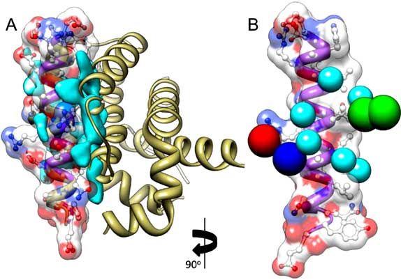
282 Current Topics in Medicinal Chemistry, 2011, Vol. 11, No. 3                                                             Mustata et al.

and their activation via formation of multimeric pore like          where 1-4 are hydrophobic residues,  designates a small
structures on the mitochondrial outer membrane, leading to          residue, Z is usually an acidic residue, L and D are conserved
mitochondrial dysfunction and caspase activation Fig. (1).          leucine and aspartate.
PUMA is implicated in many pathological and physiological
                                                                        As shown in Fig. (2), the sequence and structure align-
processes including cancer, tissue injury, neurodegenerative        ments of PUMA against the pro-survival proteins A1 and
diseases, immune response and bacterial or viral infection
                                                                    Mcl-1 reveal conserved interactions between three pairs of
[11]. Recent work in mice indicates that PUMA is the pri-
                                                                    residues, all found in the BH3 domain: a hydrophobic inter-
mary, if not the sole, mediator of p53-dependent radiation-
                                                                    action of PUMA Leu141 with MCl-1 Phe251 (or A1 Phe95);
induced apoptosis in the rapidly dividing tissues of the gas-
                                                                    and two salt bridge interactions between PUMA Arg142 and
trointestinal (GI) tract and hematopoietic (HP) system, and
                                                                    Mcl-1 Asp237 (or A1 Asp81) and between PUMA Asp146
amongst cellular targets including cells and progenitors in         and Mcl-1 Arg244 (or A1 Arg88).
the intestinal and hematopoietic systems. Genetic ablation or
inhibition of PUMA provides drastic radioprotection in mice             Note that Mcl-1 and A1 have a root-mean-square devia-
[12-15].                                                            tion (RMSD) of 1.6 Å in their backbone atoms and are most
                                                                    similar in their binding profiles [17]. Upon superimposing
                                                                    the PUMA BH3 domain in complex with Mcl-1 (PDB file
                                                                    2roc [16] with the PUMA BH3 domain in complex with A1
                                                                    (PDB file 2vof [17]), this high degree of homology, as well
                                                                    as the three conserved interactions of PUMA are clearly seen
                                                                    Fig. (2). These key interacting residues highlighted in Fig.
                                                                    (2) form the basis of pharmacophore model described below,
                                                                    which we designed for potential inhibitors/mitigators of
                                                                    PUMA activity.

Fig. (1). PUMA-mediated apoptosis. PUMA is induced by a wide
range of death stimuli, such as gamma-radiation, reactive oxygen
species (ROS) and inflammatory cytokines. Binding to the Bcl-2
like proteins by PUMA through its BH3 domain (triangle) leads to
activation of Bax/Bak at the mitochondrial membrane, permeabliza-
tion of mitochondrial outer membrane, and release of apoptogenic
proteins such as cytochrome C, SMAC and AIF. The apoptotic
proteins promote activation of caspases and nucleases leading to
irreversible cell demise.

    The 3D structures of PUMA BH3 domain in complex
with anti-apoptotic Bcl-2 proteins Mcl-1 [16] and A1 [17]
have been recently determined Fig. (2A). Based on binding
properties of BH3-only proteins with Bcl-2 like proteins,
Bcl-2 inhibitors have been developed to mimic the actions of
the proapoptotic BH3 domains [18, 19]. Considering the
importance of the interactions of PUMA/Bcl-2 like proteins
in initiating the intrinsic pathway, we describe herein the
identification of small molecules that disrupt or prevent these
key interactions and consequently suppress the apoptotic            Fig. (2). Interactions between PUMA BH3 domain and pro-
response induced by PUMA and gamma irradiation.                     apoptotic proteins A1 and Mcl-1. (A) Overlay of PUMA bound
                                                                    structures of A1 (PDB code: 2vof [17]) and Mcl-1 (PDB code: 2roc
                                                                    [16]) are shown. Panels show a closer view of key interactions be-
RESULTS
                                                                    tween PUMA and A1/Mcl-1 proteins and the locations of pharma-
Sequence and Structure Analysis                                     cophore features. The radius of the feature spheres are set to 0.75 Å
                                                                    ( of the actual size in the pharmacophore model) for display pur-
    Among the Bcl-2 pro-survival proteins, A1 is the most
                                                                    poses. (B) Sequence properties considered in designing the pharma-
similar to Mcl-1 in terms of binding profile, sharing 25.4%
                                                                    cophore model. Key conserved interactions by sequence alignment
identity and 46.2% similarity over their helical bundles [16].
                                                                    include a Leu-Phe hydrophobic interaction (PUMA Leu141.C1
Fig. (2) displays the shared mechanism of interaction be-
                                                                    with Mcl-1 Phe251.C, in blue), an Arg-Asp salt-bridge interaction
tween PUMA BH3 domain (an -helix of 22 residues) and
                                                                    (PUMA Arg142.NH1 with Mcl-1 Asp237.O2, in red), and an Asp-
these two family members. A 13-residue motif defines the            Arg salt-bridge interaction (PUMA Asp146.O1 with Mcl-1
BH binding domain, designated as 1XX2 XX3DZ4L,
                                                                    Arg244.N and A-1 Arg88.N, in green.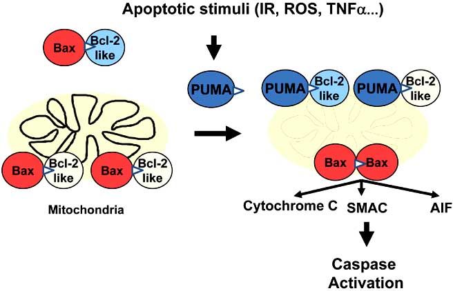
Induced Fit Simulations on Nuclear Receptors                              Current Topics in Medicinal Chemistry, 2011, Vol. 11, No. 3   283

Pharmacophore Development, Database Screening and                   tion and optimization of ADME/Tox properties early in the
Generation of Hits                                                  discovery process. An ADME/Tox filtering might be consid-
                                                                    ered premature at the hit finding stage, however, we applied
    We designed a pharmacophore model Fig. (3) on the ba-
                                                                    the in silico assessment of ADME/Tox properties to priori-
sis of the PUMA BH3 domain structure and its interactions
                                                                    tize the 142 compounds that resulted from the initial data-
in the complexes with A1 and Mcl-1, consistent with previ-          base screening process, with the understanding that those
ous biochemical measurements. Therefore our pharma-
                                                                    compounds could potentially serve as drugs. The first round
cophore model comprises three residues, two of which are
                                                                    of experiments were therefore performed on compounds that
engaged in salt bridges with PUMA BH3 domain residues,
                                                                    were predicted to have no ADME/Tox liabilities.
and the third making hydrophobic contacts, in accord with
the conserved Asp Arg, and Phe of Mcl-1 and A1, which                   The ACD ADME/TOX software [23, 24] was used to
play a key role in stabilizing the interactions with PUMA.          rationally deconvolute the set of hits to extract a set of
The 3-dimensional arrangement of these residues has been            twenty compounds that passed all the ADME/Tox filters.
deduced from the known geometry of the interfacial interac-         These compounds selected as potential lead compounds are
tions illustrated in Fig. (2). Quantitative verification of the     presented in Fig. (4).
three conserved interactions was conducted with the Fast
Contact 2.0 server [20]. This server estimates the contribu-        Druggability Calculations
tion of different pairs of interfacial residues to the overall
binding free energy, which confirmed that the ionic Asp-                Protein-protein interfaces are challenging targets. Meth-
                                                                    ods for assessing the druggability of proteins are usually
Arg, Arg-Asp and hydrophobic Phe-Leu interactions be-
                                                                    trained using proteins with well-defined active site pockets,
tween the BH3 domain of PUMA and the Mcl-1 or A1 resi-
                                                                    and hence are not applicable to protein-protein interfaces in
dues make significant contribution to the total binding en-
                                                                    general. Recently, Seco and Barril introduced a first princi-
ergy.
                                                                    ples-based method that utilizes molecular dynamics (MD)
                                                                    simulations of a binary solvent mixture to identify binding
                                                                    sites and assess their druggability [25]. The binary solvent
                                                                    was composed of 20% isopropanol and 80% water by vol-
                                                                    ume. The choice of isopropanol as the probe molecule is to
                                                                    capture polar-neutral and hydrophobic features of drug-like
                                                                    molecules. The method identifies regions enriched with iso-
                                                                    propanol molecules, hot spots, on the protein surface. These
                                                                    hot spots are then used to estimate maximal binding affinity
                                                                    that a drug-like molecule can attain by binding the corre-
                                                                    sponding site. We performed 16 ns long MD simulations for
                                                                    PUMA, using the structure resolved by Smits et al. [17]. We
                                                                    examined where, if any, binding hot spots are located on the
                                                                    PUMA binding surface.
Fig. (3). Ligand-based pharmacophore model. Two-dimensional             We observed that regions highly concentrated with iso-
representation of pharmacophore features, with inter-residue dis-   propanol fall on the binding surface of PUMA BH3 domain.
tances. Key conserved interactions derived from sequence and        The overlay of isopropanol enrichment areas (colored cyan)
structural data include an Asp-Arg salt-bridge interaction (PUMA    and the prosurvival protein A1 (light yellow ribbon diagram)
Asp146.O1 with Mcl-1 Arg244.N and A-1 Arg88.N, blue fea-         is shown in Fig. (5A). Notably, druggability calculations
ture), an Arg-Asp salt-bridge interaction (PUMA Arg142.NH1 with     based on BH3 structure only correctly identify the interface
Mcl-1 Asp237.O2, red feature), and a Leu-Phe hydrophobic inter-    with protein A1 to be a highly druggable site. This observa-
action (PUMA Leu141.C1 with Mcl-1 Phe251.C and A1                 tion further supports the view that this particular surface re-
Phe95.C1, green features).                                         gion is a promising site for small molecule binding and
                                                                    thereby block PUMA binding to prosurvival proteins.
    The resulting PUMA inhibitor pharmacophore model                    As a further analysis, we selected six hot spots (cyan)
was used to screen the ZINC 8.0 database [21] for lead-like         deduced from druggability computations, and calculated the
molecules (about 2.0 x 10⁶ of them) and extract those suffi-        maximal affinity to be 3 μM at the region spanned by these
ciently similar to the pharmacophore model. Up to 250 con-          hot spots. Selected hotspots and their location with respect to
formations were generated for each lead-like molecule using         the pharmacophores model sites (blue, red and green
MOE2008.10 [22]. The high throughput screening of this              spheres) are shown in Fig. (5B). The predicted maximal af-
large set of compounds against the pharmacophore model              finity is considerably low, yet still in the range where MD-
captured 142 hits, which were subjected to further examina-         based method is able to make reliable predictions [25]. It
tion by both computations and experiments.                          should also be noted that this method is well suited for esti-
                                                                    mating the contributions of polar/neutral and hydrophobic
ADME/Tox in silico Predictive Analyses                              groups to binding affinity, and estimates a lower bound in
                                                                    the absence of electrostatic (ionic) interactions. Ideally, a
    Most failures in clinical trials result from ADME/Tox           PUMA inhibitor can make favorable charged interactions
deficiencies. To mitigate this risk, state-of-the-art drug dis-     with the arginine and aspartic acid on PUMA by positioning
covery strategies and programs now incorporate the evalua-          suitable atoms at the acceptor and donor sites of our pharma-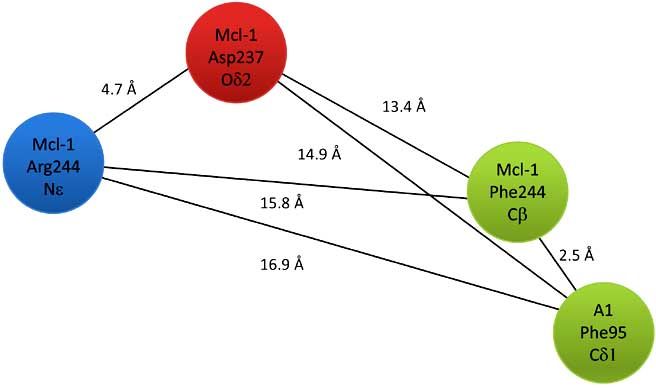
284 Current Topics in Medicinal Chemistry, 2011, Vol. 11, No. 3                                                             Mustata et al.

Fig. (4). Chemical structures of 20 hits from ZINC 8.0 DB screening using the pharmacophore model and ADME/T calculations. 20 structur-
ally diverse hits were extracted by ADME/T calculations, out of 142 identified from the ZINC 8.0 screening against the pharmacophore
model. Compounds acquired for experimental testing are marked with a black star. Structures of hit 12, 15 and 18 are not depicted due to
intellectual property protection.

                                                                      cophore. Such favorable interactions are not accounted for
                                                                      by the isopropanol probe [25]. Hence, we presume that bind-
                                                                      ing affinities better than 3 μM are achievable by targeting the
                                                                      close neighborhood of the selected hotspots with specific
                                                                      pharmacophores.

                                                                      Selected Compounds Inhibited PUMA-Induced Apopto-
                                                                      sis
                                                                          We obtained 13 structurally diverse molecules identified
                                                                      above, or candidate PUMA inhibitors, from commercial
                                                                      sources for testing marked with asterisks in Fig. (4). Various
                                                                      cell-based and cell free systems were used to determine
                                                                      whether these computationally identified PUMA inhibitor
                                                                      had desired biological and biochemical activities. We have
                                                                      previously shown that PUMA induces apoptosis through a
Fig. (5). Druggability calculations for PUMA BH3 domain. (A)          Bax and mitochondria-dependent manner in the intestinal
Regions enriched by isopropanol are shown by the cyan cloud           epithelium and colon cancer cells [9, 13, 26, 27]. We first
around PUMA. The displayed volume corresponds to enrichment in        analyzed their effects on apoptosis induced by PUMA in
isopropanol concentration by 4 or more folds over expected concen-    DLD1 cells using an adenovirus expressing PUMA [27, 28].
tration. The highly enriched regions correspond to the interface      When added at 25 μM, ten out of 13 compounds signifi-
between PUMA and interacting proteins. (B) Isopropanol binding        cantly suppressed PUMA-induced apoptosis Fig. (6A). The
hotspots are shown along with the pharmacophore features. Six         apoptosis induced by PUMA is completely dependent on its
hotspots were used to calculate a binding affinity of 3 M, in the    BH3 domain, as the BH3 domain deleted PUMA (Ad-
absence of ionic interactions.                                        BH3) was unable to induce apoptosis in DLD1 cells and
Induced Fit Simulations on Nuclear Receptors Current Topics in Medicinal Chemistry, 2011, Vol. 11, No. 3 285
several other cell lines Fig. (6A) [28]. In addition, 8 out of utes after irradiation, all three compounds displayed anti-
13 compounds significantly inhibited growth suppression apoptotic activities in p21 KO cells Fig. (7A). These com-
induced by PUMA Fig. (6B). Most (11 out of 13) com- pounds alone had little or no effects on the proliferation or
pounds had little or measurable negative effects on prolifera- apoptosis of either cell line at 25 or 50 μM Fig. (7A) and
tion or apoptosis on their own compared with vehicle at data not shown. We further examined the ability of these
doses of 1, 5, 25 and 50 μM Fig. (6C) and data not shown. three compounds on radiation-induced apoptosis in a rela-
These results suggest that several compounds identified us- tively radiosensitive germ cell line NCCIT as a stem cell
ing the pharmacophore model, are potential PUMA inhibi- model. When added at 25 uM and 15 minutes after irradia-
tors. tion, all three compounds displayed significant antiapoptotic
activities Fig. (7B).
Selected Compounds Inhibited PUMA-Dependent and We further evaluated the effects of hit 18 on radiosensi-
Radiation-Induced Apoptosis tivity of murine hematopoietic cell line 32D cl3. Hit 18 was
We previously reported that radiation or DNA damaging found to significantly suppress radiosensitivity when added
agent Adriamycin induces apoptosis in HCT116 cells defi- either one hour before or after radiation in a clonogenic as-
cient in the cyclin-dependent kinase (CDK) inhibitor p21 say in doses from 0-6 Gy, reflected by an increase in D0
(p21-KO cells) [27, 29, 30]. Radiation-induced apoptosis is (final slope of irradiation survival curve expressed in Gy)
PUMA-dependent in p21-KO cells, as deleting PUMA in and Ñ (shoulder on the survival curve) Fig. (7C). These re-
these cells (double knockout (p21/PUMA DKO) signifi- sults suggest that selected compounds were able to inhibit
cantly blocked radiation-induced apoptosis Fig. (7A). We PUMA-dependent and radiation-induced apoptosis. Interest-
selected three compounds that significantly suppressed ingly, hit 18 consistently showed the highest level of protec-
PUMA-induced apoptosis and growth suppression (Hits 12, tion against both PUMA- or radiation-induced apoptosis
15 and 18) and examined their effects on radiation-induced Figs. (6 and 7).
apoptosis in this system. When added at 25 uM and 15 min-
Fig. (6). Selected compounds inhibited PUMA-induced apoptosis and growth suppression. DLD1 cells were infected with 10 MOI of Ad-
PUMA, or a control Ad-BH3 with or without the addition of compounds. The compounds were used at 25 μM and added the same time as
Ad-PUMA. The cells were analyzed at 48 hr after treatment. (A) The effects of indicated compounds on Ad-PUMA-induced apoptosis were
analyzed by nuclear fragmentation assay at 48 hr. Right, the expression of PUMA and the BH3 deleted PUMA (BH3) was confirmed by
Western blotting. Tubulin was used as loading control. *The P values of hits 12, 15 and 18 vs. DMSO are 1.3 x 10⁻³, 1.0 x 10⁻⁵, and 1.3 x
10-6, respectively. (B) The cell growth of DLD1 cells was analyzed by MTS assays. *The P values of hits 12, 15 and 18 vs. DMSO are 1.2 x
10-7, 4.0 x 10⁻⁷ and 1.2 x 10⁻⁸, respectively. (C) The cell growth of DLD1cells without Ad-PUMA infection was analyzed by MTS assays.
The results are average ± SD of three independent experiments, and expressed as values relative to vehicle control.286 Current Topics in Medicinal Chemistry, 2011, Vol. 11, No. 3 Mustata et al.
Fig. (7). Selected compounds inhibited PUMA-dependent and radiation-induced apoptosis. (A) The indicated compounds were added to p21
KO or p21/ PUMA DKO cells 15 minutes after irradiation (15 Gy), and apoptosis was scored at 48 hr post-IR. *The P values of hits 12, 15
and 18 vs. DMSO in p21 KO cells are 3.28x10⁻⁶, 7.53x10⁻⁶ and 4.2x10⁻⁵, respectively. (B) The indicated compounds were added to NCCIT
cells 15 minutes after radiation (6 Gy), and apoptosis was scored at 48 hr post-IR. *The P values of hits 12, 15 and 18 vs. DMSO are
6.1x10⁻⁵, 2.1x10⁻⁶ and 6.1x10⁻⁷, respectively. (C) Clonogenic survival of 32D Cl3 cells with two compounds (10 μM) added 1 hr before and
after IR. Cells were plated and scored as described in the Materials and Methods. Results are the mean ± SD of each agent at each concentra-
tion.
Selected Compounds Inhibited the Interaction Between DISCUSSION
PUMA and Bcl-xL
Mitochondrial damage represents the "point of no return"
The BH3 domain of PUMA is capable of binding to all in the apoptotic pathway, and is highly regulated by a cas-
five known antiapoptotic Bcl-2 family members including cade involving mitochondrial membrane permeability, cyto-
Bcl-2, Bcl-xL, Mcl-1, Bcl-w and A1 with high affinity, chrome c leakage, caspase activation and cell demise [7, 35,
while that from most other BH3-only proteins do so selec- 36]. The Bcl-2 protein family has been identified as the
tively [31, 32]. Prior work suggests that Bcl-xL is a major gateway to mitochondria-mediated apoptosis [37]. The BH3-
antiapoptotic Bcl-2 member in human intestinal epithelium only protein PUMA integrates diverse death signals through
[33, 34]. We therefore determined whether these three com- both p53-dependent and -independent mechanisms to initiate
pounds with promising cellular activities are able to disrupt apoptosis. Unlike most BH3-only proteins, PUMA neutral-
the interactions between PUMA and Bcl-xL using a cell free izes all five known anti-apoptotic Bcl-2 members through
system established earlier [34]. In this system, PUMA and high-affinity interactions, which makes the PUMA BH3 do-
Bcl-xL are expressed separately in BAX KO HCT 116 cells main an enticing target for drug design.
first to avoid activation of caspases and degradation of cellu-
In the present study, using structural data on the con-
lar proteins, and mixed. HA-tagged PUMA and V5-tagged
served interactions of PUMA with Bcl-2-like proteins Mcl-1
Bcl-xL interacted strongly by immunoprecipitation in a BH3
and A1, we developed a pharmacophore model for a poten-
domain-dependent fashion Fig. (8A). This interaction is sig-
tial inhibitor of the interaction of PUMA with Bcl-2 family
nificantly inhibited by two of three compounds Fig. (8B).
members. Although targeting protein-protein interactions
We also did similar experiments using lysates prepared from using small molecules can be difficult, inhibitors may be
293 cells, which contains BAX protein. Similar results were
developed provided that the binding surface of the target
obtained. Interestingly, hit 18 displayed the strongest effect
protein has druggable features. The deep hydrophobic
on disrupting this interaction, correlated with its strongest
groove on the surface of Bcl-xL for example proved it feasi-
antiapoptotic effects in cells Figs. (6 and 7).
ble to develop highly specific inhibitors of BclxL-Bad inter-Induced Fit Simulations on Nuclear Receptors Current Topics in Medicinal Chemistry, 2011, Vol. 11, No. 3 287
Fig. (8). Selected compounds inhibited the interaction between PUMA and Bcl-xL. (A) The interaction of PUMA and Bcl-xL in BAX-KO
extracts were was analyzed by IP. Antibodies against HA were used to pull down PUMA. Western blotting (IB) was performed to analyze
Bcl-xL bound to PUMA (V5) and the efficiency for PUMA pull down (HA). Input represents 50% of the starting lysates used for IP. (B) The
effects of the indicated compounds on the interactions between PUMA and Bcl-xL were analyzed as in (A). Compounds were used at 100
μM and 25 μM in BAX-KO and 293 lysates, respectively. The vehicle (DMSO, V) or compounds were incubated with PUMA lysates for 15
minutes prior to the addition of V5-Bcl-xL lysates.
action [38]. The pharmacophore model developed here is Apoptosis evasion is a hall mark of cancer [46]. Resis-
based on the amino acid identities and geometry of specific tance to apoptosis is commonly associated with altered ex-
recognition residues on two proteins that bind PUMA. In pression in both pro- and anti-apoptotic Bcl-2 family mem-
silico screening of the ZINC 8.0 database, and in silico bers, and contributes to tumor formation, progression and
ADME/Toxicity profiles, complemented by druggability impaired responsiveness to anticancer therapies [11, 3, 47].
calculations in support of the adopted strategy, led to a small Expression of PUMA rapidly kills a variety of human cancer
set of lead compounds that are further screened and validated cells, and results in profound chemo- or radio-sensitization
using assays. The working concentrations of several com- and [11, 27, 48]. Therefore, an analogous strategy can be
pounds in cells are between 10-25 μM, not too far from the used to develop PUMA mimetics as Pan-Bcl-2 inhibitors. In
predicted 3 μM. fact, this possibility has been explore and led to the devel-
opment of a selective Bcl-2 and Bcl-xL inhibitor ABT-737
PUMA inhibitors can have several applications. Given a
and analogs, currently in clinical trials [18, 19]. Interestingly,
prominent role of PUMA in DNA damage induced apoptosis
resistance to ABT-737 is attributable to Mcl-1 overexpres-
in the GI tract [13, 39] and HP systems [14, 15], PUMA in-
sion in some cases [49], where Pan-Bcl-2 inhibitors might be
hibitors are expected to provide radiation protection and
useful.
mitigation. PUMA transcription induction occurs within 8 hr
following radiation while protein levels remain elevated be- In summary, the interaction of PUMA/Bcl-2 like proteins
yond 24 hours. Inhibiting PUMA binding to Bcl-2 like pro- is a critical regulatory step in apoptosis initiation. The struc-
teins is likely to provide a wider window for intervention, tural features of this interactions and its dependence on a
compared to inhibiting its transcription, a critical considera- rather small PUMA BH3 domain make it an attractive target
tion of mitigation measures administrated after a radiation for drug development. PUMA inhibitors might be effective
accident or attack. PUMA inhibitors might also prevent or and safe in radiation protection and mitigation and perhaps
mitigate intestinal damage and apoptosis induced by in- in other types of tissue injury associated with apoptosis.
flammatory cytokines, reactive oxygen species or chemo-
therapy [40, 41] Fig. (1), some of which are implicated in the METHODS
delayed phase of radiation damage [42, 43]. Furthermore, it
should not be surprising and perhaps even desirable that Ligand-Based Pharmacophore Modeling
PUMA inhibitors identified in this study can function as Ligand-based development of a pharmacophore model
Pan-BH3 inhibitors based on several highly conserved inter- uses the spatial information of known ligands (or binding
actions of the BH3 domain with Bcl-2 like proteins [16, 17]. proteins) for topological description of ligand-receptor inter-
Since p53 ablation leads to high risks in cancer [44] and ex- actions and for the subsequent discovery of new structural
acerbated IR-induced GI damage [45], inhibiting PUMA leads. In this study, pharmacophore features that comple-
might be more advantageous compared to p53 in tissue pro- ment the BH3 domain were built upon both sequence analy-
tection. Additional work and optimization of the lead struc- sis of hPUMA and structural analysis of the BH3 domain of
tures identified in this study can guide their further develop- PUMA in complex with Mcl-1 (PDB entry 2roc) [16] and
ment. A1 (PDB entry 2vof) [17]. The pharmacophore model was288 Current Topics in Medicinal Chemistry, 2011, Vol. 11, No. 3 Mustata et al.
built by applying the PGH (Polarity-Charged-Hydro- ranging from 6 to 15 Gy using a 137Cs irradiator (Mark I, JL
phobicity) scheme in MOE 8.0 software [22]. Shepherd and Associates, San Fernando, CA, USA). PUMA
inhibitors (25 M or as indicated) were added to the media
Database (DB) Searching following irradiation. Thirteen small molecules with defined
structures were purchased from either InterBioScreen (Rus-
The ZINC 8.0 DB [21] containing approximately 2.0x sia) or Chembridge (San Diego, CA, USA) and details are
106 lead-like compounds was used for screening. Compared available upon request. The three compounds used in the
to de novo design methods, known DB searching offers the validation were hits 12, 15 and 18.
advantage that hits can be purchased for testing. For each
compound in the DB, alternative conformations were gener-
Analysis of Apoptosis and Cell Growth
ated allowing for a maximum conformational energy of 20
kcal/mol above the lowest-energy found. The number of Following treatment, floating and adhering cells were
conformers generated for each compound was limited to a collected at 48 hr. For analysis of apoptosis by nuclear stain-
maximum number of 250. ing, cells were resuspended and fixed in PBS solution con-
taining 3.7% formaldehyde, 0.5% Nonidet P-40 and 10
ADME/Toxicity Predictions μg/ml Hoechst 33258 (Molecular Probes). Apoptosis was
assessed through microscopic visualization of condensed
Computational modeling tools were used to estimate the chromatin and micronucleation as previously described [48,
bioavailability, aqueous solubility, blood brain barrier poten- 55]. A minimum of 300 cells were analyzed in triplicate.
tial, human intestinal absorption, the cytochrome P450 (i.e. Cell growth was measured by the 3-(4,5-dimethyl-thiazol-
CYP2D6) enzyme inhibition potential, mutagenicity, and 2yl)-5-(3-carboxymethoxyphenyl)-2-(4-sulfophenyl)-2Htetr-
hERG inhibition of the hits obtained from the database azolium (MTS) assay in 96-well plates (2,500 cells per well)
screening. The bioavailability, aqueous solubility, and hu- using CellTiter 96 Aqueous One Solution (Promega) as de-
man intestinal absorption were estimated using the scribed [49, 57]. Each experiment was repeated at least twice
ACD/ADME Boxes software (ACD Labs, Toronto, Canada) in triplicate. For irradiation survival curves, 32D cells were
[23, 24], while mutagenicity, hERG and CYP2D6 inhibition irradiated over a total dose range of 0 to 8 Gy. The cells were
were estimated with ACD/TOX screening (ACD Labs, To- incubated with compounds 1 hr before or after irradiation at
ronto, Canada) [23, 24]. 10 μM. Irradiated cells were centrifuged to a cell pellet and
resuspended in medium then plated in 0.8% methylcellulose
Druggability Index Calculations containing medium for clonogenic survival curve assay.
We applied druggability index calculations introduced by Cells were plated at 500, 1000, or 2000 cells per plate in
Seco et al. [25]. In our work, we used NAMD [50] program triplicate at each dose, and the colonies of greater than 50
and CHARMM force field [51]. To prevent protein rotation cells were counted at day 7 according to published methods
and translation, we applied 0.01 kcal/mol harmonic restraints [53, 54]. Data are processed by software as described [54].
on -carbons. Threonine side-chain atom types and partial
charge set used by Seco and Barril was used to define iso- Antibodies and Western Blotting
propanol topology in CHARMM format [51]. Hotspot identi- The antibodies used for Western blotting include antibod-
fication and druggability index calculations were performed ies against HA (Santa Cruz (rabbit)), HA (Roche (mouse))
using the procedure described by Seco et al. [25]. and anti-V5 antibody (Invitrogen). Western blotting analysis
was performed as previously described [28, 56].
Cell Culture and Treatment
All cell lines were maintained at 37°C and with 5% CO2. Immunoprecipitation
Colon cancer cell lines DLD1 and HCT116 derivative lines BAX KO HCT 116 cells or 293 cells were transfected
(BAX KO-knock-out cells [33], p21 KO – p21 knock-out with HA-PUMA and V5-Bcl-xL by LipofectamineTM 2000
cells [52], PUMA and p21 KO cells [27]) were cultured in (Invitrogen) following the instructions of the manufacturer.
McCoy's 5A media (Invitrogen). The germ cell tumor line The expression plasmids of HA tagged PUMA and V5-
NCCIT and kidney epithelial cell line 293 were obtained tagged Bcl-xL were previously described [9]. Transfection
from ATCC and cultured with RPMI-1640 medium (Medi- and immunoprecipitation was performed as described with
atech) and DMEM (Invitrogen), respectively. The IL-3 de- minor modifications [35]. In brief, twenty-four hrs after
pendent murine hematopoietic progenitor cell line 32D cl 3 transfection, cells in one T-75 flask were harvested and re-
was grown in McCoys modified medium [53, 54]. All cell suspended in 1 ml of EBC buffer (50 mM Tris-HCl, pH 7.5,
culture media were supplemented with 10% defined fetal 100 mM NaCl, 0.5% NP-40) supplemented with protease
bovine serum (Hyclone), 100 units/ml penicillin, and 100 inhibitor cocktail (Roche Applied Sciences). The cell sus-
g/ml streptomycin (Invitrogen). pension were disrupted by sonication and then spun at
Cells were plated in 12-well plates at ~20% density and 10,000 g for 10 min to collect the cell lysates (super-
allowed to attach for 12-16 hr prior to adenovirus infection natant). The lysates from HA-PUMA and V5-Bcl-xL trans-
or irradiation. The DLD1 cells were infected with 0.01 ul of fections were mixed and incubated with a compound (100
virus, an equivalent 10 multiplicity of infection (MOI) [28]. M) with gentle rotation for 1 h at RT. Compounds or
The constructions and purification of recombinant adenovi- DMSO were incubated with PUMA extracts 15 minutes be-
ruses, Ad-PUMA and Ad-BH3 were previously described fore the addition of Bcl-xL extracts. The mixture was then
[27, 48]. Cells were irradiated at dose rate 100 cGy/min incubated with 50 μl Dynabeads® Protein A (Invitrogen)Induced Fit Simulations on Nuclear Receptors Current Topics in Medicinal Chemistry, 2011, Vol. 11, No. 3 289
with antibody bound for 1 hr at RT following the instructions Tomaselli, K. J.; Wang, B.; Wendt, M. D.; Zhang, H.; Fesik, S. W.;
of the manufacturer. For immunoprecipitation (IP), 1 μg of Rosenberg, S. H. An inhibitor of Bcl-2 family proteins induces re-
gression of solid tumours. Nature, 2005, 435(7042), 677-681.
IP antibodies (HA) were added to cell lysates diluted with [19] Zhang, L.; Ming, L.; Yu, J. BH3 mimetics to improve cancer ther-
the binding buffer in a total volume of 200 ul. Following the apy; mechanisms and examples. Drug Resist. Updat, 2007, 10(6),
final wash in a new tube, the immunocomplexes on the 207-217.
beads were eluted with 50 μl of 2 Laemmli sample buffer, [20] Camacho, C. J.; Zhang, C. FastContact: rapid estimate of contact
and binding free energies. Bioinformatics, 2005, 21(10), 2534-
heated at 95ºC for 10 min, and analyzed by Western blotting. 2536.
[21] Irwin, J. J.; Shoichet, B. K. ZINC--a free database of commercially
ACKNOWLEDGEMENT available compounds for virtual screening. J. Chem. Inf. Model.,
2005, 45(1), 177-182.
This work is supported by NIH U19A1068021) (JSG, IB [22] Molecular Operating Environment (MOE), http://www.chemcomp.
and JY), 1R01GM086238-01 (IB), and UO1DK085570 (JY). com, 2008.
[23] Japertas, P.; Didziapetris, R.; Petrauskas, A. Fragmental methods in
the analysis of biological activities of diverse compound sets, Mini.
REFERENCES Rev. Med. Chem., 2003, 3(8), 797-808.
[1] Thompson, C.B. Apoptosis in the pathogenesis and treatment of [24] ACD/Labs, http://www.acdlabs.com, ADME/TOX Boxes, 2009.
[25] Seco, J. Luque, F. J.; Barril, X. Binding site detection and drugga-
disease. Science, 1995, 267(5203), 1456-1462.
[2] Fulda, S.; Debatin, K.M. Extrinsic versus intrinsic apoptosis path- bility index from first principles. J. Med. Chem., 2009, 52(8), 2363-
2371.
ways in anticancer chemotherapy. Oncogene, 2006, 25(34), 4798-
4811. [26] Yu, J.; Wang, P.; Ming, L.; Wood, M.A.; Zhang, L. SMAC/Diablo
mediates the proapoptotic function of PUMA by regulating PUMA-
[3] Adams, J.M.; Cory, S. The Bcl-2 apoptotic switch in cancer devel-
opment and therapy. Oncogene, 2007, 26(9), 1324-1337. induced mitochondrial events. Oncogene, 2007, 26(29), 4189-4198.
[27] Yu, J.; Wang, Z.; Kinzler, K.W.; Vogelstein, B.; Zhang, L. PUMA
[4] Thornberry, N.A.; Lazebnik, Y. Caspases: enemies within. Science,
1998, 281(5381), 1312-1316. mediates the apoptotic response to p53 in colorectal cancer cells.
Proc. Natl. Acad. Sci. USA, 2003, 100(4), 1931-1936.
[5] Antonsson, B.; Conti, F.; Ciavatta, A.; Montessuit, S.; Lewis, S.;
Martinou, I.; Bernasconi, L.; Bernard, A.; Mermod, J.J.; Mazzei, [28] Yu, J.; Yue, W.; Wu, B.; Zhang, L. PUMA sensitizes lung cancer
cells to chemotherapeutic agents and irradiation. Clin. Cancer Res.,
G.; Maundrell, K.; Gambale, F.; Sadoul, R.; Martinou, J.C. Inhibi-
tion of Bax channel-forming activity by Bcl-2. Science, 1997, 2006, 12(9), 2928-2936.
[29] Waldman, T.; Kinzler, K.W.; Vogelstein, B. p21 Is Necessary For
277(5324), 370-372.
[6] Bagci, E.Z., Vodovotz, Y., Billiar, T.R., Ermentrout, G.B.; Bahar, the p53-Mediated G(1) Arrest In Human Cancer Cells. Cancer
Res., 1995, 55(22), 5187-5190.
I. Bistability in apoptosis: roles of bax, bcl-2, and mitochondrial
permeability transition pores. Biophysics, 2006, 90(5), 1546-1559. [30] Polyak, K.; Waldman, T.; He, T.-C.; Kinzler, K.W.; Vogelstein, B.
Genetic determinants of p53 induced apoptosis and growth arrest.
[7] Yu, J.; Zhang, L. Apoptosis in human cancer cells. Curr. Opin.
Oncol., 2004, 16(1), 19-24. Genes Devel., 1996, 10(15), 1945-1952.
[31] Chen, L.; Willis, S.N.; Wei, A.; Smith, B.J.; Fletcher, J.I.; Hinds,
[8] Puthalakath, H.; Strasser, A. Keeping killers on a tight leash: tran-
scriptional and post-translational control of the pro-apoptotic activ- M.G.; Colman, P.M.; Day, C.L.; Adams, J.M.; Huang, D.C. Differ-
ential targeting of prosurvival Bcl-2 proteins by their BH3-only
ity of BH3-only proteins. Cell Death Differ., 2002, 9(5), 505-512.
[9] Yu, J.; Zhang, L.; Hwang, P.M.; Kinzler, K.W.; Vogelstein, B. ligands allows complementary apoptotic function. Mol. Cell, 2005,
17(3), 393-403.
PUMA induces the rapid apoptosis of colorectal cancer cells. Mol.
Cell, 2001, 7(3), 673-682. [32] Kuwana, T.; Bouchier-Hayes, L.; Chipuk, J.E.; Bonzon,C.; Sulli-
van, B.A.; Green, D.R.; Newmeyer, D.D. BH3 domains of BH3-
[10] Nakano, K.; Vousden, K.H. PUMA, a novel proapoptotic gene, is
induced by p53. Mol. Cell, 2001, 7(3), 683-694. only proteins differentially regulate Bax-mediated mitochondrial
membrane permeabilization both directly and indirectly. Mol. Cell,
[11] Yu, J.; Zhang, L. PUMA, a potent killer with or without p53. On-
cogene, 2008, 27 (Suppl 1), S71-83. 2005, 17(4), 525-535.
[33] Zhang, L.; Yu, J.; Park, B.H.; Kinzler, K.W.; Vogelstein, B. Role
[12] Potten, C.S. Radiation, the ideal cytotoxic agent for studying the
cell biology of tissues such as the small intestine. Radiat. Res., of BAX in the apoptotic response to anticancer agents. Science,
2000, 290(5493), 989-992.
2004, 161(2), 123-136.
[13] Qiu, W.; Carson-Walter, E. B.; Liu, H.; Epperly, M.; Greenberger, [34] Ming, L.; Wang, P.; Bank, A.; Yu, J.; Zhang, L. PUMA dissociates
Bax and BCL-xL to induce apoptosis in colon cancer cells. J. Biol.
J. S.; Zambetti, G. P.; Zhang, L.; Yu, J. PUMA regulates intestinal
progenitor cell radiosensitivity and gastrointestinal syndrome. Cell Chem., 2006, 281(23), 16034-16042.
[35] Wang, X. The expanding role of mitochondria in apoptosis. Genes
Stem Cell, 2008, 2(6), 576-583.
[14] Wu, W. S.; Heinrichs, S.; Xu, D.; Garrison, S. P.; Zambetti, G. P.; Dev., 2001, 15(22), 2922-2933.
[36] Green, D.R.; Reed, J.C. Mitochondria and apoptosis. Science,
Adams, J. M.; Look, A. T. Slug antagonizes p53-mediated apopto-
sis of hematopoietic progenitors by repressing puma. Cell, 2005, 1998, 281(5381), 1309-1312.
[37] Leibowitz, B.; Yu, J. Mitochondrial signaling in cell death via the
123(4), 641-653.
[15] Yu, H.; Shen, H.; Yuan, Y.; Xufeng, R.; Hu, X.; Garrison, S. P.; Bcl-2 family. Cancer Biol. Ther., 2010, 9(6), 417-422.
[38] Petros, A. M.; Nettesheim, D. G.; Wang, Y.; Olejniczak, E. T.;
Zhang, L.; Yu, J.; Zambetti, G.; Cheng, T. Deletion of Puma pro-
tects hematopoietic stem cells and confers long-term survival in re- Meadows, R. P.; Mack, J.; Swift, K.; Matayoshi, E. D.; Zhang, H.;
Thompson, C. B.; Fesik, S. W. Rationale for Bcl-xL/Bad peptide
sponse to high-dose -irradiation. Blood, 2010, 115(17), 3472-
3480. complex formation from structure, mutagenesis, and biophysical
studies. Protein Sci., 2000, 9(12), 2528-2534.
[16] Day, C. L.; Smits, C.; Fan, F. C.; Lee, E. F.; Fairlie, W. D.; Hinds,
M. G. Structure of the BH3 domains from the p53-inducible BH3- [39] Qiu, W.; Leibowitz, B.; Zhang, L.; Yu, J. Growth factors protect
intestinal stem cells from radiation-induced apoptosis by suppress-
only proteins Noxa and Puma in complex with Mcl-1. J. Mol. Biol.,
2008, 380(5), 958-971. ing PUMA through the PI3K/AKT/p53 axis. Oncogene, 2010, 29,
1622-1632.
[17] Smits, C.; Czabotar, P.E.; Hinds, M.G.; Day, C.L. Structural plas-
ticity underpins promiscuous binding of the prosurvival protein A1. [40] Wu, B.; Qiu, W.; Wang, P.; Yu, H.; Cheng, T.; Zambetti, G. P.;
Zhang, L.; Yu, J. p53 independent induction of PUMA mediates in-
Structure, 2008, 16(5), 818-829.
[18] Oltersdorf, T.; Elmore, S.W.; Shoemaker, A.R.; Armstrong, R.C.; testinal apoptosis in response to ischaemia-reperfusion. Gut., 2007,
56(5), 645-654.
Augeri, D.J.; Belli, B.A.; Bruncko, M.; Deckwerth, T. L.; Dinges,
J.; Hajduk, P. J.; Joseph, M. K.; Kitada, S.; Korsmeyer, S. J.; [41] Wang, P.; Wang, P.; Qiu, W.; Dudgeon, C.; Liu, H.; Huang, C.;
Zambetti, G. P.; Yu, J.; Zhang, L. PUMA is directly activated by
Kunzer, A. R.; Letai, A.; Li, C.; Mitten, M. J.; Nettesheim, D. G.;
Ng, S.; Nimmer, P. M.; O'Connor, J. M.; Oleksijew, A.; Petros, A. NF-kappaB and contributes to TNF-alpha-induced apoptosis. Cell
Death Differ., 2009, 16(9), 1192-1202.
M.; Reed, J. C.; Shen, W.; Tahir, S. K.; Thompson, C. B.;290 Current Topics in Medicinal Chemistry, 2011, Vol. 11, No. 3 Mustata et al.
[42] Mothersill, C.; Seymour, C. B. Radiation-induced bystander ef- molecular dynamics with NAMD. J. Comput. Chem., 2005, 26(16),
fects--implications for cancer. Nat. Rev. Cancer, 2004, 4(2), 158- 1781-1802.
164. [51] Brooks, B. R.; Brooks, C. LIII.; Mackerell, A. D.Jr.; Nilsson, L.;
[43] Epperly, M. W.; Travis, E. L.; Sikora, C.; Greenberger, J. S. Man- Petrella, R. J.; Roux, B.; Won, Y.; Archontis, G.; Bartels, C.;
ganese [correction of Magnesium] superoxide dismutase (MnSOD) Boresch, S.; Caflisch, A.; Caves, L.; Cui, Q.; Dinner, A. R.; Feig,
plasmid/liposome pulmonary radioprotective gene therapy: modu- M.; Fischer, S.; Gao, J.; Hodoscek, M.; Im, W.; Kuczera, K.; Laz-
lation of irradiation-induced mRNA for IL-I, TNF-alpha, and TGF- aridis, T.; Ma, J.; Ovchinnikov, V.; Paci, E.; Pastor, R. W.; Post, C.
beta correlates with delay of organizing alveolitis/fibrosis. Biol. B.; Pu, J. Z.; Schaefer, M.; Tidor, B.; Venable, R. M.; Woodcock,
Blood Marrow Trans., 1999, 5(4), 204-214. H. L.; Wu, X.; Yang, W.; York, D. M.; Karplus, M. CHARMM:
[44] Donehower, L. A.; Harvey, M.; Slagle, B. L.; McArthur, M. J.; the biomolecular simulation program. J. Comput. Chem., 2009,
Montgomery, C. A. Jr. Butel, J.S.; Bradley, A. Mice deficient for 30(4), 1545-1614.
p53 are developmentally normal but susceptible to spontaneous [52] Wang, P.; Yu, J.; Zhang, L. The nuclear function of p53 is required
tumours. Nature, 1992, 356(6366), 215-221. for PUMA-mediated apoptosis induced by DNA damage. Proc.
[45] Komarova, E. A.; Kondratov, R. V.; Wang, K.; Christov, K.; Natl. Acad. Sci. USA, 2007, 104(10), 4054-4059.
Golovkina,T. V.; Goldblum, J. R.; Gudkov, A. V. Dual effect of [53] Epperly, M. W.; Osipov, A. N.; Martin, I.; Kawai, K. K.;
p53 on radiation sensitivity : p53 promotes hematopoietic injury, Borisenko, G. G.; Tyurina, Y. Y.; Jefferson, M.; Bernarding, M.;
but protects from gastro-intestinal syndrome in mice. Oncogene, Greenberger, J.S.; Kagan, V.E. Ascorbate as a "redox sensor" and
2004, 23(19), 3265-3271. protector against irradiation-induced oxidative stress in 32D CL 3
[46] Hanahan, D.; Weinberg, R. A. The hallmarks of cancer. Cell, 2000, hematopoietic cells and subclones overexpressing human manga-
100(1), 57-70. nese superoxide dismutase. Int. J. Radiat. Oncol. Biol. Phys., 2004,
[47] Fesik, S.W. Promoting apoptosis as a strategy for cancer drug 58(3), 851-861.
discovery. Nat. Rev. Cancer, 2005, 5(11), 876-885. [54] Epperly, M. W.; Bernarding, M.; Gretton, J.; Jefferson, M.; Nie, S.;
[48] Sun, Q.; Sakaida, T.; Yue, W.; Gollin, S. M.; Yu, J. Chemosensiti- Greenberger, J. S. Overexpression of the transgene for manganese
zation of head and neck cancer cells by PUMA. Mol. Cancer Ther., superoxide dismutase (MnSOD) in 32D cl 3 cells prevents apopto-
2007, 6(12), 3180-3188. sis induction by TNF-alpha, IL-3 withdrawal, and ionizing radia-
[49] van Delft, M. F.; Wei, A. H.; Mason, K. D.; Vandenberg, C. J.; tion. Exp. Hematol., 2003, 31(6), 465-474.
Chen, L.; Czabotar, P. E.; Willis, S. N.; Scott, C. L.; Day, C. L.; [55] Yu, J.; Zhang, L.; Hwang, P. M.; Rago, C.; Kinzler, K. W.;
Cory, S.; Adams, J. M.; Roberts, A. W. The BH3 mimetic ABT- Vogelstein, B. Identification and classification of p53-regulated
737 targets selective Bcl-2 proteins and efficiently induces apopto- genes. Proc. Natl. Acad. Sci. USA, 1999, 96(25), 14517-14522.
sis via Bak/Bax if Mcl-1 is neutralized. Cancer Cell, 2006, 10(5), [56] Sun, Q.; Ming, L.; Thomas, S. M.; Wang, Y.; Chen, Z. G.; Ferris,
389-399. R. L.; Grandis, J. R.; Zhang, L.; Yu, J. PUMA mediates EGFR ty-
[50] Phillips, J. C.; Braun, R.; Wang, W.; Gumbart, J.; Tajkhorshid, E.; rosine kinase inhibitor-induced apoptosis in head and neck cancer
Villa, E.; Chipot, C.; Skeel, R. D.; Kale, L.; Schulten, K. Scalable cells. Oncogene, 2009, 28(24), 2348-2357.
Received: April 23, 2010 Accepted: May 21, 2010You can also read