Formaldehyde Exposure and Epigenetic Effects: A Systematic Review - MDPI
←
→
Page content transcription
If your browser does not render page correctly, please read the page content below
applied
           sciences
Review
Formaldehyde Exposure and Epigenetic Effects:
A Systematic Review
Veruscka Leso * , Maria Carmela Macrini, Francesco Russo and Ivo Iavicoli
 Department of Public Health, Section of Occupational Medicine, University of Naples Federico II, Via S.
 Pansini 5, 80131 Naples, Italy; maria.carmela.macrini@hotmail.it (M.C.M.); francesco.russo.na@gmail.com (F.R.);
 ivo.iavicoli@unina.it (I.I.)
 * Correspondence: veruscka.leso@unina.it; Tel.: +39-081-746-4763; Fax: +39-081-746-2133

 Received: 29 February 2020; Accepted: 24 March 2020; Published: 28 March 2020

 Abstract: Formaldehyde (FA) is a general living and occupational pollutant, classified as carcinogenic
 for humans. Although genotoxicity is recognized as a FA mechanism of action, a potential contribution
 of epigenetic effects cannot be excluded. Therefore, aim of this review is to comprehensively assess
 possible epigenetic alterations induced by FA exposure in humans, animals, and cellular models.
 A systematic review of Pubmed, Scopus, and Isi Web of Science databases was performed. DNA global
 methylation changes were demonstrated in workers exposed to FA, and also in human bronchial
 cells. Histone alterations, i.e., the reduction in acetylation of histone lysine residues, in human
 lung cells were induced by FA. Moreover, a dysregulation of microRNA expression in human lung
 adenocarcinoma cells as well as in the nose, olfactory bulb and white blood cells of rodents and
 nonhuman primates was reported. Although preliminary, these findings suggest the role of epigenetic
 modifications as possible FA mechanisms of action that need deeper qualitative and quantitative
 investigation. This may allow to define the role of such alterations as indicators of early biological
 effect and the opportunity to include such information in future risk assessment and management
 strategies for public and occupationally FA-exposed populations.

 Keywords: formaldehyde; epigenetic; DNA methylation; histone modifications; microRNAs;
 occupational exposure; biological monitoring; risk assessment; risk management

1. Introduction
     Formaldehyde (FA) is a volatile, colorless, flammable, and highly reactive aldehyde, employed
in the production of industrial and consumer products. It is present in both indoor and outdoor
environments [1,2]. Indoor FA sources include building materials, such as furniture, particle board,
certain insulation materials, paints, varnishes, and textiles [3,4]. Direct emissions from combustion
processes occurring in motor vehicles, power plants, incinerators, refineries characterize a major
anthropogenic source of FA in outdoor environments [5]. Certain workplaces, such as industries
involved in resin, plastics, wood, insulation, paper, textile, and chemical productions, as well as
medical institutions using disinfectants and embalming chemicals experience the highest levels of the
substance [3]. Formaldehyde is also a by-product of cigarette smoke [3,6]. Additionally, it is found as a
natural product in most living systems [7,8].
     Formaldehyde inhalation has been associated with sensory irritation of the eyes, and respiratory
tract, childhood and adult asthma as well as to alterations in pulmonary function as reported in
clinical studies and epidemiological surveys in occupational and residential environments [9]. The
International Agency for Research on Cancer has classified FA as carcinogenic to humans (Group
1) because of its ability to cause cancer of the nasopharynx and leukemia and the detected positive
association with sino-nasal cancer [3]. Great efforts have been focused on understanding the mode

Appl. Sci. 2020, 10, 2319; doi:10.3390/app10072319                                 www.mdpi.com/journal/applsci
Appl. Sci. 2020, 10, 2319 2 of 18
of action for FA carcinogenesis. Currently available data strongly indicate that genotoxicity plays an
important role in the carcinogenicity of FA in humans. Numerous experiments have demonstrated
that FA exposure can induce mutations in a variety of test systems ranging from bacteria to laboratory
animals [10,11]. Moreover, micronucleus formation has been repeatedly reported to occur in cells of the
nasal and oral mucosa of FA-exposed humans [3,12,13]. Formaldehyde is a highly reactive compound.
It can covalently bind to DNA and proteins inducing adducts formation and cross-linking [8]. FA reacts
with lysine residues to produce N(6)-formyl-lysine adducts with proteins in vitro and in vivo [14,15],
and FA protein adducts have been described for albumin [16], insulin [17], and hemoglobin [18,19].
Such alterations may function as sensitive and specific exposure biomarkers (dose metric) and potential
effect indicators, as they are involved in mutations and subsequent cancer development [20].
However, exposure to genotoxic carcinogens, in addition to their genetic effects, might involve
a variety of non-genotoxic changes in cells [21]. In this perspective, it cannot be excluded that
epigenetic mechanisms may contribute to the FA toxicity [22]. Epigenetics is defined as the study
of heritable phenotypes that do not alter the DNA sequence, able to control development, tissue
differentiation, and cellular responsiveness [23,24]. DNA methylation, post-translational histone
modifications, and changes in microRNA expression are the most used mechanisms able to initiate and
sustain epigenetic information. These effects may all contribute to regulate gene expression profiles,
affecting chromatin structure, chromosome integrity, gene transcription, and genomic imprinting
therefore possibly contributing to the risk of disease development [25]. An altered epigenetic status
may cause genomic instability and loss of controlled growth signals, as typically observed in cancer
cells [26]. Epigenetic alterations, rather than specific genetic mutations per se are reported for the clonal
expansion of altered preneoplastic foci and tumor development [27]. Therefore, it seems important
to verify if epigenetic modifications may function as early cellular events in response to genotoxic
carcinogens with well-known modes of action.
Considering the ubiquitous nature of FA pollutant and the great number of job categories
potentially exposed in workplaces, the aim of our review is to provide a comprehensive overview
on possible epigenetic changes induced by FA exposure in humans, animals, and cellular models
and to address the potential health impact derived from such effects. This may allow to deeply
understand the molecular mechanisms underlying FA-induced toxicity and carcinogenicity and to
define novel, early biological alterations that may function as possible future biomarkers of effect or
susceptibility in exposed subjects. Overall, this review may provide deep insight into the relationship
between genome, FA chemical exposure, and disease risk and may be helpful to point out biological
mechanisms that should be carefully considered in future public and occupational risk assessment and
management approaches.
2. Materials and Methods
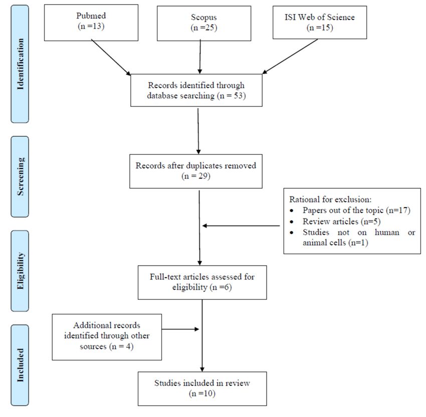
According to the Preferred Reporting Items for Systematic Reviews and Meta-Analyses Statement
(PRISMA) criteria [11], a systematic literature search was performed on PubMed, Scopus, and ISI Web
of Science databases. The search aimed to identify studies focusing on epigenetic effects induced by
formaldehyde exposure published until the 1st of November 2019 (Figure 1). The terms “formaldehyde,”
“exposure,” “epigenetic,” combined with the Boolean operator “AND,” were employed to assess
the exposure context and the outcome of the investigation. All titles and abstracts retrieved were
independently examined by two of the authors who selected articles that met the inclusion criteria.
Peer-reviewed research articles, i.e., cross-sectional, cohort, case-control studies, published in English
and exploring epigenetic alterations determined by FA exposure in humans have been included
together with studies performed on animal and cellular models. Exclusion criteria regarded articles
published in languages other than English, papers exploring epigenetic effects of endogenous FA,
studies carried out on experimental models different from cells, animals and human subjects, review
and conference papers, as well as publications not addressing the possible relationship between FA
exposure and epigenetic effects. Our preliminary search retrieved 13, 25, and 15 references throughAppl. Sci. 2020, 10, 2319 3 of 18
Appl. Sci.
PubMed, 2020, 10, xand
Scopus, FOR PEER REVIEW
ISI Web of Science databases, respectively, for a total of 53 articles. 3 of 20 After
duplicate removal, 29 articles remained. Among those, the studies that did not meet the inclusion
for a total of 53 articles. After duplicate removal, 29 articles remained. Among those, the studies that
criteria were excluded and specifically, 17 were removed because they were considered out of the
did not meet the inclusion criteria were excluded and specifically, 17 were removed because they
topicwere
from title andout
considered abstract analysis,
of the topic 5 were
from title and excluded as review
abstract analysis, 5 werearticles,
excludedand 1 as performed
as review articles, in
experimental settings different from those considered suitable for inclusion. Indeed,
and 1 as performed in experimental settings different from those considered suitable for inclusion. 6 publications
couldIndeed,
be identified in thiscould
6 publications preliminary phase.
be identified Therefore,
in this an extended
preliminary search, including
phase. Therefore, an extended the following
search,
terms: “formaldehyde
including exposure,”
the following terms: individually
“formaldehyde combined
exposure,”with “DNA methylation,”
individually combined with “histone,”
“DNA and
methylation,”
“microRNA,” was“histone,”
performed andand
“microRNA,” was performed
could retrieve the aboveand could retrieve
mentioned the above
papers. mentioned
Four further eligible
articles could be included through the assessment of the reference list accompanying publishedlist
papers. Four further eligible articles could be included through the assessment of the reference articles
accompanying
and implement thepublished articles and
pool of relevant implementfound
publications the pool
via of
therelevant publications
electronic search. Allfound
fullvia theof the
texts
electronic search. All full texts of the articles considered valuable for the aim of our review have been
articles considered valuable for the aim of our review have been obtained and a critical evaluation was
obtained and a critical evaluation was performed. Overall, our search retrieved a total of 10
performed. Overall, our search retrieved a total of 10 publications for review.
publications for review.
Figure 1. Flow diagram of literature search.
Figure 1. Flow diagram of literature search.
3. Formaldehyde-Induced Epigenetic Effects
3. Formaldehyde-Induced Epigenetic Effects
Formaldehyde-induced epigenetic changes, such as DNA methylation [21,28,29], histone
Formaldehyde-induced epigenetic changes, such as DNA methylation [21,28,29], histone
modifications [30–32], as well as alterations in non-coding microRNAs (MiRNAs) [33–36] have
modifications [30–32], as well as alterations in non-coding microRNAs (MiRNAs) [33–36] have been
been investigated both in occupational and experimental settings (Table 1). The following paragraphs
investigated both in occupational and experimental settings (Table 1). The following paragraphs will
will attempt to summarize
attempt to summarizesuchsucheffects
effects with
with thethe aim
aim to to point
point outout currently
currently available
available data data and critical
and critical
issues that require further epidemiological research in order to also conceive epigenetic contribution to
risk assessment and management strategies.Appl. Sci. 2020, 10, 2319 4 of 18
Table 1. Studies addressing epigenetic formaldehyde (FA) effects in humans, animals, and cellular models.
EXPERIMENTAL EPIGENETIC
EXPERIMENTAL DESIGN MAIN RESULTS REFERENCES
SETTING OUTCOMES
HUMAN INVESTIGATION
3 Median personal FA environmental exposure (ppm):
group A (n. 8): 0.013; group B (n. 15): 0.035; group C
Airborne FA concentrations were (n. 26): 0.07.
Brazilian hairdressers, measured through personal (samplers 3 Median biological monitoring levels of formic acid
hairdresser assistants, fitted in volunteers breathing zone) and before exposure (mg/L): group A: 25.99; group B:
receptionists, manicurists, area passive samplers (8-h work shift). 24.80; group C: 19.93. Median biological monitoring
and managers (n. 41F; 8M) Global DNA methylation was assessed levels of formic acid after exposure (mg/L): group A:
Mean age ± SD: 31.04 ± in whole blood samples were collected 21.49; group B: 20.70; group C: 22.44.
DNA Methylation Barbosa et al. [28]
10.46 years after 8-h of work. 3 Higher median levels of global DNA methylation
Years working in beauty Biological monitoring of formic acid were determined in groups B (4.55%) and C (4.29%)
salons (median: 6 years) was performed on urine samples compared to group A (3.12%).
No unexposed controls collected at the beginning of the work 3 Statistically significant positive correlation was
were enrolled. shift and again 8 h after the first found between global DNA methylation and FA in
sampling. the personal passive samplers (rs: 0.307, p: 0.032).
3 No significant increase in biological monitoring
findings could be observed between the 3 groups.
IN VIVO EXPERIMENTS
3 FA exposure affected the expression levels of 108
miRNAs in nose samples, specifically, 84, 59, and 0
miRNAs altered in the 7, 2, and 28-day plus 7
day-recovery groups, respectively.
Nose-only exposure to 2 ppm
3 In the WBCs, FA exposure altered the expression of
formaldehyde for 6 h/day; Four groups
40 miRNAs: 31, 8, and 3 miRNAs at the 3 different
of exposure were considered: 7, 28, 28
time points, respectively. In BM, no changes in
days plus a 7-day recovery period,
miRNAs were identified.
unexposed controls.
Male Fischer rats MicroRNAs 3 Transcriptomics based analysis demonstrated that Rager et al. [34]
Multiple samples were collected from
the nasal epithelium, circulating FA exposure induced differential expression of 830
mononuclear WBCs, and BM cells. and 42 genes in the nose and in 96 and 130 genes in
Microarray analysis employed to assess WBC, in the 7 and 28 day group, respectively.
695 rat known miRNAs. 3 Thirty-five of the 864 total FA responsive transcript
are immune cell specific. Seven transcripts showed
formaldehyde-induced expression in both exposure
groups, namely Cd34, Clec7a, Fcgr2b, Lpl, Lsp1,
Mustn1, and Osmr.Appl. Sci. 2020, 10, 2319 5 of 18
Table 1. Cont.
EXPERIMENTAL EPIGENETIC
EXPERIMENTAL DESIGN MAIN RESULTS REFERENCES
SETTING OUTCOMES
3 Microarray analysis: 18 and 7 miRNAs were up- and
downregulated, respectively, after exposure to 3
ppm for 6 h. greatest increase reported for
Mice treated with 3 ppm FA for 6 h for miR-199b-5p and miR-144-5p. Exposure for 7 days
either 1 or 7 consecutive days via only altered the expression of nine miRNAs: eight
inhalation; controls maintained in the increased and one decreased.
same conditions except for FA. 3 miRNAs (mir-144-5p, miR-199b-5p, miR-200a-5p
ICR male mice MicroRNAs Li et al. [35]
Olfactory bulb homogenized and RNA and miR-146b-5p) increased most after 6 h/1-day and
isolated for microarray analysis. 6 h/7 days of FA exposures confirmed by RT-PCR.
Expression levels of miRNAs tested 3 A total of 295 genes predicted to be regulated by
using quantitative RT-PCR. miR-199b-5p, 247 by miR-200a-5p and 139 by
miR-146b-5p. pathways with the highest scores
were the integrin signaling pathway and the axonal
guidance signaling pathway.
3 FA exposure significantly decreased the expression
Animals exposed via inhalation to 0, 2,
levels of 3 miRNA following 2 ppm and disrupted
or 6 ppm FA for 6 hr/day for two
the expression of 13 miRNAs following 6 ppm.
consecutive days.
3 Three miRNAs that were significantly decreased in
Small RNAs extracted from nasal
response to 2 ppm FA (i.e., miR-142-3p, miR-145,
Male cynomolgus samples, collected within 3 h from the
and miR-203) were also significantly decreased in
macaques (Macaca MicroRNAs last exposure, assessed for Rager et al. [36]
response to 6 ppm FA.
fascicularis, n. 8) genome-wide miRNA expression levels.
Transcriptional targets of FA-affected 3 Pathways of highest significance correlated with
miRNAs analyzed. Expression levels of increased expression of miR-125b was the apoptosis
miRNAs tested using quantitative signaling (expression levels of apoptotic related
RT-PCR. genes, i.e., BAK1, CASP2, MAP2K7, MCL1 were
significantly reduced)Appl. Sci. 2020, 10, 2319 6 of 18
Table 1. Cont.
EXPERIMENTAL EPIGENETIC
EXPERIMENTAL DESIGN MAIN RESULTS REFERENCES
SETTING OUTCOMES
IN VITRO EXPERIMENTS
3 Genome of treated cells undergo a gradually global
hypomethylation with increasing exposure time
to FA: - Anti-5-methyl-C immunohistochemistry
assay: mean density of fluorescence was reduced to
32.1 following 24 weeks of treatment, for control
group the density was 61.3, after 6 weeks 54.4, after
Cell cultures treated with 10 mM FA for
12 weeks 43.5 and for BTC groups 43.5; - Flow
24 h for 24 weeks. Control cells treated
cytometric assay: fluorescence intensity of 16HBE
with serum-free MEM.
cells was reduced to 36.1 at 24 weeks of treatment,
BTC, a malignant transformed 16HBE
Human bronchial while was 76.5 for control, 62.4 after 6 weeks, 45.2
DNA methylation cell line was used as a positive control. Liu et al. [29]
epithelial (16HBE) cells after 12 weeks and 34.9 for BTC; - HPCE assay: an
Genomic DNA methylation measured
average proportion of methylated mC/(C + mC) was
by: anti-5-methyl-C immuno-
4.72% in control, 3.76% in 6 week, 3.22% in 12 week
histochemistry assay, flow cytometric
and 2.35% in 24 week groups, while 2.1% in
assay and HPCE assay.
BTC group.
3 The mRNA expression level for DNMT3a and
DNMT3b significantly and gradually decreased,
while that of DNMT1 and MBD2 significantly
increased gradually with increasing exposure time
to FA.
Cells were exposed to 3 concentrations
3 No significant DNA methylation changes were
of FA (95% cellular viability- high dose;
detected in response to FA exposure to
1/10 of high dose: medium dose; 1/100
diverse concentrations.
of high dose: low dose) for 24 h with or
3 Global mean ± SD percentages of DNA methylation
without a mix of S9 metabolic mix
were: 4.09 ± 0.23; 5.21 ± 0.57 and 4.61 ± 0.23 in low,
Human lymphoblastoid required for biotransformation of
DNA methylation medium and high dose, respectively, in absence of Tabish et al. [21]
cells (TK6) procarcinogens into active carcinogens.
S9 metabolic mix
Global DNA methylation was obtained
quantifying (5Me)dC and dC using 3 Global mean ± SD percentages of DNA methylation
ultrapressure liquid chromatography were: 4.61 ± 0.44; 4.55 ± 0.43 and 4.23 and in low,
for separation and tandem mass medium and high dose, respectively, in the presence
spectrometry for quantification. of S9 metabolic mix.Appl. Sci. 2020, 10, 2319 7 of 18
Table 1. Cont.
EXPERIMENTAL EPIGENETIC
EXPERIMENTAL DESIGN MAIN RESULTS REFERENCES
SETTING OUTCOMES
3 FA increased the phosphorylation of H3S10, and
H3S28 and gamma-H2AX with generation peaks
60–240 min after the treatment.
3 FA-induced phosphorylation of H3S10 in a
concentration dependent manner up to 1 mM.
Cell cultures treated with 0–1 mM FA for Conversely phosphorylation decreased from 3
Human lung
10–600 min mM FA.
adenocarcinoma epithelial Histone Modifications Yoshida et al. [30]
Western blotting employed to determine 3 FA decreased the acetylation of histone H3 at lysine
(A549) cells
histone modifications. 9 (H3K9), at lysine 14 (H3K14), and at the
N-terminal site (H3 global). Phosphorylation of
H3S10 plays an important role in the induction of
immediate-early genes, such as c-fos and c-jun. The
treatment with FA significantly induced both genes
in a dose-dependent manner.
Cell cultures treated with 0–0.5 mM FA
Human bronchial for 6 h. For chronic FA 0–0.1 mM for 96 h.
3 FA exposure induced a significant reduction in the
epithelial BEAS-2B cells; Protein carbonyl assays employed to
acetylation of the N-terminal tails of cytosolic
human osteosarcoma determine FA adducts with histone
Histone Modifications histones (H3K9Ac, H3K14Ac, H4K12Ac) of BEAS-2B Chen et al. [31]
UTA6 cells; human nasal proteins
cells. Levels of H4K12Ac were significantly reduced
septum quasidiploid Histone post-transcriptional
in all 3 cell lines following 100 µM FA for 24–72 h.
tumor RPMI2650 cells modifications assessed by Western blot
analysis.
3 Cigarette side-stream smoke induced a marked,
Human lung Cells were treated with various dose-dependent phosphorylation of H3S10 and
adenocarcinoma epithelial concentrations of cigarette side-stream H3S28 in all 3 cellular models.
(A549) cells; normal Histone Modifications smoke (~6.25%–100%) for ~up to 8 h. 3 The phosphorylation increased up to 60 min and Ibuki et al. [32]
human lung fibroblasts, Flow cytometric analysis was performed gradually decreased from 120 min.
MRC-5 and WI-38 to assess phosphor-H3S10 3 Acetylation of H3K9 and H3K14 slightly increased
at 60–180 min and 120–360 min, respectively.Appl. Sci. 2020, 10, 2319 8 of 18
Table 1. Cont.
EXPERIMENTAL EPIGENETIC
EXPERIMENTAL DESIGN MAIN RESULTS REFERENCES
SETTING OUTCOMES
3 The most significantly differentially expressed
miRNAs were miR-33 (FC = −5.5), miR-450 (FC =
Cell cultures treated with 1 ppm (1.2 −3.6), miR-330 (FC = −2.4), miR-181a (FC = −2.1),
mg/m3 ) gaseous FA generated by heat and miR-10b (FC = −2.1).
Human lung in 143 mg paraformaldehyde until 3 qRT-PCR validated the findings of the decreased
adenocarcinoma epithelial powder completely vaporized. miRNA expression induced by formaldehyde
MicroRNAs Rager et al. [33]
(A549) cells Microarray analysis employed to assess exposure: FC = −1.3 for miR-330; FC = −7.4 for
>500 known miRNAs. miR-181a; FC = −1.2 for miR-33; and FC = −1.5
Expression levels of miRNAs also for miR-10b.
tested using quantitative RT-PCR. 3 Signaling pathways associated with cancer,
inflammatory response, and endocrine system
regulation related to miRNA expression alterations.
BAK1, BCL2-antagonist/killer 1; BM, bone marrow; CASP2, caspase 2, apoptosis related cysteine peptidase; DNMT, DNA methyltransferase; FA, formaldehyde; FC, fold change;
gamma-H2AX, histone 2AX at serine 139; H3S10, histone 3 at serine 10 residue; H3S28, histone 3 at serine 28 residue; HBE, human bronchial epithelial; HPCE, high performance capillary
electrophoresis; MAP2K7, mitogen-activated protein kinase 7; MBD2, methyl-CpG-binding protein DNA-binding domain protein 2; MCL1, myeloid cell leukemia sequence 1; MEM,
minimum essential Eagle’s medium; RT-PCR, real time-polymerase chain reaction; WBCs, white blood cells.Appl. Sci. 2020, 10, 2319 9 of 18
3.1. DNA Methylation
DNA methylation is a major epigenetic modification in mammals [29,37]. It involves the addition
of a methyl group to the cytosine nucleotide at CpG sites via the donation from S-adenosylmethionine,
catalyzed by DNA methyltransferases (DNMTs) [37]. DNA methylation can affect the chromatin
structure, and therefore, control the selective transcription or silencing of tissue-specific genes potentially
involved in disease pathogenesis [29]. The possible effects of occupational exposure to FA on global
DNA methylation were investigated in a population of Brazilian beauty salon workers [28]. These were
exposed during hair treatments employing creams illegally added with FA as a straightening agent.
Environmental monitoring allowed to rank workers into three increasing groups (A, B, C) of exposure
according to the FA environmental workplace levels:Appl. Sci. 2020, 10, 2319 10 of 18
responsible for the diverse obtained findings and further investigations are warranted to confirm such
epigenetic effects and define the FA impact on DNA methylation.
3.2. Histone Modifications
Histone modifications occur post-transcriptionally and can affect the accessibility of DNA to
transcription factors or DNA damaging agents, thus influencing DNA transcription, damage, and
repair [43]. There are several types of histone modifications, including methylation, acetylation,
phosphorylation, and ubiquitination of specific amino acid residues on the histone tails [44]. These
molecular changes have recently attracted attention because they have been linked to many diseases
including cancer [45]. In regard to histone modifications, no human or animal data are currently
available and only few in vitro studies investigated these epigenetic effects [30–32]. Yoshida et al. [30]
examined histone modifications in human lung adenocarcinoma epithelial A549 cells following an
acute (10–600 min) treatment with FA with a focus on histone 3 (H3). Treated cells showed greater
levels of phosphorylated histone H3 at serine 10 (H3S10), and serine 28 (H3S28) residues compared
with unexposed controls. Conversely, FA induced a reduction of the acetylation of histone H3 at lysine
9 (H3K9) and 14 (H3K14), and at the N-terminal site (H3 global) maybe in relation to the reaction that
FA itself could have with lysine residues limiting acetylation. This mode of action was confirmed by
the lack of acetylation reduction observed when histone was pre-acetylated before FA treatment, thus
supporting the idea that FA could not react with acetylated lysine histone residues. Concerning the
biological consequences of these epigenetic changes, the phosphorylation of H3S10 was enhanced at
the promoter regions of the proto-oncogenes FOS and JUN. Both genes were dose-dependently induced
by FA treatment suggesting a possible relationship between FA-induced tumor promotion activity and
phosphorylation of H3S10. The biological responses induced by the alterations in histone acetylation
remain to be understood. In line with these results, also the exposure of human pulmonary epithelial
A549 cells, and normal human lung fibroblasts MRC-5 and WI-38 to cigarette side-stream smoke,
containing FA, induced a significant phosphorylation of histone 3 at the serine 10 and 28 residues
(H3S10, H3S28) [32]. As previously reported, such phosphorylation was increased in the promoter
sites of the proto-oncogenes FOS and JUN, indicating a role of the cigarette smoke in tumor promotion.
Since the phosphorylation of H3S10 was decreased in the aldehyde-removed cigarette side-stream
smoke and was significantly induced by treatment with FA, aldehydes have been suspected to have
partially contributed to this phenomenon.
Chen et al. [31] demonstrated FA’s (0.5 mM) ability to form adducts on lysine residues of histone
proteins in human bronchial epithelial BEAS-2B cells. The formation of such FA-histone adducts
prevented the acetylation of the same site by histone acetyltransferase as demonstrated by the dramatic
decrease of the lysine acetylation of the cytosolic fraction of histones H3 and H4, specifically at H3K9,
H3K14, and H4K12 residues. A comparable reduction in the acetylation of H4K12 residues was
demonstrated with a lower concentration of FA (100 µM) on the same cell line, but also on human
osteosarcoma UTA6 and nasal epithelial RPMI2650 cells, although with different time periods of
manifestation in the diverse cell lines. Such changes may affect chromatin accessibility to subsequent
gene transcription. In fact, following FA exposure, the transport of histone H3 into chromatin is
compromised affecting chromatin conformation and inducing transcriptional dysregulation. This was
confirmed by the hundreds of genes whose expression was reported to be affected by 100-µM exposure
for 48 h in BEAS-2B cells. Among those, some tumor suppressors, i.e., CDKN1A and SERPINB5, and
oncogenes, i.e., FOS and JUN, which were identified as potentially involved in head and neck cancer
and/or in hematological neoplasias. Moreover, FA-induced inhibition of chromatin assembly may
facilitate anchorage-independent growth of cells. Therefore, the ability of FA to compromise chromatin
assembly through inhibition of lysine acetylation on newly synthesized histones represents a possible
novel mechanism underlying FA-induced carcinogenesis.
The ability of FA to react with histone lysine residues reported by Yoshida et al. [30] and
Chen et al. [31] are in line with previous results obtained by Lu et al. [45] in a simplified in vitro modelAppl. Sci. 2020, 10, 2319 11 of 18
employing H4 isolated from calf thymus tissues and human recombinant H4 purified after expression
in E. Coli. The authors, in fact, demonstrated that the FA-induced lysine adducts on histone may impair
post-transcriptional histone acetylation and deacetylation balance. This could affect the recruitment
of specific proteins highly associated with the post-transcriptional pattern, and trigger a series of
abnormal cascade effects responsible for alterations in normal cell growth.
3.3. MicroRNAs
MicroRNAs are small non-coding RNAs, of about 22 nucleotides, that regulate gene expression
at the post-transcriptional level. The importance to investigate potential effects of FA on miRNAs
relies on their ability to regulate gene expression through the binding of mRNA, causing rapid
decay of the message, translational repression of the mRNA signals, and inducing cleavage of newly
translated polypeptides [46]. Concerning in vivo responses induced by FA inhalation, the same
group of researchers demonstrated a significant dysregulation in miRNA expression in the nose,
olfactory bulb and white blood cells (WBCs) of rodents [35,36], as well as in the nasal epithelium
cells of non-human primates [34]. Interestingly, Rager et al. [35] applied a multitiered approach to
define possible genome-wide miRNA responses to FA, as well as time and tissue-related alterations
in transcriptional profiles. MiRNA expression, in fact, was investigated within the nasal respiratory
epithelium, circulating WBCs, and bone marrow (BM) and were affected in rats treated with FA via
inhalation for 7 and 28 days [35]. The nose was the site with the greatest change in miRNA expression
levels, followed by the WBCs, while no changes were evident in the BM samples. When alterations
were analyzed according to the different 7 and 28 day exposures, FA-induced miRNA changes in
nose showed high stability and no significant quantitative and qualitative differences over time. Such
alterations failed to persist after a recovery period (7 days) following the 28-day treatment. Comparing
the FA-responsive miRNAs across the different tissues revealed largely distinct miRNA responses, as
only 10 alterations overlapped between nose and WBC samples and most with different directions
of expression. Overall, these findings suggest that cells in direct contact with FA exposure are more
responsive at the miRNA level than cells distant from exposure contact sites. Moreover, 7 of the
34 miRNAs with sustained decreased nose expression over time, i.e., let-7a, let-7c, let-7f, miR-10b,
miR-126, miR-21, and miR-23a, have been previously shown to have decreased expression in cultured
lung cells exposed to 1 ppm FA [33]. To integrate miRNA responses with transcriptional changes,
mRNA profiles were assessed in the nose and WBCs. FA-responsive miRNAs were able to regulate a
series of mRNAs, ranging from the 7% to 35% of those FA-related, determining an increase in immune
system/inflammation signaling in both nose and WBCs.
Considering the miRNA alterations reported in nasal epithelium of treated rats, and the potential
regulating role of miRNAs in the neurogenesis of the olfactory bulb, Li et al. [36] investigated expression
changes within olfactory bulb in mice after FA acute to subacute inhalation. Eighteen miRNAs were
upregulated and seven were down-regulated after an acute (6-h) exposure to FA via inhalation. In the
olfactory bulb of mice exposed to FA, the alterations in miRNA expression was more profound after
1 day of exposure for 6 h relative to 7 days of 6 h/day of exposure, as this last condition only altered
the expression of nine miRNAs. The differentially expressed miRNA, i.e., mir-141-5p, mir-200a-5p,
mir-199b-5p, are believed to be involved in axon guidance and MAPK signaling, which are associated
with inter-neuronal maturation in the bulb, essential to assure a normal olfactory function [47,48].
Additionally, also other pathways related to the immune system, such as toll-like receptor signaling
and communication between innate and adaptive immune cells, molecular mechanisms of cancer, as
well as cell cycle regulation have been also associated to the miRNA alterations detected after both 1-
and 7-days of exposure.
Potential changes in miRNA expression profiles have been also investigated in the nasal epithelial
cells from the maxilla-turbinate region of Cynomolgus macaques, as the area with the maximum adsorption
of FA, collected from animals treated via inhalation [34]. Microarray analysis identified three miRNAs
with significantly decreased expression levels upon exposure to 2 ppm FA and 13 miRNAs with affectedAppl. Sci. 2020, 10, 2319 12 of 18
expression when exposure was to 6 ppm (four significantly increased and nine significantly decreased).
Interestingly, two of the 13 FA-responsive miRNAs were among those reported as affected in vitro by FA,
namely miR-26b and miR-140-5p [33]. The two most increased (miR-125b and miR-152) and decreased
(miR-145 and miR-142-3p) miRNAs in response to 6 ppm FA were primarily correlated with pathways
involved in sphingolipid metabolism, apoptosis, and ILK signaling. Concerning sphingolipids, recent
work has demonstrated that their metabolites are involved in the regulatory signaling of various
biological processes, including apoptosis, cell cycle arrest, inflammation, necrosis, and senescence [49].
The epigenetic FA-mediated control on apoptotic mechanisms that may be associated with cellular
transformation and cancer development could be demonstrated by the decreased expression of four
apoptosis-related mRNAs predicted to be regulated by miR-125b. ILK signaling is involved in a variety
of processes within epithelial cells, including cell survival, cell proliferation, and cell adhesion to the
extracellular matrix [50]. Additionally, many other FA-responsive miRNAs have known relationship
with cancers, and particularly were differentially expressed in human nasopharyngeal carcinomas.
These effects demonstrate the FA ability to significantly disrupt miRNA expression profiles within the
nasal epithelium that may influence cellular disease state.
The expression levels of miRNAs have been demonstrated to be dysregulated by FA exposure
in vitro [33]. In particular, when A549 type II lung human carcinoma epithelial cells were acutely
treated with gaseous FA, using a direct air-liquid interface that physically mimics the human respiratory
tract, microarray analysis demonstrated that 89 miRNAs were significantly down-regulated compared
to untreated controls, while no significant increased expression could be detected. Functional and
molecular network analysis of the predicted miRNA transcript targets revealed that FA exposure altered
the signaling pathways associated with cellular growth/development and proliferation. Moreover, as a
confirmation that FA treatment could activate inflammatory response, IL-8 pathway was demonstrated
to be increased by FA-miRNA-induced alterations, with increased protein expression levels compared
to controls.
4. Discussion
The general living and occupational ubiquitous nature of exposure to FA requires a deep
understanding of the molecular mechanisms potentially underlying its induced toxicity and
carcinogenicity. In this regard, although the limited number of studies and the lack of robust
epidemiological data on occupational exposed populations still prevent the extrapolation of definite
conclusions, preliminary evidences are emerging concerning the possibility that epigenetic changes
may condition biological response to FA.
Workers exposed to FA demonstrated a global increase in DNA methylation in whole blood
samples, positively related to FA workplace levels, to the number of procedures involving such
chemical exposure, as well as to have been directly employed in such specific job tasks [28]. Conflicting
evidence in DNA methylation emerged when these human findings were compared with previously
published in vitro results. In fact, these reported a significant decrease in global methylation in
bronchial epithelial cells chronically treated with FA, or the absence of significant DNA methylation
changes in acutely exposed human lymphoblastoid cells [29,32]. This may be due to the different
biological matrices investigated, i.e., the whole blood in humans [28] vs. bronchial and lymphoblastoid
cells in the experimental in vitro settings [29,32], which may be biased by cell-specific methylation
patterns. Variable experimental designs, i.e., concerning doses and time periods of FA exposure, as
well as the diverse analytical techniques adopted, i.e., high-performance liquid chromatography-diode
array detector equipment [28], high-performance capillary electrophoresis assay [29], and tandem
mass spectrometry [32], may all be responsible for quite different results. In this scenario, to define
the role of DNA methylation changes as possible FA biomarkers of early effect, future in-field and
epidemiological investigations seem absolutely necessary.
However, although in vitro experimental settings may provide a limited representativeness of
real human exposure contexts, such investigations may contribute to identify target genes whoseAppl. Sci. 2020, 10, 2319 13 of 18
expression changes may be involved in FA-induced methylation effects and toxic manifestations. This
idea is supported by the specific patterns of DNMT1, DNMT3a, DNMT3b expression responses found
in human bronchial epithelial cells that may suggest specific FA-induced alterations, in turn reflecting
the methylation levels determined in the samples [42]. Such data, once confirmed in epidemiological
investigations, may provide further support to the role of such specific patterns of changes as possible
FA biomarkers of early effect to be considered in suitable risk assessment processes.
Scientific evidence has been emerging concerning the role of histone post-translational
modifications in a multitude of DNA related processes, such as DNA damage repair, DNA replication,
and transcription of tumor-related genes [51,52]. In particular, the dysregulation of histone H3
phosphorylation, reported following FA exposure, may perturb cellular pathways involved in human
cancer development [30,32]. In fact, it is known that in interphase cells, such phosphorylation
phenomena are involved in the chromatin remodeling of the promoter regions of specific loci, i.e.,
FOS and JUN, implicated in tumorigenic processes, while in mitotic cells, they are responsible
for the maintaining of chromosome stability and their accurate segregation [53]. Histone H3
phosphorylation was reported in aflatoxin B1-transformed hepatocytes, benzo(a)pyrene- and coke
oven emissions-transformed human bronchial epithelial cells in which such modification performed
a mediation role in cellular carcinogenic transformation [51]. The phosphorylation of H3S10 has
been reported also to be induced by several environmental factors, such as arsenite [54], nickel [55],
and UVB [56,57].
Histone modification changes induced by FA exposure, included also the formation of FA-lysine
histone adducts and the consequent reduction in lysine acetylation. In general, acetylation neutralizes
the positive charge of lysine residues, resulting in a weaker histone-DNA interaction, in a subsequent
relaxation of the chromatin, and transcriptional activation [58]. In this context, FA may impact
post-transcriptional modifications disturbing the recruitment of regulatory complexes and evolving
into an aberrant chromatin assembly that may facilitate anchorage-independent growth of cells. These
data suggest that compromising chromatin assembly, through inhibition of lysine acetylation, may
dysregulate cancer-related gene expression, as a possible new mechanism underlying FA-induced
carcinogenesis [31]. The identification of FA reactive sites on histones is an initial step in understanding
additional mechanisms of FA toxicity and carcinogenicity [59]. However, many issues remain to
be clarified concerning the structure and fate of FA lysine adducts, the biological impact of such
modifications on lysine residues, which are the exact sites for the formation of adducts, and their exact
modes of action as well as how these changes may be recovered. Overall, to fully understand these
aspects may be important to define the role of such alterations as possible biomarkers of early effect or
susceptibility to the adverse FA impact.
MicroRNA alterations have been detected in both in vitro [33] and in vivo models [34–36]. From a
biological monitoring perspective, some conclusions, emerged from the study by Rager et al. [35], need
to be carefully considered. In different, simultaneously analyzed tissues, in fact, the authors determined
the greatest and more time-stable miRNA alterations (from 7 to 28 days of treatment) in the “most
directly exposed” nose epithelium compared to the “less directly exposed” WBCs. Moreover, miRNA
changes showed a tissue-specific expression as only a minimal part of the alterations overlapped
between these two different target sites. This underlines the importance to preliminary evaluate the
relevance of individual biological matrices in the assessment of epigenetic biomarkers in relation to the
routes of exposure as well as to the biological cascade intended to be investigated. Most of the miRNAs
affected in nose epithelium and WBCs, in fact, were related to the expression of genes involved in
nasopharyngeal carcinomas as well as in inflammatory/immune responses. Comparably, miRNA
alterations detected in nose-epithelium of nonhuman primates were predicted to affect the balance
between cell death and survival-affecting apoptosis, cell cycle progression, inflammation, therefore
being potentially implicated in cellular transformation and cancer development [34,36].
Then, an altered expression of miRNAs may not only be useful as a biomarker, but also as an
active component or tumor development. Evidence has been pointed out concerning the miRNAAppl. Sci. 2020, 10, 2319 14 of 18
alterations in all types of human cancer, both induced by genotoxic carcinogens, i.e., polycyclic
aromatic hydrocarbon, aflatoxin B1, n-nitroso compounds, as well as by not genotoxic carcinogens, i.e.,
arsenic [27]. However, the specific role of miRNA deregulation in the etiology of cancers remain to be
elucidated. To establish a link between miRNAs and carcinogenesis, some essential issues need to be
addressed, such as if the altered expression of miRNA occurs at the early stages of carcinogenesis in
the target organ and if such early changes are also evident in full-fledged tumors; if a mechanistic link
exists between aberrant miRNA expression and cancer development; and whether altered miRNA
expression is sufficient to initiate neoplastic cell transformation and cancer development.
Interestingly, it was estimated that between 7% and 35% of the FA-responsive transcripts were
regulated by FA-associated miRNAs. These data, on the one side, suggest that the miRNAs may be
operating through mechanisms other than mRNA degradation [46,60], but also that, on the other side,
other transcriptional regulators, such as DNA methylation and histone modifications [61], may play a
role in FA-induced genomic responses. It may be argued that, DNMTs may cooperate with histone
modifying enzymes to repress specific gene region expression, conversely, histone modifications
may impact DNA methylation patterns, and also a bidirectional influence between miRNA and
DNA methylation can exist [42]. Therefore, to assess the relevance of epigenetic effects in biological
monitoring will require further research comprehensive of a deep analysis of all the three principal
mechanisms and their possible interplay.
Finally, some limitations emerged from our revision that should be pointed out in order to
plan future methodologically adequate investigations able to provide more informative data. The
availability of only one study on occupationally FA-exposed subjects, underlines the relevance to
plan in-field, and epidemiological investigations to define the role of epigenetic changes as possible
biomarkers of early FA effects on humans. Although in vitro and in vivo studies, may be important
to extrapolate preliminary information, caution should be applied to adequately interpret results
obtained with high-doses of exposure (i.e., up to 6 ppm FA in vivo, almost greater than concentrations
found in workplace settings) [3,12] and for periods of treatment not resembling those experienced by
occupationally FA-involved populations (i.e., maximum 28 days in vivo) [34–36]. Future investigations
should confirm epigenetic effects following experimental conditions better resembling those realistically
occurring in workplace settings, generally characterized by low-dose, long-term exposures. This seems
even more important considering that some changes in DNA methylation have been determined
in subjects exposed at levels lower than those commonly adopted as occupational exposure limits.
Longitudinal studies are also strongly pursued. Diverse miRNA modifications, in fact, were evident
after shorter acute or subacute exposures compared to longer ones, suggesting that variable duration
of treatment may differently affect target biological responses and therefore biological monitoring
findings [35,36]. A deeper research, aimed to overcome such knowledge gaps, may be useful to
understand the relevance of epigenetic early biological alterations in risk assessment and management
remodeling in public and occupational exposure settings which may include also the re-assessment of
currently available occupational exposure limits to better protect the health of involved workers.
5. Conclusions
Epigenetics may provide a useful tool to understand the molecular mechanisms underlying
FA toxicological profile and to point out the relationship between FA chemical exposure,
genome/epigenome changes, and disease risk. Although preliminary evidence demonstrated DNA
methylation, histone and miRNA changes may be induced by FA exposure in workers, as well
as in in vivo and in vitro experimental settings; the limited number of available studies prevent
definite conclusions. Therefore, in vivo and in vitro studies, performed under conditions better
resembling those experienced in occupational and general living environments, may provide data on
qualitative and quantitative epigenetic changes as well as proper biological matrices to be analyzed
and time-dependent trends to be verified in human biological monitoring investigations. This kind of
research may further contribute to the understanding of new mechanisms underlying FA-inducedAppl. Sci. 2020, 10, 2319 15 of 18
toxicity, and particularly, carcinogenicity. Finally, in-field and epidemiological investigations should be
pursued to confirm such experimental results and define their possible role as FA early biological effect
indicators. More definite, future knowledge on this topic may allow to include epigenetic information
in suitable risk assessment and management processes to assure a better protection of the health of
public and occupationally FA-exposed populations.
Author Contributions: Conceptualization, V.L., I.I.; methodology, V.L., M.C.M., F.R.; writing—original draft
preparation, V.L., M.C.M., F.R.; writing—review and editing, V.L. and I.I.; supervision, V.L., I.I. All authors have
read and agreed to the published version of the manuscript.
Funding: This research received no external funding.
Conflicts of Interest: The authors declare no conflict of interest.
References
1. Salthammer, T.; Mentese, S.; Marutzky, R. Formaldehyde in the indoor environment. Chem. Rev. 2010, 110,
2536–2572. [CrossRef]
2. World Health Organization (WHO). WHO guidelines for indoor air quality: Selected pollutants. WHO Guide
2010, 9, 454.
3. IARC, Working Group on the Evaluation of Carcinogenic Risks to Humans Chemical Agents and Related
Occupations. Formaldehyde. IARC Monogr. Eval. Carcinog. Risks Hum. 2012, 100, 9–562.
4. National Toxicology Program (NTP). NTP 12th report on carcinogens. Rep. Carcinog. Carcinog. Profiles 2011,
12, iii-499.
5. WHO Regional Office for Europe Copenhagen. Formaldehyde. In Air Quality Guidelines for Europe—Second
Edition; 2000; chapter 5.8; pp. 87–91. Available online: http://www.euro.who.int/__data/assets/pdf_file/0005/
74732/E71922.pdf (accessed on 26 March 2020).
6. Godish, T. Formaldehyde exposures from tobacco smoke: A review. Am. J. Public Health 1989, 79, 1044–1045.
[CrossRef] [PubMed]
7. Dahl, A.R.; Hadley, W.M. Formaldehyde production promoted by rat nasal cytochrome P-450-dependent
monooxygenases with nasal decongestants, essences, solvents, air pollutants, nicotine, and cocaine as
substrates. Toxicol. Appl. Pharmacol. 1983, 67, 200–205. [CrossRef]
8. Yang, M.; Ospina, M.; Tse, C.; Toth, S.; Caudill, S.P.; Vesper, H.W. Ultraperformance liquid chromatography
tandem mass spectrometry method to determine formaldehyde hemoglobin adducts in humans as biomarker
for formaldehyde exposure. Chem. Res. Toxicol. 2017, 30, 1592–1598. [CrossRef]
9. Liteplo, R.G.; Meek, M.E. Inhaled formaldehyde: Exposure estimation, hazard characterization, and
exposure-response analysis. J. Toxicol. Environ. Health B Crit. Rev. 2003, 6, 85–114. [CrossRef] [PubMed]
10. IARC, Working Group on the Evaluation of Carcinogenic Risks to Humans Chemical Agents and Related
Occupations. Formaldehyde, 2-butoxyethanol and 1-tertbutoxypropan-2-ol. IARC Monogr. Eval. Carcinog.
Risks Hum. 2006, 88, 1–478.
11. Schmid, O.; Speit, G. Genotoxic effects induced by formaldehyde in human blood and implications for the
interpretation of biomonitoring studies. Mutagenesis 2007, 22, 69–74. [CrossRef]
12. Orsière, T.; Sari-Minodier, I.; Iarmarcovai, G.; Botta, A. Genotoxic risk assessment of pathology and anatomy
laboratory workers exposed to formaldehyde by use of personal air sampling and analysis of DNA damage
in peripheral lymphocytes. Mutat. Res. 2006, 605, 30–41. [CrossRef] [PubMed]
13. Ye, X.; Yan, W.; Xie, H.; Zhao, M.; Ying, C. Cytogenetic analysis of nasal mucosa cells and lymphocytes from
high level long-term formaldehyde exposed workers and low-level short-term exposed waiters. Mutat. Res.
2005, 588, 22–27. [CrossRef] [PubMed]
14. Edrissi, B.; Taghizadeh, K.; Dedon, P.C. Quantitative analysis of histone modifications: Formaldehyde is a
source of pathological n-formyllysine that is refractory to histone deacetylases. PLoS Genet. 2013, 9, e1003328.
[CrossRef] [PubMed]Appl. Sci. 2020, 10, 2319 16 of 18
15. Edrissi, B.; Taghizadeh, K.; Moeller, B.C.; Kracko, D.; Doyle-Eisele, M.; Swenberg, J.A.; Dedon, P.C. Dosimetry
of N-formyllysine adducts following [C H]-formaldehyde exposures in rats. Chem. Res. Toxicol. 2013, 26,
1421–1423. [CrossRef] [PubMed]
16. Bogdanffy, M.S.; Morgan, P.H.; Starr, T.B.; Morgan, K.T. Binding of formaldehyde to human and rat nasal
mucus and bovine serum albumin. Toxicol. Lett. 1987, 38, 145–154. [CrossRef]
17. Metz, B.; Kersten, G.F.; Baart, G.J.; de Jong, A.; Meiring, H.; ten Hove, J.; van Steenbergen, M.J.; Hennink, W.E.;
Crommelin, D.J.; Jiskoot, W. Identification of formaldehyde-induced modifications in proteins: Reactions
with insulin. Bioconjugate Chem. 2006, 17, 815–822. [CrossRef]
18. Guthe, K.F. The formaldehyde−hemoglobin reaction. J. Biol. Chem. 1959, 234, 3169–3173.
19. Hoberman, H.D.; San George, R.C. Reaction of tobacco smoke aldehydes with human hemoglobin. J. Biochem.
Toxicol. 1989, 3, 105–119. [CrossRef]
20. Swenberg, J.A.; Lu, K.; Moeller, B.C.; Gao, L.; Upton, P.B.; Nakamura, J.; Starr, T.B. Endogenous versus
exogenous DNA adducts: Their role in carcinogenesis, epidemiology, and risk assessment. Toxicol. Sci. 2011,
120 (Suppl. 1), S130–S145. [CrossRef]
21. Tabish, A.M.; Poels, K.; Hoet, P.; Godderis, L. Epigenetic factors in cancer risk: Effect of chemical carcinogens
on global DNA methylation pattern in human TK6 cells. PLoS ONE 2012, 7, e34674. [CrossRef]
22. Swenberg, J.A.; Moeller, B.C.; Lu, K.; Rager, J.E.; Fry, R.C.; Starr, T.B. Formaldehyde carcinogenicity research:
30 years and counting for mode of action, epidemiology, and cancer risk assessment. Toxicol. Pathol. 2013, 41,
181–189. [CrossRef] [PubMed]
23. Egger, G.; Liang, G.; Aparicio, A.; Jones, P.A. Epigenetics in human disease and prospects for epigenetic
therapy. Nature 2004, 429, 457–463. [CrossRef] [PubMed]
24. Arimondo, P.B.; Barberousse, A.; Pontarotti, G. The Many Faces of Epigenetics Oxford, December 2017.
Epigenetics 2019, 14, 623–631. [CrossRef] [PubMed]
25. Feinberg, A. The key role of epigenetics in human disease. N. Engl. J. Med. 2018, 379, 400–401.
26. Karpinets, T.V.; Foy, B.D. Tumorigenesis: The adaptation of mammalian cells to sustained stress environment
by epigenetic alterations and succeeding matched mutations. Carcinogenesis 2005, 26, 1323–1334. [CrossRef]
27. Pogribny, I.P.; Muskhelishvili, L.; Tryndyak, V.P.; Beland, F.A. The role of epigenetic events in genotoxic
hepatocarcinogenesis induced by 2-acetylaminofluorene. Mutat. Res. 2011, 722, 106–113. [CrossRef]
28. Barbosa, E.; Dos Santos, A.L.A.; Peteffi, G.P.; Schneider, A.; Müller, D.; Rovaris, D.; Bau, C.H.D.; Linden, R.;
Antunes, M.V.; Charão, M.F. Increase of global DNA methylation patterns in beauty salon workers exposed
to low levels of formaldehyde. Environ. Sci Pollut. Res. Int. 2019, 26, 1304–1314. [CrossRef]
29. Liu, Q.; Yang, L.; Gong, C.; Tao, G.; Huang, H.; Liu, J.; Zhang, H.; Wu, D.; Xia, B.; Hu, G.; et al. Effects of
long-term low-dose formaldehyde exposure on global genomic hypomethylation in 16HBE cells. Toxicol.
Lett. 2011, 205, 235–240. [CrossRef]
30. Yoshida, I.; Ibuki, Y. Formaldehyde-induced histone H3 phosphorylation via JNK and the expression of
proto-oncogenes. Mutat. Res. 2014, 770, 9–18. [CrossRef]
31. Chen, D.; Fang, L.; Mei, S.; Li, H.; Xu, X.; Des Marais, T.L.; Lu, K.; Liu, X.S.; Jin, C. Regulation of Chromatin
Assembly and Cell Transformation by Formaldehyde Exposure in Human Cells. Environ. Health Perspect.
2017, 125, 097019. [CrossRef]
32. Ibuki, Y.; Toyooka, T.; Zhao, X.; Yoshida, I. Cigarette sidestream smoke induces histone H3 phosphorylation
via JNK and PI3K/Akt pathways, leading to the expression of proto-oncogenes. Carcinogenesis 2014, 35,
1228–1237. [CrossRef] [PubMed]
33. Rager, J.E.; Smeester, L.; Jaspers, I.; Sexton, K.G.; Fry, R.C. Epigenetic changes induced by air toxics:
Formaldehyde exposure alters miRNA expression profiles in human lung cells. Environ. Health Perspect.
2011, 119, 494–500. [CrossRef] [PubMed]
34. Rager, J.E.; Moeller, B.C.; Doyle-Eisele, M.; Kracko, D.; Swenberg, J.A.; Fry, R.C. Formaldehyde and epigenetic
alterations: microRna changes in the nasal epithelium of nonhuman primates. Environ. Health Perspect. 2013,
121, 339–344. [CrossRef] [PubMed]
35. Rager, J.E.; Moeller, B.C.; Miller, S.K.; Kracko, D.; Doyle-Eisele, M.; Swenberg, J.A.; Fry, R.C.
Formaldehyde-associated changes in microRNAs: Tissue and temporal specificity in the rat nose, white
blood cells, and bone marrow. Toxicol. Sci. 2014, 138, 36–46. [CrossRef] [PubMed]Appl. Sci. 2020, 10, 2319 17 of 18
36. Li, G.; Yang, J.; Ling, S. Formaldehyde exposure alters miRNA expression profiles in the olfactory bulb. Inhal.
Toxicol. 2015, 27, 387–393. [CrossRef] [PubMed]
37. Esteller, M. Relevance of DNA methylation in the management of cancer. Lancet Oncol. 2003, 4, 351–358.
[CrossRef]
38. Pierce, J.S.; Abelmann, A.; Spicer, L.J.; Adams, R.E.; Glynn, M.E.; Neier, K.; Finley, B.L.; Gaffney, S.H.
Characterization of formaldehyde exposure resulting from the use of four professional hair straightening
products. J. Occup. Environ. Hyg. 2011, 8, 686–699. [CrossRef]
39. Norma Regulamentadora 15 (NR 15). Atividades e operações insalubres. D.O.U. Portaria MTb 1978, 3, 214.
40. OSHA, Occupational Safety and Health Administration. Formaldehyde. Available online: https://www.osha.
gov/OshDoc/data_General_Facts/formaldehyde-factsheet.pdf (accessed on 25 November 2019).
41. ECHA, European Chemicals Agency. Worker Exposure to Formaldehyde and Formaldehyde Releasers.
2019. Available online: https://echa.europa.eu/documents/10162/13641/investigationreport_formaldehyde_
workers-exposure_final_en.pdf/ac457a0c-378d-4eae-c602-c7cd59abc4c5 (accessed on 19 March 2020).
42. Moore, L.D.; Le, T.; Fan, G. DNA methylation and its basic function. Neuropsychopharmacology 2013, 38, 23–38.
[CrossRef]
43. Chappell, G.; Pogribny, I.P.; Guyton, K.Z.; Rusyn, I. Epigenetic alterations induced by genotoxic occupational
and environmental human chemical carcinogens: A systematic literature review. Mutat. Res. Rev. Mutat.
Res. 2016, 768, 27–45. [CrossRef]
44. Weber, D.; Heisig, J.; Kneitz, S.; Wolf, E.; Eilers, M.; Gessler, M. Mechanisms of epigenetic and cell-type
specific regulation of Hey target genes in ES cells and cardiomyocytes. J. Mol. Cell. Cardiol. 2015, 79, 79–88.
[CrossRef] [PubMed]
45. Lu, K.; Boysen, G.; Gao, L.; Collins, L.B.; Swenberg, J.A. Formaldehyde-induced histone modifications
in vitro. Chem. Res. Toxicol. 2008, 21, 1586–1593. [CrossRef] [PubMed]
46. Filipowicz, W.; Bhattacharyya, S.N.; Sonenberg, N. Mechanisms of post-transcriptional regulation by
microRNAs: Are the answers in sight? Nat. Rev. Genet. 2008, 9, 102–114. [CrossRef] [PubMed]
47. Choi, P.S.; Zakhary, L.; Choi, W.Y.; Caron, S.; Alvarez-Saavedra, E.; Miska, E.A.; McManus, M.; Harfe, B.;
Giraldez, A.J.; Horvitz, H.R.; et al. Members of the miRNA-200 family regulate olfactory neurogenesis.
Neuron 2008, 57, 41–55. [CrossRef] [PubMed]
48. Imai, T.; Sakano, H. Axon-axon interactions in neuronal circuit assembly: Lessons from olfactory map
formation. Eur. J. Neurosci. 2011, 34, 1647–1654. [CrossRef] [PubMed]
49. Bartke, N.; Hannun, Y.A. Bioactive sphingolipids: Metabolism and function. J. Lipid Res. 2009, 50, S91–S96.
[CrossRef]
50. Gilcrease, M.Z. Integrin signaling in epithelial cells. Cancer Lett. 2007, 247, 1–25. [CrossRef]
51. Zhu, X.; Li, D.; Zhang, Z.; Zhu, W.; Li, W.; Zhao, J.; Xing, X.; He, Z.; Wang, S.; Wang, F.; et al. Persistent
phosphorylation at specific H3 serine residues involved in chemical carcinogen-induced cell transformation.
Mol. Carcinog. 2017, 56, 1449–1460. [CrossRef]
52. Faucher, D.; Wellinger, R.J. Methylated H3K4, a transcription associated histone modification, is involved in
the DNA damage response pathway. PLoS Genet. 2010, 6, e1001082. [CrossRef]
53. Thompson, L.L.; Guppy, B.J.; Sawchuk, L.; Davie, J.R.; McManus, K.J. Regulation of chromatin structure
via histone post-translational modification and the link to carcinogenesis. Cancer Metastasis Rev. 2013, 32,
363–376. [CrossRef]
54. Li, J.; Gorospe, M.; Barnes, J.; Liu, Y. Tumor promoter arsenite stimulates histone H3 phosphoacetylation of
proto-oncogenes c-fos and c-jun chromatin in human diploid fibroblasts. J. Biol. Chem. 2003, 278, 13183–13191.
[CrossRef] [PubMed]
55. Ke, Q.; Li, Q.; Ellen, T.P.; Sun, H.; Costa, M. Nickel compounds induce phosphorylation of histone H3 at
serine 10 by activating JNK-MAPK pathway. Carcinogenesis 2008, 29, 1276–1281. [CrossRef] [PubMed]
56. Keum, Y.S.; Kim, H.G.; Bode, A.M.; Surh, Y.J.; Dong, Z. UVB-induced COX-2 expression requires histone H3
phosphorylation at Ser10 and Ser28. Oncogene 2013, 32, 444–452. [CrossRef] [PubMed]
57. He, Z.; Cho, Y.Y.; Ma, W.Y.; Choi, H.S.; Bode, A.M.; Dong, Z. Regulation of ultraviolet B-induced
phosphorylation of histone H3 at serine 10 by Fyn kinase. J. Biol. Chem. 2005, 280, 2446–2454. [CrossRef]
58. Banister, C.E.; Koestler, D.C.; Maccani, M.A.; Padbury, J.F.; Houseman, E.A.; Marsit, C.J. Infant growth
restriction is associated with distinct patterns of DNA methylation in human placentas. Epigenetics 2011, 6,
920–927. [CrossRef]You can also read