The Histone Variant H3.3 Marks Active Chromatin by Replication-Independent Nucleosome Assembly
←
→
Page content transcription
If your browser does not render page correctly, please read the page content below
Molecular Cell, Vol. 9, 1191–1200, June, 2002, Copyright 2002 by Cell Press
The Histone Variant H3.3 Marks Active Chromatin
by Replication-Independent Nucleosome Assembly
Kami Ahmad and Steven Henikoff1 ment are not clear. A study in Tetrahymena concluded
Fred Hutchinson Cancer Research Center that no protein difference between histone H3 variants
1100 Fairview Avenue North was required for replacement histone deposition and
Seattle, Washington 98109 that expression of either variant outside of S phase ap-
peared to be sufficient (Yu and Gorovsky, 1997). In con-
trast, by examining the dynamics of histone proteins in
Summary Drosophila nuclei we show that the major histone H3
and the replacement histone H3.3 proteins have distinct
Two very similar H3 histones—differing at only four properties during in vivo chromatin assembly. Histone
amino acid positions—are produced in Drosophila H3.3 participates in replication-independent (RI) nucleo-
cells. Here we describe a mechanism of chromatin some assembly and is targeted to transcriptionally ac-
regulation whereby the variant H3.3 is deposited at tive loci throughout the cell cycle. Transcription-coupled
particular loci, including active rDNA arrays. While the deposition of H3.3-containing nucleosomes may be a
major H3 is incorporated strictly during DNA replica- general mechanism for rapidly replacing permanently
tion, amino acid changes toward H3.3 allow replica- modified nucleosomes and for heritably activating
tion-independent (RI) deposition. In contrast to repli- genes.
cation-coupled (RC) deposition, RI deposition does
not require the N-terminal tail. H3.3 is the exclusive Results
substrate for RI deposition, and its counterpart is the
only substrate retained in yeast. RI substitution of H3.3 H3.3 Is Deposited by a Replication-Independent
provides a mechanism for the immediate activation Pathway
of genes that are silenced by histone modification. To monitor histone dynamics in vivo, we constructed
Inheritance of newly deposited nucleosomes may then fusion genes encoding various histones and the green
mark sites as active loci. fluorescent protein (GFP) under the control of heat shock-
inducible promoters. These constructs were transfected
Introduction into exponentially growing Kc cells and induced as de-
scribed (Henikoff et al., 2000; Ahmad and Henikoff,
Histone octamers package the DNA of eukaryotic ge- 2001). We have previously reported that the deposition
nomes into arrays of nucleosomes. Local modifications of histone H3-GFP in the nucleus parallels that of nucleo-
of chromatin are important for gene activity and are tide analog incorporation into DNA (Ahmad and Heni-
thought to be accomplished by targeting histone-modi- koff, 2001). Localization of histone H3-GFP was com-
fying enzymes to particular segments (Jenuwein and pletely blocked by pretreatment of cells with the DNA
Allis, 2001). Acetylation, phosphorylation, and methyla- replication inhibitor aphidicolin, demonstrating that the
tion of histones can alter the conformation of nucleo- deposition of histone H3 is strictly replication depen-
somes or can function as specific binding sites for en- dent. Detection of a component of the DNA replication
zymes that alter chromatin structure (Wolffe, 1998; machinery, PCNA (Ng et al., 1990; Henderson et al.,
Marmorstein, 2001). The use of alternate histones pro- 2000), also confirms that deposition of histone H3-GFP
vides another way of modifying chromatin. For example, is coupled to DNA replication: PCNA, BrdU, and H3-
the Drosophila genome encodes three variants of his- GFP give similar labeling patterns both in early S phase
tone H3. The major H3 and replacement H3.3 histones (when euchromatic DNA is replicating; Figure 1A) and in
(Fretzin et al., 1991; Akhmanova et al., 1995) are canoni- late S phase (when heterochromatic DNA is replicating;
cal in that they are phylogenetically conserved through- Figure 1B). BrdU and H3-GFP closely overlap because
out the histone fold and N-terminal tail domains, while both are present for the entire 2 hr labeling period. PCNA
the third variant is the highly diverged centromeric his- labeling does not precisely overlap, as it provides a
tone Cid (Henikoff et al., 2000). The inclusion of any “snapshot” of replication only at the time of fixation
variant histone in a nucleosome is expected to alter the (Leonhardt et al., 2000). In subsequent labeling experi-
functional properties of chromatin. ments, we use PCNA to indicate the cell cycle stage.
The bulk of nucleosome assembly occurs as DNA is Expression of H4-GFP gave qualitatively similar repli-
replicated, and assembly factors that can accomplish cation patterns to those of H3-GFP in 60% of labeled
deposition of histone H3 have been extensively character- nuclei (Figure 2A, left and middle). These results are
ized (Mello and Almouzni, 2001). However, some histone consistent with the assembly of histone H4 and histone
deposition occurs outside of S phase. The replacement H3 into nucleosomes during DNA replication. However,
histone H3.3 slowly replaces H3 after differentiating cells the remaining 40% of cells with H4-GFP expression
have exited the cell cycle (Lennox and Cohen, 1988; showed five to nine discrete labeled foci in gap phase
Pina and Suau, 1987) and during spermatogenesis be- nuclei (Figure 2A, right). These foci were typically found
fore DNA becomes repackaged with protamines (Akh- in or near heterochromatin and nucleoli. Pretreatment
manova et al., 1997). The mechanics of histone replace- of cells with aphidicolin completely abolished H4-GFP,
PCNA, and deoxynucleotide analog replication patterns,
1
Correspondence: steveh@fhcrc.org but did not prevent the localization of H4-GFP to discrete
Molecular Cell
1192

                                                                       taining Cid and H4 at these sites. We reasoned that
                                                                       the remaining H4 sites must be incorporating the final
                                                                       histone H3 variant, H3.3. Indeed, expression of H3.3-
                                                                       GFP in cells demonstrated that this variant does un-
                                                                       dergo both replication-coupled (RC) and RI deposition
                                                                       (Figures 2C and 2E). None of the H3.3-GFP foci coin-
                                                                       cided with centromeres, showing that centromeres ex-
                                                                       clusively use the Cid histone.
                                                                          We confirmed that the H3.3-GFP is tightly bound to
                                                                       chromatin by extracting cells with 1.5 M salt before fixa-
                                                                       tion. After this treatment, nuclei retain 48% of the H3.3-
                                                                       GFP but only 22% of the H2B-GFP (p ⫽ 0.001). Such
                                                                       differential extraction is expected from the biochemical
                                                                       properties of these histones (Wolffe, 1998), and the
                                                                       proper behavior of GFP-tagged histones has been ex-
                                                                       tensively documented (Kimura and Cook, 2001).

                                                                       Replication-Independent Deposition at rDNA Arrays
                                                                       To map the locations of the sites in the nucleus where RI
                                                                       deposition of histone H3.3 and H4 occurs, we examined
                                                                       mitotic figures from cells transfected with histone-GFP
                                                                       constructs. The G2 phase in Kc cells is 4–6 hr long
                                                                       (Dolfini et al., 1970); thus, mitotic figures with H3-GFP
                                                                       labeling first appear 4–6 hr after heat-shock induction
                                                                       (Figure 3A) and show patterns consistent with histone-
                                                                       GFP production in late S phase, when heterochromatin
                                                                       was replicating (Ahmad and Henikoff, 2001). In contrast,
                                                                       labeled mitotic figures with H3.3-GFP and H4-GFP ap-
                                                                       pear within 2 hr of induction (Figures 3B and 3C). H4-
                                                                       GFP showed prominent labeling at a single extended
Figure 1. Histone H3-GFP Deposition Coincides with DNA Syn-
                                                                       site near the middle of an X chromosome (Figure 3B).
thesis                                                                 The pattern of H3.3-GFP was very similar to that of H4-
Drosophila nuclei contain a chromocenter of heterochromatic DNA.       GFP, showing the greatest labeling over an extended
Euchromatic DNA replicates early in S phase and is largely nonover-    site on the X chromosome and at low levels specifically
lapping with late replication in heterochromatin. Cells were induced   in euchromatin (Figures 3C and 3D). These cells must
to produce H3-GFP, and BrdU was added to the culture media for         have been in the G2 phase of the cell cycle when histone-
a period of 2 hr. Detection of PCNA in fixed cells gives a snapshot
                                                                       GFP was produced. This was confirmed by the presence
of replication foci at that time.
(A) A nucleus in early S phase shows similar patterns of labeling      of H3.3 labeling on mitotic chromosomes that showed
with anti-BrdU antibody, histone-GFP fluorescence, and anti-PCNA       no incorporation of pulse-labeled nucleotides (Figure
antibody.                                                              3E) and by observing mitotic figures from aphidicolin-
(B) A nucleus in late S phase shows overlapping patterns of BrdU       treated cultures that nevertheless displayed H3.3-GFP
and histone-GFP, and a similar but not identical pattern of anti-
                                                                       labeling (Figure 3F). Thus, these mitotic labeling patterns
PCNA staining due to the progress of DNA replication over the time
course. In the merged images, BrdU is blue, H3-GFP is green, PCNA      with H3.3-GFP and H4-GFP must have resulted from RI
is red, and DNA staining (DAPI) is in gray.                            deposition.
                                                                          The extended appearance and proximal location of
                                                                       the prominent H3.3 and H4 site on the labeled X chromo-
foci (Figure 2B). These must be sites in the genome at                 some suggested that it coincides with the large rDNA
which RI deposition of histone H4 occurs.                              gene repeat array on this chromosome. In situ hybridiza-
   Since histone H3 deposition is strictly replication de-             tion with probes to the 28S rDNA gene confirmed that
pendent, we reasoned that RI deposition of histone H4                  this is so (Figure 3G). Quantitative measurements of GFP
might be accompanied by the deposition of H3 variants                  signal over the rDNA array and over all of the chromo-
to form variant nucleosomes. Centromeric histones are                  somes indicated that ⵑ40% of all histone H3.3 in the
thought to be included in nucleosomes at centromeres,                  cell is deposited at the rDNA locus (n ⫽ 5 spreads).
and we have previously demonstrated that the Drosoph-                  In Tetrahymena, a histone H3 replacement variant is
ila centromeric H3 variant Cid localizes to centromeres                enriched in the transcriptionally active macronucleus,
by a RI pathway (Ahmad and Henikoff, 2001). Thus, we                   suggesting that this Tetrahymena variant potentiates
expected that some sites showing H4 RI deposition                      active chromatin (Allis and Wiggins, 1984). We presume
would be centromeres. Detection of centromeres in                      that the high intensity of histone H3.3-GFP staining at
H4GFP-transfected cells demonstrates that four to six                  the rDNA locus in Drosophila is due to the combination
of the H4 RI foci were indeed centromeres (Figure 2D),                 of its densely repeated genes with high transcriptional
consistent with the assembly of nucleosomes con-                       activity.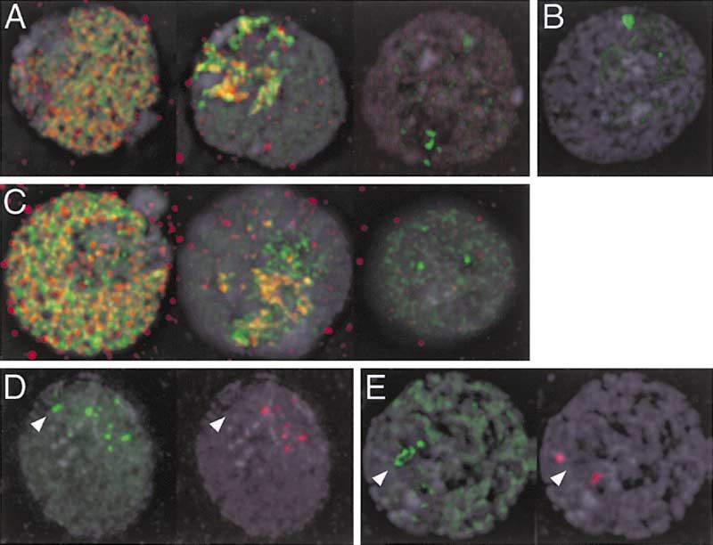
H3.3 Marks Active Chromatin
1193
Figure 2. RC and RI Deposition of Histone-
GFP Fusion Proteins
(A) Histone H4-GFP (green) deposits at repli-
cating DNA, indicated by localization of PCNA
(red) in both early (left) and late S phase (mid-
dle) cells. Some gap phase cells that lack
detectable PCNA (right) show localized RI de-
position of histone H4-GFP.
(B) Blocking DNA synthesis by aphidicolin
treatment before producing H4-GFP elimi-
nates replication patterns, but nuclei con-
tinue to display foci of RI deposition.
(C) Histone H3.3-GFP (green) localizes to rep-
licating DNA, indicated by localization of
PCNA (red), as well as to foci in gap phase
cells that lack detectable PCNA (right).
(D) In gap phase cells, many H4-GFP foci
(green), but not all, correspond to centro-
meres, which are marked by anti-Cid anti-
body (red). The arrowhead indicates a spot
of H4-GFP deposition that does not coincide
to a centromere.
(E) In gap phase cells, H3.3-GFP deposits at
sites in the nucleolus (arrowhead), but not at
centromeres (red). DNA staining (DAPI) is in
gray.
H3.3 Incorporates De Novo into Growth-Induced The origin of XL has been attributed to an expansion of
rDNA Arrays the rDNA locus on this chromosome, presumably as
Notably, we often observed labeling with H3.3-GFP and these cells adapted to culture conditions. We observed
H4-GFP of only one X chromosome. This is not due to that the rDNA array on XL was always labeled by H3.3-
absence of rDNA from other X chromosomes in these GFP (Figure 4A), consistent with this locus being active
cells because the detection of 28S rDNA by in situ hy- in all cells. However, in some experiments, variable num-
bridization confirmed rDNA arrays are present on each bers of cells had additional labeling on XS chromosomes
of the three X chromosomes (Figure 3G). Other studies (Figure 4B). To test whether some of this variability be-
have pointed out that many Drosophila cell lines (includ- tween experiments was due to differences in growth
ing Kc) carry two distinguishable kinds of X chromo- conditions, we transfected cells with the histone H3.3-
somes: a short one (XS) that resembles the normal X of GFP construct and then induced expression in samples
flies, and a longer X (XL) (Privitera, 1980; Echalier, 1997). of this culture 16 or 24 hr later. We found that many
Figure 3. RI Deposition Occurs at the rDNA
Locus and in Euchromatin
(A) Scheme for examining mitotic spreads
from cells induced in the G2 phase of the
cell cycle. G2 in Kc cells is 4–6 hr long; thus
mitotics observed 2 hr after induction were
in G2 when induced. Many mitotic spreads
observed 5 hr after induction were in late S
phase when induced.
(B) Mitotic figures labeled with histone H4-
GFP (green) appear within 2 hr of induction.
GFP signal localizes to a large site on an X
chromosome (arrow). Centromeres are de-
tected with anti-Cid (red).
(C) RI deposition of H3.3-GFP (green) resem-
bles that of H4-GFP.
(D) Increased gain of the green channel from
(C) shows that H3.3-GFP labels the euchro-
matic arms of all chromosomes at a low level.
(E) Lack of nucleotide labeling (red) after puls-
ing cells with nucleotide analog immediately
before induction of H3.3-GFP (green) con-
firms that this mitotic spread is from a cell that
was in G2 and that deposition is replication
independent.
(F) Mitotic figures labeled with H3.3-GFP (green) continue to appear even when DNA replication is blocked with aphidicolin shortly before
induction, indicating that these cells had completed S phase before induction.
(G) In situ hybridization detects a large rDNA array (28S probe, red) that corresponds to the intense site of H3.3-GFP RI deposition on the XL
chromosome (arrow). Additional rDNA genes are present on XS chromosomes (arrowheads).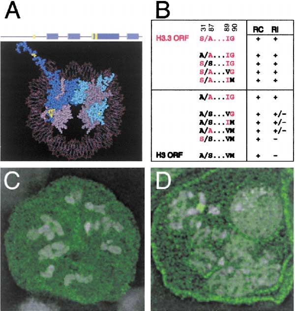
Molecular Cell
1194

Figure 4. H3.3 Marks Activated rDNA Arrays
Some mitotic spreads (A) show intense H3.3-GFP labeling (green)
only on the large rDNA array on the elongated XL chromosome
(arrow), but not on XS chromosomes (arrowhead), which also carry
rDNA genes. Another spread (B) shows H3.3-GFP labeling on both
XL and XS chromosomes. The inset shows an enlargement of the
proximal part of the XS chromosome. Wherever H3.3-GFP is pres-
ent, there is a gap in heterochromatin (antibody to H3di-MethylK9, red).
(C) RI H3.3-GFP labeling shows little overlap with heterochromatin
in interphase nuclei. DNA staining (DAPI) is in gray.

cells from exponentially growing cultures showed RI
labeling on both XL and XS chromosomes (mean num-                          Figure 5. Amino Acid Changes in H3.3 Determine Assembly
                                                                           Pathways
ber of labeled loci/metaphase spread ⫽ x ⫽ 1.66, SD ⫽
0.63), while metaphase spreads from the later time point,                  (A) Structure of histone H3 and the (H3•H4)2 tetramer. A schematic
                                                                           of the H3 protein is shown (top), with ␣ helices of the protein indi-
when culture growth had slowed, showed labeling on
                                                                           cated with blue cylinders. The (H3•H4)2 tetramer (bottom) is drawn
only the one XL (x ⫽ 1, SD ⫽ 0, p ⫽ 0.004). This change                    with Cn3D (NCBI). The two H3 chains are in shades of blue, and H4
in frequency suggests that the smaller rDNA arrays on                      chains are in shades of gray. Only one N-terminal tail of H3, starting
XS chromosomes are maintained in a transcriptionally                       at residue 20, is shown. The four positions that distinguish major
silent state but can be activated.                                         histone H3 from H3.3 are highlighted in yellow.
   We considered that the silencing of XS rDNA arrays                      (B) Site-directed mutations in the H3 and H3.3 ORFs and their activity
                                                                           when expressed in cells. RC deposition was scored in interphase
might be due to heterochromatin-mediated silencing.
                                                                           cells, and RI deposition by the labeling of the rDNA locus on meta-
Indeed, staining of metaphase spreads from cells ex-                       phase spreads 2 hr after induction (efficient [⫹], weak [⫹/⫺], or
pressing histone H3.3-GFP for the heterochromatin                          none [⫺]). The efficiency of RI deposition (the ratio of GFP intensity
marker H3di-MethylK9 (H3Me) revealed that rDNA arrays la-                  at labeled rDNA arrays to the background intensity) for mutants
beled by RI deposition of H3.3-GFP are depleted for                        scored as weak was ⬍5 (H3.3 gave a ratio of 29). Red residues indicate
H3Me, in spite of being flanked on both sides by hetero-                   identities in H3.3, and black indicates identities found in H3.
                                                                           (C) H3-GFP protein (green) does not localize to chromatin when
chromatin (Figures 4A and 4B). In every XS chromosome
                                                                           induced in gap phase cells. H3-GFP protein is distributed throughout
where the proximal region was labeled with H3.3-GFP,                       the cytoplasm.
a corresponding gap in the H3Me pattern was found (Fig-                    (D) The H3A/S...IM-GFP protein (green) localizes poorly to the rDNA
ure 4B, inset). That sites heavily labeled with H3.3-GFP                   locus. DNA staining (DAPI) is in gray.
were largely unlabeled with H3Me was confirmed in in-
terphase nuclei (Figure 4C). We conclude that the chro-
matin state of rDNA arrays can be reversed in response                     after S phase, it does not deposit onto DNA (Figure
to changes in growth conditions, and H3.3 accumulates                      5C). To identify which of the four differences between
de novo at activated genes.                                                Drosophila H3 and H3.3 are responsible for differential
                                                                           deposition, we used site-directed mutagenesis to alter
Amino Acid Changes toward H3.3 Allow                                       the histone-GFP fusion genes (Figure 5B). Single muta-
Replication-Independent Deposition                                         tions were introduced into the H3.3-GFP fusion gene
Characteristic amino acid substitutions distinguish ma-                    (with amino acid residues S31 A87 I89 G90; abbreviated
jor histone H3 proteins from their replacement histone                     “S/A...IG”) to match each of the H3 identities (A31 S87
H3 paralogs (Figure 5A), suggesting that some or all of                    V89 M90; “A/S...VM”). Each of these permutated tem-
these substitutions are responsible for differences in                     plates was transfected and expressed in cells, and the
deposition (Waterborg and Robertson, 1996). However,                       ability of the resultant fusion proteins to participate in
the RI deposition of replacement histone H3 variants                       RC and RI deposition examined. All mutant proteins
has been attributed entirely to its availability in the gap                were efficiently deposited onto replicating DNA, demon-
phases of the cell cycle (Yu and Gorovsky, 1997). In-                      strating that these changes did not interfere with chro-
stead, our results producing H3-GFP and H3.3-GFP from                      matin assembly. However, none of the mutations pre-
the same inducible promoter argue that at least some of                    vented deposition at the rDNA array, and thus we
the characteristic substitutions specify which assembly                    conclude that no single identity is necessary for the RI
pathway is used. It is clear that H3-GFP fails to undergo                  pathway. We also introduced the converse mutations
RI deposition, because when the protein is produced                        into the H3-GFP fusion gene to match each of the H3.3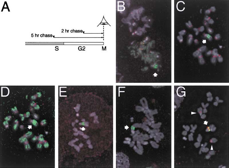
H3.3 Marks Active Chromatin
1195

identities. Strikingly, each of three mutations was suffi-     beling (Figure 6E). This deletion extends into a critical
cient by itself to confer partial RI activity (Figures 5B      region of histone H3 that passes through the DNA gyres
and 5D). All three of these positions lie in the core of       in the nucleosome (Luger et al., 1997). A similar deletion
the histone (Figure 5A). To further confirm that these         is lethal in yeast (Mann and Grunstein, 1992), suggesting
residues specify assembly pathways, we converted all           that the region is required to form a proper nucleosomal
three positions in H3 to the H3.3 identities (A/A...IG).       particle. We conclude that RI nucleosome assembly ma-
As expected, this mutant undergoes both RC and RI              chinery can deposit a truncated histone H3.3 protein.
deposition. Since any one change at these positions in            Because the N-terminal tail is essential for RC but not
H3 allows some RI deposition, it appears that this is          for RI deposition, deletion constructs of histone H3.3-
a default ability of H3 variants. We conclude that the         GFP separate these two pathways of nucleosome as-
identities at these positions specify assembly pathways        sembly. Staining for PCNA in cells producing a truncated
and that the combination of residues in the major H3           histone H3.3-GFP revealed that nucleosome assembly
histone actively prevents RI assembly.                         even in S phase cells is not limited to replicating DNA:
                                                               some RI deposition of histone H3.3 occurs in euchroma-
Replication-Coupled Deposition Requires                        tin and in the nucleolus (Figures 6F and 6G). Thus, al-
the Conserved N-Terminal Tail                                  though the bulk of nucleosome assembly uses the vastly
The above analysis demonstrates the existence of RC            more abundant histone H3 and is coupled to DNA repli-
and RI deposition pathways that use different histone H3       cation, at some sites nucleosomes are assembled con-
variants. These pathways may be mediated by different          tinually throughout the cell cycle.
nucleosome assembly machines, raising the possibility
that the conserved portions of canonical H3 variants are       Discussion
important in one pathway but not the other. The histone-
fold domain of histone H3 is essential for correct folding     Differences between H3 and H3.3 Specify
of the protein in the nucleosome (Arents and Moudria-          the Nucleosome Assembly Pathway
nakis, 1995); thus it is unlikely to be dispensable in any     Two very similar forms of histone H3 are produced in
histone H3 variant. A role for the extended N-terminal         Drosophila cells. The major histone H3 genes are greatly
tail of H3 in nucleosome assembly has been examined            upregulated during S phase for the assembly of newly
both in vivo (Ling et al., 1996; Freeman et al., 1996) and     replicated chromatin (Osley, 1991), and the two orphan
in vitro (Shibahara et al., 2000; Quintini et al., 1996) and   genes encoding the variant histone H3.3 are expressed
has been found to be dispensable.                              at constitutive levels throughout the cell cycle (Akhma-
   We examined whether the N-terminal tail regions of          nova et al., 1995). We show that there are two pathways
histone H3 and H3.3 are required for either nucleosome         for chromatin assembly in Drosophila cells: one that
assembly pathway in Drosophila cells. A series of dele-        assembles nucleosomes during DNA replication, and a
tions was generated that removed portions of the his-          second that assembles them only at particular loci by
tone tail from GFP fusion constructs (Figure 6A). We           a RI mechanism. The existence of an RI pathway is
transfected these constructs into cells and induced ex-        demonstrated by multiple lines of evidence: (1) tagged
pression as before. The distribution of histone-GFP was        histones deposit in interphase cells that lack replication
compared to the PCNA pattern in individual nuclei to           foci; (2) they deposit when DNA replication is blocked; (3)
determine whether RC deposition with the deleted pro-          they label mitotic chromosomes when produced during
tein would still occur. In this experimental system, we        G2; and (4) histone H3.3, but not H3, is the substrate
find that the N-terminal tail of histone H3 is essential for   for RI deposition.
in vivo RC nucleosome assembly. Histone H3 proteins               Before nucleosome assembly, histones form stable
deleted for this region localize poorly to replicating DNA     (H3•H4)2 tetramers and H2A•H2B dimers (Krude and
or remain diffuse throughout the nucleus (Figures 6B           Keller, 2001). Our results indicate that both H3- and
and 6C). Deletion of the N-terminal tail does not inhibit      H3.3-containing tetramers can be deposited during DNA
tetramer formation with histone H4 in vitro (Shibahara         replication. Since H3 and H3.3 are identical across the
et al., 2000), and H3 continues to be imported into the        tetramer protein interaction surface (Luger et al., 1997),
nucleus (Figure 6), implying that these truncated pro-         and if assembly is unbiased, both homotypic and hetero-
teins are defective for a later step in RC nucleosome          typic tetramers will be produced every cell cycle.
assembly.                                                         Extensive biochemical studies have led to a step-
                                                               wise model for the assembly of nucleosomes (Krude
Replication-Independent Deposition Does Not                    and Keller, 2001). The chromatin assembly factor (CAF)
Require the N-Terminal Tail                                    includes proteins that promote the folding and assembly
Uncovering a requirement for a region in the N-terminal        of (H3•H4)2 tetramers and that subsequently deposit
tail of histone H3 for RC deposition prompted us to            histones onto newly replicated DNA (Stillman, 1986).
examine whether this region is also required for RI depo-      Components of CAF are recruited to sites of DNA repli-
sition of histone H3.3. Most truncated histone H3.3-GFP        cation in vivo by a specific interaction with PCNA (Shiba-
proteins were efficiently used for RI deposition (Figure       hara and Stillman, 1999). However, alternative assembly
6A) and were resistant to salt extraction (data not shown),    activities must also exist because CAF is nonessential
although larger deletions produced aberrant protein ag-        in both Saccharomyces (Enomoto et al., 1997; Kaufman
gregates in some nuclei (Figure 6D). Only the most proxi-      et al., 1997) and Arabidopsis (Kaya et al., 2001). At least
mal deletion (deleting 40 of the 44 residues from the          one factor that stimulates CAF activity also has nucleo-
N-terminal tail) showed a reduced intensity of rDNA la-        some assembly activity on its own (Tyler et al., 1999).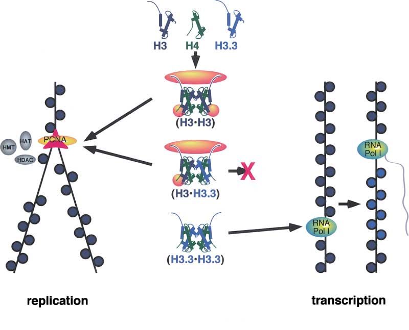
Molecular Cell
1196

                                                                                   Figure 6. The N-Terminal Tail Is Required for
                                                                                   RC but Not RI Deposition
                                                                                   (A) Schematic of deletions that remove por-
                                                                                   tions of the tail from histone-GFP fusion pro-
                                                                                   teins. Amino acid residues that are unique to
                                                                                   H3 are indicated above the consensus se-
                                                                                   quence, and those unique to H3.3 are below
                                                                                   it. The blue cylinder marks the beginning of
                                                                                   the core region. Thick horizontal lines delimit
                                                                                   the deletions made in histone H3 and H3.3
                                                                                   constructs. To score RC deposition, at least
                                                                                   20 GFP⫹ nuclei with late S phase PCNA pat-
                                                                                   terns were scored for overlap between GFP
                                                                                   and PCNA. The percentage of nuclei with
                                                                                   overlap (indicating efficient RC deposition),
                                                                                   weak overlap with general nuclear fluores-
                                                                                   cence, and only diffuse fluorescence is given.
                                                                                   The RI deposition of truncated H3.3-GFP pro-
                                                                                   teins was scored on mitotic spreads.
                                                                                   (B) H3⌬319-GFP weakly localizes to replicating
                                                                                   DNA (PCNA, red).
                                                                                   (C) H3⌬326-GFP is diffuse throughout a late S
                                                                                   phase nucleus.
                                                                                   (D) Occasional nuclei show aggregates of
                                                                                   H3.3⌬335-GFP protein.
                                                                                   (E) H3.3⌬342-GFP shows reduced localization
                                                                                   (arrow) to the rDNA locus in mitotic spreads,
                                                                                   with ⬍5-fold enrichment over background.
                                                                                   (F and G) H3.3⌬335-GFP localizes by RI deposi-
                                                                                   tion to euchromatin and the nucleolus in early
                                                                                   (F) and late (G) S phase cells (PCNA, red).

Chromatin remodeling factors are additional candidates          might then associate with RI assembly factors and be
for alternative activities since some can transfer nucleo-      recruited to active loci.
somes to DNA in vitro (Ito et al., 1997; Lorch et al., 1999).      The idea that distinct complexes mediate the two
Indeed, two remodeling complexes, RSF and ACF both              kinds of nucleosome assembly is supported by our find-
promote transcription and mediate nucleosome assem-             ing that the extreme N-terminal tail of histone H3 variants
bly (LeRoy et al., 1998; Loyola et al., 2001; Levenstein        is required for RC deposition but not RI deposition. Stud-
and Kadonaga, 2002). From our work, it is clear that            ies with histones H2A and H2B in Physarum (Thiriet
RC and RI deposition actually use different histone H3          and Hayes, 2001) reveal clear deficiencies in the RC
variants, and we suggest that histone H3.3 is the correct       deposition of tailless histones. However, in in vitro ex-
substrate for chromatin assembly factors that are tran-         periments where tailless histones are the only ones
scriptionally linked. It will be interesting to see if known    available, chromatin can be assembled (Shibahara et
assembly factors prefer this variant.                           al., 2000; Quintini et al., 1996). In our experiments and
  Histone H3 and H3.3 differ at only four amino acid            those in Physarum, tailless histones must deposit as
positions, and mutational analysis reveals that a single        efficiently as endogenous full-length histones or they
change at any one of the three positions in the histone         will be out-competed and will not appear in chromatin.
core allow some RI deposition. Thus, we interpret RI            Assembly may also be affected by predeposition modifi-
deposition as a default feature of canonical histone H3         cations (Sobel et al., 1995) or cell type differences. For
variants, and only the three residues found in H3 will          example, chromatin assembly in embryos with rapid
preclude RI deposition. Such specificity could be under-        nuclear division may be less stringent than assembly in
stood if these positions in histone H3 make contacts            somatic cells, as we have examined here. Indeed, in
that only fit with replication-specific assembly factors,       Xenopus early embryonic nuclei, truncated H3 can be
or bind an accessory protein that alters the activity of        deposited into chromatin, albeit inefficiently (Freeman
a more general nucleosome assembly factor (Figure 7).           et al., 1996).
The three positions in H3 are in solvent-accessible re-            There appear to be multiple mechanisms that ensure
gions of the nucleosome particle (Arents et al., 1991;          the utilization of different histone H3 variants by nucleo-
Luger et al., 1997) (Figure 5A) and thus contacts with          some assembly pathways. In Tetrahymena, only the re-
these positions before assembly is plausible. It is sim-        placement H3 variant gene hv2 is expressed in gap
plest to imagine that replication-specific assembly pro-        phase cells, and this histone undergoes RI deposition.
teins recognize tetramers that contain H3 and localize          Deletion of hv2 is viable but is accompanied by the
them to newly replicated DNA. All remaining tetramers           constitutive expression of a major H3 gene (Yu and Gor-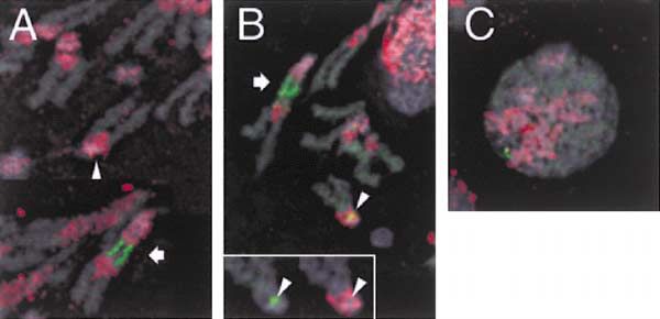
H3.3 Marks Active Chromatin
1197

                                                                                  Figure 7. Model for the Specification of
                                                                                  Nucleosome Assembly Pathways by Histone
                                                                                  H3 Variants
                                                                                  Histone proteins (blue and green) form homo-
                                                                                  typic and heterotypic tetramers. Replicative
                                                                                  chaperones or assembly factors (yellow)
                                                                                  make contacts with the N-terminal tails and
                                                                                  with a region in the core of the major H3 chain
                                                                                  (including S87 and VM89, 90). Contact with
                                                                                  both tails and with at least one core region
                                                                                  is required for RC deposition. The complex
                                                                                  is committed to assembly on newly replicated
                                                                                  DNA (left) by recruitment to replication forks,
                                                                                  and chromatin modifications are maintained
                                                                                  by enzymes (gray) that accompany DNA poly-
                                                                                  merase. Heterotypic tetramers can bind the
                                                                                  core region replicative factor through one H3
                                                                                  chain; thus H3.3 expressed during S phase
                                                                                  undergoes RC deposition as heterotetra-
                                                                                  mers. Homotypic H3.3 tetramers lack the
                                                                                  core recognition sites and fail to bind the rep-
                                                                                  licative factor. Targeting of homotypic H3.3
                                                                                  tetramers to active loci may be mediated by
                                                                                  an interaction between RI assembly factors
                                                                                  and transcription factors or may simply fill in
                                                                                  gaps in chromatin left after RNA polymerase
                                                                                  passage and nucleosome displacement
                                                                                  (right).

ovsky, 1997). Thus, the differential production of histone     cation-independent nucleosome assembly pathway is
variants in Tetrahymena appears to direct their use by         essential in all cells. This implies that, functionally, a
RC and RI pathways. It is likely that the differential pro-    replacement histone H3 has always been extant. In or-
duction of the animal histone H3 and H3.3 also contrib-        ganisms that encode only one kind of canonical histone
utes to their use by different assembly pathways be-           H3 protein that is used throughout chromatin (Thatcher
cause production of the major H3 protein is down               et al., 1994; Waterborg and Robertson, 1996; Waterborg
regulated in gap phase cells (Harris et al., 1991). At least   et al., 1995), we expect that this H3 variant must undergo
one transcriptional activator sequence that directs S          both RC and RI deposition. Fungal lineages are particu-
phase expression of the major H3 coincides with the            larly intriguing in this regard because all ascomycetes,
codons that distinguish it from H3.3 (Bowman and Hurt,         including laboratory yeasts and molds, carry only one
1995). However, in addition to differences in production,      canonical histone H3. Each of these is identical to animal
we find that nucleosome assembly pathways distinguish          H3.3 at positions 89 and 90, and often identical at posi-
between histone H3 variant proteins in Drosophila. In-         tion 31 (Baxevanis and Landsman, 1998; and data not
deed, deletion of even one of the two histone H3.3 genes       shown). Thus, by this criterion, we propose that the
in mice is semilethal, implying that animal H3.3 is not        solitary histone H3 proteins in ascomycetes are equiva-
redundant with H3 (Couldrey et al., 1999). This difference     lent to histone H3.3. Indeed, nucleosome assembly ac-
between Tetrahymena and animals may be due to the              tivity in the cell cycle gap phases has been detected in
evolutionary history of histone H3 variants: the ciliate       Saccharomyces (Altheim and Schultz, 1999). These
replacement H3 variant hv2 appears to have had a sepa-         fungi appear to have lost their ancestral H3, as we find
rate origin from that of animal and that of plant replace-     that genomes from the Basidiomycota sister clade have
ment H3 histones (Thatcher et al., 1994; Waterborg and         both H3 and H3.3 (The Institute for Genomic Research,
Robertson, 1996). Regardless of whether differential           Cryptococcus neoformans genome project at http://
production or substrate specificity direct their utiliza-      www.tigr.org; and DOE White Rot Genome Project at
tion, distinctive replacement H3 histones are the normal       http://www.jgi.doe.gov/programs/whiterot.htm). His-
substrate for RI pathways.                                     tone H2A in Saccharomyces may have an analogous
                                                               evolutionary history, since it now performs the functions
Yeast Have Retained Only the H3.3 Counterpart                  of the H2A and the H2A.X variants in other organisms
Alternate interpretations of the phylogenetic history of       (Downs et al., 2000). Thus, both histone H3 and H2A in
the histone H3 family have been proposed. One analysis         Saccharomyces appear to be evolutionary derivatives
suggested that a replacement histone H3 variant was            of replacement genes.
the common ancestor (Wells et al., 1986), but other inter-        The lack of an H3 counterpart in yeasts and molds
pretations have proposed that replacement histones             may provide insight into differences between simple
have multiple independent origins (Thatcher et al., 1994;      fungi and complex multicellular eukaryotes in main-
Waterborg and Robertson, 1996). We believe that the            taining silent chromatin. Much of the Saccharomyces
presence of paralogous histone H3 genes in many or-            genome is continually in a transcriptionally competent
ganisms may preclude delineation of which sequence             state (Sherman, 1997), similar to H3.3-containing re-
is ancestral. However, our findings suggest that a repli-      gions in complex genomes. Perhaps this relative lack
Molecular Cell
1198
of silent chromatin allowed the loss of the strictly RC Experimental Procedures
histone substrate. Heterochromatic silencing in yeast
Constructs
may be needed only at special sites, such as silent
We used the heat shock-inducible HS-H3-GFP and HS-H2B-GFP
mating type loci and telomeres, where SIR-based silenc- plasmids previously described (Henikoff et al., 2000) and con-
ing has evolved. In multicellular eukaryotes, the need structed similar fusions for the Drosophila H3.3A and H4 genes,
for maintaining most of the genome in a continuously with a six amino acid linker (SRPVAT) between GFP and the last
silent state in differentiated cells may favor maintaining residue in these ORFs. These constructs are designated HS-H3.3A-
two distinct H3 histones. GFP and HS-H4-GFP, respectively. To generate N-terminal deletions
of the H3 and H3.3 tails, primers to the internal segments of the
ORFs specified in the text that included an XbaI site and the first
Replication-Independent Deposition of H3.3 three codons of the ORF (MAR) at their 5⬘ end were used in PCR
Marks Active Chromatin with a primer to the GFP ORF with Ampli-Taq Gold (Perkin-Elmer,
What essential function might replication-independent Foster City, CA) or Platinum Taq (GIBCO-BRL, Grand Island, NY)
nucleosome assembly serve? Targeting of histone H3.3 enzymes to generate truncated ORFs from HS-H3-GFP and HS-
H3.3A-GFP templates. These products were digested with XbaI and
may be due to transcriptional activity at these sites.
EagI and cloned into the XbaI, EagI-digested HS-H2B-GFP vector.
Passage of RNA polymerase has been shown to dis- For site-directed mutagenesis of H3-GFP and of H3.3A-GFP, 2.9 kb
place nucleosomes (Pfaffle et al., 1990; Clark and fragments containing the complete genes were each subcloned into
Felsenfeld, 1992), although transcription without nucleo- pUC19, and the QuikChange kit (Stratagene, La Jolla, CA) was used
some dissociation has also been reported (Studitsky et with primers including the codon changes specified in the text.
al., 1994). Our results indicate that in a natural context, All deletion and mutation constructs were confirmed by BigDye
sequencing (ABI, Foster City, CA). These plasmids were then used
nucleosomes are indeed displaced, because newly syn-
for transfections into Kc cells.
thesized histones take their place. This has two conse-
quences: first, the rapid switching of histone modifica-
Immunostaining and DNA FISH
tions; and second, the establishment of a heritable Culture, transfection, fixation, and image collection methods have
distinction between active and bulk chromatin. been previously described (Henikoff et al., 2000; Ahmad and Heni-
While histone modifications such as phospho and koff, 2001). Transfected constructs were induced for 1 hr at 37⬚C
acetyl groups are catalytically added and removed and and returned to 25⬚C for recovery for 2 hr before fixation. The G2
may be maintained by enzymes that accompany DNA phase of the cell cycle is 4–6 hr long in this cell line (Dolfini et al.,
1970; Ahmad and Henikoff, 2001); thus all mitotic figures in these
replication machinery (Rountree et al., 2000), it appears
preparations are from cells that were induced after S phase was
that lysine methylation is irreversible (Jenuwein, 2001). complete. To mark centromeres, DNA replication forks, and hetero-
Gene silencing is associated with H3 methylation at resi- chromatin, we used rabbit antisera to the Cid histone (Henikoff et
due K9, and this would be long lived if dilution of the al., 2000), PCNA (Henderson et al., 2000), and histone H3di-MethylK9
methylated histone through DNA replication and nucleo- (UpState Biotech, Lake Placid, NY), respectively, followed by anti-
some segregation were the only method for its elimina- rabbit IgG goat antibodies conjugated with either Texas-Red or with
Cy5 fluorochromes (Jackson ImmunoResearch, West Grove, PA).
tion. However, new nucleosome assembly by an RI path-
For triple labeling of nucleoside incorporation, histone-GFP deposi-
way is a logical method for rapid gene activation, as tion, and PCNA distribution, cells transfected with the HS-H3-GFP
all modified histones can be replaced within one cell plasmid were induced, and BrdU and deoxycytidine were added to
generation (Figure 7). The rapid turnover of a replace- the culture media for a final concentration of 100 g/ml at the start
ment histone H3 variant in alfalfa (Waterborg, 1993) sup- of the recovery period. Cells were fixed and immunostained for GFP
ports this view. and PCNA using mouse monoclonal anti-GFP antibody (Molecular
Probes, Eugene, OR) and rabbit anti-PCNA antibody, respectively,
The regeneration of nucleosomes by RI assembly also
followed by anti-mouse Rhodamine-Red-conjugated monovalent
alters chromatin, because a variant histone is incorpo- Fab fragments (Jackson ImmunoResearch) and anti-rabbit Cy5-con-
rated. Histone H3.3 includes a serine at residue 31, and jugated antibody. Slides were then refixed with Carnoy’s fixative
modifications of this site would provide unique regula- and processed as described (Van Hooser and Brinkley, 1999), except
tion of active chromatin. Modification of (H3.3•H4)2 tetra- that DNA was denatured with 0.07 N NaOH for 30 min at 25⬚C.
mers before deposition could also effectively target Incorporated BrdU was detected using mouse FITC-conjugated an-
tibody (Roche, Pleasanton, CA).
modifications to active regions. Finally, the inheritance
For DNA FISH and GFP detection, we immunostained for GFP
of variant nucleosomes through cell division might pre- using a mouse monoclonal anti-GFP antibody (Molecular Probes),
dispose regions to be transcriptionally active again. It followed by anti-mouse FITC-conjugated antibodies (Jackson Im-
is striking that histone H3.3 mostly localizes to an rDNA munoResearch). Slides were then refixed and denatured as above.
array, and numerous cases are known where the activity A probe to the 28S rDNA gene was prepared using the primers
of one rDNA array is heritable (nucleolar dominance) CGAAAGACCAATCGAACCATCTAG and GAACCGTATTCCCTTTC
GTTCAA. These were used to amplify a 1 kb fragment in PCR, and
(Reeder, 1985; Pikaard, 2000). Nucleolar dominance has
this product was used with the BioPrime DNA labeling kit (GIBCO-
been observed in cell lines and in interspecific hybrids BRL). Hybridization was performed overnight at 25⬚C, and bound
and may be an example of regulation when rDNA gene probe was detected using Texas-Red-conjugated streptavidin
copy number varies. Since some RNA polymerase I com- (Pierce Chemical Co., Rockford, IL).
ponents remain associated with active rDNA arrays
through mitosis, the inheritance of these proteins has Construct Evaluation and Image Quantitation
been suggested as the basis for nucleolar dominance Images were analyzed using DeltaVision software (Applied Preci-
(Roussel et al., 1996). Similarly, the inheritance of variant sion, Issaquah, WA). Transfection efficiencies were estimated as
the fraction of interphase cells with GFP fluorescence and were
nucleosomes provides an obvious mechanism for epi-
typically ⵑ70%. Each construct was tested at least four times. RC
genetic inheritance at rDNA arrays and at euchromatic deposition was evaluated in transfected and induced cells with a
genes. Thus, transcription would allow RI deposition of late S phase (heterochromatic) PCNA pattern, which allows a close
H3.3, which would in turn maintain the active state. assessment of the GFP and PCNA patterns. RI deposition was as-H3.3 Marks Active Chromatin
1199
sayed by counting the fraction of metaphase spreads 2 hr after plex formation triggered by DNA damage occurs independent of
induction with GFP labeling at the X chromosome rDNA locus. The the ATM product in human cells. Nucleic Acids Res. 29, 1341–1351.
frequency of GFP fluorescence in interphase cells served as the Baxevanis, A.D., and Landsman, D. (1998). Histone sequence data-
expected frequency for labeled metaphase figures if the histone- base: new histone fold family members. Nucleic Acids Res. 26,
GFP protein could undergo RI deposition. At least 20 metaphase 372–375.
figures were examined for each sample. For quantitative measure-
Bowman, T.L., and Hurt, M.M. (1995). The coding sequences of
ments of histone H3.3-GFP deposition, we used DeltaVision object-
mouse H2A and H3 histone genes contains a conserved seven nu-
building software to define DAPI-stained chromosomes in meta-
cleotide element that interacts with nuclear factors and is necessary
phase spreads from cultures transfected with HS-H3.3A-GFP. The
for normal expression. Nucleic Acids Res. 23, 3083–3092.
integrated pixel intensities in the GFP channel over these chromo-
somes was then measured, as was deposition specifically at the Clark, D.J., and Felsenfeld, G. (1992). A nucleosome core is trans-
rDNA locus, defined as the intensely labeled contiguous segment ferred out of the path of a transcribing polymerase. Cell 71, 11–22.
of the XL chromosome. Background intensity was subtracted from Couldrey, C., Carlton, M.B.L., Nolan, P.M., Colledge, W.H., and Ev-
each of these, and the ratio of the two measures estimates the ans, M.J. (1999). A retroviral gene trap insertion into the histone
fraction of histone H3.3 that is targeted to the rDNA locus. The ratio 3.3A gene causes partial neonatal lethality, stunted growth, neuro-
between the peak pixel intensity at a labeled rDNA locus and the muscular deficits and male sub-fertility in transgenic mice. Hum.
mean background intensity was used as an estimate of the efficiency Mol. Genet. 8, 2489–2495.
at which mutated H3.3-GFP proteins underwent RI deposition. The Dolfini, S., Courgeon, A.M., and Tiepolo, L. (1970). The cell cycle
unmutated H3.3A-GFP construct gave an efficiency ratio of 29 (n ⫽ of an established line of Drosophila melanogaster cells in vitro.
7). Mutations that were scored as defective for RI deposition gave Experientia 26, 1020–1021.
ratios less than five. Downs, J.A., Lowndes, N.F., and Jackson, S.P. (2000). A role for
Saccharomyces cerevisiae histone H2A in DNA repair. Nature 408,
In Situ Salt Extraction 1001–1004.
Cells were transfected with HS-H2B-GFP, and HS-H3.3-GFP con-
Echalier, G. (1997). Drosophila Cells in Culture, First Edition. (New
structs were grown on coverslips, induced, and allowed to recover
York: Academic Press).
for 2 hr. H3.3-GFP histone in nuclear preparations was undetectable
by Western analysis with anti-H3 antibodies (data not shown). Ex- Enomoto, S., McCune-Zierath, P.D., Gerami-Nejad, M., Sanders,
traction of nuclear proteins was performed as described (Balajee M.A., and Berman, J. (1997). RLF2, a subunit of yeast chromatin
and Geard, 2001), except that we used 1.5 M NaCl in order to extract assembly factor-I, is required for telomeric chromatin function in
H2B, but not H3 and H4. Parallel sets of cells were mock treated vivo. Genes Dev. 11, 358–370.
with extraction buffer with only 130 mM NaCl. Cells were then fixed Freeman, L., Kurumizaka, H., and Wolffe, A.P. (1996). Functional
as above, and we measured the GFP fluorescence intensities in domains for assembly of histones H3 and H4 into the chromatin of
seven to eight random fields of nuclei to quantitate the amount of Xenopus embryos. Proc. Natl. Acad. Sci. USA 93, 12780–12785.
histone-GFP proteins in mock-treated cells and the amount retained Fretzin, S., Allan, B.D., van Daal, A., and Elgin, S.C. (1991). A Dro-
after extraction. sophila melanogaster H3.3 cDNA encodes a histone variant identical
with the vertebrate H3.3. Gene 15, 341–342.
Acknowledgments
Harris, M.E., Bohni, R., Schneiderman, M.H., Ramamurthy, L.,
Schumperli, D., and Marzluff, W.F. (1991). Regulation of histone
We thank Harmit Malik, Pauline Ng, Paul Talbert, and Danielle Ver-
mRNA in the unperturbed cell cycle: evidence suggesting control
maak for helpful comments.
at two posttranscriptional steps. Mol. Cell. Biol. 11, 2416–2424.
Received: December 12, 2001 Henderson, D.S., Wiegand, U.K., Norman, D.G., and Glover, D.M.
Revised: April 16, 2002 (2000). Mutual correction of faulty PCNA subunits in temperature-
sensitive lethal mus209 mutants of Drosophila melanogaster. Genet-
ics 154, 1721–1733.
References
Henikoff, S., Ahmad, K., Platero, J.S., and van Steensel, B. (2000).
Ahmad, K., and Henikoff, S. (2001). Centromeres are specialized Heterochromatic deposition of centromeric histone H3-like proteins.
replication domains in heterochromatin. J. Cell Biol. 153, 101–110. Proc. Natl. Acad. Sci. USA 97, 716–721.
Akhmanova, A.S., Bindels, P.C.T., Xu, J., Miedema, K., Kremer, H., Ito, T., Bulger, M., Pazin, M.J., Kobayashi, R., and Kadonaga, J.T.
and Hennig, W. (1995). Structure and expression of histone H3.3 (1997). ACF, an ISWI-containing and ATP-utilizing chromatin assem-
genes in Drosophila melanogaster and Drosophila hydei. Genome bly and remodeling factor. Cell 90, 145–155.
38, 586–600. Jenuwein, T. (2001). Re-SET-ting heterochromatin by histone meth-
Akhmanova, A.S., Miedema, K., Wang, Y., van Bruggen, M., Berden, yltransferases. Trends Cell Biol. 11, 266–273.
J.H.M., Moudrianakis, E.N., and Hennig, W. (1997). The localization Jenuwein, T., and Allis, C.D. (2001). Translating the histone code.
of histone H3.3 in germ line chromatin of Drosophila males as estab- Science 293, 1074–1080.
lished with a histone H3.3-specific antiserum. Chromosoma 106, Kaufman, P.D., Kobayashi, R., and Stillman, B. (1997). Ultraviolet
335–347. radiation sensitivity and reduction of telomeric silencing in Sacchar-
Allis, C.D., and Wiggins, J.C. (1984). Histone rearrangements accom- omyces cerevisiae cells lacking chromatin assembly factor-I. Genes
pany nuclear differentiation and dedifferentiation in Tetrahymena. Dev. 11, 345–357.
Dev. Biol. 101, 282–294. Kaya, H., Shibahara, K.I., Taoka, K.I., Iwabuchi, M., Stillman, B., and
Altheim, B.A., and Schultz, M.C. (1999). Histone modification gov- Araki, T. (2001). FASCIATA genes for chromatin assembly factor-1
erns the cell cycle regulation of a replication-independent chromatin in Arabidopsis maintain the cellular organization of apical meri-
assembly pathway in Saccharomyces cerevisiae. Proc. Natl. Acad. stems. Cell 104, 131–142.
Sci. USA 96, 1345–1350. Kimura, H., and Cook, P.R. (2001). Kinetics of core histones in living
Arents, G., and Moudrianakis, E.N. (1995). The histone fold: a ubiqui- human cells: little exchange of H3 and H4 and some rapid exchange
tous architectural motif utilized in DNA compaction and protein di- of H2B. J. Cell Biol. 153, 1341–1353.
merization. Proc. Natl. Acad. Sci. USA 92, 11170–11174. Krude, T., and Keller, C. (2001). Chromatin assembly during S phase:
Arents, G., Burlingame, R.W., Wang, B.C., Love, W.E., and Moudria- contributions from histone deposition, DNA replication and the cell
nakis, E.N. (1991). The nucleosomal core histone octamer at 3.1 Å division cycle. Cell. Mol. Life Sci. 58, 665–672.
resolution: a tripartite protein assembly and a left-handed superhe- Leonhardt, H., Rahn, H.P., Weinzierl, P., Sporbert, A., Cremer, T.,
lix. Proc. Natl. Acad. Sci. USA 88, 10148–10152. Zink, D., and Cardoso, M.C. (2000). Dynamics of DNA replication
Balajee, A.S., and Geard, C.R. (2001). Chromatin-bound PCNA com- factories in living cells. J. Cell Biol. 149, 271–279.Molecular Cell
1200
Lennox, R.W., and Cohen, L.H. (1988). The production of tissue- Sobel, R.E., Cook, R.G., Perry, C.A., Annunziato, A.T., and Allis, C.D.
specific histone complements during development. Biochem. Cell (1995). Conservation of deposition-related acetylation sites in newly
Biol. 66, 636–649. synthesized histones H3 and H4. Proc. Natl. Acad. Sci. USA 92,
LeRoy, G., Orphanides, G., Lane, W.S., and Reinberg, D. (1998). 1237–1241.
Requirement of RSF and FACT for transcription of chromatin tem- Stillman, B. (1986). Chromatin assembly during SV40 DNA replica-
plates in vitro. Science 282, 1900–1904. tion in vitro. Cell 45, 555–565.
Levenstein, M.E., and Kadonaga, J.T. (2002). Biochemical analysis Studitsky, V.M., Clark, D.J., and Felsenfeld, G. (1994). A histone
of chromatin containing recombinant Drosophila core histones. J. octamer can step around a transcribing polymerase without leaving
Biol. Chem. 277, 8749–8754. the template. Cell 76, 371–382.
Ling, X., Harkness, T.A., Schultz, M.C., Fisher-Adams, G., and Thatcher, T.H., MacGaffey, J., Bowen, J., Horowitz, S., Shapiro,
Grunstein, M. (1996). Yeast histone H3 and H4 amino termini are D.L., and Gorovsky, M.A. (1994). Independent evolutionary origin of
important for nucleosome assembly in vivo and in vitro: redundant histone H3.3-like variants of animals and Tetrahymena. Nucleic
and position-independent functions in assembly but not in gene Acids Res. 22, 180–186.
regulation. Genes Dev. 15, 686–699. Thiriet, C., and Hayes, J.J. (2001). A novel labeling technique reveals
Lorch, Y., Zhang, M., and Kornberg, R.D. (1999). Histone octamer a function for histone H2A/H2B dimer tail domains in chromatin
transfer by a chromatin-remodeling complex. Cell 96, 389–392. assembly in vivo. Genes Dev. 15, 2048–2053.
Loyola, A., LeRoy, G., Wang, Y.H., and Reinberg, D. (2001). Reconsti- Tyler, J.K., Adams, C.R., Chen, S.R., Kobayashi, R., Kamakaka, R.T.,
tution of recombinant chromatin establishes a requirement for his- and Kadonaga, J.T. (1999). The RCAF complex mediates chromatin
tone-tail modifications during chromatin assembly and transcrip- assembly during DNA replication and repair. Nature 402, 555–560.
tion. Genes Dev. 15, 2837–2851. Van Hooser, A.A., and Brinkley, W.R. (1999). Methods for in situ
Luger, K., Mader, A.W., Richmond, R.K., Sargent, D.F., and Rich- localization of proteins and DNA in the centromere-kinetochore
mond, T.J. (1997). Crystal structure of the nucleosome core particle complex. In Methods in Cell Biology, First Edition, Volume 61, (New
at 2.8 Å resolution. Nature 389, 251–260. York: Academic Press), pp. 57–80.
Mann, R.K., and Grunstein, M. (1992). Histone H3 N-terminal muta- Waterborg, J.H. (1993). Histone synthesis and turnover in alfalfa.
tions allow hyperactivation of the yeast GAL1 gene in vivo. EMBO Fast loss of highly acetylated replacement histone variant H3.2*. J.
J. 11, 3297–3306. Biol. Chem. 268, 4912–4917.
Marmorstein, R. (2001). Protein modules that manipulate histone Waterborg, J.H., and Robertson, A.J. (1996). Common features of
tails for chromatin regulation. Nat. Rev. Mol. Cell Biol. 2, 422–432. analogous replacement Histone H3 genes in animals and plants. J.
Mol. Evol. 43, 194–206.
Mello, J.A., and Almouzni, G. (2001). The ins and outs of nucleosome
assembly. Curr. Opin. Genet. Dev. 11, 136–141. Waterborg, J.H., Robertson, A.J., Tatar, D.L., Borza, C.M., and Davie,
J.R. (1995). Histones of Chlamydomonas reinhardtii: synthesis, acet-
Ng, L., Prelich, G., Anderson, C.W., Stillman, B., and Fisher, P.A.
ylation, and methylation. Plant Physiol. 109, 393–407.
(1990). Drosophila proliferating cell nuclear antigen. Structural and
functional homology with its mammalian counterpart. J. Biol. Chem. Wells, D., Bains, W., and Kedes, L. (1986). Codon usage in histone
265, 11948–11954. gene families of higher eukaryotes reflects functional rather than
phylogenetic relationships. J. Mol. Evol. 23, 224–241.
Osley, M.A. (1991). The regulation of histone synthesis in the cell
cycle. Annu. Rev. Biochem. 60, 827–861. Wolffe, A.P. (1998). Chromatin: Structure and Function, Third Edition.
(San Diego: Academic Press).
Pfaffle, P., Gerlach, V., Bunzel, L., and Jackson, V. (1990). In vitro
Yu, L., and Gorovsky, M.A. (1997). Constitutive expression, not a
evidence that transcription-induced stress causes nucleosome dis-
particular primary sequence, is the important feature of the H3 re-
solution and regeneration. J. Biol. Chem. 265, 16830–16840.
placement variant hv2 in Tetrahymena thermophila. Mol. Cell. Biol.
Pikaard, C.S. (2000). The epigenetics of nucleolar dominance. 17, 6303–6310.
Trends Genet. 16, 495–500.
Pina, B., and Suau, P. (1987). Changes in histones H2A and H3
variant composition in differentiating and mature rat brain cortical
neurons. Dev. Biol. 123, 51–58.
Privitera, E. (1980). A Drosophila melanogaster cell line tested for
the presence of active NORs by silver staining. Chromosoma 81,
431–437.
Quintini, G., Treuner, K., Gruss, C., and Knippers, R. (1996). Role of
amino-terminal histone domains in chromatin replication. Mol. Cell.
Biol. 16, 2888–2897.
Reeder, R.H. (1985). Mechanisms of nucleolar dominance in animals
and plants. J. Cell Biol. 101, 2013–2016.
Rountree, M.R., Bachman, K.E., and Baylin, S.B. (2000). DNMT1
binds HDAC2 and a new co-repressor, DMAP1, to form a complex
at replication foci. Nat. Genet. 25, 269–277.
Roussel, P., Andre, C., Comai, L., and Hernadez-Verdun, D. (1996).
The rDNA transcription machinery is assembled during mitosis in
active NORs and absent in inactive NORs. J. Cell Biol. 133, 235–246.
Sherman, F. (1997). Yeast genetics. In The Encyclopedia of Molecu-
lar Biology and Molecular Medicine, First Edition, Volume 6, R.A.
Meyers, ed. (Weinheim, Germany: VCH), pp. 302–325.
Shibahara, K., and Stillman, B. (1999). Replication-dependent mark-
ing of DNA by PCNA facilitates CAF-1-coupled inheritance of chro-
matin. Cell 96, 575–585.
Shibahara, K., Verreault, A., and Stillman, B. (2000). The N-terminal
domains of histones H3 and H4 are not necessary for chromatin
assembly factor-1-mediated nucleosome assembly onto replicated
DNA in vitro. Proc. Natl. Acad. Sci. USA 97, 7766–7771.You can also read