Generation of a safe and efficacious llama single-domain antibody fragment (vHH) targeting the membrane-proximal region of 4- 1BB for ...
←
→
Page content transcription
If your browser does not render page correctly, please read the page content below
Open access Original research
Generation of a safe and efficacious
J Immunother Cancer: first published as 10.1136/jitc-2020-002131 on 25 June 2021. Downloaded from http://jitc.bmj.com/ on July 11, 2021 by guest. Protected by copyright.
llama single-domain antibody fragment
(vHH) targeting the membrane-proximal
region of 4-1BB for engineering
therapeutic bispecific antibodies
for cancer
Tianhang Zhai ,1 Chao Wang,2 Yifeng Xu,3 Weifeng Huang,3 Zhijun Yuan,2
Tao Wang,2 Shuang Dai,3 Shaogang Peng,3 Tuling Pang,2 Wenchao Jiang,2
Yuhua Huang,2 Yuefeng Zou,2 Yingda Xu,2 Joanne Sun,2 Xinjiang Gong,2
Jinping Zhang,4 Andy Tsun,2 Bin Li,1 Xiaoniu Miao2,4
To cite: Zhai T, Wang C, Xu Y, ABSTRACT as determined by primary immune cell assays and toxicity
et al. Generation of a safe Background The discovery of checkpoint inhibitors evaluation in vivo.
and efficacious llama single- towards cytotoxic T-lymphocyte protein 4 (CTLA-4) Conclusions A unique single-domain antibody was
domain antibody fragment discovered that binds to the CRD4 domain of 4-1BB. When
and programmed cell death protein 1 (PD-1) has been
(vHH) targeting the membrane-
revolutionary for the treatment of cancers. These therapies incorporated into a 4-1BB/PD-L1 bispecific (PM1003),
proximal region of 4-1BB
for engineering therapeutic have only offered an average of 20%–30% response we have shown the potent inhibition of PD-L1 activity
bispecific antibodies for cancer. rates across the tumor spectrum and the combination with 4-1BB agonism upon cross-bridging with PD-L1 in
Journal for ImmunoTherapy of agonists towards the tumor-necrosis superfamily vitro. Antitumor activity with minimal toxicity was found in
of Cancer 2021;9:e002131. members, such as 4-1BB and CD40, has shown potent vivo. Thus, PM1003 is a uniquely differentiating and next
doi:10.1136/jitc-2020-002131 efficacy in preclinical studies; however, these agonists generation therapeutic agent for cancer therapy.
have exhibited high degrees of toxicity with limited efficacy
►► Additional supplemental in human trials. In this study, we have generated a single-
material is published online only. domain antibody towards a unique epitope of 4-1BB that INTRODUCTION
To view, please visit the journal limits its potential on-target toxicity while maintaining The ultimate goal for the immunotherapy of
online (http://dx.d oi.org/10. sufficient potency. This 4-1BB binder is ideal for use in
1136/j itc-2020-0 02131).
cancer is to activate immune responses towards
the engineering of multispecific antibodies to localize and kill tumor cells. Although these immune
4-1BB activation within the tumor microenvironment, as reactions often require the induction of inflam-
TZ and CW are joint first shown here by a anti-PD-L1/4-1BB bispecific candidate
matory responses, these activation signals must
authors. (PM1003).
be restrained to limit excess inflammation. A
Methods To determine the functional activity of the 4-
Accepted 23 May 2021 1BB- and PD-L1-binding elements of PM1003, in vitro
certain class of ‘checkpoint’ molecules mediate
luciferase reporter and primary cell assays were used this process in a spatial and temporal manner.1
to test the potency of programmed cell death 1 ligand 1 Although the CTLA-4 and PD-1/PD- L1 class
© Author(s) (or their (PD-L1) blockade and PD-L1-mediated 4-1BB activation of inhibitors spurred this revolution in cancer
employer(s)) 2021. Re-use via cross-bridging. X-ray crystallography was conducted therapy by conducting the so-called ‘releasing
permitted under CC BY-NC. No
to resolve the binding epitopes of the respective binding of the brake’ on the immune system, a large
commercial re-use. See rights
and permissions. Published by arms, and accurate binding kinetics were determined number of patients still do not respond, and
BMJ. using standard affinity measurement techniques. Human early combination strategies with chemothera-
For numbered affiliations see 4-1BB and/or PD-L1 knock-in mice were used in cancer pies have tried to raise the survival rate of cancer
end of article. models for testing the in vivo antitumor efficacy of patients.2–5 Thus, the combination of these
PM1003, and safety was evaluated further.
checkpoint inhibitors6 and the investigation/
Correspondence to Results PM1003 shows potent activation of 4-1BB
Dr Xiaoniu Miao; identification of the biological mechanisms
and blockade of PD-L1 in cell-based assays. 4-1BB
miao.xn@biotheus.com activation was exerted through the bridging of PD-L1 that determine whether a patient may respond
on target cells and 4-1BB on effector cells. No PD-L1- to these therapies7–9 was and still is of utmost
Professor Bin Li; importance.
binli@shsmu.edu.cn
independent activation of 4-1BB was observed. Through
X-ray crystallography, a unique binding epitope in the Lymphocytes express costimulatory receptors
Dr Andy Tsun; cysteine-rich domain 4 (CRD4) region was resolved that that, when activated, can improve effector and
tsun.a@b iotheus.com provides high potency and potentially low on-target toxicity memory responses. As such, the tumor necrosis
Zhai T, et al. J Immunother Cancer 2021;9:e002131. doi:10.1136/jitc-2020-002131 1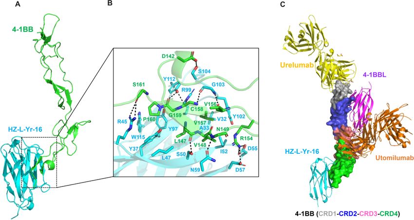
Open access
factor receptor superfamily of receptors (TNFRSF), which METHODS
J Immunother Cancer: first published as 10.1136/jitc-2020-002131 on 25 June 2021. Downloaded from http://jitc.bmj.com/ on July 11, 2021 by guest. Protected by copyright.
includes CD27, 4-1BB (CD137), OX40 (CD134) and GITR Mice
(CD357) have been targeted using agonist antibodies.10 4-1BB Human PD- L1/PD-1/4- 1BB triple knock-in mice were
is a potent stimulator of T cells and natural killer (NK) cells purchased from Gempharmatech (Jiangsu, China).
and when activated in combination with checkpoint inhibi- Human PD-L1/4-1BB double knock-in mice and human
tors may improve anticancer immune responses, especially 4-1BB knock-in mice were purchased from Biocytogen
in immunogenic- poor tumors.11 Unfortunately, inherent (Beijing, China). All mice were maintained under
on-target-related hepatotoxicity has been observed during specified pathogen-free conditions, and all studies were
the clinical development of Urelumab.12 13 The next wave of approved by the Animal Care and Use Committee of
discovery campaigns for 4-1BB agonists has been launched to HUST-Suzhou Institute for Brainsmatics.
address this issue by targeting different epitopes, optimizing
Cells and cell lines
binding affinities and through engineering different modes CT-26 and MC-38 cells knocked-in with human PD-L1
of Fc-mediated crosslinking.14–16 (CT-26-huPD-L1 and MC-38-huPD-L1 cells, respectively)
Most TNFR- superfamily members, such as 4- 1BB, were obtained from Gempharmatech (Jiangsu, China).
consist of four cysteine-rich domains (CRDs). The current Chinese hamster ovary (CHO) cells were obtained from
understanding is that agonism towards TNFR-superfamily Life technologies. Human PD-L1 (CHO-huPD-L1) and
members (modeled on CD40) is strongest when antibodies 4-1BB (CHO-hu4-1BB) overexpressing CHO cells were
bind to CRD1 (membrane distal) and weakest at CRD4 generated by the transduction of CHO cells with full
(membrane proximal).17 Interestingly, Urelumab binds to length human coding sequences. 4-1BB-NF-κB luciferase
CRD1 of 4-1BB and has shown potent agonist activity yet reporter cells (Jurkat-hu4-1BB-Luc) were generated by
induces high liver toxicity, whereas Utomilumab, which the transduction of Jurkat cells (ATCC) with lentivirus
binds between CRD3 and CRD4, has shown relative safety from a NF-κB luciferase reporter plasmid (Promega) and
but with limited activity towards 4-1BB.18 There is a concern a human 4-1BB-encoding vector.
that monospecific agonist antibodies may only offer a linear
correlation between potency and toxicity. One strategy to Antibodies
reduce their liver-related on-target effects has been to opti- The sequences of Urelumab and Utomilumab
mize FcγR binding to FcγRs that are normally enriched in were obtained from patents US8137667B2 and
US2012/0237498A1, respectively. Heavy and light chain
tumors,16 but next- generation bispecific antibodies have
genes were cloned into mammalian expression vector
an advantage through crosslinking 4-1BB receptors only in
pcDNA3.1, expressed using the ExpiCHO system (Invit-
the presence of tumor-associated antigens (TAAs) through
rogen) and protein was purified by protein A chromatog-
cross-bridging, and various programs are currently entering
raphy (GE).
the clinic targeting TAAs such as epidermal growth factor
receptor (EGFR) and human epidermal growth factor Alpaca immunization and library construction
receptor 2 (HER2).19–22 Alpacas were immunized subcutaneously with approx-
PD-L1 (CD274, B7- H1), one of the ligands for PD-1, imately 500 µg recombinant human PD- L1 (ACRO
is a checkpoint regulator that restrains T cell activation Biosystems) or 4- 1BB protein (ACRO Biosystems) on
during antigen presentation and effector function. PD-L1 days 0, 14, 28 and 42 with Freund’s adjuvant (Sigma).
expression has been identified in a wide variety of solid Blood was collected 7 days after the final immunization
tumors, which makes it a potential TAA for PD-L1/4-1BB to isolate peripheral blood mononuclear cell (PBMC).
bispecific antibody development. In preclinical studies, RNA was isolated using TRIZOL reagent (Life technol-
significant synergistic effects have been observed when ogies). cDNA was synthesized using the SuperScript IV
antibodies targeting both of these pathways are combined. First-Strand Synthesis kit (Life technologies) primed with
PD-L1/4-1BB bispecific antibodies could therefore combine oligo (dT) and subsequently used to amplify the single
PD-L1 blockade and 4-1BB agonism to provide a substantial domain antibody (vHH) repertoire. vHH libraries were
survival benefit in multiple mouse tumor models with low/ constructed by cotransforming linearized pFabVH vector
no toxicity.22 and the PCR product containing homology arms by yeast
Here, we report the discovery and development of a bispe- gap-repair. The pFab vector system allows the display of
cific antibody towards PD-L1 and 4-1BB (PM1003). PM1003 fusion proteins containing a vHH followed by Aga2p and
a Myc tag (online supplemental figure 1A).
potently blocks the interaction between PD-1 and PD-L1 and
binds to 4-1BB at the CRD4 domain allowing for effective Selection of vHH by yeast display
dose-dependent activation of 4-1BB in the presence of PD-L1, Expression of vHH on the yeast cell surface was induced
with minimal toxicity. The unique 4-1BB binding epitope has by culturing in G/RCAA (substituting 2 g/L galactose and
potential for hitting a sweet spot between efficacy and toxicity raffinose for the dextrose in the SDCAA medium), and
and we believe this discovery has resulted in the generation of expression-positive yeast cells were sorted by magnetic
a valuable 4-1BB agonist for creating more effective and safer bead aided cell sorting (Miltenyi) then 4× by fluores-
4-1BB-based multispecific therapeutics for cancer. cence activated cell sorting (FACS) (BD, Aria III) using
2 Zhai T, et al. J Immunother Cancer 2021;9:e002131. doi:10.1136/jitc-2020-002131
Open access
biotinylated antigens). Individual clones were picked for toolkit (COOT).27 Figures and structural alignments and
J Immunother Cancer: first published as 10.1136/jitc-2020-002131 on 25 June 2021. Downloaded from http://jitc.bmj.com/ on July 11, 2021 by guest. Protected by copyright.
sequencing. superpositions were generated using PyMOL.
Protein expression and purification Cell binding assay
The DNA sequences for PD-L1 (19–134) was synthesized CHO-hu4-1BB or CHO-huPD-L1 cells were incubated for
with a C-terminal 6x His-tag fusion and cloned into the 30 min with a series dilution of antibodies. After washing,
pET21b vector for expression in E. coli. Protein was puri- the cells were incubated with phycoerythrin (PE)-goat
fied on a Ni-NTA column and then polished through a (ab’)2 antihuman Fc antibody (abcam) for 30 min at
Superdex 75 Increase 10/300 GL Sciences (GL) column. 4°C. Stained cells were analyzed on a CytoFlex system
The vHH HZ-C-Ye-18 was fused to human g1-Fc (HZ-C- (Beckman). Median fluorescent intensity values were
Ye-18-Fc) in a pcDNA3.1 vector. The ExpiCHO expres- plotted against the concentration of primary antibody.
sion system (Life technologies) was used to express the
vHH- Fc antibodies according to the manufacturer’s Cell based blocking assay
instructions. Cell culture was collected 5 days after trans- Antibodies were incubated with CHO-hu4-1BB cells for
fection and purified by a Protein A column. The eluted 30 min, followed by incubation with biotinylated human
protein was concentrated and first cleaved by IdeS. The 4-
1BB ligand (ACROBiosystems) for 30 min at room
resulting mixture was passed through Protein G columns temperature. The cells were washed and subsequently
to remove Fc, and then digested by GingisKHAN to incubated with Streptavidin, R-Phycoerythrin Conjugate
generate HZ-C-Ye-18. 4-1BB (25-162) and HZ-L-Yr-16 were (SA-PE) (ThermoFisher) at 4°C for 20 min. Fluorescence
individually constructed by similar approaches as HZ-C- signals were determined on a CytoFlex system and results
Ye-18. However, a truncated Fc with only Leu- Gly-
Gly were analyzed using GraphPad7 software.
in the hinge region was cloned into the vector.23 The Biotinylated human PD- L1 (ACROBiosystems) was
encoded plasmid was transiently transfected into Expi293 incubated with a concentration gradient of antibodies
cells (Life technologies) using polyethylenimine (PEI). for 30 min. The mixtures were added to CHO-huPD-1
For 4-1BB, the eluted protein was concentrated, cleaved cells and incubated for 1 hour at 4°C. The cells were then
by IdeS and purified by a Protein A column. The 4-1BB stained with SA-PE for 20 min. The fluorescence signals
monomer was separated from 4-1BB homodimer using were determined on a CytoFlex system and results were
on a Superdex 75 Increase 10/300 GL column. For HZ-L- analyzed using GraphPad Prism software.
Yr-16, only one step purification was performed using a Production of a 4-1BB/PD-L1 bispecific antibody PM1003
Protein G column after IdeS cleavage. A PD-L1/4-1BB bispecific antibody named PM1003 was
generated by fusing the anti-4-1BB vHH to the C terminus
Crystallization, X-ray data collection and structure determination of anti-PD-L1 vHH-Fc through a 20-residue-long linker.
vHH and antigen were mixed in a 1:1 molar ratio and PM1003 incorporates LALA mutations (leucine to alanine
purified on a Superdex 75 Increase 10/300 GL column. at positions 234 and 235 according to Eu numbering)
Crystals were grown in 96-well plates by sitting drop vapor in the CH2 domain to reduce Fc receptor binding.28 29
diffusion (protein:buffer:reservoir of 0.4 µL:0.4 µL:40 µL). PM1003 was expressed transiently using HEK293F cells
The PD-L1:HZ-C-Ye-18 complex and 4-1BB:HZ-L-Yr-16 (Invitrogen). Supernatants were purified on MabSelect
complex were crystallized in 0.2 M (NH4)2SO4, 0.1 M SuRe LX Protein-A prepacked columns on an ÄKTAx-
Bis-Tris pH 5.5, 25% w/v PEG 3350 and 1.5 M Li2SO4, press instrument (GE Healthcare Life Sciences).
100 mM Tris pH 8.5, 10 mM NiSO4, respectively. For data
collection, the crystals were soaked in a reservoir solution Cell bridging
containing 20% (v/v) glycerol and flash-frozen in liquid CHO-huPD-L1 cells were stained with Cell Trace CFSE
nitrogen (Viva Biotech (Shanghai) Limited). (ThermoFisher), while CHO-hu4-1BB cells were stained
Diffraction images were collected at the Shanghai with CellTracker Deep violet (ThermoFisher) following
Synchrotron Radiation Facility beamline BL19U1 (for the manufacturer’s standard methodology. After staining,
PD-L1:HZ-C-Ye-18) and BL18U1 (for 4-1BB:HZ-L-Yr-16) cells were washed and resuspended. Labeled cells were
on a Pilatus detector and were indexed, integrated mixed and incubated for 2 hours at 37°C with titrated
and scaled using XDS.6 Resolution limits were cut antibodies. After incubation, samples were evaluated on
off at I/σ(I)=2.2 and 3.1 for PD- L1:HZ- C-Ye-18 and a CytoFlex system. Data were plotted using GraphPad
4-1BB:HZ-L-Yr-16 complex, respectively.24 Phases were Prism Software.
determined by molecular replacement with Phaser25 using
the PD-L1 structure (PDB: 5JDS) and the vHH structure Binding ELISA
(PDB: 5M2J) for the PD-L1:HZ-C-Ye-18 complex, 4-1BB Human 4-1BB, GITR, OX40 or CD40 (ACRO Biosystems)
structure (PDB: 6MGP) and the vHH structure (PDB: were coated onto a 96-well plate overnight at 4°C. The
4XTA) for 4-1BB:HZ-L-Yr-16 complex as search models. plate was washed and blocked for 1 hour in 5% bovine
Structure refinement was carried out using Refmac5.26 serum albumin/phosphate buffered saline (BSA/PBS) at
Model inspection, manual rebuilding and structure valida- room temperature. Serially diluted antibodies were added
tion was performed using crystallographic object-oriented to the plate in duplicate and incubated for 1 hour at room
Zhai T, et al. J Immunother Cancer 2021;9:e002131. doi:10.1136/jitc-2020-002131 3
Open access
temperature. After washing, Goat (ab’)2 antihuman IgG In vivo efficacy studies
J Immunother Cancer: first published as 10.1136/jitc-2020-002131 on 25 June 2021. Downloaded from http://jitc.bmj.com/ on July 11, 2021 by guest. Protected by copyright.
Fc (HRP) (Abcam) was added and incubated at room Human PD-L1/PD-1/4-1BB triple knock-in BALB/c mice
temperature for 1 hour. Plates were washed and signal aged 8–10 weeks each received 1×10⁶ CT-26-huPD-L1 cells
developed using 3,3’,5,5’-tetra-methylbenzidine substrate injected subcutaneously in the left flank. Tumor volume
solution (Solarbio) according to manufacturer’s instruc- measurements were taken with calipers to determine the
tions. Absorbance at 450 nm was read on a Molecular longest and shortest axes of the tumor and the following
Devices SpectraMax i3 system with SoftMax Pro software. formula was used to calculate tumor volume: ‘tumor
volume (mm3)=Length×Width2/2’. Dosing was initiated
Co-binding ELISA when tumors were an average of 100 mm3. Antibodies
Human 4-1BB protein was coated onto a 96-well plate over- were injected intraperitoneally into mice on days 7, 9 and
night at 4°C. After washing and blocking, serially diluted 11.
antibodies were added to the plate and incubated for
1 hour at room temperature. After washing, biotinylated Hepatotoxicity measurement
human PD-L1 protein (KACTUS) was added and incu- To study the hepatotoxic effects of our samples,
bated at room temperature for 1 hour. Plates were washed human 4-1BB knock-in mice were treated with 10 mg/
and the signal was developed using streptavidin (HRP) kg Urelumab or 6.67 mg/kg PM1003 i.p. every 5 days
(Abcam) according to manufacturer’s instructions. for four doses in total. Seven days after the final dose,
alanine aminotransferase (ALT) levels in the blood were
analyzed using the Alanine Transaminase Activity Assay
Primary T cell activation assay
Kit (abcam) following the manufacturer’s instructions.
Human primary T cells were isolated from frozen PBMC
Liver pathology was assessed by H&E and immunohisto-
(SAILYBIO) using the human total T cell isolation kit
chemistry (IHC) staining by Servicebio (Wuhan, China).
(STEMCELL) according to the manufacturer’s instruc-
tions. Anti-CD3 antibody, OKT-3 (Biolegend), was coated
(1 µg/mL) on NUNC flat-bottom plates (ThermoFisher)
RESULTS
for 2 hours at 37°C then washed twice with PBS. Seri-
Discovery of a 4-1BB-specific vHH with agonist activity
ally diluted anti-4-1BB antibodies or control antibodies
To generate vHHs specifically recognizing 4-1BB, alpacas
were either coated after OKT-3 for 2 hours followed
were immunized with recombinant human 4-1BB protein
by washing or added to the culture medium and incu-
and a yeast display library was generated. The library was
bated with human primary T cells for 4 or 5 days. Cell
screened against both human and cynomolgus (cyno)
supernatants were measured for secreted IFN-γ levels
4-1BB to identify human and cyno cross-reactive vHH
by ELISA (eBioscience). PD-L1 negative or PD-L1 posi-
sequences with a broad range of binding affinities and
tive CHO cells were co-cultured with human primary T
epitope coverage. In mind of potential efficacy versus
cells in X-VIVO15 medium (LONZA) in the presence
safety issues, clone L-Yr-16 was chosen for humanization
of plate bound 1 µg/mL OKT3 and serially diluted test
(HZ-L-Yr-16) due to its mild agonist activity, and fused
antibodies for 2 days at 37°C. After incubation, cell super-
to a human g1-Fc containing LALA mutations (online
natants were measured for secreted IL-2 levels by ELISA
supplemental figure 1B) to remove Fc effector function
(eBioscience).
(HZ-L-Yr-16-Fc).
HZ-L -
Y r-16-
F c binds to both human and cyno
Mixed lymphocyte reaction (MLR) assay 4-1 BB-overexpressing CHO cells in a dose-d ependent
Monocytes were isolated from PBMC. Monocyte-derived manner (figure 1A,B), whereas Urelumab only bound
dendritic cells (moDCs) were generated by culturing the to human 4-1 BB-e xpressing cells due to the lack of
monocytes in X-VIVO15 medium with 10 ng/mL GM-CSF cyno 4- 1 BB cross-r eactivity. ELISA- based binding
and 20 ng/mL IL-4 (R&D Systems) for 6 days. Human data revealed that HZ- L -
Y r-16-F c binds strongly to
primary T cells were isolated from a different PBMC human 4-1 BB, but not to any other TNFR-superfamily
donor source and then cocultured with the moDCs in members (figure 1C). A cell- based blocking assay
a 96-well plate and serially diluted antibodies for 3 days. was used to show that HZ-L -Y r-16-F c is a ligand non-
Cell supernatants were measured for secreted IL-2 levels blocker, while Utomilumab is a blocker and Urelumab
by ELISA (eBioscience). is a partial blocker (figure 1D).
To demonstrate that HZ- L -
Y r-16-
F c can induce
NF-κB luciferase reporter assay human 4-1 BB signaling, activated human primary T
PD-L1-negative or PD-L1-positive CHO cells were co-cul- cells (by plate-bound OKT3), were cocultured with
tured with Jurkat-hu4-1BB-NF-κB-Luc cells in the pres- anti-4-1 BB antibody either plate- bound or in solu-
ence of 1 µg/mL OKT-3 and serially diluted antibodies for tion. Both plate-bound HZ-L -Y r-16-F c and Urelumab
16 hours at 37°C. Bio-Glo Reagent (Promega), prepared showed T cell stimulation activity as measured by
according to manufacturer’s instructions, was added and IFN-γ release, while Utomilumab showed the weakest
fluorescence measured using a SpectraMax i3 system with agonism in the assay (figure 1E). HZ-L -Y r-16-Fc did not
SoftMax Pro software (Molecular Devices). show any stimulation in solution phase, indicating that
4 Zhai T, et al. J Immunother Cancer 2021;9:e002131. doi:10.1136/jitc-2020-002131
Open access
J Immunother Cancer: first published as 10.1136/jitc-2020-002131 on 25 June 2021. Downloaded from http://jitc.bmj.com/ on July 11, 2021 by guest. Protected by copyright.
Figure 1 Characterization of anti-4-1BB antibody (HZ-L-Yr-16-Fc). Binding activity of HZ-L-Yr-16-Fc to human 4-1BB (A)
or cyno 4-1BB (B) overexpressing CHO cells as revealed by flow cytometry analysis. (C) Binding of HZ-L-Yr-16-Fc to OX40,
GITR, CD40, 4-1BB determined by ELISA. (D) Effect of indicated anti-4-1BB antibodies blocking 4-1BB ligand binding to 4-1BB
measured by flow cytometry. (E) Purified T cells from human PBMC were stimulated with plated anti-CD3 and indicated anti-
4-1BB antibodies. Five days later, IFN-γ in the culture medium was analyzed by ELISA. (F) Purified T cells from human PBMC
were stimulated with plated anti-CD3 and indicated anti-4-1BB or anti-CD28 antibodies in solution. Five days later, IFN-γ in the
culture medium was analyzed by ELISA. Data are mean±SD, ***p
Open access
J Immunother Cancer: first published as 10.1136/jitc-2020-002131 on 25 June 2021. Downloaded from http://jitc.bmj.com/ on July 11, 2021 by guest. Protected by copyright.
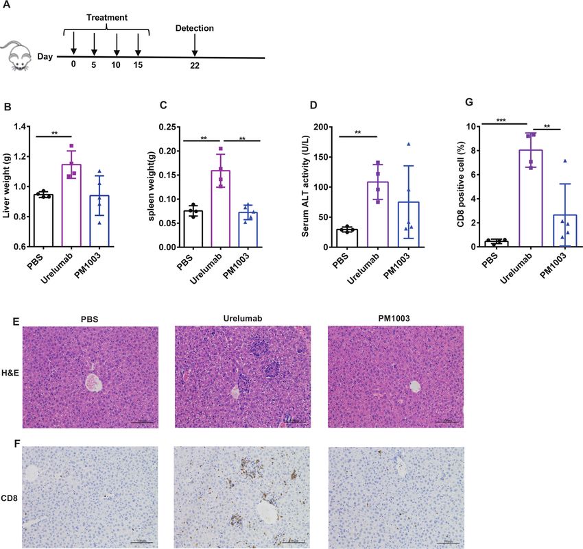
Figure 2 Detailed interaction sites between the 4-1BB and HZ-L-Yr-16 and epitope comparison between different antibody
agonists. (A) Overall structure of the 4-1BB: HZ-L-Yr-16 complex. 4-1BB is shown in green; HZ-L-Yr-16 is shown in cyan. (B)
The interactions between 4-1BB and HZ-L-Yr-16. Hydrogen bonds are shown as black dashed lines. (C) Epitope comparison of
HZ-L-Yr-16 (in cyan), Urelumab (yellow) and Utomilumab (orange). 4-1BBL (in magenta) is also aligned for comparison.
Structural determination of the PD-L1:anti-PD-L1 vHH hydrophobic interface between HZ-C-Ye-18 and PD-L1,
complex as well as Y59 from CDR2 and P100/Y103 from CDR3.
To investigate the binding epitope of our anti- PD-
L1 The binding epitope for HZ- C-Ye-18 shows similarity
vHH the structure of the PD-L1:HZ-C-Ye-18 complex was with FDA-approved PD-L1 antibodies, including Atezoli-
resolved to a 1.64 Å resolution. The overall structure of zumab (PDB: 5X8 L),30 Avelumab (PDB: 5GRJ)31 and
the PD- L1:HZ-C-
Ye-18 complex is shown in figure 3A. Durvalumab (PDB: 5X8M)30 (online supplemental figure
Analysis of the PD-L1:HZ-C-Ye-18 binding interface with 5A). The low contact area did not affect its in vitro cell-
proteins, interfaces, structures and assemblies (PISA)
based binding and blocking activities (online supple-
shows how each protein buries within a ~838 Å2 surface
mental figure 4A,B), even though Atezolizumab possesses
area. All three CDRs of HZ-C-Ye-18 contain contact resi-
dues with PD-L1 through a number of hydrogen bonds almost twice the area at the interface (~1646 Å2 by PISA).
and hydrophobic interactions (figure 3B). The CDR1, Comparison of HZ- C-
Ye-18 with another PD- L1 vHH
CDR2 and CDR3 loops were observed to form hydrogen termed KN035 (PDB: 5JDS)32 shows that both these mole-
bonds with the residues Y56, N63, H69, D73, K75, S117 cules contact with several PD-1 interaction sites on PD-L1,
and G119 of PD- L1. Additionally, Y32, W33 and Y35 including I54, Y56, M115 and A121 (online supplemental
from the highly hydrophobic CDR1 are involved in the figure 5B,C).
Figure 3 Detailed interaction sites between PD-L1 and HZ-C-Ye-18. (A) Overall structure of the PD-L1:HZ-C-Ye-18 complex
(PDB: 7CZD). PD-L1 is shown in magenta, while HZ-C-Ye-18 is shown in purple. The CDR1, CDR2 and CDR3 domains of
HZ-C-Ye-18 are colored green, cyan and yellow, respectively. (B) The interactions between PD-L1 and HZ-C-Ye-18. Hydrogen
bonds are shown as black dashed lines.
6 Zhai T, et al. J Immunother Cancer 2021;9:e002131. doi:10.1136/jitc-2020-002131
Open access
J Immunother Cancer: first published as 10.1136/jitc-2020-002131 on 25 June 2021. Downloaded from http://jitc.bmj.com/ on July 11, 2021 by guest. Protected by copyright.
Figure 4 Structure of PM1003 and the binding activity of the bispecific antibody to both 4-1BB and PD-L1. (A) Structural
representation of PM1003. (B) Binding of PM1003 to human PD-L1 overexpressed on CHO cells measured by flow cytometry.
(C) Binding of PM1003 to human 4-1BB overexpressed on CHO cells measured by flow cytometry. (D) PM1003 blocks the
interaction between PD-1/PD-L1 determined by flow cytometry. (E) PM1003 simultaneous binding to both human PD-L1
and human 4-1BB as determined by ELISA. (F) PM1003 simultaneous binding to both human PD-L1 and human 4-1BB
overexpressing cells as determined by a cell bridging flow cytometry assay.
Design and characterization of an anti-PD-L1/4-1BB bispecific PM1003-mediated T cell activation is dependent on
therapeutic crosslinking via PD-L1 cross-bridging
We proceeded to generate a bispecific molecule by To demonstrate that PM1003 is an effective PD- L1-
fusing HZ- L-Yr-16 to the C terminus of HZ- C-Ye-18-
Fc mediated 4-1BB agonist, Jurkat-4-1BB-NF-κB-Luc cells
via a 20-residue-long linker (PM1003, figure 4A). The were cocultured with empty CHO or CHO-huPD-L1 cells
binding activity of each arm was similar to the parent and OKT-3/PM1003, then tested for NF- kB signaling
antibodies in cell- based assays (figure 4B,C). Biolayer through detection of luciferase activity. Maximum acti-
interferometry (BLI) analyses demonstrated high affinity vation/luciferase signals were detected upon co- cul-
binding to PD- L1 and 4- 1BB (online supplemental ture with CHO-huPD-L1 cells and PM1003 (figure 5A).
figure 6A,B). More importantly, PM1003 did not show Similarly, human primary T cells were stimulated by
any non-specific binding to non-transfected CHO cells plate-bound OKT-3 mAb, then co-cultured with CHO-
(online supplemental figure 6C) or 293F cells (online
huPD- L1 or empty CHO cells with serially diluted
supplemental figure 6D). Functional assays showed that
PM1003 or HZ-L-Yr-16-Fc. T cell activation induced by
PM1003 efficiently inhibited PD-L1 binding to PD-1 on
PM1003 was only observed upon co- incubation with
CHO-huPD-1 cells (figure 4D). The ability of PM1003 to
CHO-huPD-L1 cells (figure 5B,C). These data indicate
facilitate co-binding of PD-L1 and 4-1BB was also evalu-
ated by both BLI (online supplemental figure 6E) and that PM1003-mediated T cell activation was dependent
ELISA (figure 4E). To evaluate if PM1003 facilitated cell on crosslinking via PD-L1-mediated cross-bridging. The
bridging through simultaneously binding to both recep- activity of PM1003 was tested via an MLR, which utilizes
tors, we fluorescently labeled CHO- huPD-L1 cells and human primary T cells and moDC expressing endoge-
CHO-hu4-1BB cells with different dyes and then treated nous levels of both targets. PM1003 showed stronger T
them with PM1003 followed by flow cytometry anal- cell activation than the combination of anti-PD-L1 and
ysis (online supplemental figure 6F). PM1003 induced anti-4-1BB antibodies (figure 5D), whereas HZ-L-Yr-16-Fc
the formation of doublets of CHO-huPD-L1 and CHO- alone did not elicit any activation showing that PD-L1-
hu4-1BB cells, while the combination of HZ-C-Ye-18-Fc mediated cross-bridging is a major contributor for the
and HZ-L-Yr-16-Fc did not (figure 4F). agonist activity of PM1003.
Zhai T, et al. J Immunother Cancer 2021;9:e002131. doi:10.1136/jitc-2020-002131 7Open access
J Immunother Cancer: first published as 10.1136/jitc-2020-002131 on 25 June 2021. Downloaded from http://jitc.bmj.com/ on July 11, 2021 by guest. Protected by copyright.
Figure 5 4-1BB agonism via PM1003 is dependent on crosslinking via PD-L1-mediated cross-bridging. (A) PM1003 activity of
Jurkat-hu4-1BB-NF-κB luciferase cells incubated with CHO-huPD-L1 cells or PD-L1-negative CHO cells. (B) PM1003 activity
in a human primary T cell activation assay with non-transfected CHO cells. (C) PM1003 activity in a human primary T cell
activation assay with CHO-huPD-L1 cells. (D) PM1003 activity in a mixed lymphocyte reaction with monospecific components
and in combination.
PM1003 suppresses tumor growth in syngeneic tumor models PM1003 shows negligible hepatotoxicity
To study the antitumor efficacy of PM1003 in vivo, Since 4-1BB agonists have been shown to induce hepa-
CT-26- huPD-
L1 cells and MC38- huPD-L1 cells were totoxicity, we compared the toxicity profile of PM1003
injected subcutaneously into the flank of human PD-1/ with Urelumab in mice. Human 4-1BB knock-in mice
PD-L1/4-1BB triple knock-in BALB/c mice and human
were injected i.p. with PBS, Urelumab, or PM1003
PD-L1/4-1BB double knock- in C57BL/6 mice, respec-
every 5 days for four doses in total then euthanized
tively. In the CT-26-huPD- L1 tumor model, PM1003
was shown to substantially reduce tumor growth 1 week later (figure 7A). Urelumab treatment induced a
(figure 6A–C). Tumor inhibition was dose- dependent significant increase in liver and spleen weight, whereas
and significantly higher than that of the equimolar dose PM1003 itself had no substantial effect (figure 7B,C).
of Urelumab or the combination of the single agents. Serum levels of ALT increased on Urelumab treatment
Dose- dependent antitumor efficacy was also observed but not with PM1003 (figure 7D). Liver-infiltrating
in a MC38-huPD-L1 tumor model (figure 6D). In order lymphocytes were also evaluated by H&E and CD8
to determine whether these complete responses (CRs) IHC staining. Urelumab treatment induced significant
resulted in the induction of immune memory, mice with
levels of lymphocyte/CD8+ T cell infiltration into liver
CRs were rechallenged with the same cell line in the
inguinal mammary fat pad on the opposite flank. The tissues, whereas PM1003 did not (figure 7E–G). These
inhibition of the ‘rechallenged’ tumor growth indicated results were consistent with the clinical toxicity profile
that immune memory was induced in the PM1003-treated of Urelumab. Collectively, PM1003 showed limited anti-
group (figure 6E). 4-1BB-mediated hepatotoxicity.
8 Zhai T, et al. J Immunother Cancer 2021;9:e002131. doi:10.1136/jitc-2020-002131Open access
J Immunother Cancer: first published as 10.1136/jitc-2020-002131 on 25 June 2021. Downloaded from http://jitc.bmj.com/ on July 11, 2021 by guest. Protected by copyright.
Figure 6 In vivo efficacy of PM1003 in CT-26-huPD-L1 and MC38-huPD-L1 mouse model. (A) Human PD-1/PD-L1/4-1BB
triple knock-in mice were injected subcutaneously with 1×10⁶ CT-26-huPD-L1 tumor cells, then molar equivalent doses of
indicated antibodies were administrated on days 7, 9, 11. Tumor size was measured every other day. (B) Images of tumors
(left) and tumor weight from mice (right). (C) Individual tumor growth spaghetti plots. Data are mean±SEM, *pOpen access
J Immunother Cancer: first published as 10.1136/jitc-2020-002131 on 25 June 2021. Downloaded from http://jitc.bmj.com/ on July 11, 2021 by guest. Protected by copyright.
Figure 7 PM1003 was tested for liver pathology in a 4-1BB knock-in C57 mouse model. (A) Experimental protocol for the
toxicity model. Human 4-1BB knock-in C57 mice (n=4 or 5/group) were treated with 10 mg/Kg Urelumab, 6.67 mg/Kg PM1003
or PBS on days 0, 5, 10 and 15. 7 days after the final treatment, (B) liver weight, (C) spleen weight, (D) serum ALT activity, (E)
liver tissue HE staining, (F) liver tissue CD8-positive T cell IHC staining and (G) percentage of CD8-positive cell infiltration, were
measured. Data are mean±SEM, **pOpen access
antibodies that bind to the membrane distal domain of have helped to de-risk any potential toxicity issues before
J Immunother Cancer: first published as 10.1136/jitc-2020-002131 on 25 June 2021. Downloaded from http://jitc.bmj.com/ on July 11, 2021 by guest. Protected by copyright.
CD40, but weaker for those binding to membrane distal entering clinical trials.
domain of OX40.17 37 Although we do not have access to There are a number of anti- PD-
L1/4-
1BB programs
a panel of antibodies that bind to every domain of 4-1BB, under development at present,22 towards other tumor
it is clear that different clones exhibit very distinct agonist antigens/stromal markers, or using 4- 1BBL instead of
profiles, and that the current evidence suggest that the an antibody binder to activate 4- 1BB.20 38 39 Recently,
binding to the distal N-terminal domain of 4-1BB may RO7122290, an antibody targeting fibroblast activation
naturally offer a higher degree of agonist activity with protein (FAP), fused to 4-1BBL, was well-tolerated up to
potential toxicity issues (as with Urelumab). It is also 2000 mg as a single-agent and 1000 mg in combination
understood that a fast association/dissociation (fast-on/ with atezolizumab, without reaching a maximum toler-
fast-off) binding profile may improve the agonist activity ated dose, showing the potential safety of this class of
of mAbs by offering a constant on/off binding flux. Thus, agent (DOI: https://doi.org/10.1016/j.annonc.2020.08.
the strong binding strength and ligand blocking activities 1145). From the current literature and information from
of Utomilumab might contribute to reduced activity. the public domain, there is yet to be a fully characterized
To ensure that the toxicity related to 4-1BB agonists is bispecific antibody that binds to the CRD4 domain of
restricted to the local tumor microenvironment, investi- 4-1BB. We believe that this mild agonist activity is suitable
gators have engineered bispecific molecules to bind to for a PD-L1-based 4-1BB bispecific due to the high doses
tumors with one arm and bridge over to 4-1BB receptors used for current anti-PD-L1 treatments. We also believe
using the other.19–22 This bridging effect between the that PD-L1, which is upregulated after T cell activation
effector and target cells ensures that 4-1BB is only activated and IFN-γ exposure, would provide a safer cross-bridging
in the presence of tumor cells. Here, we fused HZ-L-Yr-16 pair that is localized to the local tumor microenvironment.
to the C-terminus of an anti-PD-L1 vHH that contains an
effector-silenced human g1 Fc (HZ-C-Ye-18-Fc) resulting
in the therapeutic candidate PM1003. HZ-C-Ye-18 is a
potent blocker of the interaction between PD-1 and PD-L1 CONCLUSION
and through structural determination has an overlapping PM1003 is a highly differentiating drug candidate
binding epitope similar to Atezolizumab. Both the PD-L1 that binds to a unique epitope on 4-1BB and facilitates
and 4-1BB binding sites of PM1003 retain their potency receptor agonism with a lowered risk for toxicity issues.
in this bispecific form. Moreover, the bispecific antibody PM1003 has been shown to block potently the interaction
was shown to cross-bridge PD-L1- and 4-1BB-expressing between PD-1 and PD-L1 and to induce agonism towards
cells and can bind to both antigens simultaneously at 4-
1BB only in the presence of PD- L1-mediated cross-
the protein level. These properties were reflected in cell- bridging. Thus, PM1003 is a promising bispecific agent to
based activation assays using luciferase reporter cell lines be developed for the treatment of multiple solid tumors.
and primary cells with and without PD- L1-expressing
targets. Author affiliations
1
The synergy of combining both PD- L1 and 4- 1BB Shanghai Institute of Immunology, Department of Immunology and Microbiology,
binders in PM1003 was displayed in a tumor model where Shanghai Jiao Tong University School of Medicine, Shanghai, China
2
Discovery Biology & Discovery Technology, Biotheus Inc, Zhuhai, China
CT-26 cells with human PD-L1 knocked-in to replace the 3
Discovery Biology, Biotheus (Suzhou) Co., Ltd, Suzhou, China
murine sequence were implanted into human PD-1/ 4
Institutes of Biology and Medical Sciences, Soochow University, Suzhou, China
PD-L1/4-1BB triple knock-in mice. Our results show that
PM1003 was much superior compared with the combi- Acknowledgements We would like to thank the Biotheus Discovery Biology and
nation of anti-PD-L1+anti-4-1BB antibodies at medium Discovery Technology teams; Weihui Fu for the protein formulation study; Liping
(4 mg/kg) and high (12 mg/kg) doses. Dose-dependent Wan for drug developability analysis; Xiaolin Liu and Yi Luo for scientific contribution
and review; Tiantian Dong for preclinical assessment.
antitumor efficacy was also confirmed in MC38-huPD-L1
Contributors TZ and CW contributed equally. XM and YFX constructed the yeast
knock- in cells implanted into human PD- L1/4-1BB display library and conducted the vHH selection. TW constructed the bispecific
double knock-in mice. PM1003 was also able to induce antibody and generated the protein material. TZ and SD designed and performed
memory as shown in rechallenged mice. Finally, we cell-based assays. CW, YH, YZ and WJ generated the materials for crystallization
showed that hepatotoxicity was kept at a minimum in and determined the structures. ZY and TP designed and performed the BLI
experiments. WH and SP performed the in vivo experiments. JS, XG, BL, AT and
mice treated with PM1003 compared with Urelumab, YDX provided guidance and support throughout. JZ, AT, BL and YDX assisted with
where liver weight, spleen weight and CD8+ T cell infiltra- manuscript revisions. TZ, CW, YFX, AT and XM generated figures and wrote the
tion were significantly lower than the Urelumab group. manuscript with input from all the authors.
Non-human primates are not a suitable model for eval- Funding The authors have not declared a specific grant for this research from any
uating the toxicity of Urelumab due to its known low to funding agency in the public, commercial or not-for-profit sectors.
minimal cross-reactivity. As PM1003 has cross-reactivity Competing interests The research was funded by Biotheus Inc. All authors are
to both cynomolgus PD-L1 and 4-1BB, we conducted a current employees of Biotheus. Inc, with the exception of JZ, TZ and BL who declare
non-good laboratory practice (GLP) safety assessment in no competing interests.
cynomolgus monkeys at 60 mg/kg without any signs of Patient consent for publication Not required.
untoward toxicity (data in revision). These safety studies Provenance and peer review Not commissioned; externally peer reviewed.
Zhai T, et al. J Immunother Cancer 2021;9:e002131. doi:10.1136/jitc-2020-002131 11Open access
Data availability statement Data are available in a public, open access 15 Kotanides H, Sattler RM, Lebron MB, et al. Characterization of 7A5:
J Immunother Cancer: first published as 10.1136/jitc-2020-002131 on 25 June 2021. Downloaded from http://jitc.bmj.com/ on July 11, 2021 by guest. Protected by copyright.
repository. All data relevant to the study are included in the article or uploaded as a human CD137 (4-1BB) receptor binding monoclonal antibody with
supplementary information. Structural information and atomic coordinates have differential agonist properties that promotes antitumor immunity. Mol
Cancer Ther 2020;19:988–98.
been deposited into the Protein Data Bank (PDB) with the accession codes 7CZD
16 Qi X, Li F, Wu Y, et al. Optimization of 4-1BB antibody for cancer
and 7D4B. Other data that support the findings of this study are available from the immunotherapy by balancing agonistic strength with FcγR affinity.
corresponding author upon request. Nat Commun 2019;10:2141.
Supplemental material This content has been supplied by the author(s). It has 17 Yu X, Chan HTC, Orr CM, et al. Complex interplay between epitope
not been vetted by BMJ Publishing Group Limited (BMJ) and may not have been specificity and isotype dictates the biological activity of anti-human
CD40 antibodies. Cancer Cell 2018;33:664–75.
peer-reviewed. Any opinions or recommendations discussed are solely those 18 Chin SM, Kimberlin CR, Roe-Zurz Z, et al. Structure of the 4-1BB/4-
of the author(s) and are not endorsed by BMJ. BMJ disclaims all liability and 1BBL complex and distinct binding and functional properties of
responsibility arising from any reliance placed on the content. Where the content utomilumab and urelumab. Nat Commun 2018;9:4679.
includes any translated material, BMJ does not warrant the accuracy and reliability 19 Compte M, Harwood SL, Muñoz IG, et al. A tumor-targeted trimeric
of the translations (including but not limited to local regulations, clinical guidelines, 4-1BB-agonistic antibody induces potent anti-tumor immunity
terminology, drug names and drug dosages), and is not responsible for any error without systemic toxicity. Nat Commun 2018;9:4809.
and/or omissions arising from translation and adaptation or otherwise. 20 Claus C, Ferrara C, Xu W, et al. Tumor-Targeted 4-1BB agonists for
combination with T cell bispecific antibodies as off-the-shelf therapy.
Open access This is an open access article distributed in accordance with the Sci Transl Med 2019;11:eaav5989.
Creative Commons Attribution Non Commercial (CC BY-NC 4.0) license, which 21 Hinner MJ, Aiba RSB, Jaquin TJ, et al. Tumor-Localized
permits others to distribute, remix, adapt, build upon this work non-commercially, costimulatory T-cell engagement by the 4-1BB/HER2
and license their derivative works on different terms, provided the original work is bispecific Antibody-Anticalin fusion PRS-343. Clin Cancer Res
2019;25:5878–89.
properly cited, appropriate credit is given, any changes made indicated, and the use
22 Lakins MA, Koers A, Giambalvo R, et al. FS222, a CD137/PD-L1
is non-commercial. See http://c reativecommons.org/licenses/by-nc/4.0 /. tetravalent bispecific antibody, exhibits low toxicity and antitumor
activity in colorectal cancer models. Clinical Cancer Research
ORCID iD 2020;26:4154–67.
Tianhang Zhai http://orcid.org/0000-0002-5 285-5823 23 Novarra S, Grinberg L, Rickert KW, et al. A hingeless Fc fusion
system for site-specific cleavage by ides. MAbs 2016;8:1118–25.
24 Karplus PA, Diederichs K. Assessing and maximizing data quality in
macromolecular crystallography. Curr Opin Struct Biol 2015;34:60–8.
25 McCoy AJ, Grosse-Kunstleve RW, Adams PD, et al. Phaser
crystallographic software. J Appl Crystallogr 2007;40:658–74.
REFERENCES 26 Murshudov GN, Vagin AA, Dodson EJ. Refinement of
1 Pardoll DM. Immunology beats cancer: a blueprint for successful macromolecular structures by the maximum-likelihood method. Acta
translation. Nat Immunol 2012;13:1129–32. Crystallogr D Biol Crystallogr 1997;53:240–55.
2 Hodi FS, O'Day SJ, McDermott DF, et al. Improved survival with 27 Emsley P, Lohkamp B, Scott WG, et al. Features and development of
ipilimumab in patients with metastatic melanoma. N Engl J Med Coot. Acta Crystallogr D Biol Crystallogr 2010;66:486–501.
Overseas Ed 2010;363:711–23. 28 Hezareh M, Hessell AJ, Jensen RC, et al. Effector function activities
3 Robert C, Thomas L, Bondarenko I, et al. Ipilimumab plus of a panel of mutants of a broadly neutralizing antibody against
dacarbazine for previously untreated metastatic melanoma. N Engl J human immunodeficiency virus type 1. J Virol 2001;75:12161–8.
Med 2011;364:2517–26. 29 Bruhns P, Iannascoli B, England P, et al. Specificity and affinity of
4 Topalian SL, Hodi FS, Brahmer JR, et al. Safety, activity, and human Fcγ receptors and their polymorphic variants for human IgG
immune correlates of Anti–PD-1 antibody in cancer. N Engl J Med subclasses. Blood 2009;113:3716–25.
2012;366:2443–54. 30 Lee HT, Lee JY, Lim H, et al. Molecular mechanism of PD-1/PD-L1
5 Hamid O, Robert C, Daud A, et al. Safety and tumor responses blockade via anti-PD-L1 antibodies atezolizumab and durvalumab.
with lambrolizumab (Anti–PD-1) in melanoma. N Engl J Med Sci Rep 2017;7:5532.
2013;369:134–44. 31 Liu K, Tan S, Chai Y, et al. Structural basis of anti-PD-L1 monoclonal
6 Wolchok JD, Kluger H, Callahan MK, et al. Nivolumab plus antibody avelumab for tumor therapy. Cell Res 2017;27:151–3.
ipilimumab in advanced melanoma. N Engl J Med 2013;369:122–33. 32 Zhang F, Wei H, Wang X, et al. Structural basis of a novel PD-
7 Mahoney KM, Atkins MB. Prognostic and predictive markers for the L1 nanobody for immune checkpoint blockade. Cell Discovery
new immunotherapies. Oncology 2014;28 Suppl 3:39–48. 2017;3:17004.
8 Alexandrov LB, Nik-Zainal S, Wedge DC, et al. Signatures of 33 Bartkowiak T, Jaiswal AR, Ager CR, et al. Activation of 4-1BB on
mutational processes in human cancer. Nature 2013;500:415–21. liver myeloid cells triggers hepatitis via an Interleukin-27–Dependent
9 Le DT, Uram JN, Wang H, et al. Pd-1 blockade in tumors pathway. Clin Cancer Res 2018;24:1138–51.
with mismatch-repair deficiency. N Engl J Med Overseas Ed 34 Cheng L, Wang J, Li X, et al. Interleukin-6 induces Gr-1+CD11b+
2015;372:2509–20. myeloid cells to suppress CD8+ T cell-mediated liver injury in mice.
10 Schaer DA, Hirschhorn-Cymerman D, Wolchok JD. Targeting tumor- PLoS One 2011;6:e17631.
necrosis factor receptor pathways for tumor immunotherapy. J 35 Wang J, Zhao W, Cheng L, et al. CD137-mediated pathogenesis from
Immunother Cancer 2014;2:7. chronic hepatitis to hepatocellular carcinoma in hepatitis B virus-
11 Chen S, Lee L-F, Fisher TS, et al. Combination of 4-1BB agonist transgenic mice. J.i. 2010;185:7654–62.
and PD-1 antagonist promotes antitumor effector/memory CD8 T 36 Li Y, Tan S, Zhang C, et al. Limited cross-linking of 4-1BB by 4-1BB
cells in a poorly immunogenic tumor model. Cancer Immunol Res ligand and the agonist monoclonal antibody Utomilumab. Cell Rep
2015;3:149–60. 2018;25:909–20.
12 Segal NH, Logan TF, Hodi FS, et al. Results from an integrated safety 37 Griffiths J, Hussain K, Smith HL, et al. Domain binding and isotype
analysis of Urelumab, an agonist Anti-CD137 monoclonal antibody. dictate the activity of anti-human OX40 antibodies. Journal for
Clin Cancer Res 2017;23:1929–36. ImmunoTherapy of Cancer 2020;8:e001557.
13 Ho SK, Xu Z, Thakur A, et al. Epitope and Fc-mediated cross- 38 Trüb M, Uhlenbrock F, Claus C, et al. Fibroblast activation protein-
linking, but not high affinity, are critical for antitumor activity of targeted-4-1BB ligand agonist amplifies effector functions of
CD137 agonist antibody with reduced liver toxicity. Mol Cancer Ther intratumoral T cells in human cancer. J Immunother Cancer
2020;19:1040–51. 2020;8:e000238.
14 Eskiocak U, Guzman W, Wolf B, et al. Differentiated agonistic 39 You G, Lee Y, Kang Y-W, et al. B7-H3×4-1BB bispecific antibody
antibody targeting CD137 eradicates large tumors without augments antitumor immunity by enhancing terminally differentiated
hepatotoxicity. JCI Insight 2020;5. CD8 + tumor-infiltrating lymphocytes. Sci Adv 2021;7:eaax3160.
12 Zhai T, et al. J Immunother Cancer 2021;9:e002131. doi:10.1136/jitc-2020-002131You can also read