HDAC1 is required for GATA-1 transcription activity, global chromatin occupancy and hematopoiesis
←
→
Page content transcription
If your browser does not render page correctly, please read the page content below
Published online 27 August 2021                                                    Nucleic Acids Research, 2021, Vol. 49, No. 17 9783–9798
                                                                                                         https://doi.org/10.1093/nar/gkab737

HDAC1 is required for GATA-1 transcription activity,
global chromatin occupancy and hematopoiesis
Bowen Yan1 , Jennifer Yang1 , Min Young Kim1 , Huacheng Luo2 , Nicholas Cesari2 , Tao Yang1 ,
John Strouboulis4 , Jiwang Zhang5 , Ross Hardison 6 , Suming Huang 2,3 and Yi Qiu 1,3,*
1
 Department of Cellular and Molecular Physiology, Hershey, PA 17033, USA, 2 Department of Pediatrics, Hershey,
PA 17033, USA, 3 Penn State Cancer Institute, Pennsylvania State University College of Medicine, Hershey, PA
17033, USA, 4 Comprehensive Cancer Center, School of Cancer and Pharmaceutical Sciences, Faculty of Life
Sciences and Medicine, King’s College London, London SE5 9NU, UK, 5 Department of Pathology, Loyola University

                                                                                                                                                                  Downloaded from https://academic.oup.com/nar/article/49/17/9783/6358684 by guest on 12 October 2021
Medical Center, Maywood, IL 60153, USA and 6 Departments of Biochemistry and Molecular Biology, Pennsylvania
State University, University Park, PA 16802, USA

Received March 08, 2021; Revised August 05, 2021; Editorial Decision August 10, 2021; Accepted August 16, 2021

ABSTRACT                                                                          INTRODUCTION
The activity of hematopoietic factor GATA-1 is mod-                               GATA-1 is a tissue specific transcription factor that is
ulated through p300/CBP-mediated acetylation and                                  essential for terminal differentiation of erythrocytes and
FOG-1 mediated indirect interaction with HDAC1/2                                  megakaryocytes (1). GATA binding sites are present in the
containing NuRD complex. Although GATA-1 acety-                                   regulatory regions of virtually all erythroid-specific genes,
                                                                                  suggesting that GATA-1 represents an erythroid-specific
lation is implicated in GATA-1 activation, the role of
                                                                                  master regulator (1–4). Genetic mutations in GATA-1 are
deacetylation is not studied. Here, we found that the                             associated with various forms of anemia and myeloid ma-
FOG-1/NuRD does not deacetylate GATA-1. However,                                  lignancy (5–10). GATA-1 has been reported to interact with
HDAC1/2 can directly bind and deacetylate GATA-1.                                 a variety of coactivators and corepressors, including the
Two arginine residues within the GATA-1 linker re-                                HDAC1/2-containing NuRD or MeCP1 corepressor com-
gion mediates direct interaction with HDAC1. The                                  plexes, through its interaction with FOG-1 (2,11–13). The
arginine to alanine mutation (2RA) blocks GATA-1                                  NuRD complexes appear to be important for GATA-1 me-
deacetylation and fails to induce erythroid differenti-                           diated activation and repression (11,12,14). We recently re-
ation. Gene expression profiling and ChIP-seq anal-                               ported that HDAC1 is acetylated during erythropoiesis and
ysis further demonstrate the importance of GATA-1                                 acetylated HDAC1 loss deacetylase activity (15,16). More
deacetylation for gene activation and chromatin re-                               importantly, acetylated HDAC1 converts the NuRD core-
cruitment. GATA-12RA knock-in (KI) mice suffer mild                               pressor complex to coactivator complexes (15,17), support-
                                                                                  ing the notion that NuRD is required for GATA-1 activa-
anemia and thrombocytopenia with accumulation of                                  tion (14). Therefore, HDAC1 is a critical component in reg-
immature erythrocytes and megakaryocytes in bone                                  ulating GATA-1 function.
marrow and spleen. Single cell RNA-seq analysis of                                   GATA1 undergoes acetylation by p300/CBP on sev-
Lin− cKit+ (LK) cells further reveal a profound change                            eral clusters of lysine residues adjacent to the two DNA-
in cell subpopulations and signature gene expres-                                 binding zinc fingers (18,19). Mutating these sites dramat-
sion patterns in HSC, myeloid progenitors, and                                    ically impairs GATA-1 function, suggesting that acetyla-
erythroid/megakaryocyte clusters in KI mice. Thus,                                tion of GATA-1 plays a role in its transcriptional activ-
GATA-1 deacetylation and its interaction with HDAC1                               ity. The mechanistic basis for GATA-1 regulation by acety-
modulates GATA-1 chromatin binding and transcrip-                                 lation remains controversial; while one study showed di-
tional activity that control erythroid/megakaryocyte                              rect enhancement of DNA binding by acetylation (18),
commitment and differentiation.                                                   another study showed enrichment in chromatin binding
                                                                                  without affecting DNA binding (20). GATA-1 acetylation
                                                                                  may also mediate recruitment of the BET family bromod-
                                                                                  omain factor Brd3, which plays a critical role in promoting

* To   whom correspondence should be addressed. Tel: +1 717 531 0003 (Ext 321489); Fax: +1 717 531 7667; Email: yqiu1@pennstatehealth.psu.edu
Present address: Tao Yang, Department of Physiology and Pharmacology, College of Medicine and Life Sciences, The University of Toledo, Toledo, OH 43614,
USA.

C The Author(s) 2021. Published by Oxford University Press on behalf of Nucleic Acids Research.
This is an Open Access article distributed under the terms of the Creative Commons Attribution-NonCommercial License
(http://creativecommons.org/licenses/by-nc/4.0/), which permits non-commercial re-use, distribution, and reproduction in any medium, provided the original work
is properly cited. For commercial re-use, please contact journals.permissions@oup.com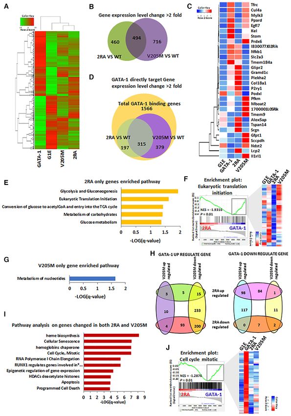
9784 Nucleic Acids Research, 2021, Vol. 49, No. 17

GATA-1 occupancy of erythroid target genes (21). How-           Chromatin immunoprecipitation (ChIP) assays and ChIP-
ever, it remains unknown whether and how GATA-1                 seq
acetylation is regulated in contributing to its activities in
                                                                ChIP assays were performed as previously described (15).
hematopoiesis.
                                                                ChIP primers are listed in Supplemental Table S1. ChIP-seq
   In this study, we found that GATA-1 can be deacety-
                                                                data were mapped to the reference mouse genome (mm10)
lated by HDAC1 through direct interaction independently
                                                                using Bowtie2 (25). Peaks were identified using the MACS
of FOG-1. This interaction is required for GATA-1 deacety-
                                                                program (26). Binding sites annotation were performed us-
lation and GATA-1 chromatin occupancy. Furthermore,
                                                                ing the ChIPseeker (27). The ChIP-seq dataset has been de-
the disruption of the GATA-1-HDAC1 interaction re-
                                                                posited to GEO (accession number GSE161608). The de-
presses GATA-1 mediated differentiation programs, includ-
                                                                tailed method is described in Supplementary Methods.
ing HSC, myeloid progenitors, erythroid and megakary-
ocytic differentiation, suggesting essential roles for the
GATA-1 and HDAC1 interaction in modulating GATA-1               Colony forming assays

                                                                                                                                    Downloaded from https://academic.oup.com/nar/article/49/17/9783/6358684 by guest on 12 October 2021
function.
                                                                Primary mouse bone marrow and spleen cells were har-
                                                                vested from 2RA KI or C57 mice. Methylcellulose mix was
MATERIALS AND METHODS                                           prepared follow the manufacture’s instruction (Stem cell
Acetylation and deacetylation in vitro                          Technology, Cat # M3134). 1 × 10⁵ bone marrow cells or
                                                                5 × 10⁵ spleen cells were added to methylcellulose medium
Flag-tagged p300 and HDAC1 were expression and puri-            and plated in triplicate. The cells were incubate at 37◦ C for
fied from baculoviral vector. The in vitro acetylation and      6 days and colonies were scored by light microscopy.
deacetylation assays were described previously (16).
                                                                RESULTS
Affinity pull-down assay
                                                                HDAC1, but not the FOG-1/NuRD corepressor complex,
Bacterial-expressed GST-tagged wild type GATA-1 and             deacetylates acetylated GATA-1
GATA-1 mutants were purified immobilized. The protein
level were determined by SDS-PAGE. The GST pull-down            It has been well documented that FOG-mediated inter-
assay was performed as previously described (17).               action with the HDAC1/2-containing NuRD corepressor
                                                                complex is required for GATA-1 mediated repression and
                                                                activation (2,11–15). Since GATA-1 is an acetylated protein
Generation of GATA-1 2RA knock-in mice                          and acetylation is positively linked to its activity (21), we ex-
A targeting vector harboring GATA-1 R243, 247A (2RA)            amined whether the FOG-1-associated NuRD complex me-
mutation was generated for gene targeting by homologous         diates GATA-1 deacetylation. The NuRD complex was im-
recombination. The detail method is provided in Supple-         munoprecipitated with FOG-1 antibody from undifferen-
mentary Methods.                                                tiated MEL cells and subsequently incubated with in vitro
                                                                acetylated GATA-1. To our surprise, the NuRD-containing
                                                                FOG-1 complex did not deacetylate GATA-1 (Figure 1A).
RNA sequencing (RNA-seq) and single cell RNA-seq
                                                                As a positive control, the NuRD complex deacetylated hi-
Total RNA was isolated and DNAse I treated using RNA            stone H4 effectively (Figure 1B), indicating that the pu-
purification kit (Genesee Scientific, San Diego). RNA li-       rified NuRD complex exhibits active deacetylase activity.
braries were prepared using the TruSeq RNA sample prep          Of note, recombinant HDAC1 or HDAC2 can deacety-
kit (Illumina, San Diego) and sequenced on the Illu-            late acetylated GATA-1 and histone efficiently (Figure 1C,
mina HiSeq 2000 according to the manufacturer’s instruc-        Supplemental Figure S1A and B). Thus, these results indi-
tions. Reads were analyzed with Tophat (22), Cuffdiff (23)      cate that HDAC1/2 can deacetylate GATA-1 in vitro in a
pipeline. Raw data and normalized gene expression data          FOG1/NuRD complex independent manner.
were deposited to Gene Expression Omnibus (GEO) under              Since HDAC1 deacetylates a number of transcription
accession number GSE161607. RT-qPCR were performed              factors through direct interaction (28), we hypothesized that
to validate RNA-seq results on representative GATA-1 tar-       HDAC1 could deacetylate GATA-1 through a direct inter-
get genes. Primers used are listed in Supplemental Table S1.    action. GST pull down was carried out by incubating GST-
   For scRNA-seq, Lin− cKit+ mice bone marrow cells were        GATA-1 with 35 S-labeled HDAC1 to show that GATA-1
sorted and loaded to 10× Genomics platform with a target        physically and directly interacts with HDAC1 in vitro (Fig-
output of 7000 cells. scRNA-seq libraries were constructed      ure 1D). The interaction surface in GATA-1 mediating in-
using chromium single cell 3 reagent kits v2 (10× Ge-          teraction with HDAC1, was further determined by using a
nomics) and sequenced on the NovaSeq 6000 Sequencing            series of GST-GATA-1 deletion mutants. In order to elimi-
System (Illumina). The analysis was performed using Cell        nate potential contamination of FOG-1 complex in reticu-
Ranger pipeline (10× Genomics) and ‘Seurat’ package (24)        locyte lysates when used to synthesize 35 S-labeled HDAC1,
in R language. The scRNA-seq dataset has been deposited         Flag-tagged HDAC1 purified from baculovirus expressing
to GEO (accession number GSE161729).                            insect cells was used for subsequent pull down assays. The
   Details of the methods and analysis are described in Sup-    interaction region was found to be located within the linker
plementary Methods.                                             region between the N- and C- zinc-fingers of GATA-1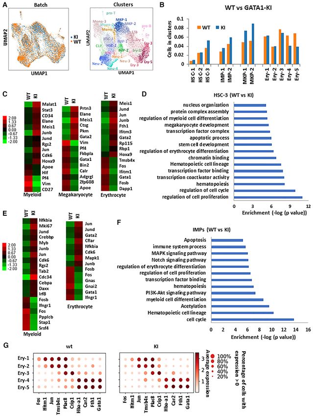
Nucleic Acids Research, 2021, Vol. 49, No. 17 9785

                                                                                                                                                               Downloaded from https://academic.oup.com/nar/article/49/17/9783/6358684 by guest on 12 October 2021

Figure 1. GATA-1 can directly interact with HDAC1 and be deacetylated by HDAC1/2 but not the FOG-1 complex. (A) GST-GATA-1 was in vitro
acetylated by recombinant p300 and then incubated with increasing level of FOG-1 complex purified from MEL cells by immunoprecipitation with FOG-
1 antibody. The acetylation level of GATA-1 is evaluated by western blot with anti-acetyl-lysine antibody. BSA is as a control. (B) ac-histone was incubated
with increasing level of FOG1 complex. (C) ac-GATA-1 was incubated with increasing level of HDAC1. (D) GST or GST GATA-1 was incubated with 35 S-
labeled HDAC1. Associated HDAC1 were subjected to SDS-PAGE and autoradiography. (E) Schematic representation of GATA-1 and mutant constructs.
(F) GST tagged GATA-1 and mutant constructs were incubated with Flag tagged recombinant HDAC1. (G) Conservation of HDAC1 binding sites. Two
arginines which mediate HDAC1 binding are in red. Lysines in green are major acetylation sites at the linker region. (H) GST-GATA-1 or mutant were
incubated with recombinant HDAC1. The associated Flag-HDAC1 was subjected to western blot with Flag antibody. (I) GATA-1 and FOG-1 expression
plasmids were transfected into 293 cell and nuclear extracts were immunoprecipitated with anti GATA-1 antibody. (J) Acetylated GST-GATA-1 or mutant
were incubated with recombinant HDAC1 or BSA.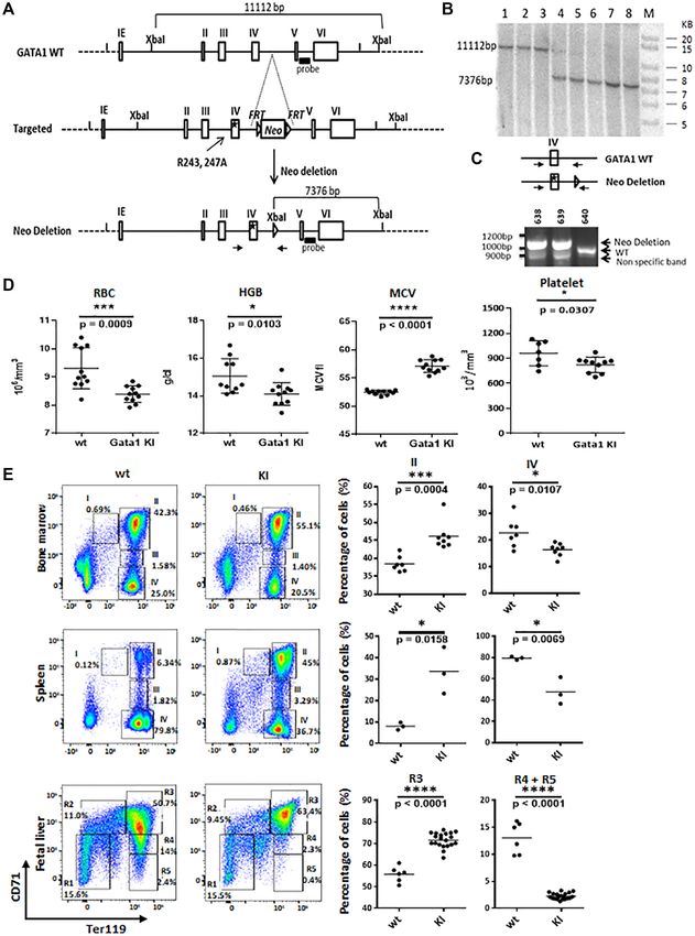
9786 Nucleic Acids Research, 2021, Vol. 49, No. 17

(Figure 1E, F). Through secondary structure analysis, po-        pressing wild type GATA-1 (Figure 2D, Supplemental Fig-
tential charged surface residues were mutated and two evo-       ure S2B). Similar effects were shown for the V205M mu-
lutionally conserved arginine residues at aa 243 and 247 of      tation, as previously observed (11,32) (Figure 2D, Supple-
GATA-1 were identified as being important for binding to         mental Figure S2B). The 2RA/V205M double mutant also
HDAC1 (Figure 1G). Mutating arginine to alanine at 243           prevents erythroid differentiation (Figure 2D, Supplemen-
and 247 (2RA mutant), significantly reduced the interaction      tal Figure S2B). Therefore, blocking GATA-1 and HDAC1
with HDAC1 (Figure 1H) without affecting GATA-1 DNA              direct interaction results in inhibition of GATA-1 mediated
binding in vitro, or its association with TAL1 (Supplemen-       erythroid differentiation in the G1E cellular model. We next
tal Figure S1C and D). The 2RA mutation is also defective        examined protein stability of 2RA mutant. The experiment
in HDAC2 binding (Supplemental Figure S1E), suggesting           shows that 2RA mutation results in the increase of protein
that both HDAC1 and HDAC2 bind to this linker region.            degradation after cycloheximide (CHX) treatment (Supple-
To investigate whether 2RA mutation affects interaction          mental Figure S2C, D). This result is consistent with previ-
with FOG-1, wild type GATA-1 and 2RA mutant, as well             ous finding that GATA-1 acetylation reduces protein stabil-

                                                                                                                                    Downloaded from https://academic.oup.com/nar/article/49/17/9783/6358684 by guest on 12 October 2021
as the FOG-1 binding defective GATA-1 V205M mutant,              ity (33).
were overexpressed with FOG-1 in HEK293 cells. GATA-1
or 2RA mutant, but not V205M, can co-precipitate FOG-            Blocking the HDAC1/GATA-1 direct interaction alters ex-
1(Figure 1I), indicating the 2RA mutation, which lost di-        pression patterns of both GATA-1 activated and repressed
rect interaction with HDAC1, is still able to interact with      genes
FOG-1. These results indicate that the 2RA mutant only
affects the direct interaction with HDAC1, but not the in-       We next performed RNA-seq to examine the effects of
teraction with FOG-1/NuRD. Furthermore, the GATA-1               disrupting HDAC1/GATA-1 direct interactions on global
V205M mutant, which is defective in FOG-1/NuRD bind-             gene expression in G1E GATA-1 2RA cells, compared to
ing, maintains in vitro binding to HDAC1 using nuclear           expression patterns in control G1E cells and G1E cells ex-
extract from murine erythroleukemic (MEL) cells (Supple-         pressing WT GATA-1 or the GATA-1 V205M mutant. A
mental Figure S1F). Finally, we tested whether the 2RA           total of 1685 genes exhibited at least two-fold differential
mutant affects GATA-1 deacetylation. The GATA-1 2RA              expression in G1E cells expressing WT GATA-1 compared
protein can be acetylated as the wild type protein, however,     to control G1E cells (Figure 3A) and, among these GATA-
the 2RA mutation blocks deacetylation (Figure 1J). There-        1 regulated genes, approximately one thousand genes were
fore, our results indicate that there are two distinct binding   affected by either the 2RA or the V205M mutation (Fig-
modes of GATA-1 to HDAC1: while the V205M mutation               ure 3B). The altered gene expression profile in GATA-1
affects indirect interaction with HDAC1/NuRD through             2RA cells is largely different from that of cells expressing
FOG-1, the 2RA mutation blocks direct binding to HDAC1           GATA-1 V205M, suggesting that GATA-1 regulates a dis-
and subsequent GATA-1 deacetylation.                             tinct set of target genes by differentially interacting directly
                                                                 with HDAC1/2 or via FOG-1 associated NuRD complex
                                                                 (Figure 3A–C). We further narrowed down the gene list
HDAC1/GATA-1 direct interaction is required for GATA-1
                                                                 to GATA-1 directly regulated genes that possess GATA-1
mediated erythroid differentiation
                                                                 binding sites in their enhancers/promoters (34). Over 30%
In order to test the role of FOG-1 independent HDAC1             of the GATA-1 bound genes in the G1E GATA-1 cells was
recruitment in GATA-1 mediated erythroid differentia-            affected by the 2RA mutation, whilst over 40% of the genes
tion, we carried out functional complementation assays in        were affected by the V205M mutation. Within these affected
the GATA-1 null G1E proerythroblastic cell line, a well-         genes, about half of them are differentially affected by the
established in vitro model system to study terminal ery-         2RA and V205M mutations (Figure 3D, Supplemental Fig-
throid differentiation following restoration of GATA-1 ex-       ure S3A). We further analyzed pathways affected by these
pression, for example, by retroviral transduction (29,30).       mutations. Thus, the 2RA mutation affects genes involved
We stably expressed in G1E cells estrogen inducible GATA-        in pathways regulating glucose metabolism and translation
1 constructs by fusing an estrogen receptor ligand binding       initiation (Figure 3E and F), whereas V205M affects nu-
domain to wild type GATA-1, V205M or 2RA mutants                 cleotide metabolism (Figure 3G). These results suggest the
and to the 2RA/V205M double mutant (Figure 2A, Supple-           non-redundant functions of these two interaction domains
mental Figure S2A). Cells expressing GATA-1 2RA showed           of GATA-1 in erythropoiesis.
increased GATA-1 acetylation, as this mutant is defective           There are a total of 315 GATA-1 direct target genes that
in HDAC1 binding (Figure 2B). Immunoprecipitation as-            are affected by both GATA-1 2RA and V205M, such as
says showed that the 2RA/V205M double mutant has un-             the beta-globin, GATA-2 or cKit genes (Figure 3D, Sup-
detectable interaction with HDAC1, compared to reduced           plemental Figure S3B). The majority of genes in this cat-
HDAC1 interaction in the 2RA mutant (Figure 2C), con-            egory are GATA-1 activated genes and are down regu-
firming the previous result that HDAC1 interacts with two        lated by both 2RA and V205M mutation (Figure 3H). Sim-
distinct motifs of GATA-1.                                       ilarly, within GATA-1 repressed genes which is up regu-
   Given that GATA-1 acetylation is important for the bind-      lated by 2RA, are also upregulated by the V205M mu-
ing of GATA-1 to chromatin and subsequent GATA-1                 tation with only a few exceptions (Figure 3H). Pathway
transcriptional activation (20,31), we anticipated that the      analysis shows that both interacting domains are impor-
GATA-1 2RA mutant would promote erythroid differenti-            tant for the regulation of cell cycle, apoptosis, heme bio-
ation. Surprisingly, we found that the 2RA mutation blocks       genesis and histone deacetylation (Figure 3I, J). Therefore,
erythroid differentiation when compared to G1E cells ex-         the two mutations, which abolish direct or indirect inter-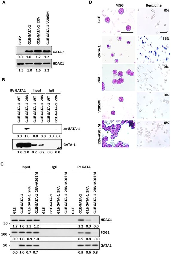
Nucleic Acids Research, 2021, Vol. 49, No. 17 9787
Downloaded from https://academic.oup.com/nar/article/49/17/9783/6358684 by guest on 12 October 2021
Figure 2. HDAC1/GATA-1 direct interaction is required for erythrocyte differentiation. ER-GATA-1 wt, 2RA, V205M and 2RA + V205M mutants were
cloned into a retroviral vector and stably expressed in G1E2 cells. Single clones were selected. Cells were treated with estradiol for 72 h. (A) The expression
of nuclear GATA-1 from G1E cells was examined by western blot. The band intensity was quantified by ImageJ. (B) Cell extracts were immunoprecipitated
with GATA-1 and western blotted with anti-acetyl-lysine and GATA-1. (C) G1E2 cells expressing ER-GATA-1 wt, 2RA and 2RA/V205M mutants were
collected after 72 h estradiol induction. Cell extracts were immunoprecipitated with anti GATA-1 antibody with cross link IP method. The resulting
products were subjected to western blot with indicated antibodies. (D) G1E cells described above were stained with MGG or Benzidine. The percentage of
hemoglobin-positive cells is indicated in the upper right corner of each Benzidine stain panel. Bar = 35 M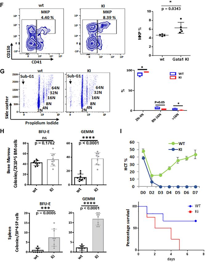
9788 Nucleic Acids Research, 2021, Vol. 49, No. 17

                                                                                                                                                         Downloaded from https://academic.oup.com/nar/article/49/17/9783/6358684 by guest on 12 October 2021

Figure 3. Differential gene expression regulation by V205M and 2RA mutants of GATA-1 in G1E cells. (A) A heat map showing genes differentially
expressed (over two fold) in G1E cells that stably express GATA-1, V205M or 2RA. (B) A Venn diagram showing overlapping and unique gene expression
pattern in G1E cells expressing GATA-1 V205M and 2RA. (C) Heatmap shows top changed genes differentially regulated by 2RA and V205M mutants.
(D) A Venn diagram showing GATA-1 direct target genes expression pattern in G1E cells expressing GATA-1 V205M and 2RA. (E) Enriched pathway in
2RA only differentially regulated genes. (F) GSEA shows 2RA only differentially regulated genes enriched in Eukaryotic translation Initiation pathway.
Heatmap shows differential gene expression pattern with 2RA and V205M. (G) Enriched pathway in V205M only differentially regulated genes. (H) Venn
diagrams showing GATA-1 up (left) and down (right) regulated genes in 2RA and V205M mutant cells compared to wild type. (I) Enriched pathway in
overlapped genes both changed in 2RA and v205M compared with wild type. * RUNX1 regulates genes involved in megakaryocyte differentiation and
platelet function. (J) GSEA shows overlapped genes enriched in cell cycle mitotic pathway. Heatmap shows similar gene expression pattern with 2RA and
V205M.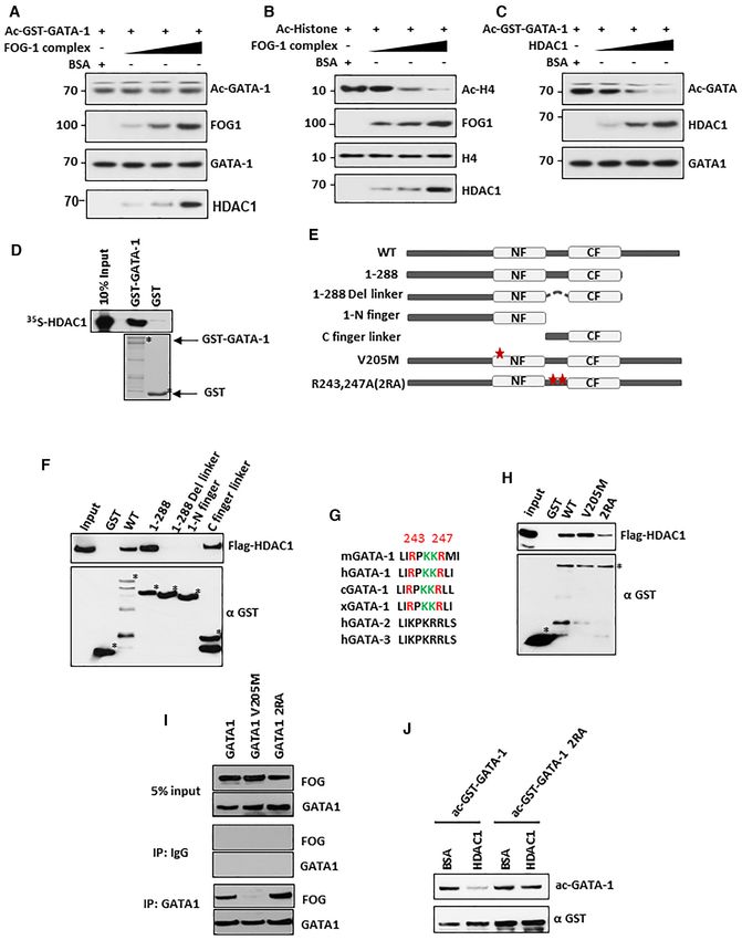
Nucleic Acids Research, 2021, Vol. 49, No. 17 9789
action with HDAC1, play distinct and overlapping roles in development, GATA-12RA knock-in mice were generated
GATA-1 mediated gene regulation, possibly through regu- using a GATA-1 2RA mutated homologous recombina-
lating GATA-1 deacetylation and chromatin deacetylation, tion targeting vector. The mice were generated by InGe-
respectively. nious Targeting Laboratory (Ronkonkoma, NY). The tar-
geting strategy is illustrated in Figure 5A. Briefly, the argi-
The HDAC1/GATA-1 direct interaction is important for nine residues at aa 243 and 247 of GATA-1 are converted
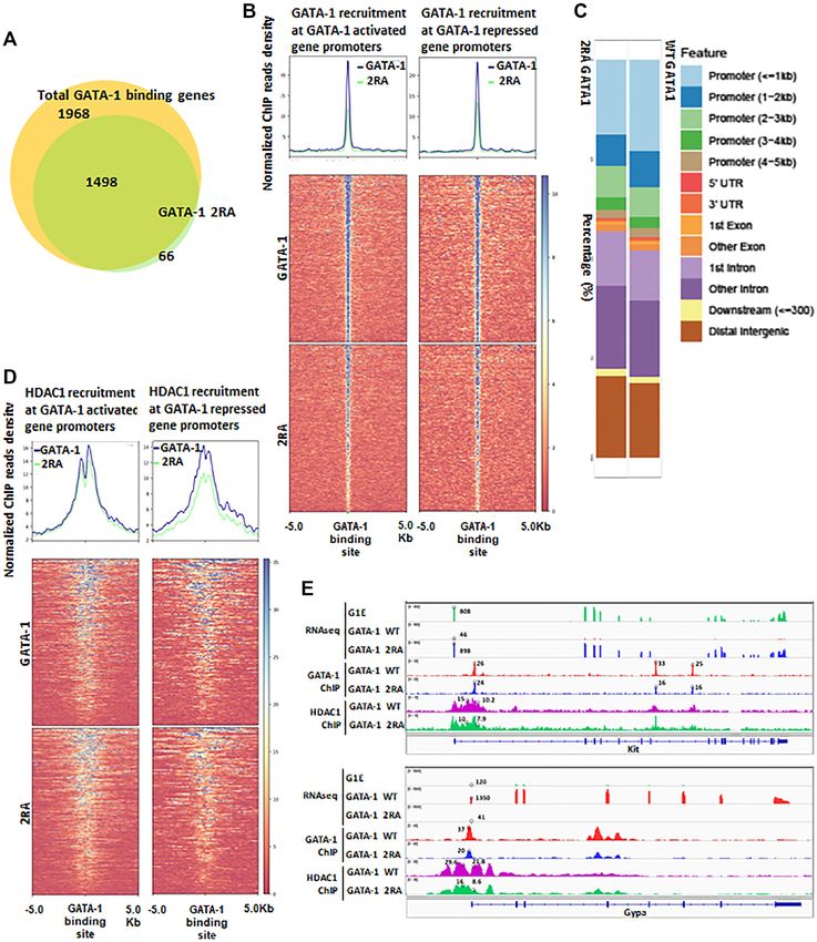
GATA-1 and HDAC1 recruitment to GATA-1 binding sites to alanine, with an FRT neo cassette for positive selection
inserted between exon IV and exon V. The targeting vec-
It has been shown that mutations in GATA-1 acetylation tor was linearized and then transfected by electroporation
sites reduce chromatin binding (20,31). Since the 2RA mu- in HF4 (129/SvEv × C57Bl/6) (Hybrid) embryonic stem
tant blocks GATA-1 deacetylation, it is conceivable that (ES) cells. The correct clones were identified through South-
the mutation will affect GATA-1 binding to targeted chro- ern blot analysis (Figure 5B). Targeted GATA-12RA ES cell
matin sites. We therefore performed ChIP-seq analysis to clones were injected into C57Bl/6 wild-type blastocysts to
Downloaded from https://academic.oup.com/nar/article/49/17/9783/6358684 by guest on 12 October 2021
analyze global binding patterns of GATA-1 2RA, as well generate GATA-12RA /wild-type chimeras. Since GATA-1
as its impact on HDAC1 recruitment. The 2RA mutation is located on the X chromosome, resulting male chimeras
abolished over 20% of GATA-1 binding (Figure 4A). For with a high percentage of agouti coat color were mated to
the sites where GATA-1 2RA remained bound, the bind- C57BL/6 WT female mice to generate germline GATA-1
ing activity was largely reduced compared to wild type 2RA knock-in hemizygote mice (Figure 5C). The GATA-1
GATA-1 (Figure 4B, Supplemental Figure S4A). Interest- 2RA knock-in allele displayed Mendelian inheritance in all
ingly, GATA-1 2RA binding is reduced in both GATA-1 offspring, indicating that the 2RA mutation does not cause
activated and repressed genes, consistent with the reduction any embryonic survival defects (Supplemental Figure S5A).
of GATA-1 mediated gene activation and repression from Peripheral blood analysis reveals that GATA-12RA
RNA seq results (Figures 3H and 4B). Although GATA- knock-in mice suffered from mild anemia and throm-
1 2RA binding is reduced at all GATA binding motifs in bocytopenia with low red cell count, low hemoglobin
different genomic regions (Supplemental Figure S4B), the and low platelet count in both genders (Figure 5D, Sup-
most profound reduction of binding was observed in prox- plemental Figure S5B). We therefore analyzed the ery-
imal promoter regions near TSSs (Figure 4C, Supplemen- throid and megakaryocyte differentiation profiles. Bone
tal Figure S4C). HDAC1 ChIP-seq shows that HDAC1 is marrow and spleen cells were stained with erythro-
recruited to GATA-1 binding sites and the 2RA mutation cyte differentiation markers CD71 and Ter119 and ana-
reduces HDAC1 recruitment at these sites (Supplemental lyzed by FACS (36,37) (Figure 5E). Differentiation and
Figure S4D). Furthermore, HDAC1 is recruited to both ac- maturation of erythroid progenitors consist of several
tivated and repressed gene promoters at comparable levels populations of erythroid cells, including proerythrob-
in WT G1E GATA-1 cells and, accordingly, the reduction lasts and early basophilic erythroblasts (I, CD71high ,
of HDAC1 recruitment by GATA-1 2RA is seen in both TER119low ), early and late basophilic erythroblasts (II,
GATA-1 activated and repressed genes (Figure 4D and E, CD71high , TER119high ), polychromatophilic erythroblasts
Supplemental Figure S4E). However, GATA-1 repressed (III, CD71med , TER119high ) and orthochromatophilic ery-
genes exhibit a greater reduction in HDAC1 binding com- throblasts (IV, CD71low , TER119High ) (36,38). In bone mar-
pared to activated genes (Figure 4D). These results indicate row, the population of CD71high , TER119high erythrocytes
that the GATA-1/HDAC1 interaction is required in both (Fraction II), was increased in KI mice compared to the
activated and repressed genes. wild type. The later stage Ortho EBs (IV) were decreased
Histone acetylation is associated with activated genes at in KI mice (Figure 5E, top panel) indicating a block in ery-
GATA-1 binding sites and is negatively associated with re- throid differentiation. The analysis of spleen also shows an
pressed genes (35) (Supplemental Figure S4F). We then ex- increase of premature erythroblasts and a decrease of nor-
amined how 2RA affects histone acetylation at GATA-1 ac- mal mature erythrocytes (Figure 5E, middle panel), result-
tivated and repressed gene promoters. The -globin gene ex- ing in splenomegaly (Supplemental Figure S5C). These re-
pression is not activated in GATA-1 2RA expressing cells sults indicate that the 2RA mutation causes a defect in ery-
(Supplemental Figure S3B). However, loss of HDAC1 in- thropoiesis in the bone marrow and spleen. Notably, there is
teraction in 2RA mutation results in the reduction of his- a significant increase of spleen size due to increased splenic
tone acetylation on -globin gene locus (Figure 4F). In con- erythropoiesis (extramedullary hematopoiesis) in an effort
trast, GATA-2 gene expression is not repressed by GATA-1 to compensate for the defect in bone marrow erythropoiesis
2RA (Supplemental Figure S3B). Histone acetylation level (Supplemental Figure S5C and D). We also examined ery-
remain high in GATA-2 gene locus (Figure 4G). Thus, loss thropoiesis in the fetal liver to investigate GATA-12RA ef-
of HDAC1 binding does not consistent with histone acety- fects in earlier developmental states. As expected, erythroid
lation level at gene locus. This may suggest that the recruit- progenitor cells from fetal liver fail to produce sufficient ma-
ment of HDAC1 by GATA-1 serves mainly for GATA-1 ture erythroid cells (Figure 5E, bottom panel).
deacetylation. We then characterized the megakaryocytic differentia-
tion defect in the GATA-12RA mice. Bone marrow cells
were stained with megakaryocytic differentiation markers
GATA-1 2RA knock-in mice suffers from anemia and throm-
CD41 and CD150 and analyzed by FACS. The KI mice
bocytopenia
exhibit more CD41/CD150 positive cells, which represent
In order to more comprehensively understand the role of megakaryocyte progenitor (MKP) cells (39) (Figure 5F).
the GATA-1 /HDAC1 interaction in hematopoietic lineage Furthermore, the knock-in mice have reduced polyploidiza-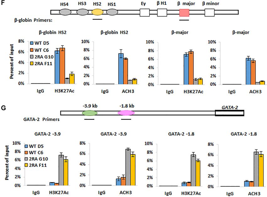
Downloaded from https://academic.oup.com/nar/article/49/17/9783/6358684 by guest on 12 October 2021
9790 Nucleic Acids Research, 2021, Vol. 49, No. 17
Nucleic Acids Research, 2021, Vol. 49, No. 17 9791
Downloaded from https://academic.oup.com/nar/article/49/17/9783/6358684 by guest on 12 October 2021
Figure 4. Disruption of GATA-1/HDAC1 interaction resulting in the decrease of chromatin recruitment of GATA-1. (A) Venn diagram shows G1E cells
expression GATA-1 2RA has reduced binding on GATA-1 binding genes compare to GATA-1 WT expressing cells. (B) ChIP-seq data shows disrupting of
GATA-1/HDAC1 interaction globally reduced GATA-1 chromatin recruitment at GATA-1 activated and repressed gene promoters. (C) Bar graph shows
GATA-1 or mutant binding distribution. (D) ChIP-seq data shows HDAC1 chromatin recruitment at GATA-1 activated and repressed gene promoters in
GATA-1 2RA mutant cells compare to WT. (E) Track view shows gene expression and chromatin recruitment at GATA-1 repressed gene kit and GATA-1
activated gene Gypa. (F and G) ChIP assay on H3K27AC or H3 acetylation at -major and -globin HS2 locus (F) and GATA-2 enhancers (G). WT D5
and WT C6 are two clones expressing wild type GATA-1, 2RA G10 and 2RA F11 are two single cell clones expressing GATA-1 2RA in G1E cells.
tion (Figure 5G), thus showing that the 2RA mutation potential of the mice to recover from anemia. All GATA-
blocks megakaryocyte differentiation at the MKP stage and 12RA mice died within 5 days after the administration of
resulting in the reduction of mature megakaryocytes. phenyl hydrazine, whereas 75% of wild-type littermates
To examine the effect of GATA-12RA in hematopoietic made a full recovery (Figure 5I). These results indicate that
stem and progenitor cells (HS/PCs), we further assessed the GATA-1 sites required for interaction with HDAC1 are
the frequencies of colony-forming unit cells (CFU-C) in the critical for GATA-1-mediated stress hematopoiesis.
bone marrow and spleen cells of WT and GATA-12RA mice
(40,41). Myeloid progenitor CFU-GEMM colonies were
GATA-12RA altered HS/PC hierarchy and inhibited
increased in the bone marrow and spleen culture in GATA-
erythroid/megakaryocytic differentiation trajectory
12RA mice (Figure 5H), indicating a compensatory expan-
sion of progenitor cells. Although there was no statistically We wished to obtain a comprehensive view of the role of the
significant difference in the number of erythroid colony- HDAC1/GATA-1 direct interaction in lineage commitment
forming units (BFU-E) in the bone marrow, the number was and differentiation. We thus performed single cell RNA-seq
increased several fold in the spleens of GATA-12RA mice analysis using Lin− cKit+ (LK) bone marrow cells which
(Figure 5H). These observations support the notion that are enriched in myeloid progenitors, to study the impact
the 2RA mutation blocks differentiation of hematopoietic of GATA-12RA on GATA-1 mediated transcription coher-
progenitors and demonstrate that there is substantial extra- ent state of HS/PC and myeloid subpopulations. Single cell
medullary erythropoiesis occurring in GATA-12RA mice. transcription profiles of over 6000 cells were analyzed from
We next investigated the ability of GATA-12RA mice to re- both wild type and GATA-12RA mice.
cover under hematopoietic stress conditions. We challenged A total of 24 transcriptional clusters were identified on
wild-type and GATA-12RA mice with phenyl hydrazine, a the basis of expressed genes previously reported (42–44)
chemical that induces hemolytic anemia, and assayed the (Figure 6A). The 2RA KI increased the frequencies of spe-Downloaded from https://academic.oup.com/nar/article/49/17/9783/6358684 by guest on 12 October 2021 9792 Nucleic Acids Research, 2021, Vol. 49, No. 17
Nucleic Acids Research, 2021, Vol. 49, No. 17 9793

                                                                                                                                                                Downloaded from https://academic.oup.com/nar/article/49/17/9783/6358684 by guest on 12 October 2021
Figure 5. Disruption of GATA-1/HDAC1 interaction leads to hematologic defect in knock in mice, and affects erythropoietic stress response. (A) Schematic
diagram of the targeting strategy for GATA12RA knock-in allele. The arginine residue at aa 243 and 247 of GATA-1 is converted to alanine and FRT neo
cassette for positive selection was inserted between exon IV and exon V. The targeting vector was linearized and then transfected by electroporation of
HF4 (129/SvEv × C57Bl/6) (Hybrid) embryonic stem cells. After selection with G418 antibiotic, surviving clones were expanded for PCR analysis to
identify recombinant ES clones. The Neo cassette in targeting vector has been removed during ES clone expansion. (B) Targeting vector was transfected
by electroporation of HF4 (129/SvEv x C57Bl/6) (Hybrid) embryonic stem cells. After selection with G418 antibiotic, surviving clones were expanded.
Positive clones were further confirmed by Southern Blotting analysis using an internal probe. Clones 4, 5, 6, 7 and 8 were confirmed as correctly targeted.
(C) Screening for hemizygous male mice. Primer set as indicated was used to screen mice for the deletion of the Neo cassette. The PCR product for the wild-
type is 933 bp. After Neo deletion, one FRT site remains (85 bp). A second band with a size of 1018 indicates Neo deletion. (D) Hematocrit of peripheral
blood from Gata1 WT and 2RA mutant KI mice. (E) FACS analysis of bone marrow, spleen and fetal liver cells from the Gata1 WT and 2RA KI mice (left).
The relative number in each region as a percentage of gated cells is indicated and summarized in the right panels. (F) Mouse bone marrow cells were first
gated for Lin– ckit+ scal– population. Then CD41+ CD150+ population was gated as MKP. Percentages of MKP cell population were summarized in the
right panel. (G) Mouse spleen cells were stain with CD41 antibody and PI. CD41 positive cells were first gated then plot for PI and side scatter. Percentages
of cell population were summarized in the right panel. n = 5 (H) Colony forming assays performed with bone marrow (top) and spleen (bottom) cells. (I)
Top: Hematocrit in the blood of wild-type (WT) and 2RA knock in (KI) mice during recovery from PHZ-induced (35 mg/kg) anemia. One representative
experiment is shown. Results are shown as mean ± SD; n = 2. Bottom: Survival curve of WT and KI mice following single injection of 35 mg/kg PHZ.
n = 6 for each group.
9794 Nucleic Acids Research, 2021, Vol. 49, No. 17
Downloaded from https://academic.oup.com/nar/article/49/17/9783/6358684 by guest on 12 October 2021
Figure 6. Single-cell RNA-sequencing data integration of batches and clustering to analyze WT and Gata1-KI LK cells. (A) Uniform Manifold Approx-
imation and Projection (UMAP) two-dimensional visualization of WT and KI cells isolated from Bone marrow LK cells with the integration of batches
analysis. Different colors represent 24 clusters (cell types) defined by the k-means clustering algorithm. IMP, immature myeloid progenitor; Mono, mono-
cyte progenitor; Neu, neutrophil/granulocyte progenitor; E/B, erythroid/basophil progenitor; Ery, erythroid progenitor; MkP, megakaryocyte progenitor;
CLP, common lymphoid progenitor; Eo, eosinophil progenitor; pro-B, B-cell progenitor; pro-T, T-cell progenitor. (B) Relative cell percentage in clusters in
WT and KI samples. (C) Hierarchical clustering heatmap of differential expression genes in HSC-3 cluster comparing WT and GATA1-KI cells. (D) Top
GO terms enrichment in HSC-3 cluster comparing WT and GATA1-KI cells. (E) Hierarchical clustering heatmap of differential expression genes in IMP
cluster comparing WT and GATA1-KI cells. (F) Top GO terms enrichment in IMP cluster comparing WT and GATA1-KI cells. (G) Top differentially
expressed genes (FDR < 0.05, logistic regression with Bonferroni correction) and GATA-1 gene expression when comparing each cell cluster with the
remaining clusters corresponding to Ery-pro clusters in WT and KI cells. The dot size encodes the fraction of cells within the cluster that show detectable
expression of the gene (UMIs > 0), while the color encodes the average expression level across all cells within a cluster.Nucleic Acids Research, 2021, Vol. 49, No. 17 9795
cific cell clusters, including the HSC-3 cluster and the im- the two zinc fingers of GATA-1. Our study shows that the
mature myeloid progenitor (IMP) clusters IMP1 and IMP2 interaction between GATA-1 and HDAC1 is required for
(Figure 6B). The increase of these populations may reflect a the deacetylation of GATA-1 and for GATA-1 mediated
compensatory mechanism for the reduction of mature ery- erythroid/megakaryocytic differentiation. Introduction of
throcytes and megakaryocytes (Figure 6B). a mutation that cripples the GATA-1/HDAC1 direct inter-
The gene expression heatmap shows that the HSC-3 clus- action (2RA mutant), results in GATA-1 constitutive acety-
ter exhibits myeloid bias as it expresses signature genes lation and impairing erythroid differentiation in G1E cells,
for myeloid progenitors, megakaryocytic progenitors and by blocking both GATA-1 activated and repressed genes.
erythroid progenitors, in addition to HSC signature genes GATA-1 acetylation has been previously studied and, al-
(44) (Figure 6C, Supplemental Figure S6A). The HSC- though it is generally considered that it promotes chromatin
3 associated myeloid biased gene signatures were reduced binding and activation (2,18–20), GATA-1 acetylation was
in GATA-12RA cells, while the HSC signature and HSC-3 also shown to positively target GATA-1 for degradation
cluster frequency were markedly enhanced and expanded and subsequent suppression of GATA-1 transcriptional ac-
Downloaded from https://academic.oup.com/nar/article/49/17/9783/6358684 by guest on 12 October 2021
(Figure 6B, C Supplemental Figure S6A). GO term analy- tivity (33). In this study, 2RA mutation results constitu-
sis of differentially expressed genes in HSC-3, consistently tive acetylation and the protein in deed is less stable com-
showed the down regulation of myeloid cell differentia- pare to the wild type protein. Consistent with previous re-
tion and erythrocyte/megakaryocyte differentiation (Fig- ported (19), GATA-1 2RA mutant protein binds to DNA
ure 6D). It has been reported that GATA-1 is expressed in similarly to the wild type protein in vitro. However, chro-
some populations of HSC and directs transcription prim- matin binding is significantly reduced on a genome-wide
ing of myeloid biased differentiation (44,45). Our data sup- scale. These defects may either be due to the reduction of
port the notion that GATA-1 is expressed in the myeloid GATA-1 protein level, constitutive acetylation of GATA-1,
biased HSC-3 cluster and further showed that GATA-12RA or due to loss of interaction with HDAC1. Although our
inhibited the myeloid potential of the HSC-3 cluster by de- preliminary studies show that there is no cofactor associ-
repressing GATA-2 (Figure 6C). This is consistent with a ated with HDAC1 at this site, uncovering the detailed mech-
change in chromatin binding and coactivator recruitment anism underlying contribution of GATA-1 acetylation sta-
(Figure 4B and D). The GATA-12RA derived HSC-3 clus- tus to its chromatin binding and transcriptional regulation
ter also shows altered transcription profiles of genes associ- in hematopoiesis, warrants further investigation. Further-
ated with the cell cycle, apoptosis and cell survival (Supple- more, it remains to be determined whether the FOG-1 in-
mental Figure S6B). Accordingly, GATA-12RA , enhanced teracting surface in the N-terminal GATA1 zinc finger and
and expanded IMP signatures and frequencies, at the same the HDAC1 interacting site cooperate to regulate GATA-1
time inhibiting erythroid signatures and potential (Figure mediated transcription in hematopoiesis.
6E). GO term analysis on differentially expressed genes in The ChIP-seq data shows that HDAC1 is recruited to
the IMP1 and IMP2 clusters, supported the notion that the active promoters by GATA-1 and the level of HDAC1 on
GATA-1 2RA mutation results in inhibition of myeloid and promoters are positively correlated to high levels of histone
erythroid differentiation (Figure 6F). acetylation. Therefore, it is conceivable that HDAC1 may
Frequencies of megakaryocyte progenitor cells (MKP1, not play a role in histone deacetylation at active promot-
MKP2) are also increased in GATA-1 2RA KI mice with ers but, instead, it may deacetylate GATA-1 or other pro-
accompanying changes in the expression levels of signature tein substrates at the promoter region. This data is consis-
genes (Figure 6B, Supplemental Figure S6C), again consis- tent with an earlier report showing that highly active genes
tent with a compensatory effect observed in FACS analysis associate with high levels of HATs and HDACs, including
(Figure 5D). The frequencies of erythroid clusters 4 and 5 HDAC1 (46), as well as high levels of histone acetylation
(Ery4, Ery5) were decreased in GATA-12RA mice (Figure at promoter regions (47). Since HDAC1 and HAT can dy-
6B). These two clusters represent more mature erythrocytes namically compete for the binding to histone H3 tail (48),
as they both express high level of alpha-globin (Hba-a1) we speculate that HDAC1 at active gene promoters may
and GATA-1 (Figure 6G). The GATA-1 2RA mutation re- be important for balancing HAT activity in order to main-
duced Hba-a1 gene and Gata-1 expression, indicating an in- tain gene activation at a desired magnitude (46). In addi-
hibition of terminal erythroid differentiation (Figure 6G). tion, HAT can attenuate HDAC1 activity by acetylating
Taken together, our data demonstrate that the GATA-1 HDAC1. Our earlier studies showed that HDAC1 is grad-
deacetylation by HDAC1 regulates specific HS/PC popu- ually acetylated during erythropoiesis and that the acety-
lations, erythroid and megakaryocytic differentiation pro- lated NuRD complex positively regulates GATA-1 func-
grams by controlling GATA-1 transcription activity and tion (15). Whether HDAC1 itself is acetylated when it di-
GATA-1 mediated change of chromatin landscape (Figure rectly binds to the GATA-1 linker site to subsequently mod-
7). ulate GATA-1 activity, remains to be determined. In con-
junction with this point, it remain to be established whether
DISCUSSION GATA-1 acetylation/deacetylation is regulated during ery-
thropoiesis, however our present data suggests it does.
In this study, we found that although the NuRD complex The GATA-12RA knock-in mice suffered from mild ane-
contains active HDAC1/2, it does not deacetylate GATA- mia and thrombocytopenia, a phenotype also seen in FOG-
1. However, HDAC1/2 on their own can efficiently deacety- 1 KI mice in which the FOG-11/NuRD interaction was dis-
late GATA-1. Further study led to the identification of a rupted, suggesting multiple layers of regulation of GATA-
direct interaction between GATA-1 and HDAC1 mediated 1 function (14,49,50). The bone marrow and spleen of
through a novel interacting site at the linker region between GATA-12RA mice showed an accumulation of immature9796 Nucleic Acids Research, 2021, Vol. 49, No. 17
Downloaded from https://academic.oup.com/nar/article/49/17/9783/6358684 by guest on 12 October 2021
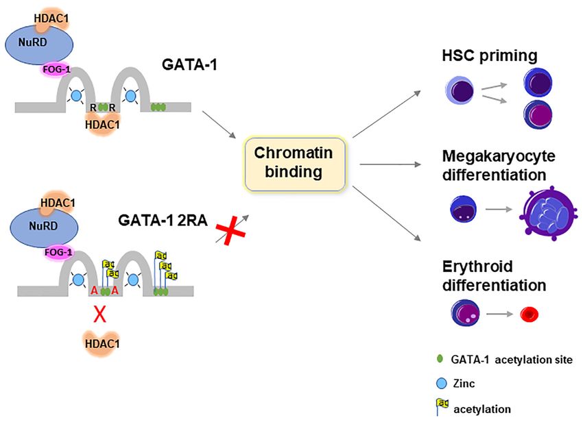
Figure 7. Graphic illustration of function of HDAC1 in regulating GATA-1 function. HDAC1 and GATA-1 direct interaction at the linker region mediates
GATA-1 deacetylation. The mutant of GATA-1 that is defective in HDAC1 binding is constitutively acetylated and resulting reduction of chromatin binding
at GATA-1 target genes and blockage of GATA-1 mediated gene transcription. In development, loss of HDAC1 binding results in reduction of myeloid
biased HS/PC, defective megakaryocyte differentiation and erythroid differentiation.
erythrocytes and megakaryocytes, whereas the single cell scRNA-seq dataset has been deposited to GEO (accession
RNA-seq analysis from mice bone marrow LK cells re- number GSE161729). The ChIP-seq dataset has been de-
vealed profound changes in HSC, myeloid progenitor and posited to GEO (accession number GSE161608).
erythroid/megakaryocyte lineage cell populations and ac-
companying signature gene expression. GATA-1 gene ex-
pression is reduced in all of these populations. These obser- SUPPLEMENTARY DATA
vations suggest that bone marrow progenitors in GATA- Supplementary Data are available at NAR Online.
12RA mice fail to give rise to sufficient numbers of mature
erythrocytes and megakaryocytes. Therefore, the GATA-1
/HDAC1 interaction is essential for GATA-1 deacetylation ACKNOWLEDGEMENTS
and hematopoiesis. Given that the FOG-1 complex also
The authors thank Wei Jian and Ruiqing Zhou for excellent
contains HDAC1, it is possible that FOG-1- dependent and
technical support.
independent HDAC1s coordinate GATA-1 acetylation and
Author contributions: B.Y. performed most of the exper-
chromatin acetylation status in the GATA-1 target genes in
iments; J.Y. and N.C. performed some experiments on
hematopoiesis.
knock-in mice, M.K. and T.Y. performed some of the ex-
It has been reported that GATA-1 is expressed in HS/PCs
periments; H.L. contributed to analysis of ChIP-seq and
(45). However, the role of GATA-1 in HS/PC develop-
scRNA-seq database; and J.Z., J.S., R.H, S.H. and Y.Q.
ment remain elusive. Our study demonstrates that GATA-
conceived the project, designed experiments, interpreted re-
1 specifically regulates the transcription state of a subpop-
sults and wrote the manuscript.
ulation of HS/PCs that exhibit myeloid bias. This cluster
of HS/PCs may represent short term HSCs, expressing sig-
nature genes for myeloid progenitors, erythroid progenitors FUNDING
and megakaryocyte progenitors. The expression of GATA-
12RA resulted in increased GATA-2 expression which, in National Institute of Health [R01HL144712 to Y.Q.,
turn, may result in cells remaining in a stemness state, with R01DK110108, R01HL141950 to S.H., R24DK106766 to
reduced expression of myeloid/erythroid biased signature R.H.]; Four Diamonds Fund (to S.H.). Funding for open
genes in HS/PCs. Thus, the role of GATA-1 in HS/PC func- access charge: NHLBI.
tion warrants further investigation. Conflict of interest statement. None declared.
DATA AVAILABILITY REFERENCES
1. Ferreira,R., Ohneda,K., Yamamoto,M. and Philipsen,S. (2005)
RNA-seq data generated in this study are available at NCBI GATA1 function, a paradigm for transcription factors in
GEO database with the accession number GSE161607. The hematopoiesis. Mol. Cell. Biol., 25, 1215–1227.Nucleic Acids Research, 2021, Vol. 49, No. 17 9797
2. Blobel,G.A., Nakajima,T., Eckner,R., Montminy,M. and Orkin,S.H. Differential gene and transcript expression analysis of RNA-seq
(1998) CREB-binding protein cooperates with transcription factor experiments with TopHat and Cufflinks. Nat. Protoc., 7, 562–578.
GATA-1 and is required for erythroid differentiation. Proc. Natl 24. Butler,A., Hoffman,P., Smibert,P., Papalexi,E. and Satija,R. (2018)
Acad. Sci. U.S.A., 95, 2061–2066. Integrating single-cell transcriptomic data across different conditions,
3. Bresnick,E.H., Lee,H.Y., Fujiwara,T., Johnson,K.D. and Keles,S. technologies, and species. Nat. Biotechnol., 36, 411–420.
(2010) GATA switches as developmental drivers. J. Biol. Chem., 285, 25. Langmead,B. and Salzberg,S.L. (2012) Fast gapped-read alignment
31087–31093. with Bowtie 2. Nat. Methods, 9, 357–359.
4. Crispino,J.D. (2005) GATA1 in normal and malignant 26. Zhang,Y., Liu,T., Meyer,C.A., Eeckhoute,J., Johnson,D.S.,
hematopoiesis. Semin. Cell Dev. Biol., 16, 137–147. Bernstein,B.E., Nusbaum,C., Myers,R.M., Brown,M., Li,W. et al.
5. Kerenyi,M.A. and Orkin,S.H. (2010) Networking erythropoiesis. J. (2008) Model-based analysis of ChIP-Seq (MACS). Genome Biol., 9,
Exp. Med., 207, 2537–2541. R137.
6. Wong,P., Hattangadi,S.M., Cheng,A.W., Frampton,G.M., 27. Yu,G., Wang,L.G. and He,Q.Y. (2015) ChIPseeker: an
Young,R.A. and Lodish,H.F. (2011) Gene induction and repression R/Bioconductor package for ChIP peak annotation, comparison and
during terminal erythropoiesis are mediated by distinct epigenetic visualization. Bioinformatics, 31, 2382–2383.
changes. Blood, 118, e128–e138. 28. Juan,L.J., Shia,W.J., Chen,M.H., Yang,W.M., Seto,E., Lin,Y.S. and
7. Kastner,P. and Chan,S. (2008) PU.1: a crucial and versatile player in Wu,C.W. (2000) Histone deacetylases specifically down-regulate
Downloaded from https://academic.oup.com/nar/article/49/17/9783/6358684 by guest on 12 October 2021
hematopoiesis and leukemia. Int. J. Biochem. Cell Biol., 40, 22–27. p53-dependent gene activation. J. Biol. Chem., 275, 20436–20443.
8. Koschmieder,S., Rosenbauer,F., Steidl,U., Owens,B.M. and 29. Welch,J.J., Watts,J.A., Vakoc,C.R., Yao,Y., Wang,H., Hardison,R.C.,
Tenen,D.G. (2005) Role of transcription factors C/EBPalpha and Blobel,G.A., Chodosh,L.A. and Weiss,M.J. (2004) Global regulation
PU.1 in normal hematopoiesis and leukemia. Int. J. Hematol., 81, of erythroid gene expression by transcription factor GATA-1. Blood,
368–377. 104, 3136–3147.
9. Moreau-Gachelin,F., Wendling,F., Molina,T., Denis,N., Titeux,M., 30. Pilon,A.M., Ajay,S.S., Kumar,S.A., Steiner,L.A., Cherukuri,P.F.,
Grimber,G., Briand,P., Vainchenker,W. and Tavitian,A. (1996) Wincovitch,S., Anderson,S.M., Mullikin,J.C., Gallagher,P.G.,
Spi-1/PU.1 transgenic mice develop multistep erythroleukemias. Mol. Hardison,R.C. et al. (2011) Genome-wide ChIP-Seq reveals a
Cell. Biol., 16, 2453–2463. dramatic shift in the binding of the transcription factor erythroid
10. Steidl,U., Steidl,C., Ebralidze,A., Chapuy,B., Han,H.J., Will,B., Kruppel-like factor during erythrocyte differentiation. Blood, 118,
Rosenbauer,F., Becker,A., Wagner,K., Koschmieder,S. et al. (2007) A e139–e148.
distal single nucleotide polymorphism alters long-range regulation of 31. Lamonica,J.M., Deng,W., Kadauke,S., Campbell,A.E.,
the PU.1 gene in acute myeloid leukemia. J. Clin. Invest., 117, Gamsjaeger,R., Wang,H., Cheng,Y., Billin,A.N., Hardison,R.C.,
2611–2620. Mackay,J.P. et al. (2011) Bromodomain protein Brd3 associates with
11. Rodriguez,P., Bonte,E., Krijgsveld,J., Kolodziej,K.E., Guyot,B., acetylated GATA1 to promote its chromatin occupancy at erythroid
Heck,A.J., Vyas,P., de Boer,E., Grosveld,F. and Strouboulis,J. (2005) target genes. Proc. Natl Acad. Sci. U.S.A., 108, E159–E168.
GATA-1 forms distinct activating and repressive complexes in 32. Letting,D.L., Chen,Y.Y., Rakowski,C., Reedy,S. and Blobel,G.A.
erythroid cells. EMBO J., 24, 2354–2366. (2004) Context-dependent regulation of GATA-1 by friend of
12. Hong,W., Nakazawa,M., Chen,Y.Y., Kori,R., Vakoc,C.R., GATA-1. Proc. Natl Acad. Sci. U.S.A., 101, 476–481.
Rakowski,C. and Blobel,G.A. (2005) FOG-1 recruits the NuRD 33. Hernandez-Hernandez,A., Ray,P., Litos,G., Ciro,M., Ottolenghi,S.,
repressor complex to mediate transcriptional repression by GATA-1. Beug,H. and Boyes,J. (2006) Acetylation and MAPK
EMBO J., 24, 2367–2378. phosphorylation cooperate to regulate the degradation of active
13. Snow,J.W., Kim,J., Currie,C.R., Xu,J. and Orkin,S.H. (2010) GATA-1. EMBO J., 25, 3264–3274.
Sumoylation regulates interaction of FOG1 with C-terminal-binding 34. Cheng,Y., Wu,W., Kumar,S.A., Yu,D., Deng,W., Tripic,T., King,D.C.,
protein (CTBP). J. Biol. Chem., 285, 28064–28075. Chen,K.B., Zhang,Y., Drautz,D. et al. (2009) Erythroid GATA1
14. Miccio,A., Wang,Y., Hong,W., Gregory,G.D., Wang,H., Yu,X., function revealed by genome-wide analysis of transcription factor
Choi,J.K., Shelat,S., Tong,W., Poncz,M. et al. (2010) NuRD mediates occupancy, histone modifications, and mRNA expression. Genome
activating and repressive functions of GATA-1 and FOG-1 during Res., 19, 2172–2184.
blood development. EMBO J., 29, 442–456. 35. Hardison,R.C., Zhang,Y., Keller,C.A., Xiang,G., Heuston,E.F.,
15. Yang,T., Jian,W., Luo,Y., Fu,X., Noguchi,C., Bungert,J., Huang,S. An,L., Lichtenberg,J., Giardine,B.M., Bodine,D., Mahony,S. et al.
and Qiu,Y. (2012) Acetylation of histone deacetylase 1 regulates (2020) Systematic integration of GATA transcription factors and
NuRD corepressor complex activity. J. Biol. Chem., 287, epigenomes via IDEAS paints the regulatory landscape of
40279–40291. hematopoietic cells. IUBMB Life, 72, 27–38.
16. Qiu,Y., Zhao,Y., Becker,M., John,S., Parekh,B.S., Huang,S., 36. Socolovsky,M., Nam,H., Fleming,M.D., Haase,V.H., Brugnara,C.
Hendarwanto,A., Martinez,E.D., Chen,Y., Lu,H. et al. (2006) and Lodish,H.F. (2001) Ineffective erythropoiesis in Stat5a(-/-)5b(-/-)
HDAC1 acetylation is linked to progressive modulation of steroid mice due to decreased survival of early erythroblasts. Blood, 98,
receptor-induced gene transcription. Mol. Cell, 22, 669–679. 3261–3273.
17. Luo,Y., Jian,W., Stavreva,D., Fu,X., Hager,G., Bungert,J., Huang,S. 37. Fraser,S.T., Isern,J. and Baron,M.H. (2007) Maturation and
and Qiu,Y. (2009) Trans-regulation of histone deacetylase activities enucleation of primitive erythroblasts during mouse embryogenesis is
through acetylation. J. Biol. Chem., 284, 34901–34910. accompanied by changes in cell-surface antigen expression. Blood,
18. Boyes,J., Byfield,P., Nakatani,Y. and Ogryzko,V. (1998) Regulation of 109, 343–352.
activity of the transcription factor GATA-1 by acetylation. Nature, 38. Kalfa,T.A., Pushkaran,S., Zhang,X., Johnson,J.F., Pan,D., Daria,D.,
396, 594–598. Geiger,H., Cancelas,J.A., Williams,D.A. and Zheng,Y. (2010) Rac1
19. Hung,H.L., Lau,J., Kim,A.Y., Weiss,M.J. and Blobel,G.A. (1999) and Rac2 GTPases are necessary for early erythropoietic expansion in
CREB-Binding protein acetylates hematopoietic transcription factor the bone marrow but not in the spleen. Haematologica, 95, 27–35.
GATA-1 at functionally important sites. Mol. Cell. Biol., 19, 39. Guo,Y., Niu,C., Breslin,P., Tang,M., Zhang,S., Wei,W., Kini,A.R.,
3496–3505. Paner,G.P., Alkan,S., Morris,S.W. et al. (2009) c-Myc-mediated
20. Lamonica,J.M., Vakoc,C.R. and Blobel,G.A. (2006) Acetylation of control of cell fate in megakaryocyte-erythrocyte progenitors. Blood,
GATA-1 is required for chromatin occupancy. Blood, 108, 3736–3738. 114, 2097–2106.
21. Gamsjaeger,R., Webb,S.R., Lamonica,J.M., Billin,A., Blobel,G.A. 40. Patel,B., Kang,Y., Cui,K., Litt,M., Riberio,M.S., Deng,C., Salz,T.,
and Mackay,J.P. (2011) Structural basis and specificity of acetylated Casada,S., Fu,X., Qiu,Y. et al. (2014) Aberrant TAL1 activation is
transcription factor GATA1 recognition by BET family mediated by an interchromosomal interaction in human T-cell acute
bromodomain protein Brd3. Mol. Cell. Biol., 31, 2632–2640. lymphoblastic leukemia. Leukemia, 28, 349–361.
22. Trapnell,C., Pachter,L. and Salzberg,S.L. (2009) TopHat: discovering 41. Deng,C., Li,Y., Zhou,L., Cho,J., Patel,B., Terada,N., Bungert,J.,
splice junctions with RNA-Seq. Bioinformatics, 25, 1105–1111. Qiu,Y. and Huang,S. (2016) HoxBlinc RNA recruits Set1/MLL
23. Trapnell,C., Roberts,A., Goff,L., Pertea,G., Kim,D., Kelley,D.R., complexes to activate hox gene expression patterns and mesoderm
Pimentel,H., Salzberg,S.L., Rinn,J.L. and Pachter,L. (2012) lineage development. Cell Rep., 14, 103–114.9798 Nucleic Acids Research, 2021, Vol. 49, No. 17
42. Giladi,A., Paul,F., Herzog,Y., Lubling,Y., Weiner,A., Yofe,I., 46. Wang,Z., Zang,C., Cui,K., Schones,D.E., Barski,A., Peng,W. and
Jaitin,D., Cabezas-Wallscheid,N., Dress,R., Ginhoux,F. et al. (2018) Zhao,K. (2009) Genome-wide mapping of HATs and HDACs reveals
Single-cell characterization of haematopoietic progenitors and their distinct functions in active and inactive genes. Cell, 138, 1019–1031.
trajectories in homeostasis and perturbed haematopoiesis. Nat. Cell 47. Wang,Z., Zang,C., Rosenfeld,J.A., Schones,D.E., Barski,A.,
Biol., 20, 836–846. Cuddapah,S., Cui,K., Roh,T.Y., Peng,W., Zhang,M.Q. et al. (2008)
43. Paul,F., Arkin,Y., Giladi,A., Jaitin,D.A., Kenigsberg,E., Combinatorial patterns of histone acetylations and methylations in
Keren-Shaul,H., Winter,D., Lara-Astiaso,D., Gury,M., Weiner,A. the human genome. Nat. Genet., 40, 897–903.
et al. (2016) Transcriptional heterogeneity and lineage commitment in 48. Li,X., Yang,H., Huang,S. and Qiu,Y. (2014) Histone deacetylase 1
myeloid progenitors. Cell, 164, 325. and p300 can directly associate with chromatin and compete for
44. Izzo,F., Lee,S.C., Poran,A., Chaligne,R., Gaiti,F., Gross,B., binding in a mutually exclusive manner. PLoS One, 9, e94523.
Murali,R.R., Deochand,S.D., Ang,C., Jones,P.W. et al. (2020) DNA 49. Gregory,G.D., Miccio,A., Bersenev,A., Wang,Y., Hong,W.,
methylation disruption reshapes the hematopoietic differentiation Zhang,Z., Poncz,M., Tong,W. and Blobel,G.A. (2010) FOG1 requires
landscape. Nat. Genet., 52, 378–387. NuRD to promote hematopoiesis and maintain lineage fidelity within
45. Singh,R.P., Grinenko,T., Ramasz,B., Franke,K., Lesche,M., Dahl,A., the megakaryocytic-erythroid compartment. Blood, 115, 2156–2166.
Gassmann,M., Chavakis,T., Henry,I. and Wielockx,B. (2018) 50. Gao,Z., Huang,Z., Olivey,H.E., Gurbuxani,S., Crispino,J.D. and
Hematopoietic stem cells but not multipotent progenitors drive Svensson,E.C. (2010) FOG-1-mediated recruitment of NuRD is
Downloaded from https://academic.oup.com/nar/article/49/17/9783/6358684 by guest on 12 October 2021
erythropoiesis during chronic erythroid stress in EPO transgenic required for cell lineage re-enforcement during haematopoiesis.
mice. Stem Cell Rep., 10, 1908–1919. EMBO J., 29, 457–468.You can also read