Role of HDAC6 inhibition in sepsis induced acute respiratory distress syndrome (Review)
←
→
Page content transcription
If your browser does not render page correctly, please read the page content below
EXPERIMENTAL AND THERAPEUTIC MEDICINE 21: 422, 2021

                 Role of HDAC6 inhibition in sepsis‑induced acute
                      respiratory distress syndrome (Review)
                   QINGHUA ZHANG, YAN WANG, DANHUA QU, JINYAN YU and JUNLING YANG

                           Department of Respiratory and Critical Care Medicine, The Second Hospital
                                   of Jilin University, Changchun, Jilin 130041, P.R. China

                                       Received August 21, 2020; Accepted February 1, 2021

                                                      DOI: 10.3892/etm.2021.9866

Abstract. Acute respiratory distress syndrome (ARDS)                   tion protects against sepsis‑induced ARDS, thereby making
induced by sepsis contributes remarkably to the high mortality         HDAC6 a promising therapeutic target. However, HDAC inhi‑
rate observed in intensive care units, largely due to a lack of        bition may be associated with adverse effects on the embryo
effective drug therapies. Histone deacetylase 6 (HDAC6) is             sac and oocyte, necessitating further studies.
a class‑IIb deacetylase that modulates non‑nuclear protein
functions via deacetylation and ubiquitination. Importantly,
HDAC6 has been shown to exert anti‑cancer, anti‑neurode‑               Contents
generation, and immunological effects, and several HDAC6
inhibitors have now entered clinical trials. It has also been          1. Introduction
recently shown to modulate inflammation, and HDAC6 inhibi‑             2. Sepsis‑induced ARDS and HDAC6
tion has been demonstrated to markedly suppress experimental           3. HDAC6 inhibition protects against inflammation
sepsis. The present review summarizes the role of HDAC6 in             4.
                                                                          H DAC6 in h ibition restores endothelial ba r r ier
sepsis‑induced inflammation and endothelial barrier dysfunc‑              hyper‑permeability
tion in recent years. It is proposed that HDAC6 inhibition             5. A promising and highly selective HDAC6 inhibitor, TubA
predominantly ameliorates sepsis‑induced ARDS by directly              6. Conclusion
attenuating inflammation, which modulates the innate and
adaptive immunity, transcription of pro‑inflammatory genes,
and protects endothelial barrier function. HDAC6 inhibi‑               1. Introduction

                                                                       Acute respiratory distress syndrome (ARDS) was first
                                                                       described in 1967 by Ashbaugh et al (1) in a patient suffering
                                                                       from sudden onset of dyspnea that was resistant to standard
Correspondence to: Dr Jinyan Yu and Dr Junling Yang,
                                                                       oxygen therapy, accompanied by a loss of lung compliance and
Department of Respiratory and Critical Care Medicine, The
Second Hospital of Jilin University, 218 Ziqiang Street, Changchun,
                                                                       diffuse alveolar infiltrates. The original definition of ARDS
Jilin 130041, P.R. China                                               has since been updated, and the disease is now commonly
E‑mail: yujinyan1988@163.com                                           defined according to the ‘Berlin’ definition proposed by the
E‑mail: junling@jlu.edu.cn                                             European Society of Intensive Care Medicine ARDS Definition
                                                                       Task Force in 2012 (2). ARDS is considered a significant
Abbreviations: ARDS, acute respiratory distress syndrome;              health and economic burden. The incidence of ARDS in
PEEP, positive and end‑expiratory pressure; HDAC6, histone 6;          the intensive care units (ICUs) of 50 countries was 10.4%.
HAT, histone acetyltransferase; HSP90, heat shock protein 90;          Furthermore, the incidence per ICU bed for four weeks in
LPS, lipopolysaccharide; Edn, endothelin; IL, interleukin; CLP,        Europe was 0.48; North America, 0.46; South America, 0.31;
cecal ligation and puncture; TNF‑α, tumor necrosis factor;             Asia, 0.27; Africa, 0.32; and Oceania, 0.57 cases, accounting
MPO, myeloperoxidase; CCL‑2, C‑C motif ligand 2; CXC,
                                                                       for 23.4% of all patients who need mechanical ventilation.
C‑X‑C motif; MAPK, mitogen‑activated protein kinase; ERK,
extracellular‑signal‑regulated kinase; JNK, c‑Jun N‑terminal kinase;
                                                                       Despite advances in supportive treatment, the mortality
NF-κ B, nuclear factor-κ B; AP, activator protein; HIV, human          of ARDS is still high. As the severity of ARDS increases,
immunodeficiency virus; Tat, transactivator of transcription; IRF‑3,   40% of patients with ARDS died in hospitals (3). In China,
interferon regulatory factor 3; IFN, interferon; MLC, myosin long      the prevalence of ARDS among patients in ICUs can be as
chain; EBD, endothelial barrier dysfunction; AJ, adherens junction;    high as 4.5%, with a mortality rate of 52% (4,5). Moreover,
TJ, tight junction; VE, vascular endothelial; TubA, tubastatin A       the long‑term prognosis for ARDS survivors is poor; in
                                                                       fact, a previous study showed that at 5 years, up to 28% of
Key words: HDAC6, sepsis, ARDS, inflammation, EBD                      survivors exhibited a decreased capacity for self‑care, as well
                                                                       as various physiological and psychological sequelae, thereby
                                                                       requiring continuous treatment (4). Clinical ARDS treatment
2 ZHANG et al: HDAC6: A PROMISING THERAPEUTIC TARGET FOR ARDS mainly comprises mechanical ventilation in a prone position Notably, HDAC6 deregulation has been shown to mediate and/or extracorporeal membrane oxygenation (6). Ventilator disease processes including carcinogenesis, neurodegen‑ management varies with the severity of ARDS:35.1% eration and autoimmunity (14,15,23). Accordingly, several of patients generally have tidal volumes above 8 ml/kg HDAC6 and pan‑HDAC inhibitors have been approved for predicted body weight, whereas 82.6% received positive and anti‑neoplastic clinical trials (24‑26). Recent studies have end‑expiratory pressure (PEEP) of
EXPERIMENTAL AND THERAPEUTIC MEDICINE 21: 422, 2021 3
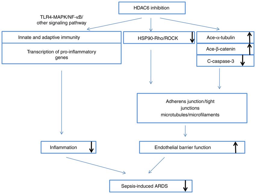
Figure 1. HDAC6 inhibition ameliorates sepsis‑induced ARDS. The inhibition occurs mainly by two ways: i) Modulation of innate and adaptive immune
cells, and the transcription of pro‑inflammatory genes, via the TLR4‑MAPK/NF‑κ B pathway, which directly attenuates inflammation; and ii) protection of
endothelial barrier function via HSP90, α‑tubulin, β‑catenin and caspase‑3, thereby enhancing adherents and strengthening the cytoskeleton. HDAC6, histone
deacetylase 6; ARDS, acute respiratory distress syndrome; TLR, Τoll‑like receptor; NF‑κ B, nuclear factor‑κ B; HSP90, heat shock protein 90.
factor in the progression of sepsis‑induced ARDS/ALI. On 3. HDAC6 inhibition protects against inflammation
one hand, it directly destroys the integrity of the endothelial
barrier through endothelial cell apoptosis, and on the other, Halili et al (27) first demonstrated that the HDAC6 inhibitor
it has been reported that the activity of apoptosis‑associated 17a effectively inhibits LPS‑induced pro‑inflammatory endo‑
proteins such as caspase‑3 can lead to the rearrangement of thelin (Edn)‑1 and interleukin (IL)‑12p40 RNA expression in
cell junction proteins (such as VE‑cadherin and claudin‑5). macrophages. Various studies have since shown that HDAC6
Lastly, in recent years, research on the function of endothe‑ directly mediates inflammation and both the innate (i.e.,
lial cell barrier in ALI has gradually increased. At present, pathogen sensing and destruction) and adaptive immunity, as
one of the main signaling pathways found to be involved in well as the modulation of the transcription of pro‑inflamma‑
endothelial cell barrier function is the S1P‑SIP1 signaling tory genes (47,48).
pathway; with the activation of Gi protein‑combined SIP1
receptor and downstream small GTPase Rac molecule, S1P Role of HDAC6 in innate and adaptive immunity cells.
induces cortactin transfer and peripheral myosin long chain Inflammation is a protective innate immune response to
(MLC) phosphorylation, which can strengthen the rearrange‑ invading pathogens and helps to prevent tissue damage. It
ment of cytoskeleton structure and form a cortical actin ring, acts like a ‘double‑edged sword’ and, upon overactivation
and strengthen the adhesion connection, tight connection, or improper activation, causes serious immune disorders,
and local adhesion complex on the cell surface, thereby systemic inflammatory response syndrome, compensatory
decreasing pulmonary edema induced by LPS in ALI (33). anti‑inflammatory response syndrome, innate immune and
In addition, the following are some pathways and systems adaptive immune disorders, and subsequently leads to multiple
that are also involved: Ang‑2‑Tie‑2‑MLC, renin‑angiotensin organ dysfunction such as ARDS. The process of inflammation
system (34), Rho‑ROCK‑LIMK1‑MLC, eNOS‑Cav1‑MLC2 begins with the influx of neutrophils, followed by the recruit‑
signaling (35), PAR1 moesin (36), HDAC6‑Hsp90 and ment of monocytes, which then differentiate into inflammatory
α‑tubulin‑β‑catenin (37‑39). macrophages or dendritic cells. These cells are the key effector
HDAC6 is extensively expressed in normal airways, where cells in the inflammatory site, recognizing and phagocytizing
it promotes microtubule destabilization and endothelial barrier pathogens and necrotic cells to help eliminate infection. In
hyper‑permeability (40). Accordingly, HDAC6 inhibition has addition, they activate the adaptive immune response through
been shown to improve survival in murine sepsis models, communication and coordination with other immune cells.
either by blocking endothelial barrier hyper‑permeability or Macrophages are the main effector cells in the inflammatory
by attenuating airway inflammation (41‑46), as will be further response and play a key role in initiation, regression, tissue
discussed (Fig. 1). repair, and regeneration of the inflammatory response (49).4 ZHANG et al: HDAC6: A PROMISING THERAPEUTIC TARGET FOR ARDS
Monocytes and macrophages, as the most efficient pathogen CXCL10 mediated by the former. Another study confirmed
scavengers and the main source of inflammatory cytokines, that hindsiipropane B inhibits the expression of CCL2,
are the key effector cells that modulate the innate immune CXCL8 and CXCL10 by inhibiting the HDAC6‑NADPH
response of the body. However, the progressive dysfunction of oxidase‑ROS‑MAPK‑NF‑κ B‑AP‑1 axis in astrocytes (56).
monocytes and macrophages also leads to immune dysfunc‑ Zhang et al (50,57) similarly found that selective HDAC6
tion during severe sepsis and septic shock. Thus, HDAC6 inhibition decreases TNF‑ α, IL‑1β and IL‑6 expression in
inhibition attenuates macrophage‑induced inflammation by both LPS‑activated RAW264.7 cells and a murine model of
inhibiting the overproduction of reactive oxygen species (ROS) acute liver failure by modulating oxidative stress and toll‑like
and modulating the expression of pro‑inflammatory cytokines receptor 4(TLR4)‑MAPK‑NF‑κ B signaling. Liu et al (58) and
induced by LPS (50). A study has also shown that HDAC6 Wang et al (59) have since confirmed that HDAC6 inhibition
deficiency impairs macrophage recruitment to the inflamma‑ protects against LPS‑induced inflammation by suppressing
tion site in a murine model of acute peritonitis, which may NF‑κ B signaling. It also decreases IL‑1β expression, as well as
suppress the phagocytic capacity of macrophages, but at the caspase‑1 cleavage and activation.
same time serves as a promising therapeutic strategy for the Interferon regulatory factor 3 (IRF‑3) is a transcription
treatment of macrophage‑associated immune diseases (49); factor that critically mediates interferon (IFN) secretion during
nonetheless, further research is still needed. The increased inflammation (60). Interestingly, Nusinzon and Horvath (61)
preoperative neutrophil lymphocyte ratio and high and middle showed that HDAC6 modulates IRF‑3 activity by deacetylating
ratio of neutrophils can have poor prognosis in patients with its coactivator, β‑catenin. Moreover, Chattopadhyay et al (62)
cancer. In addition, the proportion of neutrophils are found demonstrated that IRF‑3, β ‑catenin and CBP form a stable
to be significantly higher, with the lymphocyte count signifi‑ complex in response to viral or bacterial infection‑stimulated
cantly lower, in critical patients with severe sepsis or septic TLR‑3 signaling to induce IFN production. Importantly,
shock. According to the sequential organ failure assessment HDAC6 is required for the formation of this complex, since
(SOFA) and acute physiological and chronic health assessment its inhibition modulates β ‑catenin acetylation, thereby
II (APACHE II) scores, the severity of the clinical course is suppressing the interaction of IRF‑3 with CBP.
associated with the difference in the percentage of neutro‑
phils and lymphocytes in leukocytes. A selective HDAC6 4. HDAC6 inhibition restores endothelial barrier
inhibitor, tubastatinA (TubA) has been demonstrated to alter hyper‑permeability
the composition of blood cells, restore the lymphocyte popu‑
lation, and decrease the granulocyte‑to‑lymphocyte ratio by The endothelial barrier is a semi‑permeable membrane that
improving the monocyte and granulocyte count in a murine maintains the intra‑ and extravascular balance of water and
model of cecal ligation and puncture (CLP), providing another proteins (63). Its composition has previously been reported in
explanation for the increase insurvival outcome in septic death detail; in brief, it comprises the cytoskeleton and adherents
model. Furthermore, selective HDAC6 inhibition is needed to such as adherens junction (AJ) and tight junction (TJ) proteins,
restore innate immune cells in the bone marrow, lower stress including claudins and occludins, as well as zonula occludens
responses, immune organ atrophy and apoptosis (43,48,51,52). (ZO) (52) and local gap junction proteins (64). Endothelial
barrier dysfunction plays an important role in the pathogenesis
HDAC6 modulates pro‑inflammatory gene transcrip‑ of sepsis‑induced organ dysfunction; thus, endothelial barrier
tion. Cytokines, chemokines and adhesion molecules are protection is a proposed therapeutic modality to treat sepsis.
important pro‑inflammatory mediators, and HDAC6 overex‑ Nam et al (65) previously demonstrated that Clostridium
pression increases the production of pro‑inflammatory factors difficile toxin A induces microtubule instability in a murine
in macrophages. Conversely, HDAC6 inhibition suppresses model of C. Difficile infection by activating HDAC6, which
their expression and circulation, such as tumor necrosis in turn modulates α‑tubulin deacetylation, thereby inducing
factor (TNF)‑ α and IL‑6 in peritoneal fluids, decreases acute inflammation. Moreover, it is now well known that
myeloperoxidase (MPO) production, and increases IL‑10 HDAC6 inhibition promotes endothelial barrier hyper‑perme‑
production (45,50,53). HDAC6 inhibition has also been ability (37‑39,66,67).
reported to decrease the expression of adhesion molecules
and chemokines, including C‑C motif ligand 2 (CCL‑2), HDAC6 modulates HSP90‑dependent Rho‑associated kinase
C‑X‑C motif (CXC)L‑8 and CXCL‑10. Additionally, HDAC6 (Rho‑ROCK) activity. Hsp90 protein is a member of the heat
represses extracellular signal‑regulated kinase (ERK), c‑Jun shock molecular chaperone family that regulates protein
N‑terminal kinase (JNK), p38 and nuclear factor (NF)‑κ B. conformation and activity, and regulates a variety of cell
HDAC6 also represses activator protein (AP)‑1 activation in signaling pathways by controlling the abundance and activity
astrocytes responding to the human immunodeficiency virus of several important protein kinases and cell cycle‑associated
(HIV)‑1 transactivator of transcription (Tat) protein, by modu‑ proteins, such as Rho GTPases and actin, which is a tripartite
lating ROS homeostasis, mitogen‑activated protein kinase framework for effective vesicular transport. Rho GTPase
(MAPK), NF‑κ B and AP‑1 signaling (54,55). In summary, is a subfamily of small GTP binding proteins in the Ras
HDAC6 mediates HIV‑1 Tat‑induced pro‑inflammatory super family, composed of Cdc42, Rac1 and RhoA, which
response by regulating the MAPK, NF‑κ B and AP‑1 signaling regulates actin dynamics in endothelial cells. For example,
pathways in astrocytes. Studies showed that hindsiipropane B RhoA induces endothelial hyper‑permeability by modulating
inhibits the expression of HDAC6 induced by HIV‑1 Tat, and actin stress fiber formation and cell contraction (68). ROCK
subsequently inhibiting the expression of CCL2, CXCL8 and increases actin filament crosslinking activity of myosin IIEXPERIMENTAL AND THERAPEUTIC MEDICINE 21: 422, 2021 5
through the phosphorylation‑induced activation of MLC and as evidenced by the fact that caspase‑3 inhibition restores
inactivation of MLC phosphatase. Inhibition of Rho kinase, the endothelial barrier permeability in the lung tissue of septic
downstream effector of RhoA, can protect HLMVECS from mice (83). Increased caspase‑3 activity likely contributes to
LPS‑mediated high permeability and eliminate LPS‑induced endothelial barrier dysfunction by inducing endothelial cell
phosphorylation of MLC. Hsp90 plays an important and apoptosis; however, it has also been reported to trigger the
special role in regulating Rho activity and Rho‑dependent rearrangement of connexins, including VE‑cadherin, ZO‑1and
actin cytoskeleton remodeling. Hsp90 can indirectly activate claudin‑5 (84‑86).
Rho GTPase by protecting functional activators from protea‑
some degradation (69). Consistent with these findings, the 5. A promising and highly selective HDAC6 inhibitor, TubA
phosphorylation of MLC induced by RhoA is also inhibited
by 17‑AAG, a Hsp90 inhibitor. Studies have demonstrated In recent years, with an increase in the understanding of
that inhibition of Hsp90 can prevent and repair LPS‑induced HDAC6 and its roles in different diseases, a large number of
pulmonary endothelial barrier dysfunction by inhibiting experimental studies on HDAC6 inhibitors have been reported.
RhoA activity and signal transduction (70). It is important to There is an increasing number of studies on the role of HDAC6
note that HSP90 is a substrate of HDAC6. A previous study inhibition in inflammation, one of which involves the following:
by Joshi et al (37) proposed that HDAC6 inhibition protects Computer‑aided identification of new selective inhibitors of
against pulmonary endothelial barrier dysfunction and ALI HDAC6 with anti‑sepsis activity, development of new HDAC
by modulating HSP90; HDAC6 inhibition facilitates the inhibitors after virtual screening based on chemical data‑
acetylation of HSP90 at Lys294, which in turn suppresses bases, compound 9A [(E)‑n‑hydroxy‑4‑(2‑styrylthiazol‑4‑yl]
HSP90‑dependent Rho‑ROCK activity and blocks MLC was identified as a HDAC6 selective inhibitor (IC50 value of
phosphorylation to provide protection against LPS‑induced HDAC6 is 0.199 µm; HDAC, 8 µm). Compound 9A signifi‑
endothelial barrier dysfunction. However, more studies are cantly increased the survival of mice with sepsis induced by
required to verify this mechanism. LPS and inhibited the increase of TNF‑α and IL‑6 mRNA
expression induced by LPS (46).
HDAC6 modulates the acetylation of α‑tubulin and β‑catenin. CAY is also a highly selective HDAC6 inhibitor, which
Vascular endothelial (VE)‑cadherin and β‑catenin are major blocks the activation of NF‑ κ B by inhibiting Iκ B phos‑
AJ components. The majority of β‑catenin molecules localize phorylation in LPS‑induced ALI, can decrease the activity of
to the cytoplasmic side of the membrane, where they contact inflammatory corpuscles induced by LPS and decrease the
VE‑cadherin (71). In parallel, β ‑catenin degradation is cleavage and activation of IL‑1β and caspase‑1 (58).
controlled by its N‑terminal phosphorylation and ubiquitina‑ TubA is considered to be a potent and highly selective
tion; for example, ubiquitination at Lys49 triggers proteasomal HDAC6 inhibitor. Its IC50 value is 15 nm, 1,000 times lower
β‑catenin degradation (72). VE‑cadherin contains five extra‑ than that of other subtypes, except HDAC8 (57 times lower).
cellular cadherin repeats, a transmembrane region, and a TubA can improve the survival of mice with septicemic
highly conserved cytoplasmic tail that has been shown to form induced by lethal CLP (42). In a hemorrhagic shock model
a complex with β ‑catenin to maintain endothelial barrier combined with CLP, the survival time of mice in HDAC6
integrity (73‑75). group was also prolonged (45). The mechanism included
Microtubules and microfilaments are interacting cyto‑ changing the composition of circulating blood cells in lethal
skeletal components (76,77). Microtubules are comprised of septicemia model (48). Compared with the CLP Group, the
two globular proteins, α‑ and β‑tubulin, which polymerize to TubA treatment group recovered B lymphocytes and signifi‑
regulate endothelial barrier function. Decreasing α‑tubulin cantly increased the percentage of innate immune cells and
acetylation has been shown to affect microtubule assembly, macrophages. In addition, TubA could significantly decrease
cytoskeletal stability and cell mobility (78). Conversely, tubulin the bacterial load in the spleen and increase the phagocytic
acetylation prolongs the microtubule half‑life, increases cyto‑ capacity of macrophages in RAW264.7 cells. In this lethal
skeletal stability, and renders the cytoskeleton more resistant sepsis model, TubA significantly decreased the stress response
to drug‑induced depolymerization and disassembly (79). and thymus and bone marrow atrophy (51). The apoptosis of
Previous studies have suggested that microtubule disassembly spleen cells also decreased. WT mice treated with TubA were
stimulates actin formation and increases MLC phosphoryla‑ partially protected from vascular leakage and inflammation
tion; in contrast, microtubule depolymerization likely blocks caused by HKSA or methicillin‑resistant Staphylococcus
these effects (80,81). aureus (MRSA) (40). Thus, TubA plays an important function
HDAC6 inhibition has been demonstrated to decrease in endothelial cell barrier protection, and the mechanisms
sepsis‑induced endothelial barrier hyper‑permeability include an increase in the acetylation of α‑tubulin and
by restoring normal α‑tubulin and β ‑catenin acetylation β ‑catenin and a decrease in the activation of caspase‑3 as
patterns (39,44). Specifically, HDAC6 inhibition allows previously mentioned.
β‑catenin to be acetylated at Lys49, preventing its ubiquitina‑
tion and increasing its accumulation at the cell membrane, A brief introduction to the role of TubA in ARDS. As mentioned
thus promoting the formation of the VE‑cadherin‑β‑catenin above, most of the experiments on HDAC6 inhibitors in vitro
complex (82). Furthermore, HDAC6 inhibition has been and in vivo have been conducted with TubA. Currently, TubA
suggested to modulate α‑tubulin and β ‑catenin acetyla‑ has been reported to prolong the survival period of septic mice,
tion by decreasing caspase‑3 cleavage (39,44). Decreased decrease the ALI from sepsis, and lower the bacterial load
caspase‑3 activation protects endothelial hyper‑permeability, in spleen. It brings about these effects mainly by decreasing6 ZHANG et al: HDAC6: A PROMISING THERAPEUTIC TARGET FOR ARDS
the inflammatory response, restoring endothelial cell barrier cell‑cell junctions, and inhibits actin stress fiber formation
dysfunction, and decreasing the oxidative stress response, and decreases MLC phosphorylation via modulating HSP90.
as well as pulmonary fibrosis, by inhibiting the apoptosis of These findings support that HDAC6 inhibition is a promising
pulmonary vascular endothelial cells (87). potential therapeutic target to treat sepsis‑induced ARDS.
However, the negative effects, if any, of HDAC6 inhibitors on
Putative side effects of TubA. As the number of TubA studies embryo sac and oocytes need to be taken into account.
increases, increasingly more attention is being given to its
possible side effects. In one study, a relatively high dose was Acknowledgements
used (100 mg/kg body weight of mice) and no significant
adverse effects were observed (88). However, in another study Not applicable.
it was found that TubA exposure significantly decreased
the formation of blastocysts in early embryos of mice, and Funding
confocal microscopy showed that chromosome aggregation
failed to occur in mice embryos treated with HDAC6 inhibi‑ This work was supported by the National Natural Science
tors. In addition, the inhibition of HDAC6 induced an excessive Foundation of China (grant no. 81800080).
production of ROS. An increase in the accumulation of phos‑
phorylated γ‑H2AX was also observed in the embryos after Availability of data and materials
TubA intervention indicating an increase in DNA damage and
blastocyst cell apoptosis (89). TubA interfered with and halted All data generated or analyzed during this study are included
mouse oocyte meiosis by regulating several key histones in this published article.
(H4K16 acetylation and h3t3 and H3S10 phosphorylation)
and messenger RNA (ccnb1, CDK2, Smad3 and YWHAZ Authors' contributions
methylation‑associated genes DNMT1 and DNMT3b), and by
interfering with the organization of spindles of chromosomes, JYu analyzed and interpreted the data and final approval of
as well as the attachment of mitotic microtubules. The first the version to be published. JYa analyzed and interpreted the
polar body of mouse blastocyst oocyte could not be expelled data and was a major contributor in writing the manuscript.
after TubA treatment. However, it has been proved that homo‑ QZ was a contributor in writing the manuscript. YW and DQ
zygous knockout of HDAC6 (KO) mice are viable and fertile. contributed to literature review. All authors read and approved
Further studies have confirmed that the HDAC6 protein and the final manuscript.
mRNA levels in mouse oocytes treated with TubA were signif‑
icantly lower. The SIRT2, HDAC6, SIRT6 and SIRT7 mRNA Ethics approval and consent to participate
levels were also significantly decreased. The mRNA expres‑
sion levels of Cdk1, Cdk2, Cdk4, Cdk6, Cdc25B, and other Not applicable.
cell cycle‑associated kinases decreased significantly. These
abnormalities were associated with meiosis and cell aging. Patient consent for publication
The abnormal meiotic maturation and cell senescence induced
by TubA may be the result of its interaction with HDAC and Not applicable.
sirtuin (90). It is presumed that TubA can be used in the treat‑
ment of ARDS induced by sepsis, but effects of HDAC6 on the Competing interests
reproductive function of female should be of caution.
The authors declare that they have no competing interests.
6. Conclusion
References
Inflammation and endothelial barrier dysfunction are important
processes that contribute to the pathogenesis of sepsis‑induced 1. Ashbaugh DG, Bigelow DB, Petty TL and Levine BE: Acute
ARDS. HDAC6 has been reported to mediate inflammation, respiratory distress in adults. Lancet 2: 319‑323, 1967.
2. Ferguson ND, Fan E, Camporota L, Antonelli M, Anzueto A,
as well as both the innate and adaptive immunity, including Beale R, Brochard L, Brower R, Esteban A, Gattinoni L, et al:
inflammatory cell recruitment and bacterial clearance. The Berlin definition of ARDS: An expanded rationale, justi‑
Accordingly, HDAC6 inhibition using an inhibitor such as fication, and supplementary material. Intensive Care Med 38:
1573‑1582, 2012.
TubA increases the survival of septic mice by decreasing the 3. Bellani G, Laffey JG, Pham T, Fan E, Brochard L, Esteban A,
LPS‑induced macrophage and epithelial cell inflammation. It Gattinoni L, van Haren F, Larsson A, McAuley DF, et al; LUNG
is proposed that HDAC6 inhibition predominantly ameliorates SAFE Investigators; ESICM Trials Group: Epidemiology,
patterns of care, and mortality for patients with acute respi‑
sepsis‑induced ARDS by modulating the innate and adaptive ratory distress syndrome in intensive care units in 50 countries.
immunity and the transcription of pro‑inflammatory genes JAMA 315: 788‑800, 2016.
to directly attenuate inflammation. HDAC6 inhibition also 4. Her r idge MS, Ta nsey CM, Matté A, Tom linson G,
Diaz‑Granados N, Cooper A, Guest CB, Mazer CD, Mehta S,
protects endothelial barrier function via its effects on its target Stewart TE, et al; Canadian Critical Care Trials Group:
substrates by inducing α‑tubulin and β ‑catenin acetylation Functional disability 5 years after acute respiratory distress
and increasing membrane localization of β‑catenin, thereby syndrome. N Engl J Med 364: 1293‑1304, 2011.
5. Han S and Mallampalli RK: The acute respiratory distress
leading to the stabilization of microtubule and AJs. It also syndrome: From mechanism to translation. J Immunol 194:
prevents caspase‑3 activation, maintains lung endothelial 855‑860, 2015.EXPERIMENTAL AND THERAPEUTIC MEDICINE 21: 422, 2021 7
6. Griffiths MJD, McAuley DF, Perkins GD, Barrett N, Blackwood B, 25. Yee AJ, Bensinger WI, Supko JG, Voorhees PM, Berdeja JG,
Boyle A, Chee N, Connolly B, Dark P, Finney S, et al: Guidelines Richardson PG, Libby EN, Wallace EE, Birrer NE, Burke JN, et al:
on the management of acute respiratory distress syndrome. BMJ Ricolinostat plus lenalidomide, and dexamethasone in relapsed
Open Respir Res 6: e000420, 2019. or refractory multiple myeloma: A multicentre phase 1b trial.
7. Walkey AJ, Del Sorbo L, Hodgson CL, Adhikari NKJ, Wunsch H, Lancet Oncol 17: 1569‑1578, 2016.
Meade MO, Uleryk E, Hess D, Talmor DS, Thompson BT, et al: 26. Tu Y, Hershman DL, Bhalla K, Fiskus W, Pellegrino CM,
Higher PEEP versus Lower PEEP Strategies for Patients with Andreopoulou E, Ma kower D, Kalinsky K, Fehn K,
Acute Respiratory Distress Syndrome. A Systematic Review and Fineberg S, et al: A phase I‑II study of the histone deacetylase
Meta‑Analysis. Ann Am Thorac Soc 14 (Suppl 4): S297‑S303, inhibitor vorinostat plus sequential weekly paclitaxel and doxo‑
2017. rubicin‑cyclophosphamide in locally advanced breast cancer.
8. Hodgson CL, Cooper DJ, Arabi Y, King V, Bersten A, Bihari S, Breast Cancer Res Treat 146: 145‑152, 2014.
Brickell K, Davies A, Fahey C, Fraser J, et al: Maximal 27. Halili MA, Andrews MR, Labzin LI, Schroder K, Matthias G,
Recruitment Open Lung Ventilation in Acute Respiratory Cao C, Lovelace E, Reid RC, Le GT, Hume DA, et al:
Distress Syndrome (PHARLAP). A Phase II, Multicenter Differential effects of selective HDAC inhibitors on macrophage
Randomized Controlled Clinical Trial. Am J Respir Crit Care inflammatory responses to the Toll‑like receptor 4 agonist LPS.
Med 200: 1363‑1372, 2019. J Leukoc Biol 87: 1103‑1114, 2010.
9. Steinberg KP, Hudson LD, Goodman RB, Hough CL, Lanken PN, 28. Estenssoro E and Dubin A: Acute respiratory distress syndrome.
Hyzy R, Thompson BT and Ancukiewicz M; National Heart, Medicina (B Aires) 76: 235‑241, 2016 (In Spanish).
Lung, and Blood Institute Acute Respiratory Distress Syndrome 29. Kasznica J, Helmann M, Collins JP and Akhtar R: Bilateral
(ARDS) Clinical Trials Network: Efficacy and safety of corti‑ Ebstein‑like anomaly with atrial septal defect. Jpn Heart J 36:
costeroids for persistent acute respiratory distress syndrome. N 119‑125, 1995.
Engl J Med 354: 1671‑1684, 2006. 30. Fu P, Murley JS, Grdina DJ, Birukova AA and Birukov KG:
10. Peck TJ and Hibbert KA: Recent advances in the understanding Induction of cellular antioxidant defense by amifostine improves
and management of ARDS. F1000 Res 8: 8, 2019. ventilator‑induced lung injury. Crit Care Med 39: 2711‑2721,
11. Villar J, Ferrando C, Martínez D, Ambrós A, Muñoz T, Soler JA, 2011.
Aguilar G, Alba F, González‑Higueras E, Conesa LA, et al; dexa‑ 31. Kratzer E, Tian Y, Sarich N, Wu T, Meliton A, Leff A and
methasone in ARDS network: Dexamethasone treatment for the Birukova AA: Oxidative stress contributes to lung injury and
acute respiratory distress syndrome: A multicentre, randomised barrier dysfunction via microtubule destabilization. Am J Respir
controlled trial. Lancet Respir Med 8: 267‑276, 2020. Cell Mol Biol 47: 688‑697, 2012.
12. Rubenfeld GD, Caldwell E, Peabody E, Weaver J, Martin DP, 32. Zhao B, Gao W, Gao X, Leng Y, Liu M, Hou J and Wu Y:
Neff M, Stern EJ and Hudson LD: Incidence and outcomes of Sulforaphane attenuates acute lung injury by inhibiting oxidative
acute lung injury. N Engl J Med 353: 1685‑1693, 2005. stress via Nrf2/HO‑1 pathway in a rat sepsis model. Int J Clin
13. Ito K, Caramori G, Lim S, Oates T, Chung KF, Barnes PJ and Exp Pathol 10: 9021‑9028, 2017.
Adcock IM: Expression and activity of histone deacetylases 33. Peng X, Hassoun PM, Sammani S, McVerry BJ, Burne MJ,
in human asthmatic airways. Am J Respir Crit Care Med 166: Rabb H, Pearse D, Tuder RM and Garcia JG: Protective effects
392‑396, 2002. of sphingosine 1‑phosphate in murine endotoxin‑induced inflam‑
14. Sadoul K, Boyault C, Pabion M and Khochbin S: Regulation matory lung injury. Am J Respir Crit Care Med 169: 1245‑1251,
of protein turnover by acetyltransferases and deacetylases. 2004.
Biochimie 90: 306‑312, 2008. 34. Kong J, Zhu X, Shi Y, Liu T, Chen Y, Bhan I, Zhao Q,
15. Dekker FJ and Haisma HJ: Histone acetyl transferases as Thadhani R and Li YC: VDR attenuates acute lung injury by
emerging drug targets. Drug Discov Today 14: 942‑948, 2009. blocking Ang‑2‑Tie‑2 pathway and renin‑angiotensin system.
16. Zou H, Wu Y, Navre M and Sang BC: Characterization of the two Mol Endocrinol 27: 2116‑2125, 2013.
catalytic domains in histone deacetylase 6. Biochem Biophys Res 35. Thangavel J, Malik AB, Elias HK, Rajasingh S, Simpson AD,
Commun 341: 45‑50, 2006. Sundivakkam PK, Vogel SM, Xuan YT, Dawn B and
17. Zhang Y, Gilquin B, Khochbin S and Matthias P: Two catalytic Rajasingh J: Combinatorial therapy with acetylation and meth‑
domains are required for protein deacetylation. J Biol Chem 281: ylation modifiers attenuates lung vascular hyperpermeability in
2401‑2404, 2006. endotoxemia‑induced mouse inflammatory lung injury. Am J
18. Schäfer S, Saunders L, Eliseeva E, Velena A, Jung M, Pathol 184: 2237‑2249, 2014.
Schwienhorst A, Strasser A, Dickmanns A, Ficner R and 36. Xu Q, Liu J, Wang Z, Guo X, Zhou G, Liu Y, Huang Q and Su L:
Schlimme S: Phenylalanine‑containing hydroxamic acids as Heat stress‑induced disruption of endothelial barrier function is
selective inhibitors of class IIb histone deacetylases (HDACs). via PAR1 signaling and suppressed by Xuebijing injection. PLoS
Bioorg Med Chem 16: 2011‑2033, 2008. One 10: e0118057, 2015.
19. Zhang X, Yuan Z, Zhang Y, Yong S, Salas‑Burgos A, Koomen J, 37. Joshi AD, Barabutis N, Birmpas C, Dimitropoulou C,
Olashaw N, Parsons JT, Yang XJ, Dent SR, et al: HDAC6 Thangjam G, Cherian‑Shaw M, Dennison J and Catravas JD:
modulates cell motility by altering the acetylation level of Histone deacetylase inhibitors prevent pulmonary endothelial
cortactin. Mol Cell 27: 197‑213, 2007. hyperpermeability and acute lung injury by regulating heat shock
20. Bali P, Pranpat M, Bradner J, Balasis M, Fiskus W, Guo F, protein 90 function. Am J Physiol Lung Cell Mol Physiol 309:
Rocha K, Kumaraswamy S, Boyapalle S, Atadja P, et al: L1410‑L1419, 2015.
Inhibition of histone deacetylase 6 acetylates and disrupts the 38. Borgas D, Chambers E, Newton J, Ko J, Rivera S, Rounds S
chaperone function of heat shock protein 90: A novel basis for and Lu Q: Cigarette Smoke Disrupted Lung Endothelial Barrier
antileukemia activity of histone deacetylase inhibitors. J Biol Integrity and Increased Susceptibility to Acute Lung Injury via
Chem 280: 26729‑26734, 2005. Histone Deacetylase 6. Am J Respir Cell Mol Biol 54: 683‑696,
21. Parab S, Shetty O, Gaonkar R, Balasinor N, Khole V and Parte P: 2016.
Correction to: HDAC6 deacetylates alpha tubulin in sperm and 39. Yu J, Ma Z, Shetty S, Ma M and Fu J: Selective HDAC6 inhibition
modulates sperm motility in Holtzman rat. Cell Tissue Res 371: prevents TNF‑α‑induced lung endothelial cell barrier disruption
375, 2018. and endotoxin‑induced pulmonary edema. Am J Physiol Lung
22. Wang XX, Wan RZ and Liu ZP: Recent advances in the discovery Cell Mol Physiol 311: L39‑L47, 2016.
of potent and selective HDAC6 inhibitors. Eur J Med Chem 143: 40. Karki P, Ke Y, Tian Y, Ohmura T, Sitikov A, Sarich N,
1406‑1418, 2018. Montgomer y CP and Bi r u kova A A: Staphylococcus
23. Jia YJ, Liu ZB, Wang WG, Sun CB, Wei P, Yang YL, You MJ, aureus‑induced endothelial permeability and inflammation
Yu BH, Li XQ and Zhou XY: HDAC6 regulates microRNA‑27b are mediated by microtubule destabilization. J Biol Chem 294:
that suppresses proliferation, promotes apoptosis and target 3369‑3384, 2019.
MET in diffuse large B‑cell lymphoma. Leukemia 32: 703-711, 41. Rosenjack J, Hodges CA, Darrah RJ and Kelley TJ: HDAC6
2018. depletion improves cystic fibrosis mouse airway responses to
24. Vogl DT, Raje N, Jagannath S, Richardson P, Hari P, Orlowski R, bacterial challenge. Sci Rep 9: 10282, 2019.
Supko JG, Tamang D, Yang M, Jones SS, et al: Ricolinostat, the 42. Deng Q, Zhao T, Pan B, Dennahy IS, Duan X, Williams AM,
first selective histone deacetylase 6 inhibitor, in combination Liu B, Lin N, Bhatti UF, Chen E, et al: Protective Effect of
with bortezomib and dexamethasone for relapsed or refractory Tubastatin A in CLP‑Induced Lethal Sepsis. Inflammation 41:
multiple myeloma. Clin Cancer Res 23: 3307‑3315, 2017. 2101‑2109, 2018.8 ZHANG et al: HDAC6: A PROMISING THERAPEUTIC TARGET FOR ARDS
43. Li Y, Zhao T, Liu B, Halaweish I, Mazitschek R, Duan X and 63. Trani M and Dejana E: New insights in the control of vascular
Alam HB: Inhibition of histone deacetylase 6 improves long‑term permeability: Vascular endothelial‑cadherin and other players.
survival in a lethal septic model. J Trauma Acute Care Surg 78: Curr Opin Hematol 22: 267‑272, 2015.
378‑385, 2015. 64. Rodrigues SF and Granger DN: Blood cells and endothelial
44. Yu J, Ma M, Ma Z and Fu J: HDAC6 inhibition prevents barrier function. Tissue Barriers 3: e978720, 2015.
TNF‑α‑induced caspase 3 activation in lung endothelial cell and 65. Nam HJ, Kang JK, Kim SK, Ahn KJ, Seok H, Park SJ, Chang JS,
maintains cell‑cell junctions. Oncotarget 7: 54714‑54722, 2016. Pothoulakis C, Lamont JT and Kim H: Clostridium difficile
45. Cheng X, Liu Z, Liu B, Zhao T, Li Y and Alam HB: Selective toxin A decreases acetylation of tubulin, leading to microtubule
histone deacetylase 6 inhibition prolongs survival in a lethal depolymerization through activation of histone deacetylase 6,
two‑hit model. J Surg Res 197: 39‑44, 2015. and this mediates acute inflammation. J Biol Chem 285:
46. Yoo J, Kim SJ, Son D, Seo H, Baek SY, Maeng CY, Lee C, 32888‑32896, 2010.
Kim IS, Jung YH, Lee SM, et al: Computer‑aided identification 66. Saito S, Lasky JA, Guo W, Nguyen H, Mai A, Danchuk S,
of new histone deacetylase 6 selective inhibitor with anti‑sepsis Sullivan DE and Shan B: Pharmacological inhibition of HDAC6
activity. Eur J Med Chem 116: 126‑135, 2016. attenuates endothelial barrier dysfunction induced by thrombin.
47. Moreno‑Gonzalo O, Mayor F Jr and Sánchez‑Madrid F: Biochem Biophys Res Commun 408: 630‑634, 2011.
HDAC6 at Crossroads of Infection and Innate Immunity. Trends 67. Wang F, Zheng L, Yi Y, Yang Z, Qiu Q, Wang X, Yan W, Bai P,
Immunol 39: 591‑595, 2018. Yang J, Li D, et al: SKLB‑23bb, A HDAC6‑Selective Inhibitor,
48. Zhao T, Li Y, Liu B, Pan B, Cheng X, Georgoff P and Alam HB: Exhibits Superior and Broad‑Spectrum Antitumor Activity
Inhibition of histone deacetylase 6 restores innate immune cells via Additionally Targeting Microtubules. Mol Cancer Ther 17:
in the bone marrow in a lethal septic model. J Trauma Acute Care 763‑775, 2018.
Surg 80: 34‑40, discussion 40‑41, 2016. 68. Majolée J, Pronk MCA, Jim KK, van Bezu JSM, van der Sar AM,
49. Yan B, Xie S, Liu Y, Liu W, Li D, Liu M, Luo HR and Zhou J: Hordijk PL and Kovačević I: CSN5 inhibition triggers inflam‑
Histone deacetylase 6 modulates macrophage infiltration during matory signaling and Rho/ROCK‑dependent loss of endothelial
inflammation. Theranostics 8: 2927‑2938, 2018. integrity. Sci Rep 9: 8131, 2019.
50. Zhang WB, Yang F, Wang Y, Jiao FZ, Zhang HY, Wang LW 69. Croisé P, Estay‑Ahumada C, Gasman S and Ory S: Rho GTPases,
and Gong ZJ: Inhibition of HDAC6 attenuates LPS‑induced phosphoinositides, and actin: A tripartite framework for efficient
inflammation in macrophages by regulating oxidative stress vesicular trafficking. Small GTPases 5: e29469, 2014.
and suppressing the TLR4‑MAPK/NF‑κ B pathways. Biomed 70. Joshi AD, Dimitropoulou C, Thangjam G, Snead C, Feldman S,
Pharmacother 117: 109166, 2019. Barabutis N, Fulton D, Hou Y, Kumar S, Patel V, et al: Heat
51. Zhao T, Li Y, Bronson RT, Liu B, Velmahos GC and Alam HB: shock protein 90 inhibitors prevent LPS‑induced endothelial
Selective histone deacetylase‑6 inhibition attenuates stress barrier dysfunction by disrupting RhoA signaling. Am J Respir
responses and prevents immune organ atrophy in a lethal septic Cell Mol Biol 50: 170‑179, 2014.
model. Surgery 156: 235‑242, 2014. 71. Valenta T, Hausmann G and Basler K: The many faces and
52. González‑Mariscal L, Tapia R and Chamorro D: Crosstalk of functions of β‑catenin. EMBO J 31: 2714‑2736, 2012.
tight junction components with signaling pathways. Biochim 72. Winer IS, Bommer GT, Gonik N and Fearon ER: Lysine residues
Biophys Acta 1778: 729‑756, 2008. Lys‑19 and Lys‑49 of beta‑catenin regulate its levels and function
53. Di Liddo R, Valente S, Taurone S, Zwergel C, Marrocco B, in T cell factor transcriptional activation and neoplastic transfor‑
Turchetta R, Conconi MT, Scarpa C, Bertalot T, Schrenk S, et al: mation. J Biol Chem 281: 26181‑26187, 2006.
Histone deacetylase inhibitors restore IL‑10 expression in 73. Sauteur L, Krudewig A, Herwig L, Ehrenfeuchter N, Lenard A,
lipopolysaccharide‑induced cell inflammation and reduce IL‑1β Affolter M and Belting HG: Cdh5/VE‑cadherin promotes endo‑
and IL‑6 production in breast silicone implant in C57BL/6J thelial cell interface elongation via cortical actin polymerization
wild‑type murine model. Autoimmunity: Jan 20, 2016 (Epub during angiogenic sprouting. Cell Rep 9: 504‑513, 2014.
ahead of print). 74. Dejana E and Vestweber D: The role of VE‑cadherin in vascular
54. Youn GS, Lee KW, Choi SY and Park J: Overexpression of morphogenesis and permeability control. Prog Mol Biol Transl
HDAC6 induces pro‑inflammatory responses by regulating Sci 116: 119‑144, 2013.
ROS‑MAPK‑NF‑κ B/AP‑1 signaling pathways in macrophages. 75. Huber AH and Weis WI: The structure of the beta‑catenin/
Free Radic Biol Med 97: 14‑23, 2016.
55. Youn GS, Ju SM, Choi SY and Park J: HDAC6 mediates E‑cadherin complex and the molecular basis of diverse ligand
HIV‑1 tat‑induced proinflammatory responses by regulating recognition by beta‑catenin. Cell 105: 391‑402, 2001.
MAPK‑NF‑kappaB/AP‑1 pathways in astrocytes. Glia 63: 76. D'Alessandro M, Hnia K, Gache V, Koch C, Gavriilidis C,
1953‑1965, 2015. Rodriguez D, Nicot AS, Romero NB, Schwab Y, Gomes E, et al:
56. Jo H, Jang HY, Youn GS, Kim D, Lee CY, Jang JH, Choi SY, Amphiphysin 2 orchestrates nucleus positioning and shape by
Jun JG and Park J: Hindsiipropane B alleviates HIV‑1 Tat‑induced linking the nuclear envelope to the actin and microtubule cyto‑
inflammatory responses by suppressing HDAC6‑NADPH skeleton. Dev Cell 35: 186‑198, 2015.
oxidase‑ROS axis in astrocytes. BMB Rep 51: 394‑399, 2018. 77. Coles CH and Bradke F: Coordinating neuronal actin‑micro‑
57. Zhang WB, Zhang HY, Jiao FZ, Wang LW, Zhang H and tubule dynamics. Curr Biol 25: R677‑R691, 2015.
Gong ZJ: Histone deacetylase 6 inhibitor ACY‑1215 protects 78. Hubbert C, Guardiola A, Shao R, Kawaguchi Y, Ito A, Nixon A,
against experimental acute liver failure by regulating the Yoshida M, Wang XF and Yao TP: HDAC6 is a microtubule‑asso‑
TLR4‑MAPK/NF‑ κ B pathway. Biomed Pharmacother 97: ciated deacetylase. Nature 417: 455‑458, 2002.
818‑824, 2018. 79. Matsuyama A, Shimazu T, Sumida Y, Saito A, Yoshimatsu Y,
58. Liu L, Zhou X, Shetty S, Hou G, Wang Q and Fu J: HDAC6 inhi‑ Seigneurin‑Berny D, Osada H, Komatsu Y, Nishino N,
bition blocks inflammatory signaling and caspase‑1 activation Khochbin S, et al: In vivo destabilization of dynamic micro‑
in LPS‑induced acute lung injury. Toxicol Appl Pharmacol 370: tubules by HDAC6‑mediated deacetylation. EMBO J 21:
178‑183, 2019. 6820‑6831, 2002.
59. Wang J, Zhao L, Wei Z, Zhang X, Wang Y, Li F, Fu Y and 80. Shivanna M and Srinivas SP: Microtubule stabilization opposes
Liu B: Inhibition of histone deacetylase reduces lipopoly‑ the (TNF‑alpha)‑induced loss in the barrier integrity of corneal
saccharide‑induced‑inf lammation in primary mammary endothelium. Exp Eye Res 89: 950‑959, 2009.
epithelial cells by regulating ROS‑NF‑кB signaling pathways. 81. Bogatcheva NV and Verin AD: The role of cytoskeleton in the
Int Immunopharmacol 56: 230‑234, 2018. regulation of vascular endothelial barrier function. Microvasc
60. Hiscott J: Convergence of the NF‑kappaB and IRF pathways in Res 76: 202‑207, 2008.
the regulation of the innate antiviral response. Cytokine Growth 82. Iaconelli J, Huang JH, Berkovitch SS, Chattopadhyay S,
Factor Rev 18: 483‑490, 2007. Mazitschek R, Schreiber SL, Haggarty SJ and Karmacharya R:
61. Nusinzon I and Horvath CM: Positive and negative regulation of HDAC6 inhibitors modulate Lys49 acetylation and membrane
the innate antiviral response and beta interferon gene expression localization of β‑catenin in human iPSC‑derived neuronal cells.
by deacetylation. Mol Cell Biol 26: 3106‑3113, 2006. ACS Chem Biol 10: 883‑890, 2015.
62. Chattopadhyay S, Fensterl V, Zhang Y, Veleeparambil M, 83. Matsuda N, Takano Y, Kageyama S, Hatakeyama N, Shakunaga K,
Wetzel JL and Sen GC: Inhibition of viral pathogenesis and Kitajima I, Yamazaki M and Hattori Y: Silencing of caspase‑8
promotion of the septic shock response to bacterial infection by and caspase‑3 by RNA interference prevents vascular endothelial
IRF‑3 are regulated by the acetylation and phosphorylation of its cell injury in mice with endotoxic shock. Cardiovasc Res 76:
coactivators. MBio 4: 4, 2013. 132‑140, 2007.EXPERIMENTAL AND THERAPEUTIC MEDICINE 21: 422, 2021 9
84. Sawant DA, Tharakan B, Tobin RP, Reilly J, Hunter FA, Newell MK, 88. Wang X, Tang X, Zhou Z and Huang Q: Histone deacetylase 6
Smythe WR and Childs EW: Microvascular endothelial cell inhibitor enhances resistance to Mycobacterium tuberculosis
hyperpermeability induced by endogenous caspase 3 activator infection through innate and adaptive immunity in mice. Pathog
staurosporine. J Trauma Acute Care Surg 74: 516‑523, 2013. Dis 76: 76, 2018.
85. Lopez‑Ramirez MA, Fischer R, Torres‑Badillo CC, Davies HA, 89. Wang H, Ling L, Ai L and Bai L: HDAC6 inhibition induces
Logan K, Pfizenmaier K, Male DK, Sharrack B and Romero IA: the failure of mouse early embryonic development. J Cell
Role of caspases in cytokine‑induced barrier breakdown in Physiol 234: 8752‑8759, 2019.
human brain endothelial cells. J Immunol 189: 3130‑3139, 2012. 90. Choi YJ, Kang MH, Hong K and Kim JH: Tubastatin A inhibits
86. Zehendner CM, Librizzi L, de Curtis M, Kuhlmann CR HDAC and Sirtuin activity rather than being a HDAC6‑specific
and Luhmann HJ: Caspase‑3 contributes to ZO‑1 and Cl‑5 inhibitor in mouse oocytes. Aging (Albany NY) 11: 1759‑1777,
tight‑junction disruption in rapid anoxic neurovascular unit 2019.
damage. PLoS One 6: e16760, 2011.
87. Leyk J, Daly C, Janssen‑Bienhold U, Kennedy BN and
Richter‑Landsberg C: HDAC6 inhibition by tubastatin A is
protective against oxidative stress in a photoreceptor cell line
and restores visual function in a zebrafish model of inherited
blindness. Cell Death Dis 8: e3028, 2017.You can also read