The actin nucleator WASp is required for myoblast fusion during adult Drosophila myogenesis
←
→
Page content transcription
If your browser does not render page correctly, please read the page content below
RESEARCH ARTICLE 2347

Development 138, 2347-2357 (2011) doi:10.1242/dev.055012
© 2011. Published by The Company of Biologists Ltd

The actin nucleator WASp is required for myoblast fusion
during adult Drosophila myogenesis
Priyankana Mukherjee1,2, Boaz Gildor2, Ben-Zion Shilo2, K. VijayRaghavan1,* and Eyal D. Schejter2,*

SUMMARY
Myoblast fusion provides a fundamental, conserved mechanism for muscle fiber growth. We demonstrate here that the
functional contribution of Wsp, the Drosophila homolog of the conserved actin nucleation-promoting factor (NPF) WASp, is
essential for myoblast fusion during the formation of muscles of the adult fly. Disruption of Wsp function results in complete
arrest of myoblast fusion in all muscles examined. Wsp activity during adult Drosophila myogenesis is specifically required for
muscle cell fusion and is crucial both for the formation of new muscle fibers and for the growth of muscles derived from
persistent larval templates. Although Wsp is expressed both in fibers and individual myoblasts, its activity in either one of these
cell types is sufficient. SCAR, a second major Arp2/3 NPF, is also required during adult myoblast fusion. Formation of fusion-
associated actin ‘foci’ is dependent on Arp2/3 complex function, but appears to rely on a distinct, unknown nucleator. The
comprehensive nature of these requirements identifies Arp2/3-based branched actin polymerization as a universal mechanism
underlying myoblast fusion.

KEY WORDS: Actin, Drosophila, Myoblast fusion, WASp

INTRODUCTION                                                                        myoblasts fuses with a set of persistent larval fibers that survives
Myoblast fusion constitutes a fundamental aspect of muscle fiber                    the general wave of histolysis, and serves as a template for the
growth, underlying the formation of multinucleated contractile                      adult muscle structures (Fernandes et al., 1991; Roy and
units from pools of individual myoblasts (Rochlin et al., 2009). The                VijayRaghavan, 1998).
musculature of adult Drosophila shares many morphological and                          The different developmental modes leading to the formation
developmental characteristics with vertebrate somatic muscles,                      of the adult Drosophila flight musculature thus present
thereby providing an appealing model system for the elucidation of                  opportunities to examine both fusion between individual
universal molecular and cellular mechanisms governing                               myoblasts and between myoblasts and maturing fibers. Although
myogenesis (Dutta et al., 2004; Fernandes and Keshishian, 1999).                    classic genetic approaches have proven to be a highly successful
A prominent example is provided by the establishment of the                         tool to study myoblast fusion during embryonic Drosophila
thoracic indirect flight muscles (IFMs) of the adult fly. These large               myogenesis (Abmayr et al., 2008; Chen and Olson, 2004), their
muscles, which mediate flight by contraction and expansion of the                   application to the study of fusion during adult fly muscle
thoracic cuticle, are composed of several sets of bundled fibers,                   development has been limited. This is due both to functional
closely resembling vertebrate muscle organization.                                  requirements earlier in development for many of the genes
   Two distinct myogenic programs are employed during IFM                           potentially involved in myoblast fusion in the adult and to the
formation. The first of these follows a plan similar to that which                  syncytial nature of muscles, which restricts the usefulness of
governs muscle development of the Drosophila embryo (Beckett                        clonal analysis.
and Baylies, 2006): single ‘pioneer’ or ‘founder’ myoblasts seed                       We have used a combination of genetic approaches to
formation and differentiation of the mature fibers, which grow                      circumvent these difficulties and identify essential contributors
via repetitive rounds of fusion with neighboring myoblasts                          to adult myoblast fusion. We report an essential requirement for
(Fernandes et al., 1991; Rivlin et al., 2000). This mode of                         Wsp (WASp – FlyBase), a member of the WASp family of actin
myogenesis is common to most of the adult musculature, which                        nucleation-promoting factors (NPFs), which has previously been
has to form anew following destruction of nearly all somatic                        linked to myoblast fusion in Drosophila embryos (Kim et al.,
muscles of the Drosophila larva during the early stages of                          2007; Massarwa et al., 2007; Schafer et al., 2007). Wsp is
pupal development. A separate program is employed during                            required for all forms of myoblast fusion that lead to the growth
construction of the twelve dorsal-longitudinal muscles (DLMs),                      of adult somatic muscle fibers. In addition to Wsp, the SCAR
which are prominent flight muscles that span the length of the                      (also known as WAVE) NPF and associated elements are also
thorax. In this case, a large proliferative population of migratory                 implicated in adult myoblast fusion. Furthermore, we describe
                                                                                                                                                           DEVELOPMENT

                                                                                    transient F-actin structures that form in myoblasts near the onset
                                                                                    of fusion, under the influence of the Arp2/3 actin polymerization
1
 National Centre for Biological Sciences, Tata Institute of Fundamental Research,
                                                                                    machinery, but independently of the Wsp and SCAR nucleating
Bangalore 560065, India. 2Department of Molecular Genetics, The Weizmann            systems. These findings underscore the significant roles played
Institute of Science, Rehovot 76100, Israel.                                        by the actin-based cytoskeleton in the myoblast fusion process
*Authors for correspondence (vijay@ncbs.res.in; eyal.schejter@weizmann.ac.il)
                                                                                    (Onel and Renkawitz-Pohl, 2009; Richardson et al., 2008), and,
                                                                                    in particular, generalize and accentuate the myogenic role of the
Accepted 28 February 2011                                                           WASp pathway.
2348 RESEARCH ARTICLE Development 138 (11)
MATERIALS AND METHODS systems and processed using Adobe Photoshop CS3. Area determination
Drosophila genetics of actin foci was performed as previously described (Gildor et al., 2009;
UAS-dsRNA constructs were commonly expressed together with UAS- Richardson et al., 2007).
Dicer2 to enhance RNAi activity (Dietzl et al., 2007). Where necessary,
the GAL80ts/TARGET system (McGuire et al., 2004) was used for RESULTS
temporal control of UAS-based transgene expression. Developing flies
Adult Wsp mutant flies possess poorly developed
were maintained at 18°C, allowing for GAL80-based inhibition of GAL4
activity, and shifted (commonly at 0 hours APF) to 29°C, to inactivate the flight muscles
GAL80ts element. Although Wsp performs essential roles throughout Drosophila
development, flies lacking zygotic Wsp function survive until late
Mutant alleles pupal/early adult stages because maternally contributed Wsp gene
Wsp: Wsp1 and Df(3R)3450 (Ben-Yaacov et al., 2001). D-WIP (Vrp1 or products are sufficient for proper embryonic and larval
sltr): D-WIPD30 (Massarwa et al., 2007) and sltrS1946 (Kim et al., 2007).
development. The external morphology of Wsp mutant flies is
GAL4 drivers generally normal at the time of eclosion, except for the absence of
1151-GAL4 (myoblasts) (Roy and VijayRaghavan, 1997); rp298 (duf)- many mechanosensory bristles (Ben-Yaacov et al., 2001). To assess
GAL4 (founder cells) (Menon and Chia, 2001); Mef2-GAL4 (myogenic whether myogenesis proceeds properly in these flies, we
mesoderm) (Ranganayakulu et al., 1996). incorporated the muscle-specific reporter construct MHC-TauGFP
UAS-Wsp constructs (Chen and Olson, 2001) into a Wsp mutant background. In contrast
UAS-Wsp, UAS-Wsp-GFP, UAS-WspCA and UAS-Wspmyr have been to the very bright GFP signal normally provided by the substantial
described previously (Ben-Yaacov et al., 2001; Bogdan et al., 2005; flight musculature housed within the adult fly thorax, Wsp mutant
Massarwa et al., 2007; Tal et al., 2002). flies displayed a faint signal indicative of under developed or
Other UAS-based transgenes
missing muscles (Fig. 1A,B). To verify that the defects in adult
UAS-RedStinger (nuclear marker) (Barolo et al., 2004); UAS-myr-mRFP muscle organization arose during pupal development, we made use
(membrane marker) (Kandachar et al., 2008); UAS-Moesin-GFP of WspCA, a dominant-negative construct that lacks the crucial
(microfilament marker) (Chihara et al., 2003; Dutta et al., 2002); UAS- C-terminal Arp2/3-binding domain of Wsp and has been shown to
Rac1DN- (T17N, a dominant-negative version of Rac1) (Luo et al., 1994); strongly disrupt Wsp function during embryonic myoblast fusion
UAS-Arp3-GFP and UAS-Sop2-GFP (Hudson and Cooley, 2002). (Massarwa et al., 2007; Tal et al., 2002). Limiting expression of
UAS-dsRNA lines
UAS-WspCA to muscle tissue only, following the onset of
Arp2 (JF02785); kette (JF03342); SCAR (JF01599); Sop2 (GD42172); D- pupariation, was achieved by using the myogenic driver Mef2-
WIP (GD32888); Wsp (GD13759). JF lines are from the TRiP (Harvard) GAL4 (Ranganayakulu et al., 1996) in combination with the
collection and GD lines are from the VDRC (Vienna) collection. GAL80ts/TARGET system for temporally restricted GAL4 activity
(McGuire et al., 2004). Such flies were able to eclose but were
Other lines
incapable of flight and displayed a general reduction of thoracic
MHC-TauGFP (GFP expression in muscles driven by MHC regulatory
sequences) (Chen et al., 2003); Actin 88F-lacZ (-galactosidase expression
muscle mass, similar to that seen in the Wsp zygotic mutants (Fig.
in adult flight muscles driven by regulatory sequences from the IFM- 1C). These observations are consistent with a specific requirement
specific Actin 88F) (Hiromi et al., 1986). for Wsp during adult muscle development.
We initially chose to concentrate on the DLMs, the most
Tissue preparation and histology prominent class of Drosophila thoracic flight muscles. In the adult
Dissected muscle preparations were obtained from staged pupae (collected fly, the DLMs comprise two sets of six massive groups of fibers.
as white pre-pupae at 0 hours APF). Pupae of the desired age were
Sets of DLMs dissected from Wsp pupae were unusually small and
removed from the pupal case, pinned down on Sylgard plates and dissected
in cold PBS (see also Fernandes et al., 1991). Fixation was carried out with
lacked coherent organization (Fig. 1D,E). These abnormalities were
4% paraformaldehyde (PFA) for 30 minutes. Following washes in PBS, accentuated in matched histological sections of wild-type and Wsp
tissue was incubated with antibodies diluted in PBS containing 0.1% Triton pupal thoraces (Fig. 1F,G). Striking differences in both the size and
X-100 and 0.1% bovine serum albumin as blocking reagent. Stained pupal organization of the DLM patterns between late wild-type and Wsp
preparations were mounted in 70% glycerol. pupae are thus readily apparent, consistent with the general
Primary antibodies used included: Mab 22C10 (mouse, 1:50, reduction in adult muscle mass observed in Wsp mutant flies.
Developmental Studies Hybridoma Bank); anti-Ewg [rabbit, 1:1000
(DeSimone and White, 1993)]; anti-Twist (rabbit, 1:5000, kindly provided The Wsp flight muscle defects result from failure
by S. Roth, University of Cologne); anti-Wsp [rabbit, 1:100 (Ben-Yaacov of myoblast fusion
et al., 2001)]; anti-D-WIP [guinea pig, 1:500 (Berger et al., 2008)]; anti-
To determine the basis for the abnormalities in Wsp mutant flight
GFP (chicken, 1:500, Aves); anti-DsRed (rabbit, 1:500, Clontech); and anti-
-galactosidase (chicken, 1:200, Abcam). Secondary antibodies conjugated
muscle structure, we followed their formation during the early
to Alexa Fluor 488 and 568 (Molecular Probes) were used at 1:200. stages of pupal development. The adult DLMs develop from sets
Rhodamine-phalloidin (Molecular Probes) was used at 1:200. of three mesothoracic larval myofibers, which survive the wave of
For histological sections, thoraces of staged 96 hour APF pupae were histolysis that initiates in the early pupa and destroys almost all of
separated from the head and abdomen, fixed in 4% PFA overnight, the musculature generated during the embryonic and larval stages
DEVELOPMENT
dehydrated in an alcohol series and embedded in JB-4 (Electron (Fernandes et al., 1991). Sets of DLM progenitor fibers of normal
Microscopy Sciences). Sections (10 m) were cut using a microtome, shape and size were found in Wsp pupae 12 hours after puparium
stained with Toluidine Blue and mounted in DPX (Fluka). formation (APF), demonstrating that the templates upon which the
Immunofluorescence and microscopy adult DLMs are constructed form appropriately in the mutants (Fig.
Fluorescent images of the (GFP-labeled) thoracic musculature of live pupae 2A,B). The extensive growth of the DLM fibers initiates at ~12
were collected using a Leica MZ16 F stereomicroscope equipped with a hours APF and is accomplished by fusion of the persistent DLM
Nikon Digital Sight camera. Images of immunofluorescent samples were progenitors with numerous individual myoblasts, which migrate to
acquired using Zeiss LSM 510 and Olympus FV1000 confocal scanning the thorax from their original position near the wing imaginal disc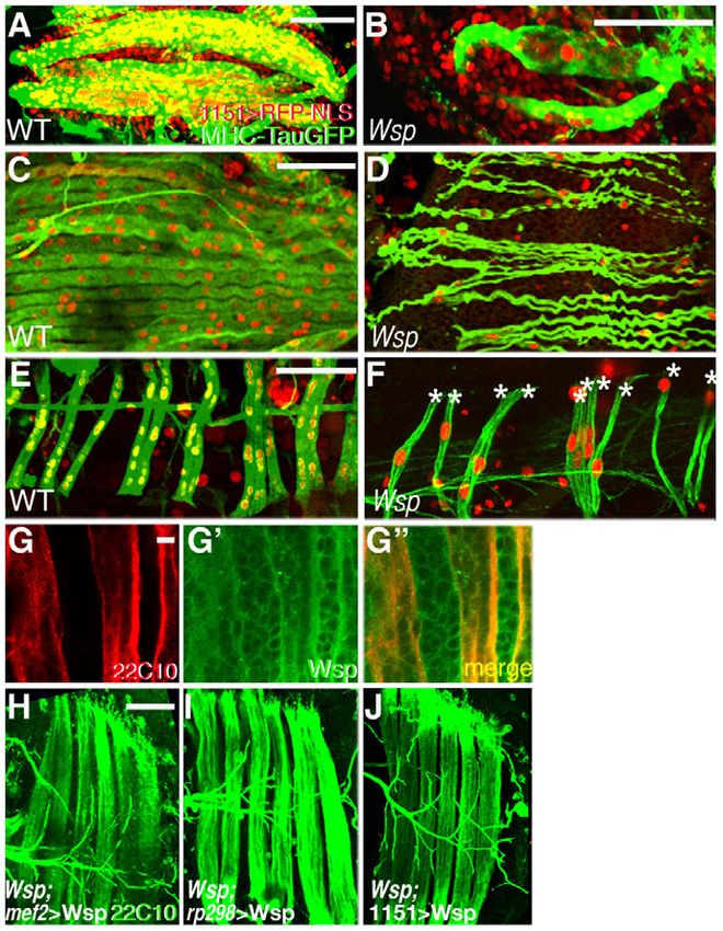
WASp in adult Drosophila myoblast fusion RESEARCH ARTICLE 2349
Fig. 1. Defective musculature of Wsp mutant flies.
(A-C)Thoracic flight muscles of intact, live pupae close to eclosion,
visualized with MHC-TauGFP. (A)Robust musculature of a wild-type
(WT) Drosophila pupa. (B,C)Poorly developed flight muscles are
characteristic of Wsp1/Df(3R)3450 pupae (B) and of pupae in which
the dominant-negative construct UAS-WspCA is expressed in
myogenic tissue only (via the Mef2-GAL4 driver) following the
onset of pupariation (C). (D,E)Dissected sets of MHC-TauGFP-
expressing DLMs from hemithoraces of wild-type (D) and
Wsp1/Df(3R)3450 (E) pupae at 72 hours APF. (F,G)Transverse
sections through pupal thoraces counterstained with Toluidine Blue.
Large groups of bundled flight muscle fibers are present in wild-
type pupae (F). Major muscle groups are indicated: DLM, dorsal
longitudinal flight muscles; DVM, dorsoventral flight muscles; JM,
leg jump muscle. Thoracic muscle bundles are markedly absent
from Wsp1/Df(3R)3450 pupae (G), in which only a few thin groups
of larval flight muscle templates remain (asterisk). Scale bars:
200m in A; 50m in D; 100m in F.
(Bate et al., 1991; Fernandes et al., 1991). Visualization using the wild-type DLMs are filled with many Ewg-positive nuclei (Fig.
myoblast-specific marker Twist demonstrated that Wsp mutants 3C). Most of these are myoblast-derived, whereas a handful of
harbor a normally sized array of proliferating wing disc-associated larger, polyploid nuclei represent the original larval set (Roy and
myoblasts (Fig. 2C,D). Taken together, these observations imply VijayRaghavan, 1998). DLMs from 18 hour APF Wsp mutant
that the two cell populations from which DLM fibers are pupae contained only 9±2.2 (n8) large nuclei (Fig. 3D), which is
constructed – larval template fibers and myoblasts – undergo similar to the established number (8-13) of template nuclei prior to
normal specification and morphogenesis in Wsp zygotic mutants. the onset of fusion (Dutta et al., 2004) and indicative of a complete
Once initiated, the process of DLM growth involves multiple arrest of myoblast fusion in Wsp mutants. Failure of fusion between
rounds of template-myoblast cell fusion and is accompanied by
splitting of the fibers, generating a mature fiber set by ~24 hours
APF (Fernandes et al., 1991; Roy and VijayRaghavan, 1998).
Examination of the DLMs at this stage revealed dramatic
morphological differences between the wild-type thoracic muscles
and those that form in Wsp mutants (Fig. 2E-F⬙). As readily
observed by monitoring the expression of MHC-TauGFP, Wsp
DLMs did not increase in size and failed to split (Fig. 2E,F). Thus,
each Wsp hemithorax contained three immature fibers at 24 hours
APF, in contrast to the enlarged six-fiber set of corresponding wild-
type DLMs. Staining for 22C10 (Futsch), a microtubule-associated
protein expressed in both mature DLM fibers and peripheral nerves
(Dutta et al., 2004; Hummel et al., 2000a), underscored the poor
differentiation of DLMs in Wsp mutants, which were nonetheless
innervated.
The observations described above define the period of 12-24
hours APF as a time window during which the absence of zygotic
Wsp function leads to abnormal flight muscle formation. We
therefore observed, simultaneously, the developing DLM fibers and
the population of myoblasts that contributes to DLM growth during
this period (Fig. 3). Fibers were viewed using template-specific
markers and myoblasts were visualized with the aid of 1151-GAL4,
a myoblast-specific GAL4 driver (Roy and VijayRaghavan, 1997).
An initial experiment utilized 1151-GAL4-based expression of
UAS-RedStinger (Barolo et al., 2004), an RFP variant that localizes Fig. 2. Flight muscle defects in Wsp mutants arise during early
to the nucleus (Fig. 3A-B⬙). Wild-type DLMs at 18 hours APF pupal development. (A,B)DLM templates expressing MHC-TauGFP at
contained many RFP-expressing nuclei (Fig. 3A-A⬙), reflecting the 12 hours APF. Templates in Wsp1/Df(3R)3450 Drosophila pupae (B)
massive incorporation of myoblasts into the larval templates via resemble those found in wild-type pupae (A). (C,D)Dissected third
DEVELOPMENT
cell fusion. In stark contrast, the DLMs in age-matched Wsp pupae, instar larval wing imaginal discs stained for the nuclear myoblast
although surrounded by myoblasts, were completely devoid of marker Twist. A large population of myoblasts is found throughout the
notum of both wild-type (C) and Wsp1/Df(3R)3450 (D) discs.
RFP-expressing nuclei and retained their larval stage morphology
(E-F⬙) Visualization of DLMs at 24 hours APF in wild-type (E-E⬙) and
and size (Fig. 3B-B⬙). Wsp1/Df(3R)3450 (F-F⬙) pupae via MHC-TauGFP expression (green) and
To provide an alternative fusion assay, we made use of the staining with Mab 22C10 (red). Whereas the wild-type fiber set splits in
transcription factor Erect wing (Ewg), which localizes to the nuclei two and grows in size to yield six mature fibers in each hemithorax, the
of both larval muscle templates and fusing myoblasts (DeSimone underdeveloped DLM templates of Wsp1/Df(3R)3450 pupae remain thin
et al., 1996; Fernandes and Keshishian, 1996). At 18 hours APF, and fail to split. Scale bars: 50m.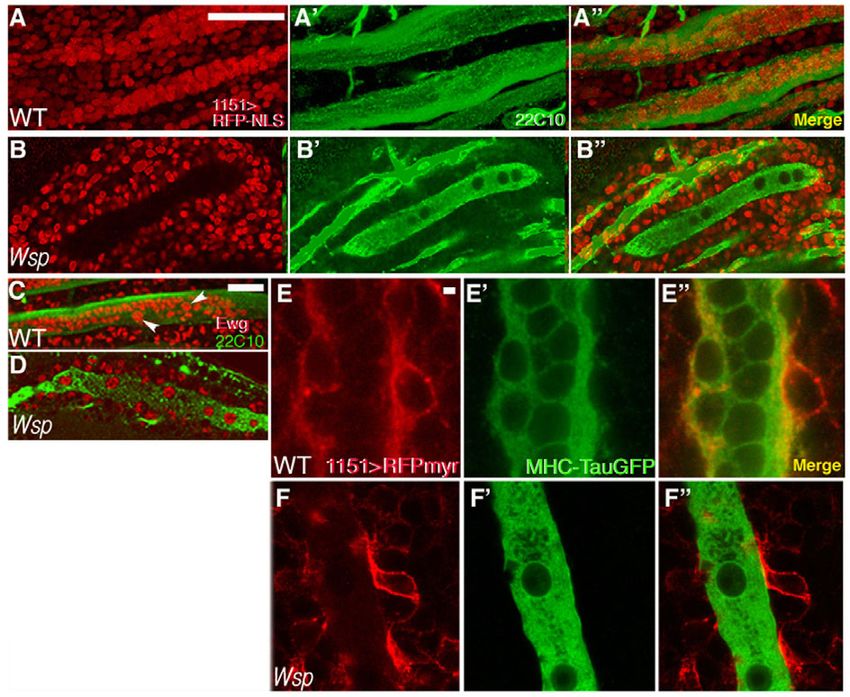
2350 RESEARCH ARTICLE Development 138 (11)
Fig. 3. Failure of fusion between template
fibers and surrounding myoblasts in Wsp
mutant pupae. (A-B⬙) Myoblast nuclei (red,
visualized by 1151-GAL4>UAS-RFP-
NLS/RedStinger) and DLM fibers (green,
visualized with Mab 22C10) in wild-type (A-A⬙)
and Wsp1/Df(3R)3450 (B-B⬙) Drosophila pupae
at 18 hours APF. Wild-type DLMs contain many
myoblast nuclei, whereas the immature fibers
in Wsp mutant pupae have not incorporated
any myoblast nuclei. The black circular figures
within these fibers correspond to the enlarged
polyploid nuclei of larval muscles (Dutta et al.,
2004). (C,D)DLM preparations from wild-type
(C) and Wsp1/Df(3R)3450 (D) pupae at 18
hours APF, stained with anti-Ewg (red) and
Mab 22C10 (green). Arrowheads (C) point to
the large, persisting larval nuclei. Such nuclei
are the only ones present in Wsp mutant fibers
(D). (E-F⬙) Myoblasts (red, visualized by 1151-
GAL4>UAS-myr-mRFP) and DLM fibers (green,
visualized with MHC-TauGFP) in wild-type (E-
E⬙) and Wsp1/Df(3R)3450 (F-F⬙) pupae at 18
hours APF. Incorporation of the membrane
marker into DLM membranes, as is observed in
wild type, does not appear to take place in
Wsp mutants. Scale bars: 50m in A,C; 2m
in E.
myoblasts and the DLM templates was further illustrated by Significantly, these muscles displayed established morphological
myoblast-specific expression of UAS-myr-mRFP (Kandachar et al., characteristics of DVM founder myoblasts, including an elongated
2008), a membrane-tethered RFP construct (Fig. 3E-F⬙). shape and an enlarged nucleus (Fernandes et al., 2005; Rivlin et al.,
Incorporation of the myoblast-derived myr-mRFP into wild-type 2000), consistent with the proper initiation of their specialized
DLM membranes was readily apparent at 18 hours APF (Fig. 3E- myogenic differentiation program, followed by arrest at the onset
E⬙), whereas no such incorporation was observed in Wsp mutant of fiber growth via fusion.
pupae, despite the close apposition of myoblasts to the template Since DVMs in Wsp mutants were often difficult to identify,
fibers (Fig. 3F-F⬙). we examined the abdominal muscles as a more reliable
Taken together, these phenotypes imply that Wsp myoblasts preparation for quantification of fusion events during de novo
migrate from the imaginal wing disc and attach to the DLM fibers, muscle formation. Both lateral and dorsal abdominal muscles
but that the fibers and adhered myoblasts fail to fuse. The adult were similarly underdeveloped in Wsp mutant pupae (Fig. 4C-
flight muscle abnormalities characteristic of zygotic Wsp mutants F). Both abdominal muscle types were properly arranged in
therefore result from a specific and apparently complete disruption parallel arrays, but the fibers that formed were very thin and
of myoblast fusion capabilities in these flies. commonly contained only a single nucleus (1.1±0.3, n10 dorsal
muscle nuclei), in contrast to the thicker, multinucleated
Wsp is a general mediator of adult myoblast (9.7±1.8, n13) wild-type fibers. These observations suggest
fusion, acting in both muscle fibers and that, as for the thoracic flight muscles, the specification and
myoblasts differentiation aspects of the adult abdominal myogenic program
Whereas DLM fiber growth is based on fusion between wing disc- are properly initiated in Wsp zygotic mutants, but that fiber
associated myoblasts and persistent larval templates, most adult growth and maturation are arrested owing to an inability to
Drosophila muscles are formed by an alternative (‘de novo’) incorporate myoblasts through cell fusion.
myogenic program, in which individual founder or pioneer The fusion events that underlie all programs of muscle growth
myoblasts seed formation of fibers via fusion with neighboring in Drosophila occur between distinct cell types, namely growing
myoblasts (Dutta et al., 2004; Fernandes and Keshishian, 1999). syncytial fibers and individual myoblasts. The employment of
Prominent examples include the thoracic dorsoventral muscles separate genetic programs by fibers and myoblasts is a general,
(DVMs), which constitute a second major set of IFMs, and the established feature of myogenesis (Buckingham et al., 2003;
various groups of abdominal muscles, which are derived from Estrada et al., 2006), raising the issue of a differential mechanistic
DEVELOPMENT
nerve-associated myoblasts (Currie and Bate, 1991; Fernandes and contribution to the fusion process by the pairs of fusing cells
Keshishian, 1999; Rivlin et al., 2000; Atreya and Fernandes, 2008). (Chen and Olson, 2004). We therefore sought to determine
We monitored the development of these different muscle types in whether any muscle cell type bias exists in the utilization of Wsp
Wsp mutant pupae and observed severe defects in the construction during pupal muscle cell fusion. We chose to study this issue
of all muscles examined (Fig. 4A-F). Wsp DVMs were during growth of the DLM fibers, as fibers and myoblasts are
considerably smaller than their wild-type counterparts and clearly defined in this muscle system from the outset. We first
contained a minimal number of nuclei, as opposed to the dozens of used anti-Wsp antibodies to assess the expression pattern of Wsp
nuclei present within the wild-type muscles (Fig. 4A,B). protein in developing DLM flight muscles, and observed a general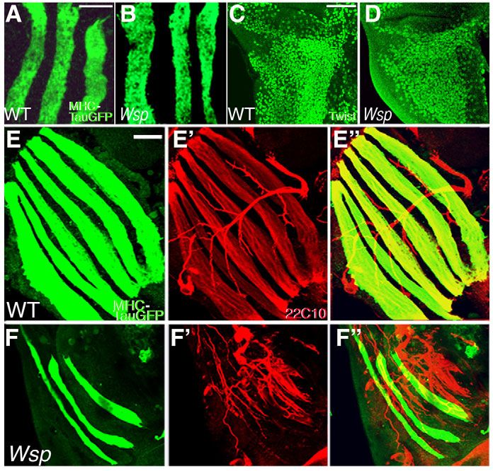
WASp in adult Drosophila myoblast fusion RESEARCH ARTICLE 2351
restriction of GAL4 activity to pupal stages (see Fig. S1 in the
supplementary material). Robust rescue of DLM growth in Wsp
mutant pupae was similarly observed following expression of Wsp
via the myoblast-specific driver 1151-GAL4 (Fig. 4J). These
experiments therefore suggest that Wsp activity in either fibers or
myoblasts is sufficient to ensure fusion-based growth of DLM
muscles.
Taken together, the Wsp fusion-arrest mutant phenotype
observed in all pupal muscles, the general expression pattern of
endogenous Wsp protein, and the full functionality of Wsp
regardless of cell type source, imply that Wsp provides a general
fusion-related function that is common among myogenic cells.
Furthermore, the ability to obtain full rescue when supplying Wsp
from either of the fusion partners is likely to have important
mechanistic implications.
Wsp acts at adult myoblast cell membranes prior
to fusion pore formation
Arp2/3-based actin polymerization in cells commonly takes place
in the vicinity of the plasma membrane, and the forces generated
by this activity mediate a variety of membrane-associated processes
(Takenawa and Suetsugu, 2007). To monitor the subcellular
localization pattern of Wsp in fusing myoblasts, we expressed a
functional Wsp-GFP fusion protein (Massarwa et al., 2007)
together with a dominant-negative form of the small GTPase Rac1
(RacDN), the expression of which in myoblasts leads to the arrest
of muscle cell fusion (Dutta et al., 2004; Fernandes et al., 2005).
Such an approach is commonly employed in studies of embryonic
myoblast fusion to allow for the accumulation of fusion-related
elements at their sites of activity (Galletta et al., 2004; Massarwa
Fig. 4. Wsp function is required in all fusing muscles and is et al., 2007; Menon et al., 2005). A prominent concentration of
sufficient when provided from either myofibers or myoblasts. Wsp-GFP was observed at the myoblast attachment sites in 1151-
(A-F)DVM thoracic flight muscles at 24 hours APF (A,B), and lateral GAL4>UAS-RacDN pupae (Fig. 5A), suggesting that Wsp localizes
(C,D) or dorsal (E,F) abdominal muscles at 48 hours APF of wild-type normally to the membrane regions that are actively involved in
(A,C,E) and Wsp1/Df(3R)3450 (B,D,F) Drosophila pupae. Fibers are fusion. Furthermore, expression in myogenic cells of Wspmyr, a
visualized with MHC-TauGFP and myoblast nuclei are visualized using
plasma membrane-tethered form of Wsp (Bogdan et al., 2005),
1151-GAL4>UAS-NLS-RFP/RedStinger. DVMs and abdominal muscles of
Wsp mutant pupae assume characteristic morphologies but fail to fuse
substantially rescued the Wsp mutant phenotype, underscoring the
with surrounding myoblasts. Asterisks (F) mark individual muscle fibers. functional significance of targeting Wsp to muscle cell membranes
(G-G⬙) Maturing DLMs at18 hours APF stained with Mab 22C10 (red) to (Fig. 5B).
mark the larval templates and with anti-Wsp (green). Wsp expression is D-WIP (Vrp1 or Sltr), the sole Drosophila member of the WIP
observed in both growing fibers and adjacent myoblasts. (H-J)Rescue protein family (Moreau et al., 2000; Sasahara et al., 2002), acts in
of the Wsp mutant DLM defects, as visualized by Mab 22C10, can be a membrane recruitment capacity to mediate the involvement of
achieved by driving UAS-Wsp in all muscle cells using Mef2-GAL4 (H) as Wsp in embryonic myoblast fusion (Kim et al., 2007; Massarwa et
well as by myofiber-specific expression using rp298-GAL4 (a GAL4 al., 2007). Furthermore, disruption of D-WIP function during pupal
insertion in the duf locus) from 0 hours APF (I), or by myoblast-specific development generates a Wsp-like fusion-arrest phenotype (see Fig.
expression obtained with the myoblast driver 1151-GAL4 (J). Scale bars:
S2 in the supplementary material). To assess whether D-WIP is
50m in A-C,E,H; 5m in G.
required for fusion site localization of Wsp, we used the 1151-
GAL4 driver to express Wsp-GFP in the D-WIP mutant
distribution in both the growing fibers and the adjacent myoblasts background. Surprisingly, Wsp-GFP remained prominently
(Fig. 4G-G⬙), suggesting that both muscle cell types could serve localized to the membrane attachment sites between myoblasts and
as a source of Wsp. immature DLM fibers (Fig. 5C), suggesting that Wsp accumulates
To address the possibility of a functional bias, we used different at these sites independently of D-WIP function. Taken together,
muscle cell type GAL4 drivers to express UAS-Wsp in a Wsp these observations imply an essential role for D-WIP function
mutant background. Phenotypic rescue of the Wsp DLM mutant during adult myoblast fusion, which is somewhat distinct, however,
DEVELOPMENT
phenotype was readily achieved using the pan-myogenic Mef2- from its embryonic role as a Wsp-recruiting element.
GAL4 driver (Fig. 4H). Rescue of the mutant phenotype was The demonstration of a conserved requirement for Wsp function
similarly obtained (Fig. 4I) when Wsp was specifically expressed during adult myoblast fusion raises the issue of the specific cellular
in the persistent larval templates, under rp298 (duf)-GAL4 (also tasks that this element performs. Observations made on Wsp
known as kirre – FlyBase) (Menon and Chia, 2001). Although a mutant embryos have suggested a role in fusion pore expansion, as
previous detailed study concluded that this driver is not active in fusion arrests at a relatively late stage, following the formation of
myoblasts (Atreya and Fernandes, 2008), we again used the nascent fusion pores (Berger et al., 2008; Gildor et al., 2009;
GAL80ts/TARGET system (McGuire et al., 2004) to ensure Massarwa et al., 2007). To address the possibility that this role is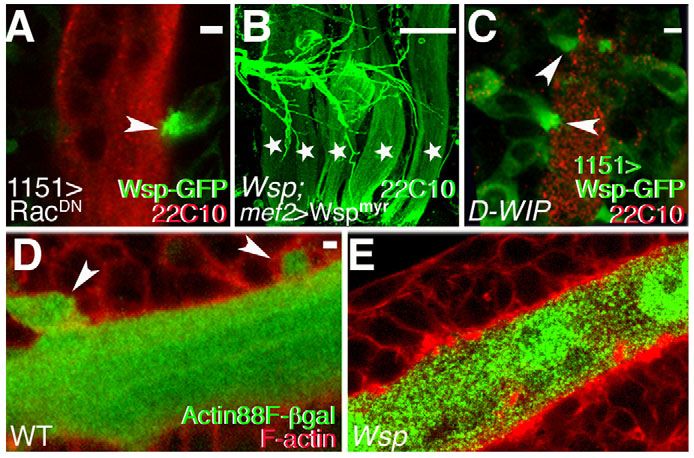
2352 RESEARCH ARTICLE Development 138 (11)
homologs for WASp (Wsp) and SCAR/WAVE (SCAR), which
generally operate in separate developmental and cellular settings
(Zallen et al., 2002). Embryonic myoblast fusion is exceptional in
this regard, as both NPFs have been ascribed essential roles in the
fusion process (Kim et al., 2007; Massarwa et al., 2007;
Richardson et al., 2007; Schafer et al., 2007; Schroter et al., 2004).
Direct comparisons, however, reveal separate and temporally
distinct roles for the two Arp2/3 NPFs in which SCAR acts prior
to Wsp, raising questions regarding the mechanistic basis by which
such functional distinctions are achieved (Berger et al., 2008;
Gildor et al., 2009). To determine the functional interplay of these
factors during the establishment of adult Drosophila muscles, we
first sought to complement the above analysis of Wsp mutants by
disrupting the activity of SCAR and its associated molecular
machinery in developing pupae.
Unlike Wsp, the maternal contribution of SCAR and related
Fig. 5. Wsp acts at fusing myoblast cell membranes prior to elements is not sufficient to overcome zygotic requirements during
fusion pore initiation. (A)In 1151-GAL4>RacDN Drosophila pupae, embryonic and larval stages. We therefore chose to pursue restricted
Wsp-GFP accumulates at the interface (arrowhead) between fusion- tissue and developmental stage expression of UAS-based RNA-
arrested myoblasts and larval templates (red, Mab 22C10). interference (RNAi) transgenes (Dietzl et al., 2007; Schnorrer et al.,
(B)Expression of Wspmyr, a membrane-tethered form of Wsp, almost 2010), as an alternative method of disrupting gene activity during
fully restores the normal DLM pattern (green, Mab 22C10) in establishment of the adult fly musculature. To verify the usefulness
Wsp1/Df(3R)3450 pupae. Individual DLMs are marked with stars.
of this approach in the context of adult myoblast fusion, we first
(C)Wsp-GFP (green) accumulates at attachment sites (arrowheads)
between fusion-arrested myoblasts and larval templates (red, Mab assessed the effects of expressing UAS-Wsp-RNAi and UAS-D-
22C10) in a fusion-arrested D-WIPD30/sltrS1946 pupa. (D,E)Lack of WIP-RNAi constructs using the muscle-specific Mef2-GAL4 driver.
cytoplasmic transfer in Wsp mutant pupae. (D)-galactosidase (green), Severe myogenic phenotypes resembling those obtained with loss-
specifically expressed in DLM templates by the Actin 88F enhancer trap, of-function alleles were observed in both cases (Fig. 6A-C).
transfers and accumulates in attached myoblasts (arrowheads) upon Incorporation of UAS-Dicer2, an established tool for enhancing
the onset of fusion. Myoblast contours (cortical F-actin) are visualized RNAi activity (Dietzl et al., 2007), proved useful in this context as
with phalloidin (red). (E)No such transfer is observed in the many it led to more severe and consistent defects in flight muscle
fusion-arrested myoblasts attached to the DLM templates of formation. We therefore applied this protocol to examine the
Wsp1/Df(3R)3450 pupae. Scale bars: 2m in A,C,D; 50m in B. involvement of SCAR and associated elements in adult myogenesis.
Although a number of transgenic RNAi constructs directed against
SCAR and functionally associated elements failed to elicit an effect,
conserved during adult myoblast fusion, we monitored cytoplasmic Mef2-GAL4-based expression of a SCAR-RNAi construct from the
transfer between myoblasts as a criterion for the stage at which the potent VALIUM series (Ni et al., 2008; Ni et al., 2009) strongly
fusion process is impaired. We made use of the Actin 88F-lacZ disrupted flight muscle development, generating a characteristic
enhancer trap transgene, in which -galactosidase (-gal) myoblast fusion-arrest phenotype of unfused myoblasts congregated
expression is limited to growing fibers, but is absent from around thin, underdeveloped DLM templates (Fig. 6D-E⬙). Such
individual myoblasts (Fernandes et al., 1991; Hiromi et al., 1986). myogenic defects (Fig. 6F-F⬙) were also observed following similar
Close examination of wild-type Actin 88F-lacZ pupae during DLM utilization of a VALIUM series RNAi construct targeting kette,
maturation revealed incorporation of -gal into a small population which encodes the conserved Nap1 (Hem – FlyBase) component of
of attached myoblasts (Fig. 5D), demonstrating that cytoplasmic the SCAR regulatory complex (Hummel et al., 2000b). Zygotic loss-
transfer between partially fused cells could be detected in this of-function mutations in this element result in a strong arrest of
setting. Transfer of this type was never observed, however, in Wsp embryonic myoblast fusion and have been used as a major tool in the
mutant pupae bearing Actin 88F-lacZ, as none of the numerous study of SCAR function in this context (Richardson et al., 2007;
unfused myoblasts attached to the persistent templates appeared to Schroter et al., 2004). The specificity of the kette-RNAi construct
contain -gal (Fig. 5E). These observations imply that the fusion was verified by restoration of normal myogenesis following co-
process in Wsp mutant pupae arrests before the initiation of expression of UAS-kette (see Fig. 6). As expected, strong fusion-
cytoplasmic continuity. arrest phenotypes were observed following RNAi-based targeting of
Sop2 and Arp2 (Hudson and Cooley, 2002), which encode two
The Arp2/3 nucleation-promoting factor SCAR is subunits of the Arp2/3 complex (Fig. 6G-H⬙), the downstream target
required for adult myoblast fusion of both the Wsp and SCAR NPFs. Quantification of DLM nuclear
WASp family proteins act as NPFs, stimulating the capacity of the content (Fig. 6J) underscored the strong inhibition of myoblast fusion
DEVELOPMENT
conserved Arp2/3 protein complex to nucleate actin polymerization by RNAi targeting the Arp2/3 complex or either of its major NPF
and generate branched microfilament arrays (Goley and Welch, systems.
2006). Metazoan cells commonly employ WASp family proteins
and the related SCAR elements as the major Arp2/3 NPFs (Pollitt Arp2/3, but neither of its major NPFs, mediates
and Insall, 2009). Although the two classes of NPFs share a formation of fusion-associated actin foci
common mode of interaction with the Arp2/3 complex, they The demonstrated involvement of actin polymerizing factors
recognize and are activated by distinct molecular machineries during adult myoblast fusion implies the formation of
(Derivery and Gautreau, 2010). Drosophila possesses single microfilament structures that contribute to the fusion process. To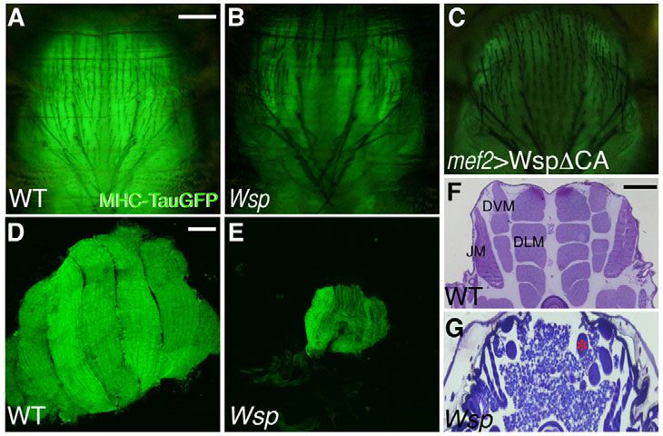
WASp in adult Drosophila myoblast fusion RESEARCH ARTICLE 2353
Fig. 6. Muscle-specific RNAi expression reveals a requirement for the SCAR NPF in adult myoblast fusion. (A-C)Dissected Drosophila
DLMs at 24 hours APF visualized with Mab 22C10. DLM templates (stars) fail to grow and split following expression of UAS-RNAi targeted against
Wsp (B) and D-WIP (C) via Mef2-GAL4. (D-H⬙) DLMs at 24 hours APF following Mef2-GAL4-based expression of transgenic UAS-RNAi constructs.
Fibers are visualized with Mab 22C10 (green) and nuclei with anti-Ewg (red). Ewg is expressed in all nuclei within the fibers as well as in nuclei of
surrounding myoblasts. The wild-type images (D-D⬙) represent the driver on its own. RNAi constructs used were targeted against SCAR (E-E⬙), kette
(F-F⬙), Sop2 (G-G⬙) and Arp2 (H-H⬙). (I)The extent of DLM fusion was quantified by counting Ewg-positive nuclei within single DLM fibers at 24
hours APF. Wild-type fibers contain 187±30.7 nuclei (n4). Quantification of Ewg-positive nuclei within DLMs of Wsp1/Df(3R)3450 mutant pupae
serves as the measure of fiber nuclear content in the complete absence of fusion (see also Fig. 3D). Fusion is strongly inhibited following expression
in muscles of D-WIP-RNAi (26.8±5 nuclei, n10), Wsp-RNAi (34.5±6.8 nuclei, n8), SCAR-RNAi (29.9±6.6, n10), kette-RNAi (34.2±5.6, n10),
Arp2-RNAi (11.6±4.7, n9) and Sop2-RNAi (14.3±2.8, n6). Co-expression of UAS-kette (Hummel et al., 2000b) together with the kette-RNAi
construct significantly rescues the fusion defect (129.1±13.5 nuclei, n9), verifying the specificity of the RNAi construct. The minimal degree of
fusion observed for all RNAi transgenes, including the Wsp-targeting construct, attests to the difficulty of obtaining complete disruption of gene
activity by this approach. Error bars indicate s.d. Scale bar: 50m.
try to identify such structures, we monitored the subcellular and phalloidin (19/20 foci examined), whereas overlap of rp298-
distribution of phalloidin-stained microfilaments during DLM GAL4-expressed Moe-GFP and phalloidin was rarely seen (2/18
formation. This analysis revealed that single, prominent, foci), and even then it was restricted to a minimal portion of the
spherical F-actin structures form at the interface between DLM structure (Fig. 7C-C⬙). The strong bias obtained using this
fibers and attached myoblasts, on the verge of fusion (Fig. 7A). approach suggests, therefore, that the actin foci reside primarily,
We refer to these structures as actin ‘foci’, a term used to if not exclusively, within myoblasts.
describe F-actin structures of similar appearance and size (2-3 We next sought to determine whether the Arp2/3 complex and
m2 in area) that are associated with myoblast fusion during its associated NPFs contribute to formation of the fusion-associated
Drosophila embryogenesis (Gildor et al., 2009; Kesper et al., actin foci. We first monitored the localization pattern of two
2007; Kim et al., 2007; Richardson et al., 2007; Sens et al., subunits of the Arp2/3 complex, Arp3 and Sop2 (Arpc1), by
2010). Such foci were also observed during construction of the expression of GFP-tagged forms (Hudson and Cooley, 2002) in
DEVELOPMENT
adult de novo forming DVMs (see Fig. S3 in the supplementary fusing adult myoblasts. Both constructs were found to strongly co-
material), suggesting that they are a general feature of myoblast localize with the actin foci, consistent with a role for Arp2/3 in
fusion in Drosophila. To assess the manner in which the foci are their establishment (Fig. 7D-E⬙). Indeed, following expression of
distributed between DLM fibers and fusing myoblasts, we RNAi constructs targeted against the Arp2/3 subunits Arp2 and
visualized them following cell type-specific expression of the Sop2 via Mef2-GAL4, the bright, spherical foci were replaced by
GFP-tagged F-actin-binding domain of Moesin (Moe) (Chihara small and diffuse accumulations of actin (Fig. 7G-I), implying that
et al., 2003; Dutta et al., 2002). Full overlap (Fig. 7B-B⬙) was Arp2/3 function is essential for the proper construction of the
commonly observed between 1151-GAL4-expressed Moe-GFP fusion-associated foci.2354 RESEARCH ARTICLE Development 138 (11)
Fig. 7. Arp2/3-dependent F-actin foci form during adult myoblast fusion. (A)Spherical foci containing F-actin (arrows) are present at the
interface between DLM templates (visualized with Mab 22C10, green) and attached myoblasts in an 18 hour APF Drosophila pupa. Foci and
myoblast outlines are visualized with phalloidin (red). (B-B⬙) An 18 hour APF pupa expressing the F-actin-binding protein Moe-GFP under the control
of the myoblast-specific driver 1151-GAL4. The anti-GFP (B, green) and phalloidin (B⬘, red) staining patterns are entirely coincident (B⬙, merge).
(C-C⬙) By contrast, when expressing Moe-GFP via the fiber-specific driver rp298 (duf)-GAL4, the anti-GFP (C, green) and phalloidin (C⬘, red) staining
patterns are mostly separate (C⬙, merge) in the rare instances in which any overlap is detected at all. The actin focus therefore resides primarily, if
not exclusively, within myoblasts. (D-E⬙) Pupae at 18 hours APF expressing GFP-tagged versions of the Arp2/3 subunits Arp3 (D-D⬙) and Sop2 (E-E⬙).
Both subunits, visualized using anti-GFP (green), strongly co-localize with F-actin (phalloidin, red) at focus structures (arrows in merges).
(F-F⬙) Endogenous D-WIP in an 18 hour APF pupa, as visualized with anti-D-WIP antibodies (green), is also found to co-localize with the F-actin
(phalloidin, red) focal structures (arrows in merge). (G-O)Morphology of 18 hour APF actin foci (visualized with phalloidin, red) following RNAi-
based targeting of Arp2/3 and related NPFs via the Mef2-GAL4 driver. DLM template muscles are visualized with Mab 22C10 (green) in all panels.
(G)Wild-type-sized focus in the absence of RNAi construct expression. Weak, diffuse foci are observed following targeting of the Arp2/3 subunits
Arp2 (H) and Sop2 (I). Targeting of Wsp (J) and D-WIP (K) does not affect focus size or morphology, whereas enlarged somewhat irregular foci are
DEVELOPMENT
found in pupae expressing RNAi targeting SCAR (L), the SCAR complex element kette (M) and both Wsp and SCAR (N) or Wsp and kette (O).
(P)The areas of actin foci in pupae expressing different RNAi constructs. Values represent the average area of ~25 foci for each genotype. Asterisks
indicate foci size values that differ significantly (PWASp in adult Drosophila myoblast fusion RESEARCH ARTICLE 2355
that expression of RNAi constructs targeting either SCAR or the 2007; Massarwa et al., 2007; Schafer et al., 2007), DLM growth
SCAR complex element kette led to the formation of enlarged, presents a distinct paradigm. The incorporation of myoblasts into
irregularly shaped actin foci (Fig. 7L,M). Enlarged foci were pre-existing fibers is characteristic of several aspects of skeletal
similarly generated following simultaneous RNAi-based targeting of muscle growth in vertebrates, including the repair of injured muscle
both the Wsp- and SCAR-based NPF systems (Fig. 7N,O). Thus, by satellite cell-derived myoblasts (Charge and Rudnicki, 2004)
neither separate nor combined Wsp or SCAR complex NPF function and the initiation of the second wave of mammalian embryonic
appears to directly mediate the contribution of the Arp2/3 complex myogenesis (Jansen and Pavlath, 2008). The essential and
to actin focus construction, implying the involvement of a distinct, functionally conserved involvement of Wsp in diverse programs of
as yet unknown system for stimulation of Arp2/3 actin nucleation Drosophila somatic muscle formation and growth leads us to
activity in this context. propose that the actin nucleation-promoting activity of WASp
As discussed below, the nature of the involvement of the WASp family proteins constitutes a fundamental aspect of the cell fusion
and SCAR pathways in actin focus formation in adult myoblasts events that accompany myogenesis.
mirrors observations made during myoblast fusion in Drosophila
embryos. The ability to directly monitor Arp2/3 complex function Involvement of the Arp2/3 nucleation system in
is, however, unique to the adult system and leads us to suggest that the formation of fusion-associated actin foci
the fusion-associated foci are Arp2/3-dependent branched Spherical F-actin-rich structures closely associated with myoblast
microfilament structures and that a novel NPF mediates the fusion pore formation during Drosophila embryogenesis have been
involvement of Arp2/3 in this process. recently described and extensively characterized (Onel and
Renkawitz-Pohl, 2009; Richardson et al., 2008; Richardson et al.,
DISCUSSION 2007; Sens et al., 2010). Our observation of very similar structures
The universal nature of muscle fiber formation and growth via during the period of adult muscle growth via fusion now suggests
myoblast fusion suggests that common molecular mechanisms that these foci are a universal feature of the myoblast fusion
underlie the fusion process. The myogenic processes leading to the process. Although a variety of alterations in embryonic actin focus
formation of adult Drosophila muscles provide a promising, yet morphology have been described for various mutants, the
generally unexplored, setting in which conserved elements of this molecular mechanism governing their formation is still unknown.
type can be identified and characterized. This is particularly true A key lingering question is the identity of the actin nucleation
for establishment of the prominent thoracic IFMs, which exhibit machinery that is responsible for establishment of the foci. The
similarities in their arrangement and program of differentiation to Arp2/3 nucleation system is an obvious candidate for such a role,
both Drosophila embryonic myogenesis and muscle formation in given the well-documented involvement of its different
vertebrates, including mammals (Fernandes and Keshishian, 1999; components in embryonic myoblast fusion. However, direct
Dutta and VijayRaghavan, 2006). Whereas a detailed genetic assessment of Arp2/3 complex function during embryogenesis is
analysis of myoblast fusion has been undertaken for the Drosophila difficult because the maternal contribution of complex subunits,
embryo (Beckett and Baylies, 2006; Maqbool and Jagla, 2007; which is essential for oogenesis (Hudson and Cooley, 2002; Zallen
Abmayr et al., 2008), few, if any, mutants affecting the et al., 2002), is sufficient to overcome zygotic gene disruption. The
corresponding adult process have been reported. In the current capacity of RNAi to efficiently disrupt gene function during adult
study, we have utilized a combination of genetic approaches to myogenesis thus provides a unique opportunity to study the
initiate progress along these lines via the study of the adult contribution of the Arp2/3 complex to actin focus formation. Our
myogenic requirements for Wsp, the sole Drosophila WASp family data suggest that Arp2/3 indeed plays a crucial role in this process,
member, and additional elements of the cellular actin as focus formation is significantly impaired upon expression of
polymerization machinery. RNAi targeting two of the complex subunits.
Such a requirement for Arp2/3 is somewhat surprising
Wsp function is essential during adult myoblast because, as shown above, actin foci persist or grow following
fusion separate or simultaneous disruption of the two major Arp2/3
A major finding of our study is that Wsp performs an essential role NPF systems centered on Wsp and SCAR. Furthermore, these
during adult fly myogenesis, which is crucial for the growth of behaviors closely match those reported following disruption of
muscle fibers via myoblast fusion. This requirement is highly Wsp and SCAR system activity by a variety of means during
specific to the fusion process, as other features and characteristics embryogenesis (Gildor et al., 2009; Richardson et al., 2008;
of adult myoblast development (e.g. the definition of a myoblast Richardson et al., 2007). Although the WASp and SCAR systems
pool, myoblast proliferation levels and the different patterns of continue to be regarded as primary NPFs for Arp2/3, a growing
myoblast migration) appear unaffected in the absence of Wsp list of alternative nucleators has recently emerged (Campellone
activity. The functional requirement for Wsp is comprehensive and and Welch, 2010), suggesting that a novel element might well
universal in character. A complete arrest of myoblast fusion is activate Arp2/3 in this context. Identification of this novel NPF
observed in Wsp mutants for all classes of somatic muscle groups should provide genetic tools for determining the functional
examined. Furthermore, reliance on Wsp activity during fusion is significance of the actin foci, which has so far remained elusive
DEVELOPMENT
common to both of the major adult myogenic programs, namely the owing to the lack of information regarding the mechanism by
de novo construction of muscle fibers from individual myoblasts which they are constructed.
and fiber growth by fusion of myoblasts with persistent larval
templates, as exemplified by the prominent DLM flight muscles Functional aspects of the cellular role performed
(Roy and VijayRaghavan, 1999). by Wsp in fusing muscles
Whereas de novo muscle fiber formation in the adult resembles A central and ongoing issue is the mechanistic role played by Wsp
the Drosophila embryonic program of myogenesis, in which Wsp during myoblast fusion. We find several of the observations
was previously shown to mediate myoblast fusion (Kim et al., reported in our study to be instructive in this context.2356 RESEARCH ARTICLE Development 138 (11)
The capacity to fully rescue the adult Wsp mutant phenotype by Atreya, K. B. and Fernandes, J. J. (2008). Founder cells regulate fiber number
providing functional Wsp in either fibers or individual myoblasts but not fiber formation during adult myogenesis in Drosophila. Dev. Biol. 321,
123-140.
suggests that interactions with cell type-specific factors are not a Barolo, S., Castro, B. and Posakony, J. W. (2004). New Drosophila transgenic
crucial mechanistic feature. It is therefore likely that the fusion- reporters: insulated P-element vectors expressing fast-maturing RFP.
associated microfilament dynamics nucleated by Wsp are Biotechniques 36, 436-442.
Bate, M., Rushton, E. and Currie, D. A. (1991). Cells with persistent twist
performed and utilized by mechanisms and molecular elements expression are the embryonic precursors of adult muscles in Drosophila.
common to all muscle cells. This conclusion is particularly Development 113, 79-89.
noteworthy considering the significance assigned to cell type- Beckett, K. and Baylies, M. K. (2006). The development of the Drosophila larval
specific pathways in some models of embryonic myoblast fusion. body wall muscles. Int. Rev. Neurobiol. 75, 55-70.
Ben-Yaacov, S., Le Borgne, R., Abramson, I., Schweisguth, F. and Schejter, E.
As suggested in the embryo, localization to sites of fusion D. (2001). Wasp, the Drosophila Wiskott-Aldrich syndrome gene homologue, is
between muscle cell membranes appears to be an important aspect required for cell fate decisions mediated by Notch signaling. J. Cell Biol. 152, 1-
of Wsp activity, although the means by which Wsp is targeted to 13.
Berger, S., Schafer, G., Kesper, D. A., Holz, A., Eriksson, T., Palmer, R. H.,
these sites during adult myogenesis is not clear. D-WIP appears to Beck, L., Klambt, C., Renkawitz-Pohl, R. and Onel, S. F. (2008). WASP and
provide a key membrane-targeting function during embryonic SCAR have distinct roles in activating the Arp2/3 complex during myoblast
myogenesis (Massarwa et al., 2007), but our observations suggest fusion. J. Cell Sci. 121, 1303-1313.
Bogdan, S., Stephan, R., Lobke, C., Mertens, A. and Klambt, C. (2005). Abi
that Wsp reaches adult fusion sites independently of D-WIP. activates WASP to promote sensory organ development. Nat. Cell Biol. 7, 977-
Establishment of the mechanism by which Wsp localizes to adult 984.
myoblast fusion sites thus requires further study. These findings Buckingham, M., Bajard, L., Chang, T., Daubas, P., Hadchouel, J., Meilhac, S.,
also leave open the role performed by D-WIP. A direct effect of D- Montarras, D., Rocancourt, D. and Relaix, F. (2003). The formation of
skeletal muscle: from somite to limb. J. Anat. 202, 59-68.
WIP on cytoskeletal organization, as suggested during embryonic Campellone, K. G. and Welch, M. D. (2010). A nucleator arms race: cellular
myoblast fusion (Kim et al., 2007), might be relevant in this control of actin assembly. Nat. Rev. Mol. Cell Biol. 11, 237-251.
context. Charge, S. B. and Rudnicki, M. A. (2004). Cellular and molecular regulation of
muscle regeneration. Physiol. Rev. 84, 209-238.
The transfer of cytoplasmic material between attached, fusion- Chen, E. H. and Olson, E. N. (2001). Antisocial, an intracellular adaptor protein, is
arrested muscle cells, which is a hallmark of the embryonic required for myoblast fusion in Drosophila. Dev. Cell 1, 705-715.
myoblast fusion phenotype in Wsp mutant embryos (Gildor et al., Chen, E. H. and Olson, E. N. (2004). Towards a molecular pathway for myoblast
fusion in Drosophila. Trends Cell Biol. 14, 452-460.
2009; Massarwa et al., 2007), is not observed following disruption Chen, E. H., Pryce, B. A., Tzeng, J. A., Gonzalez, G. A. and Olson, E. N.
of Wsp function during adult myogenesis. The basis for this (2003). Control of myoblast fusion by a guanine nucleotide exchange factor,
difference is unknown and will require better elucidation of the loner, and its effector ARF6. Cell 114, 751-762.
particular aspects of the fusion process that rely on Wsp activity. Chihara, T., Kato, K., Taniguchi, M., Ng, J. and Hayashi, S. (2003). Rac
promotes epithelial cell rearrangement during tracheal tubulogenesis in
One explanation is an expanded role for Wsp during adult myoblast Drosophila. Development 130, 1419-1428.
fusion. An additional functional requirement prior to fusion pore Currie, D. A. and Bate, M. (1991). The development of adult abdominal muscles
formation might mask any subsequent participation in fusion pore in Drosophila: myoblasts express twist and are associated with nerves.
Development 113, 91-102.
expansion. Finally, it is of interest to note that, in the adult, Derivery, E. and Gautreau, A. (2010). Generation of branched actin networks:
disruption of either the Wsp or SCAR NPF systems leads to the assembly and regulation of the N-WASP and WAVE molecular machines.
strong arrest of myoblast fusion, similar to the embryonic scenario. BioEssays 32, 119-131.
DeSimone, S. M. and White, K. (1993). The Drosophila erect wing gene, which
Our observations on adult myogenesis thus serve as a reaffirmation is important for both neuronal and muscle development, encodes a protein
that the fusion process requires non-overlapping functional which is similar to the sea urchin P3A2 DNA binding protein. Mol. Cell. Biol. 13,
contributions from the two major Arp2/3 NPFs, even though they 3641-3649.
operate in close spatial and temporal proximity. DeSimone, S., Coelho, C., Roy, S., VijayRaghavan, K. and White, K. (1996).
ERECT WING, the Drosophila member of a family of DNA binding proteins is
In summary, the observations reported in our study generalize required in imaginal myoblasts for flight muscle development. Development
the involvement of the Arp2/3 complex and its associated cellular 122, 31-39.
machinery during the construction of muscle fibers via myoblast Dietzl, G., Chen, D., Schnorrer, F., Su, K. C., Barinova, Y., Fellner, M., Gasser,
B., Kinsey, K., Oppel, S., Scheiblauer, S. et al. (2007). A genome-wide
fusion. In particular, a prominent and potentially universal role is transgenic RNAi library for conditional gene inactivation in Drosophila. Nature
assigned to the WASp NPF as a mediator of these events. 448, 151-156.
Dutta, D. and VijayRaghavan, K. (2006). Metamorphosis and the formation of
Acknowledgements the adult musculature. In Muscle Development in Drosophila (ed. H. Sink), pp.
We thank the Bloomington, VDRC and TRiP (Harvard) stock centers, S. 125-142. Georgetown: Landes Bioscience.
Bogdan, E. Chen, L. Cooley, J. Fernandes, C. Klambt, R. Palmer and S. Roth for Dutta, D., Bloor, J. W., Ruiz-Gomez, M., VijayRaghavan, K. and Kiehart, D. P.
providing Drosophila strains and reagents. We are grateful for the support and (2002). Real-time imaging of morphogenetic movements in Drosophila using
constructive criticism of the members of our labs at NCBS and WIS. This work Gal4-UAS-driven expression of GFP fused to the actin-binding domain of
was supported by grants from the Israel Science Foundation and the Muscular moesin. Genesis 34, 146-151.
Dystrophy Association to B.-Z.S. and E.D.S. and from the Wellcome Trust and Dutta, D., Anant, S., Ruiz-Gomez, M., Bate, M. and VijayRaghavan, K.
the Department of Biotechnology, Government of India to K.V. B.-Z.S. is an (2004). Founder myoblasts and fibre number during adult myogenesis in
Drosophila. Development 131, 3761-3772.
incumbent of the Hilda and Cecil Lewis Chair in Molecular Genetics. Deposited
Estrada, B., Choe, S. E., Gisselbrecht, S. S., Michaud, S., Raj, L., Busser, B. W.,
in PMC for release after 6 months.
Halfon, M. S., Church, G. M. and Michelson, A. M. (2006). An integrated
DEVELOPMENT
strategy for analyzing the unique developmental programs of different myoblast
Competing interests statement subtypes. PLoS Genet. 2, e16.
The authors declare no competing financial interests. Fernandes, J., Bate, M. and VijayRaghavan, K. (1991). Development of the
indirect flight muscles of Drosophila. Development 113, 67-77.
Supplementary material Fernandes, J. J. and Keshishian, H. (1996). Patterning the dorsal longitudinal
Supplementary material for this article is available at flight muscles (DLM) of Drosophila: insights from the ablation of larval scaffolds.
http://dev.biologists.org/lookup/suppl/doi:10.1242/dev.055012/-/DC1 Development 122, 3755-3763.
Fernandes, J. J. and Keshishian, H. (1999). Development of the adult
References neuromuscular system. Int. Rev. Neurobiol. 43, 221-239.
Abmayr, S. M., Zhuang, S. and Geisbrecht, E. R. (2008). Myoblast fusion in Fernandes, J. J., Atreya, K. B., Desai, K. M., Hall, R. E., Patel, M. D., Desai, A.
Drosophila. Methods Mol. Biol. 475, 75-97. A., Benham, A. E., Mable, J. L. and Straessle, J. L. (2005). A dominantYou can also read