A partial skeleton of Deinotherium (Proboscidea, Mammalia) from the late Middle Miocene Gratkorn locality (Austria)
←
→
Page content transcription
If your browser does not render page correctly, please read the page content below
Palaeobio Palaeoenv (2014) 94:49–70
DOI 10.1007/s12549-013-0140-x

 ORIGINAL PAPER

A partial skeleton of Deinotherium (Proboscidea, Mammalia)
from the late Middle Miocene Gratkorn locality (Austria)
Manuela Aiglstorfer & Ursula B. Göhlich &
Madelaine Böhme & Martin Gross

Received: 26 September 2013 / Revised: 11 November 2013 / Accepted: 28 November 2013 / Published online: 11 February 2014
# Senckenberg Gesellschaft für Naturforschung and Springer-Verlag Berlin Heidelberg 2014

Abstract A disarticulated, though still roughly associated                      Keywords Biostratigraphy . Biochronology . Styria .
partial Deinotherium skeleton from the late Middle                              Sarmatian . Central Europe . Deinotherium levius .
Miocene (late Sarmatian sensu stricto; 12.2–12.0 Ma)                            Deinotherium giganteum
Gratkorn locality (Austria) is described. Based on dimen-
sions and morphology of the material it can be determined
as a medium-sized taxon of Deinotheriidae and definitively                      Introduction
assigned to the genus Deinotherium. This specimen from
Gratkorn confirms the osteological differences in the                           Deinothere remains are frequent findings in the Miocene
postcrania between Prodeinotherium and Deinotherium.                            of Europe and a useful tool for biochronological and
As the diagnostically important p/3 is missing on the                           biostratigraphical considerations (see, e.g. Dehm 1960;
specimen it can only be assigned to Deinotherium levius                         Huttunen 2002a, b; Böhme et al. 2012; Pickford and
vel giganteum. The Gratkorn specimen is one of not many                         Pourabrishami 2013). Following recent works (Böhme et al.
skeletons of a medium-sized taxon of Deinotheriidae and                         2012; Pickford and Pourabrishami 2013) on the stratigraphic
one of only a few well-dated late Middle Miocene occur-                         range of the different species, the genus Prodeinotherium Éhik,
rences in Central Europe with associated dental and post-                       1930 can be considered as indicative for the Early to middle
cranial material.                                                               Middle Miocene, while Deinotherium Kaup, 1829 first occurs
                                                                                in Europe during the Middle Miocene (Mottl 1969; Svistun
                                                                                1974) and is recorded up to the terminal Late Miocene (Markov
                                                                                2008b). Unfortunately, in most cases the findings comprise only
                                                                                isolated remains, and very often only isolated teeth [e.g. abun-
This article is a contribution to the special issue “The Sarmatian vertebrate   dant tooth material from the famous Eppelsheim Formation
locality Gratkorn, Styrian Basin.”                                              (Eppelsheim Fm)]. In contrast to this, a fairly well preserved,
M. Aiglstorfer : M. Böhme                                                       disarticulated, partial Deinotherium skeleton (Fig. 1) of late
Department for Geosciences, Eberhard Karls Universität Tübingen,                Sarmatian age (12.2–12.0 Ma) was discovered in the clay pit
Sigwartstraße 10, 72076 Tübingen, Germany
                                                                                St. Stefan near Gratkorn (Styria, Austria; Gross et al. 2011;
M. Aiglstorfer (*) : M. Böhme                                                   2014, this issue) during geological mapping of the region in
Senckenberg Center for Human Evolution and Palaeoenvironment                    2005. It is one of very few skeleton findings of a medium-sized
(HEP), Sigwartstraße 10, 72076 Tübingen, Germany                                deinothere taxon described so far. The remains were excavated
e-mail: manuela.aiglstorfer@senckenberg.de
                                                                                by the Universalmuseum Joanneum, Graz, from 2005 to 2008.
U. B. Göhlich                                                                   All elements could be assigned to one individual except for
Geologisch-Paläontologische Abteilung, Naturhistorisches Museum                 some tooth remains detected about 30 m NW of the skeleton
Wien, Burgring 7, 1010 Vienna, Austria                                          that represent a second individual. The fragmentary preserva-
M. Gross
                                                                                tion of the latter allowed stable isotope analyses (δ18OCO3,
Universalmuseum Joanneum, Graz, Weinzöttlstraße 16,                             δ13C; see Aiglstorfer et al. 2014, this issue). The excavation of
8045 Graz, Austria                                                              the Deinotherium skeleton led to the discovery of an abundant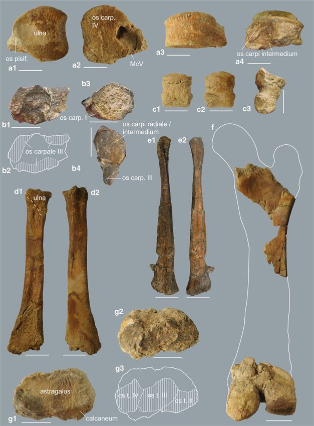
50                                                                                               Palaeobio Palaeoenv (2014) 94:49–70

Fig. 1 Sketch of the partial
skeleton of Deinotherium levius
vel giganteum from the Middle
Miocene of Gratkorn indicating
preserved remains [reconstructed
after D. proavum from Ezerovo
(Late Miocene) mounted at
PMSU (modified after Huttunen
2000; Markov 2008b)]

                                                                                                             from right part of body
                                                                                                             from left part of body
                                                                                                             vertebral column /
                                                                                                             medial part of body
                                                                                                             fragmented /
                                                                                                             not definable
                                                                                                             (costae fragments
                                                                                                             estimated)

and rich vertebrate fauna, which has been excavated in            (1930). In addition to the on-going discussion on valid genera,
continuous campaigns in a cooperative project of the              different concepts concerning species validity are also held at the
Universalmuseum Joanneum, Graz, the Eberhard Karls                moment. While some authors accept five valid morphospecies
Universität Tübingen and the Ludwig-Maximillians-Universität      (Böhme et al. 2012) or chronospecies (Pickford and
München (see other publications in this special issue).           Pourabrishami 2013), others tend to reduce the number to four
                                                                  (Gasparik 1993, 2001; Markov 2008a; Vergiev and Markov
                                                                  2010) or even only two species (Huttunen 2002a). Species
Taxonomy of European Deinotheriidae                               determination is hindered considerably by the difficulty in iden-
                                                                  tifying stratigraphically mixed faunas, the great dimensional and
Taxonomy of Deinotheriidae has been under discussion for long     morphological overlap between the species and the impossibil-
(see, e.g. Gräf 1957; Bergounioux and Crouzel 1962; Harris        ity to evaluate intraspecific variation (Huttunen 2000). Huttunen
1973; Gasparik 1993, 2001; Antoine 1994; Huttunen 2000;           (2002a), for example, synonymized Deinotherium levius
Ginsburg and Chevrier 2001; Duranthon et al. 2007; Markov         Jourdan, 1861 with Deinotherium giganteum Kaup, 1829 due
2008a, b; Vergiev and Markov 2010; Böhme et al. 2012;             to the assumed contemporaneous occurrence of D. giganteum
Pickford and Pourabrishami 2013). At the moment, one,             and D. levius morphotypes in the Eppelsheim Fm.
Deinotherium (Ginsburg and Chevrier 2001; Pickford                Furthermore, the mentioned gradual size increase
and Pourabrishami 2013), respectively two genera,                 (Gasparik 1993; Böhme et al. 2012; Pickford and
Prodeinotherium and Deinotherium (Gasparik 1993; Antoine          Pourabrishami 2013) and the stepwise morphological modifica-
1994; Huttunen 2000; Duranthon et al. 2007; Vergiev and           tion of the characteristic features (Antoine 1994; Gasparik 2001)
Markov 2010), are considered valid. While a gradual size in-      aggravate a clear species differentiation. Huttunen (2002a), like
crease within Deinotheriidae from the Early to the Late Miocene   others before her, considered Deinotherium gigantissimum
is generally accepted, Antoine (1994), Huttunen (2000), Vergiev   Stefanescu, 1892 only “a large variety of D. giganteum”
and Markov (2010) and others argue that Prodeinotherium and       (Huttunen 2002a, p. 244). Dating of deinothere findings and
Deinotherium do not only differ in size but also in dental and    identification of stratigraphically mixed faunas are the keys for
postcranial morphology. Huttunen (2000) gives an overview of      evaluation of inter- and intraspecific variations and for determi-
distinguishing characters between the smaller genus               nation of the role of sexual dimorphism or the sympatric occur-
Prodeinotherium and the larger genus Deinotherium, discussing     rence of different species. In the modern Loxodonta africana
and evaluating the characters given by Harris (1973) and others   Blumenbach, 1797, for example, the average weight of females
on specimens from Central Europe. As noted by Huttunen and        (about 2.8 t) reaches only about 56 % of the males’ average
also observed in this study (see Discussion below), genus diag-   weight (5 t; Joger 2010). Such a scope would include specimens
nostic characters can indeed be identified in the postcranial     from Prodeinotherium bavaricum von Meyer, 1831 to
material and therefore support the separation of two genera       D. giganteum. The large dimensional and morphological vari-
Prodeinotherium and Deinotherium as proposed by Éhik              ability in D. giganteum observed by Huttunen (2000) that led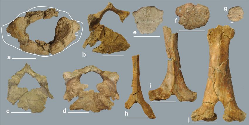
Palaeobio Palaeoenv (2014) 94:49–70                                                                                             51

her to a supposed synonymy with D. levius could thus be a            and Pickford and Pourabrishami (2013) stated that D. proavum
consequence of faunal mixing or uncertainty in stratigraphic         should have priority over D. gigantissimum Stefanescu, 1892
positions of localities, and also biased by a certain degree         and that the latter should be considered a junior synonym.
of sexual dimorphism (Huttunen 2000, 2002b). The mixed
and time-averaged faunal assemblage from the
                                                                     Nomenclature
“Dinotheriensande” (Eppelsheim Fm; at that time considered
stratigraphically uniform) in particular has biased her observa-
                                                                     The terminology for dentition used here (Fig. 2) is modified
tions and those of others for a long time. Böhme et al. (2012)
                                                                     after Gräf (1957), Tassy (1996), Harris (1973), Tobien (1988),
and Pickford and Pourabrishami (2013) were able to show,
                                                                     Huttunen (2000), Pickford and Pourabrishami (2013).
however, that the Eppelsheim Fm also covers a considerable
                                                                     Postcranial terminology follows that of Göhlich (1998).
amount of the Middle Miocene and therefore comprises sev-
eral non-co-occurring Deinotherium species. In contrast to the
observations of Huttunen (2000, 2002a, b), Gräf (1957) gives a
                                                                     Institutional abbreviations
morphospecies differentiation of D. giganteum and D. levius
based on differences in dental material. She already observed
                                                                     GPIT            Paläontologische Sammlung der
variability concerning dental features but as her comparison
                                                                                     Universität Tübingen, Tübingen, Germany
material was limited (Pickford and Pourabrishami 2013) some
                                                                     IGM             Montanuniversität Leoben, Leoben, Austria
of her features were found to be more variable than she
                                                                     MNHN            Muséum National d’Histoire Naturelle,
considered (see, for example, Huttunen 2000 for discussion),
                                                                                     Paris, France
while others show a smaller variability than she estimat-
                                                                     NHMM            Naturhistorisches Museum Mainz, Mainz,
ed due to mixed faunal assemblages (see, for example,
                                                                                     Germany
Pickford and Pourabrishami 2013 for discussion). Gräf
                                                                     NHMW            Naturhistorisches Museum Wien, Vienna,
(1957) further underestimated the dimensional range
                                                                                     Austria
sometimes (Pickford and Pourabrishami 2013). Pickford
                                                                     NMNHS           National Museum of Natural History, Sofia,
and Pourabrishami (2013) based their work on a large
                                                                                     Bulgaria
number of deinothere dental material and tried to focus
                                                                     PMSU            Paleontological Museum of Sofia University
their considerations on well-dated material and to avoid faunal
                                                                                     “St. Klimt Ochrdisky”, Department of
assemblages likely to result from a considerable extent of
                                                                                     Geology and Paleontology, Sofia, Bulgaria
faunal mixing, such as fluvial deposits. These researchers
                                                                     SMNS            Staatliches Museum für Naturkunde
classify different size groups in combination with their strati-
                                                                                     Stuttgart, Stuttgart, Germany
graphic range while being well aware that these groups cannot
                                                                     SNSB-BSPG       Staatliche Naturwissenschaftliche
be strictly separated due to a gradual size increase. Böhme et al.
                                                                                     Sammlungen Bayerns Bayerische
(2012) mention D. bavaricum, D. levius and D. giganteum as
                                                                                     Staatssammlung für Paläontologie und
morphospecies recorded from the Eppelsheim Fm based on
                                                                                     Geologie, Munich, Germany
comparisons with dental material from rich and well-
                                                                     SSN             Paläontologisches Museum Nierstein,
documented localities from Europe.
                                                                                     Nierstein, Germany
    We follow the morphospecies concept of Böhme et al.
                                                                     UMJGP           Universalmuseum Joanneum, Graz, Austria
(2012) with five European species, which differs from other
concepts, such as those of Gasparik (1993, 2001) and Vergiev
and Markov (2010) in the acceptance of the species D. levius,
based on the diagnostic features in the p/3 described by Gräf        Anatomical abbreviations
(1957) and referred to, for example, by Mottl (1969) and
Böhme et al. (2012). We could observe the generic differences        prc/prcd    protocone/protoconid
on the postcranial material from Gratkorn in comparison to           pac         paracone
Prodeinotherium from several localities, and therefore follow        mc/mcd      metacone/metaconid
the two genera concept as proposed by Éhik (1930) and used by        hyc/hycd    hypocone/hypoconid
Gasparik (1993, 2001), Huttunen (2000, 2002a, b), Duranthon          ecd         entoconid
et al. (2007), Vergiev and Markov (2010) and others, in contrast     Mc          metacarpal
to Böhme et al. (2012) and Pickford and Pourabrishami (2013).        Mt          metatarsal
In this work, we therefore consider the following European           sin.        sinistral
morphospecies to be valid: Prodeinotherium cuvieri,                  dex.        dextral
P. bavaricum, Deinotherium levius, D. giganteum and                  lmax        maximal length
D. proavum Eichwald, 1831. Codrea (1994), Gasparik (2001)            wmax        maximal width
52                                                                                         Palaeobio Palaeoenv (2014) 94:49–70

Material                                                      203601 (femur dex., fragment of proximal shaft); UMJGP
                                                              203612, 203613 (fragments of fibula dex.); UMJGP 203622
Dental and cranial material                                   (fibula sin.); UMJGP 203611 (os tarsi centrale sin.); UMJGP
                                                              203683 (os tarsi centrale dex.); UMJGP 204696 (distal troch-
UMJGP 204078 (P4/ sin.); UMJGP 203690 (P4/ dex.);             lea of metatarsal II?); UMJGP 203625 (? metatarsal IV dex.);
UMJGP 204081 (M1/ sin.); UMJGP 204079 (M2/ sin.);             UMJGP 203708 (phalanx proximalis II, III, IV dex. of pes?);
UMJGP 203628 (M2/ dex.); UMJGP 204080 (M3/ sin.);             UMJGP 203709 (os sesamoideum); UMJGP 203710 (os
UMJGP 203624 (i/2 dex.?); UMJGP 203670 (p/4 sin.);            sesamoideum); UMJGP 203620 (lateral fragment of metacarpal
UMJGP 203669 (m/1 dex.); UMJGP 203689 (m/3 sin.);             I or metatarsal I dex.?); UMJGP 203616 (metapodial?).
UMJGP 203654 (fragment of skull ?); UMJGP 203435 (p/4
sin.); 203460 (tooth fragment, buccal wall of 203435?);
UMJGP203420-21 (tooth fragments).                             Methods

Postcranial material                                          For comparison of postcranial material we used the
                                                              Prodeinotherium skeleton from Langenau (SMNS 41562;
Vertebral column and ribs                                     Germany; Early Miocene; MN 4; 17.2–17.1 Ma), the
                                                              partial Prodeinotherium skeletons from Franzensbad
UMJGP 204654 (atlas); UMJGP 203623, 204111, 203605            (NHMW2000z0047/0001; Czech Republic; Early Miocene;
(vertebrae cervicales); 203638, 203653, 203659, 203680        MN 5; 16.9 Ma) and Unterzolling (SNSB-BSPG 1977 I 229;
(vertebrae thoracicae or lumbales); UMJGP 203663 (fragment    Germany; early Middle Miocene; 15–14.5 Ma) described by
of vertebra caudalis?); UMJGP 204681 (processus spinosus of   Huttunen (2000, 2004) and Huttunen and Göhlich (2002), the
vertebra cervicalis 6 or 7); UMJGP 203693 (fragment of        partial skeleton of D. levius from Gusyatin (also Husyatyn)
processus spinosus of vertebra cervicalis 7 or vertebra       (Ukraine; Middle Miocene; early late Badenian; 13.1–
thoracica 1); UMJGP 203642 (processus spinosus of verte-      13.4 Ma; marine sediments dated with foraminifera by
brae thoracicae 1 or 2); UMJGP 203655, 203649, 203647,        Didkovsk in Svistun 1974) described by Svistun (1974) and
203602, 203694 and 203603 (processus spinosi of cranial       the skeleton of Deinotherium proavum from Ezerovo
series of vertebrae thoracicae); UMJGP 203687 (fragments      (Bulgaria; Late Miocene; MN 12; Kovachev and Nikolov
of processus spinosus (?));UMJGP 203681 (?), UMJGP            2006) mounted at the PMSU, as well as descriptions of
204684 (?), UMJGP 203716, UMJGP 203646 (?), UMJGP             postcranial elements by Huttunen (2000).
203675(?) (fragments of arcus vertebrarum); UMJGP 203604,        Comparison material for teeth comprises Prodeinotherium
203608, 203610 (two crushed costa fragments ?), 203634,       remains from Falun de la Touraine and Anjou (both France;
203643, 203644, 203648 (with fragment 203645),                early Middle Miocene; Langhian; MN 5; 15 ± 0.5 Ma),
203660(?), 203687, 203696, 203692, 203697, 203703,            Unterzolling, Sprendlingen 2 (Germany; Middle Miocene),
203717, 203666, 203658, 203629, 203630, 203635,               the Eppelsheim Formation and localities from the North
203617, 204673(?) (fragments of costae); UMJGP 203657         Alpine Foreland Basin (NAFB) described by Antoine (1994),
(costa 1/2? dex.); UMJGP 203606 (costa 2/3? dex.);            Ginsburg and Chevrier (2001), Huttunen and Göhlich (2002),
UMJGP 203639, 203650, 203695, 203633 (costae dex. of          Huttunen (2004), Duranthon et al. (2007) and Böhme et al.
central to caudal series of the thorax); UMJGP 204110,        (2012). For Deinotherium, dental material from the Middle
203618 and 203614-5 (fragment of the same costa?),            Miocene sites La Grive, St. Gaudens, Tournan (all France; late
203631, 203632, 203607 (costae sin. of central to caudal      Middle Miocene; MN 7/8; 13–11.5 Ma), Massenhausen,
series of the thorax).                                        Hinterauerbach, Sprendlingen 2 (all Germany; late Middle
                                                              Miocene; MN 7/8; 13–11.5 Ma), St. Oswald near Gratwein
Limb elements                                                 (Austria; Middle Miocene; early Badenian), Oberdorf near
                                                              Weiz (Austria; late Middle Miocene; late Sarmatian; 12.2–
UMJGP 203662, 203664, 203667, 203668, 203671, 203672,         11.6 Ma), Breitenhilm near Hausmannstetten (Austria; late
203676, 203677, 203678(?), 203679, 203691, 204103 (frag-      Middle Miocene; late Sarmatian; 12.2–11.6 Ma) and
ments of scapula?); UMJGP 203674 (humerus dex.? with part     Dietersdorfberg near Mureck (Austria; late Middle Miocene;
of scapula?); UMJGP 203665 (radius sin.); UMJGP 203621        Sarmatian; 12.7–11.6 Ma) described by Peters (1871), Depéret
(fragment of radius dex.); UMJGP 203688 (os carpi ulnare      (1887), Gräf (1957), Mottl (1969, 1970), Ginsburg and
sin.); UMJGP 203640 (os carpale secundum sin.); UMJGP         Chevrier (2001) and Böhme et al. (2012) was compared with
203685 (distal epiphysis of metacarpal II or III sin. or IV   the Gratkorn specimen. Furthermore, we considered
dex.); UMJGP 203684 (phalanx proximalis of manus?);           Deinotherium giganteum specimens described by Gräf (1957)
UMJGP 204112 (femur dex., distal epiphysis); UMJGP            and Tobien (1988) from Montredon (France; Late Miocene; late
Palaeobio Palaeoenv (2014) 94:49–70 53
Vallesian; MN 10; 9.5 Ma) and Frohnstetten (Germany; Late Description
Miocene), as well as the type of D. giganteum from Eppelsheim
(HLMD Din. 466), described by Kaup (1829, 1832). Due to the The partial deinothere skeleton from Gratkorn (Fig. 1), which
stratigraphic mixture of the rich Deinotherium material from the is preserved in a disarticulated but roughly associated situation,
Eppelsheim Formation, it is excluded besides the type of consists of elements of the vertebral column, of the anterior and
D. giganteum. Deinotherium remains from Austria described posterior limbs, and of some teeth. Most of the bones are
or referred to by Mottl (1969), Hilber (1914) and Huttunen fragmentary. This partial skeleton represents one individual,
(2000) and general observations on dental material by Tobien while a second individual can be identified by some additional
(1988), Antoine (1994), Ginsburg and Chevrier (2001) and cheek teeth fragments found 30 m NW of the skeleton.
Duranthon et al. (2007) on deinothere material from France With not fully fused epiphyses in longbones and perma-
are included in the discussion. As unfortunately no description nent, lightly worn dentition, the partial skeleton represents a
on the dental material of D. levius from Gusyatin is given in not fully grown “young” adult. It could already have reached
Svistun (1974), we only took the tooth metrics into consider- sexual maturity. A delayed fusion of the longbones and con-
ation here. Furthermore, tooth metrics of (?)D. levius from tinuation of growth beyond sexual maturity has been
Opatov (formerly Abtsdorf; Czech Republic; Middle Miocene; observed in the modern Loxodonta africana (Poole 1996; in
Badenian) given by Zázvorka (1940) are considered. males even till the age of 30–45 years).
Measurements were accomplished with a calliper (precision
if possible 0.1 mm in teeth; 1 mm in postcranial material) and Dentition and cranial material
are modified after Göhlich (1998).
Dental remains comprise ten teeth of one individual (P4/ sin.,
P4/ dex., M1/ sin., M2/ sin., M2/ dex., M3/ sin., i/2 dex.?, p/4
sin., m/1 dex., m/3 sin.) and one p/4 sin., with some cheek
Systematic palaeontology teeth fragments (UMJGP 203420, 203421, 203460) of a sec-
ond individual. A poorly preserved fragment of a pneumatized
Order Proboscidea Illiger, 1811 (?) bone (UMJGP 203654) of the skull cannot be described in
Family Deinotheriidae Bonaparte, 1845 detail due to limitations of preservation.
Genus Deinotherium Kaup, 1829
Upper dentition
Type species: Deinotherium giganteum Kaup, 1829
Valid European species: Deinotherium levius Jourdan, 1861, P4/ (P4/ sin.: UMJGP 204078; P4/ dex.: UMJGP 203690;
D. giganteum Kaup, 1829, D. proavum Eichwald, 1835 Fig. 2a–c): P4/ sin. enamel damaged anterobuccally, P4/ dex.
enamel damaged posterobuccally, both slightly worn.
Deinotherium levius vel giganteum Subrectangular in occlusal view being wider than long;
Deinotherium levius Jourdan, 1861 bilophodont; protoloph complete (reaching paracone);
metaloph incomplete (no contact with metacone); ectoloph
Lectotype: toothrow with P3/ to M3/ (Lyon, Muséum des complete with moderate ectoflexus; blunt postprotocrista
Sciences Naturelles, Nr. L.Gr. 962) weak and short; praehypocrista moderate and crenulated;
Type locality: La Grive Saint-Alban, France (late Middle median valley open lingually; anterior cingulum strong
Miocene) ascending at paracone and forming a well-developed cone;
posterior cingulum strong, ascending both to hypo- and
Deinotherium giganteum Kaup, 1829 metacone (fusion with ectoloph posterior to metacone); small
posterobuccal cingulum present at metacone; three roots.
Holotype: Left mandible with tusk, m/2 - 3, right mandible Comparison: After Gräf (1957) a P4/ with fused metaloph
fragment: symphysis with tusk fragment (HLMD Din. 466) and ectoloph is typical for D. levius. In the Gratkorn specimen,
Type locality: Eppelsheim, Germany (Miocene) metaloph and ectoloph are not fully fused, but with a fused
protoloph and a clearly developed praehypocrista they show a
similar pattern as described by Huttunen (2000) for
D. giganteum from Mannersdorf near Angern
Measurements (NHMW2000z0013/000; Austria; Late Miocene; Pannonian
H/F), which is slightly larger in dimensions than the latter or
The measurements of Deinotherium levius vel giganteum from than the range for D. levius given by Gräf (1957) or Pickford
Gratkorn are presented in Table 1. Sections of measurements and Pourabrishami (2013). D. levius from St. Oswald near
are modified after Göhlich (1998) Gratwein (Middle Miocene) described by Mottl (1969,54 Palaeobio Palaeoenv (2014) 94:49–70
Palaeobio Palaeoenv (2014) 94:49–70 55
Fig. 2 Cheek teeth of D. levius vel giganteum from Gratkorn in occlusal D. levius after Gräf (1957), but seems to be more variable
view and with dental terminology. a P4/ dex. (UMJGP 203690), b sketch following the observations of Tobien (1988) and Huttunen
of c with terminology used for upper premolars, c P4/ sin. (UMJGP
204078), d p/4 sin. (UMJGP 203670), e sketch of d with terminology (2000). Comparable to the specimen from Gratkorn, for a
used for lower premolars, f sketch of g with terminology used for upper specimen from St. Oswald near Gratwein (Middle Miocene)
molars, g M1/ sin. (UMJGP 204081), h m/1 dex. (UMJGP 203669), i Mottl (1969) observed a stronger incision on the buccal wall
sketch of h with terminology used for lower molars, j M2/ sin. (UMJGP between protoloph and metaloph than between metaloph and
204079), k m/3 sin. (UMJGP 203689), l M2/ dex. (UMJGP 203628), m
M3/ sin. (UMJGP 204080) tritoloph, which she states as common for D. levius from La
Grive (late Middle Miocene) but less common in D. giganteum.
Indeed, the incision is more pronounced in figures of
fig. 3) is heavily worn, but shows a metaloph not fully fused D. levius from La Grive (Depéret 1887, pls. 18–20),
with the ectoloph as well. It is smaller in dimensions than and can be observed as strong only in one single specimen
generally observed for D. levius. Meta- and ectoloph are also of D. giganteum figured by Tobien (1988, pl. 2, fig. 9) from
not fully fused in a Middle Miocene Deinotherium specimen Montredon (Late Miocene), but comparably strong in speci-
from Massenhausen (SNSB-BSPG 1951 I 47), which should be mens from Massenhausen (SNSB-BSPG 1951 I 47; late
D. levius following Gräf (1957), and not in all figures for Middle Miocene) and Hinterauerbach (SNSB-BSPG 1951 I
D. levius given by Depéret [1887; see, for example, D. levius 90; late Middle Miocene). The more developed incision be-
from La Grive (late Middle Miocene) figured on pl. 20, fig. 3]. tween proto- and metaloph seems to be more common in
Tobien (1988) observed fusion and non fusion of ecto- and D. levius, but is variable in its extant as well in D. levius
metaloph, as well as variability in the presence of a well- [see, for example, SSN12SP15 and 16 from Sprendlingen 2
developed praehypocrista for D. giganteum from Montredon (Middle Miocene)]. The morphology of the M1/ thus makes
(Late Miocene). Antoine (1994) and Ginsburg and Chevrier an assignation to D. levius more likely but does not exclude a
(2001) describe a rectangular shape and a weak ectoflexus as determination as D. giganteum.
being typical for P. bavaricum, a trapezoid shape and a strong M2/(anterior part of M2/ sin.: UMJGP 204079; M2/ dex.:
ectoflexus for “D. giganteum” (including D. levius). As shape UMJGP 203628; Fig. 2j, l): both slightly worn, M2/ sin.
and ectoflexus vary in D. giganteum from Montredon (Tobien incomplete (only anterior half preserved), M2/ dex. incomplete
1988) and as, for example, a P4/ of P. bavaricum from (anterolingual quarter missing). Subquadratic shape in occlusal
Sprendlingen 2 (SSN12SP10; Middle Miocene) shows a view; bilophodont; lophs complete and concave posteriorly;
stronger ectoflexus than the specimens from Montredon figured postparacrista pointing posterior and crenulated; postmetacrista
by Tobien (1988), this feature is considered variable as well. long and pointing posteromedially, crenulated as well; weak
Therefore, we agree with Huttunen (2000) that a certain vari- praemetacrista present, connected to postparacrista at its base;
ability concerning the fusion of lophs in the P4/ exists and that blunt postprotocrista long and pointing posteromedially;
the morphology of the P4/ does not provide a significant feature posthypocrista short and pointing posteriorly; weak ridge pres-
for species separation. ent posterior to metaloph at lingual side on top of large but
M1/ (M1/ sin.: UMJGP 204081; Fig. 2f–g): slightly worn, weak elevation pointing posterobuccally and fusing with
incomplete, missing anterior and lingual wall of protoloph, buc- postmetacrista by forming a small convolute and enclosing a
cal cone of tritoloph damaged posterobuccally. Subrectangular clear depression anterior to it; anterior and posterior cingula
shape and longer than wide; trilophodont; all three lophs com- strong; anterior cingulum ascends slightly at protocone forming
plete and concave posteriorly; tritoloph linguobuccally less a small elevation, but ascends strongly at paracone forming a
wide than protoloph and metatoloph; buccal posterior cristae pronounced apex; posterior cingulum descends from lingual to
(postparacrista, postmetacrista and posterior crista of the buccal ascending at metacone forming a small apex; posterior
buccal cone of the tritoloph) short and pointing posteriorly; blunt cingulum ascends lingual at hypocone; weak lingual cingulum.
lingual posterior cristae (postprotocrista, posthypocrista and pos- Comparison: The postmetaloph morphology of the
terior crista of the lingual cone of the tritoloph) pointing M2/ dex. from Gratkorn fits well in the description of Gräf
posteromedian; praecrista only present at metacone (very (1957) for D. levius and to D. levius from Sprendlingen 2
weak) and at buccal cone of tritoloph, running anteriorly and (MNHM PW2013/29-LS; Middle Miocene). With a clearly
contacting postmetacrista at its base; anterior valley present (though small) convolute and the stronger postmetaloph
anteroposteriorly wider than the posterior and with a small incision it clearly differs from the specimen assigned to
tubercle at its buccal side; buccal cingulum present ascending D. giganteum by Gräf (1957) from Frohnstetten (GPIT/1035;
occlusally at cones; posterior cingulum descends from lingual Late Miocene). Mottl (1969) describes as well the presence of a
to buccal ascending at buccal cone of tritoloph. convolute in specimens from St. Oswald near Gratwein (Middle
Comparison: Due to fragmentation it cannot be verified Miocene), but the posthypocrista in the specimens she figures
whether the metaloph in M1/ is wider than the protoloph on (Mottl 1969, pl. 3, fig.2) is more strongly developed than in the
the Gratkorn specimen, which would be characteristic for specimen from Gratkorn. Huttunen (2000) showed that the56 Palaeobio Palaeoenv (2014) 94:49–70
Table 1 Measurements of Deinotherium levius vel giganteum from Gratkorn (sections of measurements modified after Göhlich 1998)
Measurements of Deinotherium levius vel giganteum from Gratkorna, b
Dentition
Location Tooth lmax wmax want wpost w third lobe bas dm max Remarks
Upper jaw
UMJGP 204078 P4/ sin. 67.5 76.9 76.9 76
UMJGP 203690 P4/ dex. 67.5 76.5 76.5 [75] Missing enamel
UMJGP 204081 M1/ sin. [83.3-90] [74] / 74 [67] Missing anterior part
UMJGP 203628 M2/ dex. [85] / / [86] Missing anterolingual quarter
UMJGP 204079 M2/ sin. / / 86 / Only anterior half preserved
UMJGP 204080 M3/ sin. 84.5 93 93 79
Lower jaw
UMJGP 203624 i/2 dex? [90-100] Measured at most preserved
basal part
UMJGP 203670 p/4 sin. 68 61 59 61
UMJGP 203435 p/4 sin. [[65]] / / /
UMJGP 203669 m/1 dex. 85 [65] / [65] /
UMJGP 203689 m/3 sin. 92.5 79 79 73.5
lmax = maximal length; wmax = maximal width; want = anterior width; wpost = posterior width; bas dm max = maximal basal diameter
Postcranial material (measurements modified after Göhlich (1998)b
Vertebra HFr BFr BPcr DT pres cv Remarks
UMJGP 204654 Maximal BFr preserved: 130mm;
maximal width preserved at
foveae articulares craniales:
230-240mm
UMJGP 203623 [55-57] [105] [235] 260
UMJGP 204111 [75] [110-115] 245
UMJGP 203605 [60-70] 240
UMJGP 203638 ~150
UMJGP 203659 ~125
UMJGP 203680 ~110
UMJGP 203653 ~130
HFr = cranial height of foramen vertebrale; BFr = cranial width of foramen vertebrale; BPcr = width at processus articulares craniales; DT pres cv =
preserved transversal width of corpus vertebra (note: is not anatomical width!)
Processus spinosus HFr BFr BPcr DT dorsal Fr L dorsal Fr Remarks
UMJGP 204681 [40] [85] [30-40] Preserved distal width: 30mm;
preserved proximodistal
length: 200mm;
UMJGP 203642 Minimal distal width: 30mm
UMJGP 203603 [100-105] 65-70 [30-40] 137 [40]
HFr = cranial height of foramen vertebrale; BFr = cranial width of foramen vertebrale; BPcr = width at processus articulares craniales; DT dorsal Fr =
width at dorsal rim of foramen vertebrale; L dorsal Fr = craniocaudal length at dorsal rim of foramen vertebrale
Radius BD GL TD UD Bp Tp
UMJGP 203665 sin. 32 >720 77 188 112 84
BD = smallest mediodorsal width of diaphysis; GL = maximal length; TD = smallest lateropalmar width of diaphysis; UD = smallest circumference of
diaphysis; Bp = mediodorsal width at caput radii; Tp = lateropalmar width at caput radii
Os carpi ulnare TFd Tfp BFp GT
UMJGP 203688 sin. 120 107 115 ~127
TFd = dorsopalmar width of the articulation facet with carpale quartum; TFp = dorsopalmar width of the articulation facet with the ulna; BFp =
mediolateral width of the articulation facet with the ulna; GT = maximal dorsopalmar width parallel to medial plane
Os carpale secundum GB GH
UMJGP 203640 sin. 75 72
GB = maximal mediolateral width rectangular to medial plane; GH = maximal proximodistal widthPalaeobio Palaeoenv (2014) 94:49–70 57
Table 1 (continued)
Measurements of Deinotherium levius vel giganteum from Gratkorna, b
Femur DT troch min BTr
UMJGP 203601 and dex. ~60 [[230]]
UMJGP 204112
DT troch min = mediolateral width at base of trochanter minor; BTr = width of trochlea
Fibula UD
UMJGP 203622 sin. 115 Preserved maximal length:
670mm; preserved
mediolateral width distally:
120mm
UMJGP 203612-3 dex. ~120
UD = minimal circumference of diaphysis
Os tarsi centrale BFp GH Hph
UMJGP 203683 dex. [[130]]; > 125 [57] 39
UMJGP 203611 sin. [58] 40
BFp = width of articulation facet for astragalus; GH = maximal proximodistal width; Hph = central proximodistal width
Metapodial BTr TD Tp
UMJGP 203685 [70]
UMJGP 203620 45 55.7
BTr = mediolateral width of trochlea; TD = minimal dorsovolar width of diaphysis; Tp = maximal dorsovolar width
Phalanx proximalis? Bp GL BD Bd Tp TD Td
UMJGP 203684 manus ? [75]
UMJGP 203708 pes ? >68 [79] 60 70.5 56.5 35 37
Bp = proximal mediolateral width; GL = maximal proximodistal length; BD = minimal mediolateral width of diaphysis; Bd = distal mediolateral width;
Tp = proximal dorsovolar width; TD = minimal dorsovolar width of diaphysis; Td = dorsovolar width of trochlea
a
All measurements are in millimetres. Square brackets ([]) = estimated; double set of square brackets [[]] = higher degree of estimation; / = no
measurement possible
b
~ = approximately)
morphology of the postmetaloph is highly variable, that it does cingulum descending from lingual to buccal ascending at
not significantly change with tooth size and that all morpho- metacone twice forming two small peaks; weak lingual
logical variations are recorded in teeth of lengths 59–88 mm. cingulum.
Tobien (1988) even observed an intraindividual variation for Comparison: The M3/ from Gratkorn strongly resembles
D. giganteum from Montredon (Late Miocene) concerning D. giganteum from Frohnstetten (GPIT/1035; Late Miocene)
this feature (see, for example, Tobien 1988, pl. 4). Thus, the but also D. levius from Sprendlingen 2 (SSN12SP22; late
morphology of M2/ cannot be used at the moment for species Middle Miocene). It differs from the specimen from
determination of the Gratkorn specimen. St. Oswald near Gratwein (Middle Miocene) by a less strongly
M3/ (M3/ sin.: UMJGP 204080; Fig. 2m): not worn (tooth developed posthypocrista (see e.g. Mottl 1969, pl. 3, fig. 3).
germ), enamel missing at protocone. Trapezoid (widening anteri- Gräf (1957) described a long postmetacrista turning to anterior
orly) shape in occlusal view, wider than long; bilophodont; lophs at midline and tapering in the postmetaloph valley parallel to
complete and concave posteriorly; protoloph linguobuccally the posthypocrista as typical for D. levius. Tobien (1988) did not
wider than metaloph; postparacrista long, crenulated, and observe such a long postmetacrista for D. giganteum from
pointing posteriorly; postmetacrista long, crenulated, pointing Montredon (Late Miocene) and considered it a typical feature
posteromedially, and terminating at midline of tooth; for D. levius as well. In any case, the specimens of D. giganteum
postprotocrista and posthypocrista short, crenulated and pointing figured by him (Tobien 1988, pl. 4 and 5) resemble more
posteriorly; lingual half of posterior wall of protoloph and closely D. levius from Hinterauerbach (SNSB-BSPG 1951 I
metaloph with blunt elevation; anterior and posterior cingulum 90; late Middle Miocene) than the specimen from Gratkorn. In
present (anterior more strongly developed); anterior cingu- Depéret (1887) the extension and morphology of the
lum slightly ascending at protocone forming a small elevation postmetacrista seem to vary as well (see, for example,
but stronger at paracone forming a pronounced apex; anterior D. levius from La Grive (late Middle Miocene; Depéret 1887,
cingulum ascending lingually at protocone; posterior pl. 18, fig. 1 and pl. 20, fig. 3). We thus consider the58 Palaeobio Palaeoenv (2014) 94:49–70
development of the postmetacrista not useful as a diagnostic Comparison: In the p/4, the reduced metalophid compared
feature for the determination of the Gratkorn specimen. to the hypolophid is used as a character by Gräf (1957) to
distinguish D. giganteum from D. levius [although her values
Lower dentition for D. giganteum vary between 87.9 and 98.9 % and therefore
overlap with D. levius (99.4–103.2 %)]. The Deinotherium
tusk (i/2 dex.?: UMJGP 203624; Fig. 3): basal part of lower from Gratkorn fits well in morphology with D. levius from
tusk including deep pulpa, very fragmentary, missing tip and Sprendlingen 2 (MNHM PW2013/28-LS, SSN12SP34;
complete caudal wall. Basal ovoid cross section [maximal Middle Miocene) and to the specimen from Dietersdorfberg
diameter (DAP) of 90–100 mm reconstructed) with a shallow near Mureck (UMJGP 3699; late Middle Miocene; see also
longitudinal furrow along the lateral side; flattened medial description in Mottl 1969) but differs from the specimen from
side; no enamel band; no “guillochage”. St. Oswald near Gratwein (Middle Miocene; Mottl 1969, pl. 4,
Comparison: As typical for Deinotheriidae the tusk does fig. 1) by a less wide hypolophid and from one specimen from
not possess an enamel band and no “guillochage” (Göhlich Oberdorf near Weiz (UMJGP 9641; late Middle Miocene) by
1999; Duranthon et al. 2007). In terms of its size it fits well a less wide metalophid. D. giganteum from Montredon (Late
with D. levius or giganteum (see, for example, values in Miocene; Tobien 1988) shows a relatively wide metalophid in
Duranthon et al. 2007). As it is only a fragment of a young the p/4 of some specimens. Duranthon et al. 2007 observed
adult and diameters of tusks are highly variable among the two that a trapezoid shape is more frequent in D. giganteum than in
genera [for comparison, see, for example, diameter for P. bavaricum. Comparing different specimens of P. bavaricum
P. bavaricum from Unterzolling (early Middle Miocene) in (e.g. SNSB-BSPG 1952 I 36; SNSB-BSPG 1959 XIII 12;
Huttunen and Göhlich (2002)], the assignation is mainly GPIT/1035-34 and 37) and D. levius (SNSB-BSPG 1951 I 90)
based on the association with the specimen. with specimens of D. giganteum figured by Tobien (1988), it
p/4 (p/4 sin.: UMJGP 203670; Fig. 2d–e): slightly can be observed that the ratio of meta-/hypolophid width is
worn. Subrectangular shape longer than wide; bilophodont; variable and does not show any significant differences be-
metalophid and hypolophid complete and concave anteriorly, tween the species. Furthermore, Tobien (1988) showed a more
the latter being more straight and slightly longer than the first; or less constant ratio between metalophid and hypolophid
ectolophid low and descending anteriorly; strongly crenulated width (with higher variability for D. giganteum; Tobien
paracristid ascending lingually and ending in anterior cingulid; 1988, fig. 6). We therefore agree with Huttunen (2000),
cingulid present anterobuccal of paracristid; posterior cingulid who observed no morphological change for this tooth
straight and low and fusing with weak posthypocristid; low position.
buccal cingulid at median valley; two roots. m/1 (m/1 dex.: UMJGP 203669; Fig. 2h–i): slightly worn,
p/4 sin.: UMJGP 203435 (isolated tooth from different damaged anterobuccal wall of metalophid and posterolingual wall
specimen): very fragmentary, smaller and stronger worn than of tritolophid. Trilophodont; elongated anteroposterior in occlusal
UMJGP 203670. view with maximal width at second lophid; all three lophids
concave anteriorly; blunt praeprotocristid, praehypocristid and
anterior cristid of buccal tritolophid conid pointing
anteromedially; praehypocristid ending in small tubercle; anterior
cingulid weak; posterior cingulid well pronounced; both valleys
open on both sides, deeper at buccal sides; two roots.
Comparison: The feature on m/1 for distinguishing
D. levius and D. giganteum given by Gräf (1957; length of
posterior cristid/length of tritolophid) cannot be verified on the
specimen from Gratkorn as the latter misses the posterior
cristid. Taking into consideration the observations of Tobien
(1988) for D. giganteum and of Huttunen (2000) for
Deinotherium from Lower Austria, the ratios seem to show a
greater overlap than expected by Gräf. Duranthon et al. (2007)
observed a tendency of tritolophid enlargement from
P. bavaricum to D. giganteum. Though varying as well, a
general tendency can be observed upon comparison of the
different specimens of the species with the specimen from
Gratkorn (though fragmented), fitting well with D. levius from
Fig. 3 Lower tusk (i/2 dex.?) (UMJGP 203624) in caudal (a) and rostral Hinterauerbach (SNSB-BSPG 1951 I 90; late Middle
view (b). Scale bar 20 mm Miocene) and Massenhausen (late Middle Miocene).Palaeobio Palaeoenv (2014) 94:49–70 59 m/3 (m/3 sin.: UMJGP 203689; Fig. 2k): not worn (tooth was assigned to D. giganteum by Mottl (1969). However, due germ). Elongated widening anteriorly in occlusal view to the strong wear of the p/3 in the specimen an assignation to being longer than wide; bilophodont; lophs complete D. levius cannot be excluded, and based on its dimensions the and concave anteriorly; metalophid linguobuccally wider than specimen is well in accordance with this species as well [see hypolophid; praeprotocristid and praehypocristid crenulated, Fig. 6; furthermore, the well-developed anterior cingulid of the long, and pointing anteromedially; praehypocristid longer p/3 in the specimen points rather to a more primitive evolution- than praeprotocristid; praemetacristid and praeprotocristid ary stage, as it is the case in D. levius (Gräf 1957; Böhme et al. pronounced, mirror-inverted, both descending in a curve 2012)]. In the specimen from Dietersdorfberg near Mureck pointing medially recurving anteriorly to lingual and buccal (UMJGP 3699; late Middle Miocene) the posterior cingulid is side, respectively; praeentocristid pronounced but short more set off than in the specimen from Gratkorn. As the pointing anteriorly; median valley deeper at buccal side; morphology of the m/3 thus seems to be quite variable, no anterior cingulid low and very weak with small peak at buccal distinguishing characters can be recognised for species differ- side; posterior cingulid (positioned buccally) strongly developed entiation at the moment, as also observed by Huttunen (2000) with a strong apex. and Duranthon et al. (2007). Comparison: In the type of D. giganteum (Kaup 1832; add. pl. I, figs. 3, 5 and pl. IV) the posterior cingulid is wider Postcranial material and not positioned buccally as it is in the Gratkorn specimen. However, based on the figures and observations in Tobien Columna vertebralis: Of the vertebral column the atlas, eight (1988; pl. 3, fig. 20, pl. 5. figs. 23–25) for D. giganteum from fragmentary vertebrae and 12 processus spinosi/arcus Montredon (Late Miocene), the width and position of the vertebrarum are preserved (Fig. 4). posterior cingulid is variable. In comparison to other material Atlas (UMJGP 204654; Fig. 4a): poorly preserved; relative- from Styria, the m/3 from Gratkorn is similar to the specimen ly wide arcus vertebrae; on cranial side two suboval foveae from St. Oswald near Gratwein (Middle Miocene; Mottl 1969, articulares craniales for the articulation with the occipital condyles pl. 4, fig. 1), differing only in its less wide hypolophid. still visible; dorsal of articulation facets depression on each side; The m/3 in the Deinotherium from Breitenhilm near lateral median walls of foramina transversaria still observable. Hausmannstetten (UMJGP 1756; late Middle Miocene) is also Comparison: The atlas from Gratkorn is similar in dimen- similar in morphology to the Gratkorn specimen. UMJGP 1756 sions to D. giganteum from Brunn-Vösendorf (Austria; Late Fig. 4 Elements of vertebral column of D. levius vel giganteum from thoracica or lumbalis (UMJGP 203653), g fragment of vertebra caudalis? Gratkorn. a Atlas in cranial view (UMJGP204654), b vertebra cervicalis (UMJGP 203663), h processus spinosus of vertebra cervicalis 6 or 7 in cranial view (UMJGP 203605), c vertebra cervicalis in cranial view (UMJGP 204681), i processus spinosus of vertebra thoracica from cranial (UMJGP 204111), d vertebra cervicalis in cranial view (UMJGP series (UMJGP 203602), j processus spinosus of vertebra thoracica from 203623), e vertebra thoracica or lumbalis (UMJGP 203659), f vertebra cranial series (UMJGP 203603). Scale bar 10 cm (a–f, h–j), 1 cm (g)
60                                                                                                   Palaeobio Palaeoenv (2014) 94:49–70

Miocene; Pannonian E; MN 9) described by Huttunen (2000),             crest and more concave caudal side [following Huttunen and
to D. levius from Gusyatin (Middle Miocene; Svistun 1974)             Göhlich (2002) the processus spinosi become more concave
and to the specimen from Holzmannsdorfberg (UMJGP                     from caudal part of cervical vertebrae to cranial part of thoracic
61634; Austria; Late Miocene; Pannonian C/D; MN 9), but               vertebrae]; processus spinosi of vertebrae thoracicae from
it is clearly larger than Prodeinotherium from Langenau (Early        cranial series {UMJGP 203642, 203655, 203649 [with frag-
Miocene). Due to poor preservation, a morphological com-              ment of arcus vertebrae (? UMJGP 203646)], 203647, 203602,
parison is not possible.                                              203694 and 203603}: mediolaterally wider than processus
    In addition to the atlas, eight further vertebrae (more or less   spinosi of vertebrae cervicales; ordered from cranial to caudal
badly preserved) could be identified. Following comparisons           due to increase in mediolateral width [in accordance with the
with the skeletons of Prodeinotherium from Franzensbad and            skeleton of Prodeinotherium from Langenau (Early
Langenau (both Early Miocene) and the descriptions of Göhlich         Miocene)]: processus spinosus of vertebra thoracica 1 or 2
(1998) and Huttunen and Göhlich (2002), these vertebrae re-           (UMJGP 203642): with small fragment of right arcus and
mains were tentatively identified as cervicales, thoracicale or       fragmented right processus lateralis; processus spinosus with
lumbales. UMJGP 203623, 204111, 203605 comprise verte-                triangular cross section, caudally slightly concave and decreas-
brae cervicales (Fig. 4b–d): corpora vertebrarum relatively           ing in mediolateral width from proximal to distal (minimum
large and craniocaudally flat (enhanced flattening likely due to      preserved width distally: 30 mm); other processus spinosi of
sediment compaction) as typical for vertebrae cervicales, com-        vertebrae thoracicae from cranial series {UMJGP 203655,
prising more or less preserved arcus vertebrarum; UMJGP               203649 [with fragment of arcus vertebralis (? UMJGP
203605 still showing convex right cranial articulation facet,         203646)], 203647, 203602, 203694 and 203603} strongly
concave, kidney-shaped and caudoventrally facing right caudal         increase in mediolateral width; craniocaudally flattened; lon-
articulation facet, and a nearly complete arcus vertebrae; basal      gitudinal crest along the midline on the cranial surface op-
part of processus spinosus recognisable as being cranially con-       posed by a concave caudal surface; cranial crest more pro-
vex and caudally concave; UMJGP 204111 more poorly pre-               nounced in UMJGP 203655 and 203602; mediolateral width
served, slightly larger than UMJGP 203605, with complete              and dorsoventral height of arcus vertebrae increases from
arcus vertebrae and both kidney-shaped caudal articulation            UMJGP 203602 (Fig. 4i) to 203603 (Fig. 4j); UMJGP
facets still preserved; foramen vertebrae possibly slightly higher    203603 caudally not concave but with crest; fragment of one
dorsoventrally than in UMJGP 203605; concave base of                  processus spinosus with clear bite mark (UMJGP
processus spinosus inclined cranially; UMJGP 203623 largest           203694). Further fragments of processus spinosi [UMJGP
and best preserved vertebra cervicalis with both the convex           203687(?)] and arcus vertebrarum [UMJGP 203681 (?),
cranial articulation facets facing craniomedially (axis inclined      UMJGP 204684(?), UMJGP 203716, UMJGP 203675(?)] are
medially) and concave caudal articulation facets facing laterally;    preserved but cannot be assigned to specific vertebrae due to
UMJGP 203638, 203653 (with small bone fragment), 203659,              fragmentary preservation and do not allow any detailed
203680 represent vertebrae thoracicae or lumbales (Fig. 4e–f):        description.
smaller corpus vertebrae than in vertebrae cervicales with a              Costae: Most costae are fragmentary and allow no specific
subtriangular (UMJGP 203638, 203659, 203680) to                       diagnosis [UMJGP 203604, 203608, 203610 (two crushed
transverse-oval shape (UMJGP 203653) and less flattened               fragments?), 203634, 203643, 203644, 203648 (with frag-
craniocaudally than vertrebrae cervicales; UMJGP 203663 badly         ment 203645), 203660 (?), 203687, 203696, 203692,
preserved and quite small, but due to its transversal subrounded      203697, 203703, 203717, 203666, 203658, 203629,
shape and its small cranial caudal width it could be a fragment of    203630, 203635, 203617, 204673 (?)]. They were assigned
a vertebra caudalis (non-fused extremitas; Fig. 4g).                  to the Deinotherium skeleton due to their large dimensions and
    Several more or less fragmented processus spinosi                 their finding position. Eleven costae were more complete and
(Fig. 4h–j) could be tentatively assigned to certain parts of         could be determined as elements of the cranial [UMJGP
the vertebral column: processus spinosus of vertebra cervicalis       203657 (costa 1/2? dex.), UMJGP 203606 (costa 2/3? dex.),
6 or 7 (UMJGP 204681; Fig. 4h): slender processus spinosus            and central-caudal part of the thorax (costae dex.: UMJGP
[assigned to caudal part of cervical vertebral column due to          203639, 203650, 203695, 203633; costae sin.: 204110,
length and slender habitus and based on comparison with the           203631, 203618 and 203614-5 (fragment of the same rib),
skeleton of Prodeinotherium from Langenau (Early Miocene)             203632, 203607]. Costae 1/2? and 2/3? in contrast to more
and figures in Huttunen and Göhlich (2002)]; in cross section         caudal costae less curved but straight and shorter,
triangular (pointing anterior); only slight cranial inclination       craniocaudally flattened (stronger distal than proximal) and
(nearly vertical); fragment of processus spinosus of vertebra         mediolaterally expanded, widening distally; cross section of
cervicalis 7 or vertebra thoracica 1 (UMJGP 203693): slender          costa 1/2? (UMJGP 203657) proximally ovoid (pointing
and similar in dimensions to UMJGP 204681 but with stronger           caudolaterally) to distally strongly flattened and more acute
developed triangular cross section, more pronounced cranial           caudolaterally; costae of central to caudal part of thorax
Palaeobio Palaeoenv (2014) 94:49–70 61
decrease in mediolateral width from cranial to caudal diaphysis. In overall shape, the radius from Gratkorn stronger
(UMJGP 203639 mediolaterally wider than UMJGP resembles that of D. proavum from Ezerovo (Late Miocene)
203695) and gain a more rounded cross section from cranial mounted at the University of Sofia. With the latter it
to caudal; on the proximal part of corpus costae more or less also shares the generally more flattened corpus radii and the
developed sulcus costae on the cranial side and crest on caudal reduced torsion. Svistun (1974) unfortunately does not give
side; on craniolateral side ellipsoid shaped plane surface de- any information concerning the degree of the torsion of
veloped; sulcus costae more pronounced along distal part of the radius in comparison to other species. Though varying
corpus on caudal plane; costae mediolaterally flattened in its extent [in the specimen from Langenau (Early
distally. Miocene) it is more weakly developed than in the speci-
Scapula: represented by several blade-like bone fragments, mens from Unterzolling and Franzensbad] the torsion of
the largest being 100–200 mm [UMJGP 203662, 203664, the radius in the genus Prodeinotherium is stronger than in
203667, 203668, 203671, 203672, 203676, 203677, the Gratkorn specimen and in other specimens of
203678(?), 203679, 203691, 204103]. The affiliation to the Deinotherium.
scapula is due to the flatness and rather constant thickness (5– Os carpi ulnare sin. (UMJGP 203688; Fig. 5a): quite large
25 mm) of the bone-blades and due to their finding position with pronounced lateropalmar processus (mostly broken off);
(Fig. 7). All fragments are supposed to represent a single proximal articulation surface for ulna large, subtriangular
scapula, although completely compressed and fractured. No (pointing palmar) and dorsopalmar concave with a slightly
anatomical details or diagnostic characters are preserved. An convex medial half and a slightly concave lateral half
additional, small blade-like bone fragment, probably also (Fig. 5a1); triangular articulation facet for os pisiforme located
belonging to the scapula, is attached to the humerus fragment at the lateral half of palmar surface and extending on lateral
(UMJGP 203674). On fragment UMJGP 204103 chewing processus, facing lateropalmar forming a right angle with the
marks are preserved. proximal facet and tapering off medially (Fig. 5a1); distal
Fragment of humerus dex.? (UMJGP 203674): very articulation facet for articulation with os carpale quartum
fragmentary, with plane surface on one side and convex one on (damaged laterally) comprising two concave facets (axes
the other; epiphyseal surface on plane side; in size and mor- dorsopalmarly) divided by central convexity (Fig. 5a2); due
phology the convex bone fits best to a proximal articulation to fragmentariness of lateral processus only small part of
surface of a humerus; due to poor preservation a more detailed articulation facet for Mc V preserved distally on the process,
description and reasonable affiliation not possible. separated from distal facet by a distinct ridge; medial surface
Radius (radius sin. missing distal end (UMJGP 203665; with a proximal and a distal longitudinal facet for articulation
Fig. 5d): radius dex. proximal fragment with articulation facet with os carpi intermedium (Fig. 5a4).
for humerus (UMJGP 203621)): slender, tapering proximally Comparison: The distal surface of the os carpi ulnare
and bent concave laterally; distal half of corpus radii comprises two concave facets (axes dorsopalmarly) divided
mediolaterally flattened; cross-section at level of collum by central convexity as observed in Deinotherium from
subtriangular; torsion of radius not very pronounced; caput Paasdorf near Mistelbach (NHMW; Austria; Late Miocene)
radii subtriangular in proximal view; collum radii with and described by Svistun (1974) for D. levius from Gusyatin
pronounced incision dorsally; proximal articular facet for hu- (Middle Miocene). Following Huttunen (2000) this is typical
merus subdivided in two slightly concave facets, facing for the genus. It can be distinguished from the concavo-convex
proximolaterally and proximomedially, and enclosing an obtuse or concave distal surface in Prodeinotherium (Huttunen 2000;
angle (Fig. 5d1); lateropalmar on caput radii large triangular Huttunen and Göhlich 2002).
facet for articulation with ulna (Fig. 5d1; due to preservation no Os carpale secundum sin. (UMJGP 203640; Fig. 5b):
detailed description can be given, though) distally bordered by triangular shaped in proximal and distal view, narrowing
a ridge running from lateroproximal to mediodistal; medial and palmarly (here damaged); proximal articulation facet for os
lateral tuberosity on collum radii; distal to facet for the ulna on carpi radiale and intermedium large and triangular, concave
the lateropalmar side of the diaphysis longitudinal depression and tapering palmarly; facet for carpi radiale and intermedium
extending distally, becoming less deep in the middle part of the enclosing an obtuse angle with facet for os carpale tertium;
bone but deepening and widening again more distally; mini- distal articulation facet for Mc II slightly convex (preserved
mum width of the corpus radii in dorsal view in its middle part, only medially, damaged laterally); medial side damaged
broadening both distally and proximally. palmarly; round (three-quarters of circle), and slightly convex
Comparison: The radius sin. (UMJGP 203665) is facet for articulation with os carpale primum on dorsodistal
mediodorsal-lateropalmar more flattened at the proximal di- quarter of medial side (enclosing a nearly right angle with
aphysis than in P. bavaricum from Franzensbad (Early distal articulation facet); on lateral side three facets for articula-
Miocene) or Unterzolling (early Middle Miocene; Huttunen tion with the os carpale tertium not well preserved but still
and Göhlich 2002) which show a more triangular proximal recognisable (Fig. 5b1, b2): large facet located proximodorsally,62 Palaeobio Palaeoenv (2014) 94:49–70
Palaeobio Palaeoenv (2014) 94:49–70 63
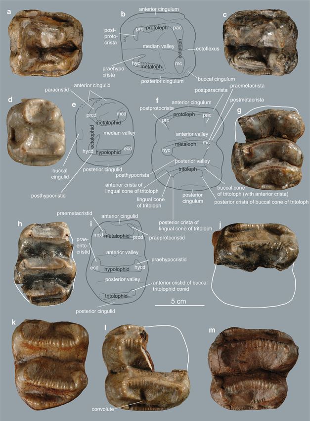
Fig. 5 Elements of anterior and posterior limbs of D. levius vel condyles on distal epiphysis damaged, the articulation surface
giganteum from Gratkorn with affiliation of articulation facets: a os carpi of the condylus lateralis femoris damaged, except for its
ulnare sin. [UMJGP 203688; 1 proximal view (os pisif. = os pisiforme), 2
distal view, 3 dorsal view, 4 medial view], b os carpale secundum sin. caudalmost part; only distal part of the trochlea ossis femoris
[UMJGP 203640; 1 lateral view, 2 sketch of lateral view with identified preserved and showing a deep distal incision between the two
articulation facets for os carpale tertium (III), 3 dorsal view (articulation condyles widening caudally; pronounced mediolateral depres-
facet for os carpale primum on medial side)], 4 proximal view with sions proximal to both condyles.
articulation facet for ossa carpi radiale and intermedium, c phalanx
proximalis of pes? (UMJGP 203708; 1 dorsal view, 2 plantar view, 3 Comparison: Due to fragmentary preservation of the
lateral/medial view), d radius sin. (UMJGP 203665; 1 lateropalmar view, Gratkorn femur no comparison to other specimens can be
2 mediodorsal view); e fibula sin. (UMJGP 203622; 1 lateroplantar view, 2 given.
mediodorsal view), f fragments of femur dex. in caudal view with sketch Fibula sin. (UMJGP 203622; Fig. 5e) and dex. (UMJGP
of outline (fragment of proximal shaft: UMJGP 203601; distal epiphysis:
UMJGP 204112), g os tarsi centrale dex. [UMJGP 203683; 1 proximal 203613 (proximal portion of shaft without facet) 203612
view, 2: distal view, 3 sketch of distal view with identified articulation (distal portion of shaft)): fibula sin. almost complete though
facets for os tarsale secundum (II), tertium (III) and quartum (IV)]. Scale lacking proximal and distal articulation facets; corpus fibulae
bar 5 cm (a–c, g), 10 cm (d–f) triangular proximally (here smallest circumference); distal
half mediolaterally flattened with slightly concave medial
side; diagonal crest running from smallest circumference
semicircular facet in proximopalmar part, only a small portion of proximodorsally along the lateral side of the proximal fourth
the elongated distal facet preserved. of the shaft.
Comparison: Comparison material for the os carpale Comparison: The morphological difference concerning
secundum consisted of one specimen of D. cf. giganteum from the fibula between Prodeinotherium and Deinotherium as
Wien XII Oswaldgasse (NHMW SK 2810; Austria; Late observed by Huttunen [“form of shaft proximally flattened
Miocene; Pannonian E; 10.4–10 Ma), which is larger and in dorsoplantar direction” in Deinotherium (Huttunen
differs morphologically from the Gratkorn specimen by a less 2000, p. 91)] cannot be confirmed based on the specimen
rounded dorsal side and the facet for articulation with os from Gratkorn, as the cross section of the proximal shaft is
carpale primum, which comprises only a semi circle in the triangular. The proximal cross section of both Gratkorn spec-
specimen from Wien XII Oswaldgasse. Following the descrip- imens is not more dorsoplantarily flattened than in
tion by Svistun (1974) the os carpale secundum of D. levius is Prodeinotherium from Langenau (Early Miocene), but its
in general of similar shape as the Gratkorn specimen but distal shaft seems to be more flattened mediolaterally than
differs from the latter as it seems to possess only two facets the latter.
for the articulation to the os carpale tertium. Os tarsi centrale sin. (UMJGP 203611) and dex. (UMJGP
Distal epiphysis with articulation facet of Mc II or III sin. 203683; Fig. 5g): both ossa tarsorum centralia badly pre-
or IV dex. (UMJGP 203685): due to its relatively large size it served and missing most of dorsal, medial and plantar
can be assigned to the manus rather than to the pes; due to surfaces; proximal articulation facet for astragalus large,
fragmentary preservation most of the articulation facet miss- concave and oval shaped (mediolaterally elongated); small,
ing; distal articulation facet dorsopalmar convex with small proximoplantar oriented facet for articulation with the
oblique ridge slightly shifted from the central line on palmar calcaneum located in the lateral half of the plantar side
part of the trochlea, but not as asymmetric as it would be forming an obtuse angle with proximal articulation facet;
expected for Mc V. on distal surface three articulation facets for the tarsals II–
Phalanx proximalis? of manus (UMJGP 203684) of IV identified (from lateral to medial for os tarsale quartum
unidentified digit: dorsal surface not preserved and phalanx (oriented distoplantolateral); os tarsale tertium; os tarsale
missing its distal part; epiphysis not entirely closed proximally; secundum); most medial distal facet for Mt I not traceable,
proximal facet for articulation with metacarpal dorsopalmarily all preserved distal facets slightly concave separated by
concave with a general inclination to proximopalmar; palmar dorsomedial-plantolateral oriented ridges diverging in
side convex. dorsomedial direction; no plantomedial process.
Comparison: Morphology alone does not allow affiliation Comparison: With only three distal facets and no articu-
to manus or pes, but dimensions in comparison with UMJGP lation facet for the Mt I the os tarsi centrale differs from that of
203708 render a determination as phalanx proximalis of Prodeinotherium (which shows four facets) but fits well with
manus more likely. the situation in Deinotherium (Huttunen 2000). Furthermore,
Femur dex. (distal epiphysis (UMJGP 204112), fragment the os tarsi centrale differs from that of P. bavaricum from
of proximal shaft (UMJGP 203601); Fig. 5f): portion of Unterzolling (early Middle Miocene) in the lack of a
proximal femur shaft with basis of trochanter minor (distinct plantomedial process (Huttunen and Göhlich 2002).
depression on shaft caudal of trochanter minor); caudolateral Distal trochlea of Mt II? (UMJGP 204696): due to its
edge of shaft subrectangular at base of trochanter minor; both smaller size in comparison to the Mc described aboveYou can also read