Molecular pathogenesis and extraovarian origin of epithelial ovarian cancer-Shifting the paradigm
←
→
Page content transcription
If your browser does not render page correctly, please read the page content below
Human Pathology (2011) 42, 918–931

                                                                                                                   www.elsevier.com/locate/humpath

Progress in pathology

Molecular pathogenesis and extraovarian origin of
epithelial ovarian cancer—Shifting the paradigm
Robert J. Kurman MD ⁎, Ie-Ming Shih MD, PhD
Division of Gynecologic Pathology, Departments of Pathology, Gynecology and Obstetrics and Oncology, The Johns Hopkins
University School of Medicine, Baltimore, MD 21231, USA

Received 25 January 2011; revised 23 March 2011; accepted 23 March 2011

 Keywords:
                                        Summary Recent morphologic, immunohistochemical, and molecular genetic studies have led to the
 Ovarian cancer;
                                        development of a new paradigm for the pathogenesis and origin of epithelial ovarian cancer based on a
 Borderline tumors;
                                        dualistic model of carcinogenesis that divides epithelial ovarian cancer into 2 broad categories
 Molecular pathogenesis;
                                        designated types I and II. Type I tumors comprise low-grade serous, low-grade endometrioid, clear cell
 Origin;
                                        and mucinous carcinomas, and Brenner tumors. They are generally indolent, present in stage I (tumor
 p53 mutations;
                                        confined to the ovary), and are characterized by specific mutations, including KRAS, BRAF, ERBB2,
 Prevention
                                        CTNNB1, PTEN, PIK3CA, ARID1A, and PPP2R1A, which target specific cell signaling pathways.
                                        Type I tumors rarely harbor TP53 mutations and are relatively stable genetically. Type II tumors
                                        comprise high-grade serous, high-grade endometrioid, malignant mixed mesodermal tumors
                                        (carcinosarcomas), and undifferentiated carcinomas. They are aggressive, present in advanced stage,
                                        and have a very high frequency of TP53 mutations but rarely harbor the mutations detected in type I
                                        tumors. In addition, type II tumors have molecular alterations that perturb expression of BRCA either by
                                        mutation of the gene or by promoter methylation. A hallmark of these tumors is that they are genetically
                                        highly unstable. Recent studies strongly suggest that fallopian tube epithelium (benign or malignant)
                                        that implants on the ovary is the source of low-grade and high-grade serous carcinoma rather than the
                                        ovarian surface epithelium as previously believed. Similarly, it is widely accepted that endometriosis is
                                        the precursor of endometrioid and clear cell carcinomas and, as endometriosis, is thought to develop
                                        from retrograde menstruation; these tumors can also be regarded as involving the ovary secondarily. The
                                        origin of mucinous and transitional cell (Brenner) tumors is still not well established, although recent
                                        data suggest a possible origin from transitional epithelial nests located in paraovarian locations at the
                                        tuboperitoneal junction. Thus, it now appears that type I and type II ovarian tumors develop
                                        independently along different molecular pathways and that both types develop outside the ovary and
                                        involve it secondarily. If this concept is confirmed, it leads to the conclusion that the only true primary
                                        ovarian neoplasms are gonadal stromal and germ cell tumors analogous to testicular tumors. This new
                                        paradigm of ovarian carcinogenesis has important clinical implications. By shifting the early events of
                                        ovarian carcinogenesis to the fallopian tube and endometrium instead of the ovary, prevention
                                        approaches, for example, salpingectomy with ovarian conservation, may play an important role in
                                        reducing the burden of ovarian cancer while preserving hormonal function and fertility.
                                        © 2011 Elsevier Inc. All rights reserved.

   This study is supported by a CDMRP grant (no. OC100517) from the US Department of Defense.
   ⁎ Corresponding author.

0046-8177/$ – see front matter © 2011 Elsevier Inc. All rights reserved.
doi:10.1016/j.humpath.2011.03.003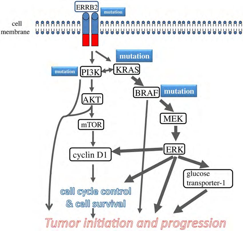
Molecular pathogenesis and extraovarian origin of EOC                                                                      919

   Paradigms, as defined by Kuhn [1] in his seminal work          1. Molecular pathogenesis of epithelial
“The Structure of Scientific Revolutions,” are the best ways      ovarian carcinoma
of explaining progress in science. Kuhn believed that
textbooks, which describe progress, as a cumulative,
                                                                     The introduction of the “borderline (low malignant
incremental process leading to a growing corpus of scientific
                                                                  potential)” category was an important step in refining the
knowledge, present an unrealistic and biased view. Instead,
                                                                  morphologic classification of EOC by identifying a group of
he felt that a more accurate depiction could be gleaned by
                                                                  tumors, defined as lacking destructive invasive growth that
looking at what scientists do most of the time, which he
                                                                  had a significantly better outcome than the invasive
termed normal science, and normal science is governed by
                                                                  carcinomas. Because it was rare to find a borderline tumor
paradigms. A paradigm generates a consensus among
                                                                  coexisting with an invasive carcinoma, it was generally
scientists working in a particular field about how work in
                                                                  believed that they were unrelated. In 1996, a relationship
that field should be done. It also identifies puzzles, assures
                                                                  between serous borderline tumor (SBT) and invasive serous
scientists that each puzzle has a solution, and provides
                                                                  carcinoma was described based on the subdivision of SBT
standards for evaluating solutions. Normal science involves
                                                                  into 2 groups. One group designated “atypical proliferative
showing how nature can be fitted into the categories provided
                                                                  serous tumor (APST)” behaved in a benign fashion, and a
by the paradigm. When puzzles arise that repeatedly resist
                                                                  second, smaller group designated “micropapillary serous
solutions a crisis of confidence occurs. During a crisis, the
                                                                  carcinoma (MPSC)” also termed “noninvasive low-grade
paradigm is subjected to testing and might be rejected. If that
                                                                  serous carcinoma” behaved like a low-grade malignant
occurs, a new paradigm replaces the previous one and a
                                                                  tumor [20]. Moreover, this latter subset was closely
scientific revolution has occurred.
                                                                  associated with invasive low-grade serous carcinoma
   The operative paradigm of ovarian carcinogenesis is that
                                                                  (LGSC), and the investigators proposed that MPSC was
epithelial ovarian cancer (EOC) is composed of several
                                                                  the immediate precursor of LGSC. The key element leading
different types, but because the vast majority is high-grade
                                                                  to this conclusion was the recognition that LGSC was a
serous carcinoma (HGSC), differences between the types are
                                                                  distinct entity that differed from HGSC in several ways (see
obscured, and therefore, EOC is regarded as a single disease.
                                                                  below). Before this, serous carcinoma was graded well,
Moreover, because carcinomas in the female pelvis tend to
                                                                  moderately, and poorly differentiated, with the implication
involve the ovary, often as the dominant mass, they have all
                                                                  that serous carcinoma was a spectrum of disease in which
been regarded as ovarian in origin. This paradigm is based on
                                                                  well-differentiated carcinoma (LGSC) progressed to poorly
taxonomy, specifically morphologic classifications, which
                                                                  differentiated carcinoma (HGSC). After this morphologic
took shape in the 1930s and 1940s [2-4], matured in the
                                                                  study linking APST to MPSC and LGSC, a series of
1950s and 1960s [5], and culminated with the World Health
                                                                  molecular genetic studies was performed, which culminated
Organization classification in 1973 [6]. The histologic
                                                                  in the proposal of a dualistic model to explain the
classifications created a structure that provided the basis
                                                                  pathogenesis of EOC [21].
for performing clinicopathologic studies, but apart from
these studies, the tools necessary to study pathogenesis were
not available, and therefore, our understanding of ovarian        1.1. Dualistic model of carcinogenesis
carcinogenesis was limited.
   Arguably, research within this paradigm has failed to             Briefly, the dualistic model accommodates and confirms
identify the precursor of EOC [7-19], and as a result, current    the heterogeneous nature of EOC and places the major
management is empirical. Despite advances in radical surgery      histologic types into 2 groups (types I and II) based on their
and cytotoxic chemotherapy, overall survival has not              distinctive clinicopathologic and molecular genetic features.
changed in more than 50 years. In the last 2 decades,             It also links specific histologic types with their putative
attention has focused on early detection, but unfortunately,      precursor lesions. Thus, type I tumors comprise LGSCs, low-
this strategy has also failed to provide a survival benefit.      grade endometrioid, clear cell, and mucinous carcinomas,
Accordingly, there are “persistent puzzles” that have resisted    which develop in a stepwise fashion from well-established
solutions, and hence, a “crisis of confidence” exists.            precursor lesions, such as borderline tumors and endometri-
   The introduction of molecular biology and the develop-         osis. They typically present as large masses that are confined
ment of new methods of tissue sampling are now ushering in        to one ovary (stage Ia), are indolent, and have a good
a paradigm shift, which can be considered “revolutionary.”        prognosis. The type I tumors are relatively genetically stable
The concepts that are emerging and shaping this new               and typically display a variety of somatic sequence mutations
paradigm are novel and highly provocative. Some of them           that include KRAS, BRAF, PTEN, PIK3CA, CTNNB1 (the
will be confirmed, and others will be modified or discarded,      gene-encoding β-catenin), ARID1A, and PPP2R1A but very
as scientists in the process of performing “normal science”       rarely TP53 [21-23]. In contrast, type II tumors comprise
within the framework of the new paradigm attempt to clarify       HGSC (usual type of serous carcinoma), high-grade
and resolve issues that have frustrated progress in reducing      endometrioid carcinoma, malignant mixed mesodermal
the burden of this disease.                                       tumors (carcinosarcomas), and undifferentiated carcinomas,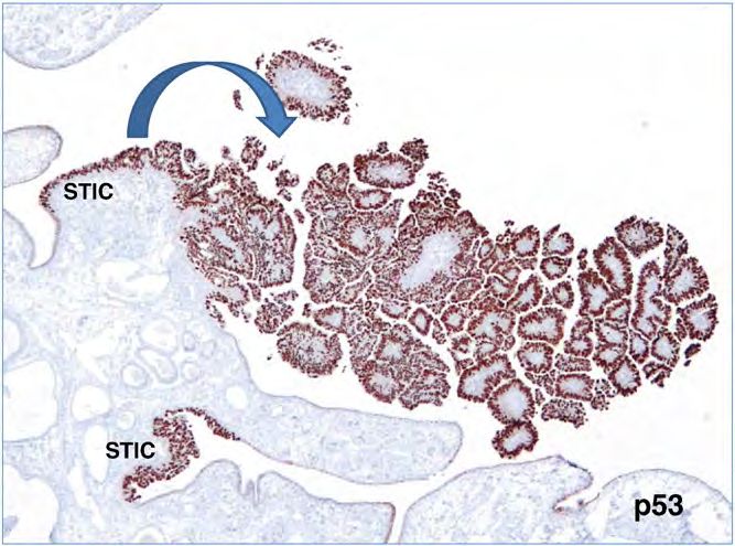
920                                                                                                       R. J. Kurman, I. -M. Shih

which present in advanced stage (stages II-IV) in more than           MAPK [37]; they rarely harbor TP53 mutations. Recent
75% of cases; they grow rapidly and are highly aggressive.            studies have further clarified the molecular pathogenesis of
Type II tumors, of which HGSC is the prototypic type, are             APST, MPSC, and LGSC. First, KRAS and BRAF
chromosomally highly unstable and harbor TP53 mutations               mutations have not been detected in serous cystadenomas,
in more than 95% of cases [24]; they rarely display the               the putative precursor of SBTs, but laser capture microdis-
mutations found in the type I tumors. BRCA inactivation,              section studies have detected these mutations in the
either by mutation or inactivation of expression of BRCA              adenoma epithelium and APST epithelium in serous
and its downstream genes via promoter methylation, occurs             cystadenomas containing small APSTs, suggesting that
in up to 40% to 50% of HGSC [25]. BRCA inactivation has               these mutations occur early in the development of APST
not been reported in the type I tumors.                               [38]. In an attempt to elucidate the relationship of APST to
                                                                      LGSC, a recent study compared the gene expression
1.2. Serous tumors                                                    profiles of APST, MPSC, and LGSC and found that
                                                                      MPSC is closer molecularly to invasive LGSC than to
   The relationship of APST and MPSC to LGSC based on                 APST [26] and that the genes involved in MAPK signaling
morphologic studies was supported by mutational analysis,             showed higher expression in MPSC than in APST. In
gene expression studies, and methylation profiling demon-             addition, a previous study reporting that MPSC harbors a
strating that these 3 tumor types shared molecular                    pattern of chromosomal imbalance distinct from that of
alterations that differed dramatically from HGSC [25-30].             APST [39] confirms the proposal that LGSC develops in a
Initial molecular genetic studies focused on individual               stepwise fashion from cystadeno(fibro)ma to APST and
genes (Fig. 1), but more recent studies have highlighted the          MPSC, supporting the biologic role of the KRAS-BRAF-
importance of molecular signaling pathways (Fig. 2). For              MEK-MAPK pathway in the development of LGSC. By
example, the MAPK signaling pathway is important for the              globally profiling the epigenetic landscape, we have
cellular response to a variety of growth and differentiation          recently reported that the methylation profiles in LGSC
factors, and activating mutations in KRAS or one of its               are closer to APST and serous cystadenoma than HGSC
downstream effectors, BRAF, (mutations of KRAS and                    [30]. This finding lends further support to the dualistic
BRAF are mutually exclusive) results in constitutive                  model of ovarian serous carcinogenesis.
activation of mitogen-activated protein kinase (MAPK)-                   In contrast to LGSC, HGSC harbors TP53 mutations in
mediated signaling in more than half of APSTs, MPSCs,                 more than 95% of cases [25] but rarely contains KRAS or
and LGSCs [31-34]. In addition, a 12-base-pair insertion              BRAF mutations. Aside from TP53 mutations, no other
mutation of ERBB2 (encoding HER2/neu), which activates                mutations are consistently found in sporadic (nonfamilial)
an upstream regulator of K-Ras, has been detected in 9% of            HGSCs including mutations of BRCA1 and BRCA2, which
these tumors. Interestingly, tumors with ERBB2 mutations              characterize familial HGSC (The Cancer Genome Atlas,
lack KRAS and BRAF mutations [35,36]. Accordingly,                    unpublished). On the other hand, inactivation of the BRCA1/2
60% to 70% of APSTs, MPSCs, and LGSCs express active                  genes by other mechanisms, such as hypermethylation of

                                 ARID1A
                                 CTNNB1
                                 PTEN                KRAS           TP53 mutation
                                 PIK3CA              Mutation
                                 PPP2R1A                            Chromosomal
                                                     Mucinous
                                 Mutation                             instability
               Type I
                                      Endometrioid                                        Inactivation
                             Clear cell                         High-grade serous         of BRCA 1/2
                                                     us
                                          ade sero                                        (Mutation or
                                 Low-gr
                                                                                          hypermethylation)
                ARID1A
                PIK3CA
                ZNF217           KRAS
                PPP2R1A
                                 BRAF
                Mutation         ERBB2                          Type II
                                 PIK3CA
                                 Mutation

                   Fig. 1   Prevalence of histologic types of EOC and their associated molecular genetic changes.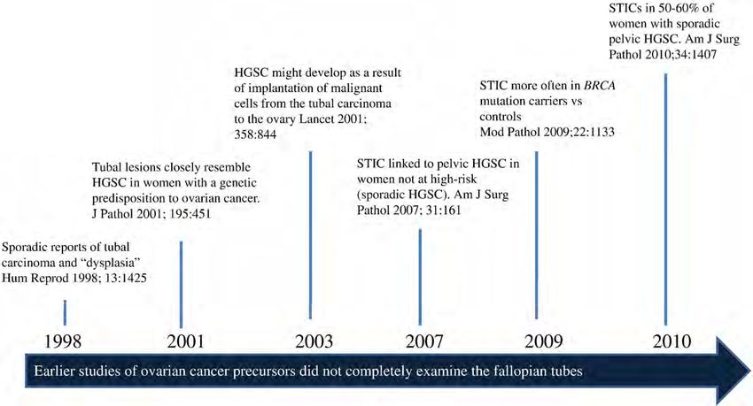
Molecular pathogenesis and extraovarian origin of EOC                                                                                     921

Fig. 2 Schematic illustration of pathway alterations involved in the development of LGSC. The cardinal molecular genetic changes include
somatic mutations in KRAS, BRAF, and occasionally ERRB2 (encoding Her2/Neu) and PIK3CA. The mutated gene products constitutively
activate the signaling pathways that regulate cellular proliferation and survival and promote tumor initiation and progression through several
mechanisms including up-regulation of glucose transporter-1. The size of the boxes containing specific genes reflects the relative frequency of
the mutation, and the thickness of the arrows indicates the relative contribution of the pathway alterations to tumor development.

the BRCA1 promoter, occurs relatively frequently, and as a                most common molecular genetic changes in clear cell
result, inactivation of BRCA1/2 by mutation or other                      carcinoma are a somatic inactivating mutation of ARID1A
mechanisms occurs in 40% to 50% of sporadic HGSCs                         [22,23] (a tumor suppressor gene detected in about 50% of
[26]. The most striking molecular feature of HGSC is diffuse              cases), an activating mutation of PIK3CA in about 50% of
and high levels of DNA copy number gains or losses, which                 tumors [42], and deletion of PTEN (a tumor suppressor
include CCNE1 (cyclin E1), NOTCH3, AKT2, RSF1, and                        gene involved in the PI3K/PTEN signaling pathway) in
PIK3CA loci [40]. Despite their distinct molecular signatures,            about 20% [43], supporting the role of an aberrant PI3K/
LGSC, and even an APST, is sometimes clonally associated                  PTEN pathway in the development of clear cell carcinoma.
with a synchronous HGSC , suggesting that such progression                In addition, single nucleotide polymorphism (SNP) array
rarely does occur [41].                                                   analysis has identified frequent amplification of the
                                                                          ZNF217 (zinc finger protein 217) locus and deletion of
1.3. Clear cell and endometrioid tumors                                   the CDKN2A/2B locus in clear cell carcinomas, suggesting
                                                                          that the pathways involving these 2 genes are also
   After serous carcinoma, endometrioid and clear cell                    important in their development.
carcinomas are the most frequent types of EOC accounting                     Like clear cell carcinoma, mutations that deregulate PI3K/
for approximately 15% to 20% of EOC in Western                            PTEN signaling are common in low-grade endometrioid
countries. The molecular genetic alterations that underlie                carcinoma, and in fact, mutation of the tumor suppressor
the development of these tumors are now beginning to                      gene PTEN, which occurs rarely in other types of EOC, has
emerge. Based on genome-wide mutational analysis, the                     been reported in approximately 20% of ovarian low-grade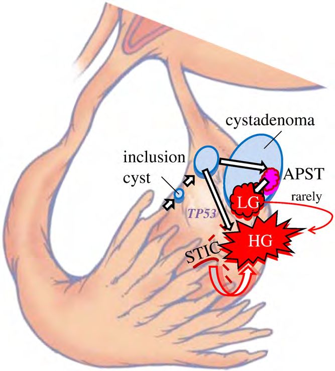
922                                                                                                R. J. Kurman, I. -M. Shih

endometrioid carcinomas [44,45]. Another mechanism by            genetic features, such as mutation of ARID1A and deletion
which activation of PI3K signaling occurs is through             of PTEN, they clearly adopt different molecular programs
activating mutations of PIK3CA, which has been detected          for their development, as is evident by their distinctly
in 20% of low-grade endometrioid carcinomas [42]. The            different morphologic phenotype and clinical behavior. For
Wnt/β-catenin signaling pathway, which is involved in the        example, canonical Wnt signaling pathway defects and
regulation of several important cellular processes including     microsatellite instability, which occur frequently in low-
proliferation, motility, and survival, is deregulated in up to   grade endometrioid carcinoma, have only rarely been
40% of ovarian endometrioid carcinomas, usually on the           detected in clear cell carcinoma [46]. Also, it has been
basis of activating mutations of CTNNB1, the gene that           recently demonstrated that compared with the other types of
encodes β-catenin [46]. Notably, mutation of CTNNB1 has          EOC, clear cell carcinoma has significantly longer telomeres,
been associated with squamous differentiation, low tumor         and this finding correlates with poor outcome [52].
grade, and a favorable outcome, features that characterize           Finally, morphologic studies have linked the endocer-
low-grade endometrioid carcinoma [47-50].                        vical-type mucinous borderline tumor, also referred to as
    Although low-grade endometrioid carcinomas are easily        “atypical proliferative seromucinous tumor,” to endome-
recognized, the distinction of high-grade endometrioid           triosis in about a third of cases [53]. We are unaware of
carcinoma from HGSC can be very difficult. Some                  any molecular genetic studies of these neoplasms;
pathologists even question the existence of high-grade           however, we have recently detected ARID1A mutations
endometrioid carcinoma, regarding it instead as a variant of     in 2 of these neoplasms, suggesting that they are more
HGSC, whereas others classify high-grade endometrioid            closely related to endometrioid than to serous or
carcinoma as “mixed high-grade endometrioid carcinoma            mucinous tumors (unpublished data) further confirming
and HGSC” or as “HGSC with features of endometrioid              the role of endometriosis as a precursor of a variety of
carcinoma.” It is therefore of interest that in a study of       “endometrioid-related” ovarian neoplasms.
ovarian endometrioid carcinomas of all grades, low-grade
endometrioid carcinomas were characterized by mutations          1.4. Mucinous tumors
that deregulate the canonical Wnt/β-catenin and PI3K/PTEN
signaling pathways and lacked TP53 mutations, whereas               These tumors have been the least studied histologic types
high-grade endometrioid carcinomas lacked Wnt/β-catenin          probably because of their relative rarity (approximately 3%
or PI3K/PTEN signaling pathway defects and frequently            of EOC). KRAS mutations occur in up to 75% of primary
harbored TP53 mutations [47]. A few high-grade endome-           mucinous carcinomas, and using KRAS as a molecular
trioid carcinomas did, in addition to TP53 mutation, display     marker, laser capture microdissection studies have shown the
molecular changes found in the low-grade endometrioid            identical KRAS mutation in mucinous carcinomas and
carcinomas, suggesting that some low-grade endometrioid          adjacent mucinous cystadenomas and borderline tumors
carcinoma may progress to high-grade carcinoma. The              [32,54,55], supporting the morphologic continuum and
findings parallel those seen in the serous tumors, namely,       tumor progression in ovarian mucinous neoplasms.
that generally, low- and high-grade tumors develop                  In summary, each of the major histologic types of EOC
independently but that rarely progression of a low-grade to      is associated with a different set of cell signaling pathways
a high-grade tumor occurs. The similar high frequency of         abnormalities, which for the type I tumors are shared with
TP53 mutations in high-grade endometrioid carcinoma as           their respective precursor lesions (borderline tumors and
in HGSC suggests that both develop in a similar fashion, via     endometriosis) supporting their stepwise progression. In
TP53 mutation, and that most high-grade endometrioid             contrast, the type II tumors, aside from a very high
carcinoma is closely related to or is a variant of HGSC.         frequency of TP53 mutations and molecular alterations of
    Morphologic studies over the past 2 to 3 decades have        BRCA1/2, are characterized by marked genetic instability
repeatedly shown an association of endometrioid and clear        and lack other mutations. The identification and character-
cell carcinoma with endometriosis, and early molecular           ization of their precursor lesions have only recently been
genetic studies demonstrated loss of hybridization (LOH) in      recognized (see below).
the same chromosomal regions in endometrioid carcinoma
and adjacent endometriosis [51], confirming a clonal
relationship between endometriosis and endometrioid carci-
noma. In addition, a recent study reported mutation of AR-       2. Origin of epithelial ovarian carcinoma
ID1A in atypical endometriosis adjacent to clear cell
carcinoma but not in distant sites of endometriosis further      2.1. Serous tumors
linking endometriosis to clear cell carcinoma and thereby
providing further evidence that endometriosis is the likely         The conventional view of the origin of serous tumor has
precursor of endometrioid and clear cell carcinoma [23].         been that they were derived from the ovarian surface
Although both clear cell and endometrioid carcinomas are         epithelium or cortical inclusion cysts. Therefore, there was
derived from endometriosis and share some molecular              surprise and skepticism when a group of Dutch investigators
Molecular pathogenesis and extraovarian origin of EOC 923
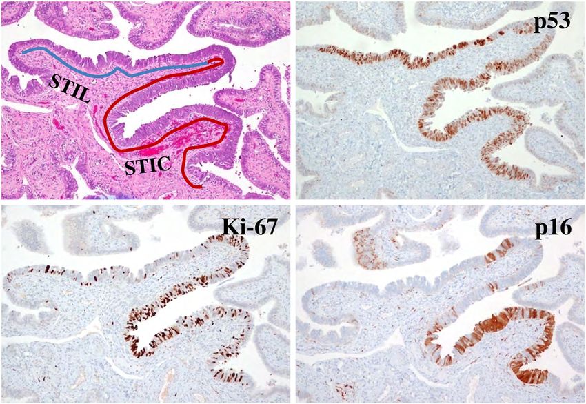
Fig. 5 Fimbria with 2 STICs and an associated papillary invasive
Fig. 3 Spread of STIC from the fimbria to the ovarian surface. HGSC highlighted by p53 immunostain.
(Reprinted with permission from: Kurman RJ, Shih IM. The origin
and pathogenesis of epithelial ovarian cancer: a proposed unifying
theory. Am J Surg Pathol 2010;34:433-43 [70].) that gives the impression that the tumor originated in the
ovary [57,58] (Fig. 3). Additional studies in which fallopian
in 2001 first described tubal intraepithelial carcinomas, later tubes were carefully examined confirmed that STICs and
designated “serous tubal intraepithelial carcinomas (STICs)” small, early invasive tubal carcinomas occurred not only in
and occult invasive HGSCs in the fallopian tube that closely women with a genetic predisposition for the development of
resembled ovarian HGSC, in women with a genetic ovarian cancer but also in 50% to 60% of women without
predisposition to ovarian cancer [56]. Similar lesions were known BRCA mutations (sporadic ovarian cancer; Fig. 4)
not found in the ovaries of the same women. In hindsight, the [59-67]. Moreover, these carcinomas were almost always
failure to identify the tubal carcinomas in the past was detected in the fimbria (Fig. 5), and it has been proposed that
because it was assumed that precursors of ovarian carcinoma earliest neoplastic change begins in secretory-type cells
would logically be in the ovaries, and therefore, the fallopian [63,66]. Further evidence supporting the proposal that STICs
tubes were not carefully examined [7-19]. It was subse- are precursors was the identification of STICs in women
quently proposed that implantation of malignant cells from without ovarian cancer as well as the presence of identical
the tubal carcinoma to the ovary develops into a tumor mass TP53 mutations in STICs and concomitant ovarian
Fig. 4 Tubal origin of ovarian HGSC. A brief history.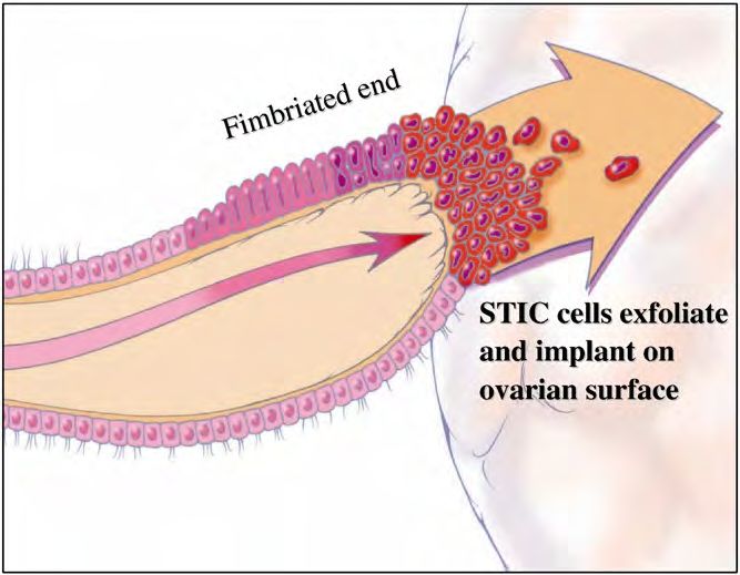
924                                                                                                     R. J. Kurman, I. -M. Shih

                                       Fig. 6   An STIL immediately adjacent to an STIC.

HGSCs, indicating a clonal relationship between them
[66,68]. A gene profiling study showing that the gene
expression profile of HGSC is more closely related to
fallopian tube epithelium than to ovarian surface epithelium
[69], and immunohistochemical studies showing that HGSC
expresses PAX8, a Müllerian marker, but not calretinin, a
mesothelial marker (ovarian surface epithelium has a
mesothelial not a Müllerian morphologic phenotype), lends
further support to the proposal that the tubal lesions are
precursors of HGSC and not the ovarian surface epithelium
[70]. Further confirmation of the link between STICs and
HGSC is the demonstration that both STICs and concomitant
ovarian HGSCs, besides coexpressing p53, also coexpress
p16, FAS, Rsf-1, and cyclin E1 [71] (Fig. 6). In addition, a
recent study showed that STICs, like other precancerous
lesions, have relatively short telomeres [72].
    As previously noted, in studies of ovarian and primary
peritoneal HGSC in which the fallopian tubes were
completely sectioned using the Sectioning and Extensively
Examining the Fimbria (SEE-FIM) protocol [63], STICs
were identified in 50% to 60% of cases [66,67]. This raises
the question as to the source of the remaining ovarian
carcinomas that lack evidence of tubal involvement. One
possibility is that small STICs were missed despite complete      Fig. 7 Development of a cortical inclusion cyst from tubal
sampling of the tubes, and in fact, it has been shown that        epithelium. (Reprinted with permission from: Kurman RJ, Shih IM.
leveling the fallopian tube blocks can detect additional          The origin and pathogenesis of epithelial ovarian cancer: a proposed
STICs not found in the original sections [67]. A second           unifying theory. Am J Surg Pathol 2010;34:433-43 [70].)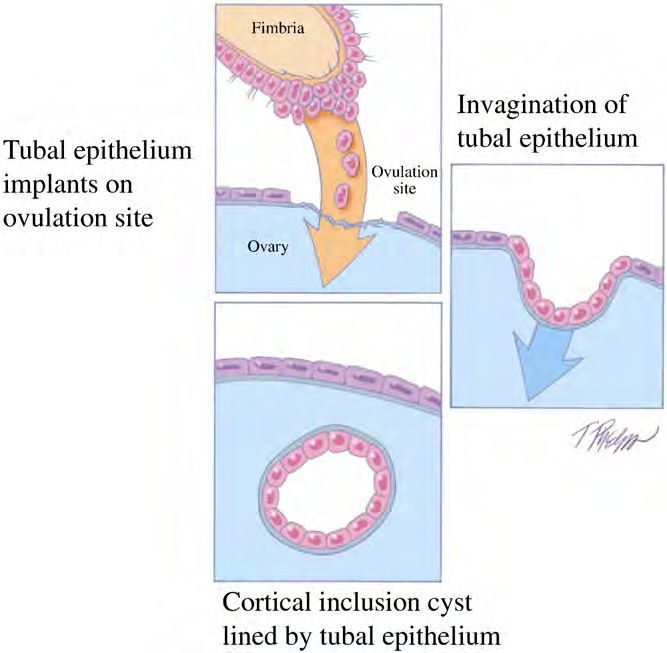
Molecular pathogenesis and extraovarian origin of EOC                                                                            925

possibility is that the invasive carcinoma overgrew and               have been discovered. These include cytologic abnormalities
obliterated the STIC. Another possibility is that the                 that fall short of STICs, which we have tentatively designated
carcinoma developed from ovarian cortical inclusion cysts.            “serous tubal intra-epithelial lesions (STILs)” (Fig. 6) and
Although it is generally stated that these cysts develop by           termed by others “tubal intraepithelial lesions in transition
invagination of ovarian surface epithelium, there is reason to        (TILT)” [76]. In addition, short stretches of normal appearing
believe that during ovulation, as the fimbria come into close         fallopian tube epithelium that strongly expresses p53, and in
contact with the ovary, tubal epithelial cells implant on the         which TP53 mutations have been identified in some cases,
disrupted ovarian surface to form a cortical inclusion cyst           have been termed “p53 signatures” [68]. Although these
[70] (Fig. 7). Parenthetically, tubal epithelial cells are easily     lesions may represent the very early events in serous
dislodged for culture in the laboratory by flushing the               carcinogenesis, it is not clear, at this time, whether STILs
fallopian tube [56] (I.-M. Shih, unpublished data). In                and p53 signatures are precursor lesions or that they are
addition, ovulation itself with the release of follicular fluid,      benign “reactive” changes that overexpress p53 and have no
which has been shown to contain reactive oxygen species               biologic relevance to neoplasia. It is conceivable that some
(free radicals), and possibly associated changes in the               are precursors and others are not; clearly, this is an area that
microenvironment, such as inflammation, may play a role               requires further investigation.
in early ovarian carcinogenesis. This is consistent with                  It appears that LGSC may also be derived from fallopian
epidemiologic evidence linking decreased ovulation (as a              tube epithelium. Careful examination of fallopian tubes in
result of either oral contraceptive usage or multiple                 women with APSTs discloses what we have recently
pregnancies) with a decreased risk of ovarian cancer                  described as “papillary tubal hyperplasia” (unpublished
[73,74]. Therefore, some HGSCs may develop from ovarian               data). This lesion is characterized by small, papillary clusters
cortical inclusion cysts [75], but these cysts could be derived       of bland-appearing tubal epithelium (both secretory and
not from the ovarian surface epithelium but from implanted            ciliated cells) that are often associated with psammoma
fimbrial tubal epithelium [70] (Fig. 8). Also, because the            bodies. Varying numbers of these clusters can be detected in
fallopian tubes are now being more carefully studied,                 the tubal lumen and appear to bud from the tubal epithelium
additional abnormalities in the fallopian tube epithelium             in a high proportion of women with APSTs (unpublished
                                                                      data). We speculate that these detached clusters of tubal
                                                                      epithelium pass through the tube and implant on the ovary
                                                                      where they can develop into APSTs or implant on the pelvic
                                                                      or abdominal peritoneum to produce noninvasive implants.

                                                                      2.2. Clear cell and endometrioid tumors

                                                                         As previously noted, it is well established by morphologic
                                                                      and, more recently, by molecular genetic studies that low-
                                                                      grade endometrioid and clear cell carcinomas develop from
                                                                      endometriotic cysts (endometriomas) and are frequently
                                                                      associated with implants of endometriosis elsewhere in the
                                                                      pelvis [77]. Although the precise origin of endometriosis has
                                                                      not been completely established, specifically, whether it
                                                                      develops in situ in the peritoneum through a process of
                                                                      metaplasia or from retrograde menstrual flow, the prepon-
                                                                      derance of data favors the latter mechanism [78]. Admittedly,
                                                                      the former theory is more difficult to prove experimentally.
                                                                      Thus, if retrograde menstruation accounts for most cases of
                                                                      endometriosis, it is logical to assume that endometrioid and
                                                                      clear cell tumors develop from endometrial tissue that
                                                                      implanted on the ovary, and therefore, the ovary is involved
                                                                      secondarily [79] (Fig. 9).
Fig. 8 Development of low-grade (type I pathway with KRAS or
                                                                         Of further interest has been the observation that the
BRAF mutation) and HGSC (type II pathway with TP53 mutation)
from tubal epithelium by way of a cortical inclusion cyst and
                                                                      eutopic endometrium in women with endometriosis exhibits
cystadenoma or an intraepithelial carcinoma (STIC) implanting         intrinsic molecular abnormalities, including activation of
directly on the ovary developing into a HGSC. (Reprinted with         oncogenic pathways [78]. Presumably, these changes permit
permission from: Kurman RJ, Shih IM. The origin and pathogenesis      the endometrial tissue to implant, survive, and invade
of epithelial ovarian cancer: a proposed unifying theory. Am J Surg   ovarian and peritoneal tissue. This hypothesis, by which
Pathol 2010;34:433-43 [70].)                                          endometrioid and clear cell carcinoma develop from
926 R. J. Kurman, I. -M. Shih
contained foci of Brenner tumor in 18% of cases [83].
Interestingly, mucinous tumors were frequently associated
with Walthard cell nests that are composed of benign
transitional-type epithelium, frequently found in paraovarian
and paratubal locations. This raises the possibility that
mucinous tumors and Brenner tumors have the same
histogenesis arising from these microscopic transitional cell
nests at the tubal-peritoneal junction, which would be
consistent with their nonMüllerian appearance [84]. The
study reported that Brenner tumors are small (median size,
0.5 cm), whereas mucinous cystadenomas are large (median
size, 9 cm). The investigators then went on to speculate that
as a small Brenner tumor grows, the mucinous component
becomes dominant, resulting in the development of a
mucinous cystadenoma that, as it enlarges, compresses and
eventually obliterates the adjacent ovary and Brenner tumor
giving the appearance that it arose in the ovary. The findings
in this study are intriguing but must be regarded as
preliminary. Additional morphologic and molecular genetic
studies are necessary to determine whether this concept is
Fig. 9 Development of low-grade endometrioid (EM) and clear valid. Another subset of gastrointestinal-type mucinous
cell (CC) carcinoma from endometriosis by retrograde menstrua- tumors can arise from ovarian mature cystic teratomas [85].
tion. (Reprinted with permission from: Kurman RJ, Shih IM. The In summary, the data support the view that serous tumors
origin and pathogenesis of epithelial ovarian cancer: a proposed
develop from the fallopian tube, that endometrioid and clear
unifying theory. Am J Surg Pathol 2010;34:433-43 [70].)
cell tumors arise from endometrial tissue passing through the
fallopian tube resulting in endometriosis, and that Brenner
and mucinous tumors develop from transitional-type epithe-
endometrial tissue implanted on the ovary, is supported by
lium located at the tubal-peritoneal junction [84]. The concept
epidemiologic evidence showing that the protective effect for
that EOC originates outside the ovary and involves it
tubal ligation is seen only for endometrioid and clear cell
secondarily has emerged only recently, because in the past,
carcinoma because tubal ligation would interrupt passage of
the default diagnosis of carcinomas involving the pelvis and
endometrial tissue from entering the peritoneal cavity but
abdomen was that these were ovarian. A carcinoma is
would not interfere with tubal cells from the fimbria
classified as tubal in origin only when the bulk of the tumor
implanting on the ovary and developing into HGSC [80].
involves the fallopian tube rather than the ovary, and there is
evidence of an intraepithelial (in situ) tubal carcinoma [86].
2.3. Mucinous tumors Similarly, HGSC that extensively involves the peritoneum,
omentum, and other abdominal organs is classified as primary
Studies over the last decade have shown that most ovarian, if there is as little as 5 mm of tumor involving the
gastrointestinal-type tumors involving the ovary are second- ovaries. Although the recent data suggesting that EOC arises
ary [81,82] and that, in fact, primary mucinous carcinomas of in extraovarian sites and involves the ovaries secondarily are
the ovary are one of the least common types of EOC compelling; serous neoplasms (low and high grade) involve
comprising about 3% of EOC. Malignant Brenner tumors are the ovaries and other pelvic and abdominal organs, such as
the least common type of EOC. The origin of these mucinous the omentum and mesentery, much more extensively than the
tumors and Brenner tumors is puzzling, because unlike fallopian tubes. Similarly, although endometrioid and clear
serous, endometrioid, and clear cell tumors, they do not cell carcinomas develop from endometriosis that frequently
display a Müllerian phenotype. Although it has been argued involve multiple sites in the pelvis, these neoplasms are
that mucinous tumors bear some relationship to the almost always confined to the ovaries. It is likely that the
endocervix, the mucinous epithelium that characterizes propensity for growth in the ovary is multifactorial, but the
them more closely resembles gastrointestinal mucosa. It precise reasons for this are unknown.
seems unlikely that they develop from cortical inclusion
cysts, because mucinous metaplasia involving cortical
inclusion cysts is a very rare finding. On the other hand, 3. The new paradigm and its
the association of Brenner tumors and mucinous tumors has clinical implications
been recognized for many years. In a provocative study of
mucinous cystadenomas and Brenner tumors, it was reported The aforementioned molecular genetic and morphologic
that after extensive sectioning, mucinous cystadenomas studies have provided new insight into the pathogenesis andMolecular pathogenesis and extraovarian origin of EOC 927
origin of EOC and, in so doing, have ushered in a new type II tumors, the goal in screening should be the
paradigm. These studies provide compelling evidence that detection of low-volume disease even if outside the ovary
contrary to what was previously believed, EOC is not rather than stage I disease (tumor confined to the ovary).
primarily ovarian in origin but rather secondary leading to This can only be accomplished by developing a panel of
the conclusion that the only true primary ovarian neoplasms sensitive and specific biomarkers that are expressed early in
are gonadal stromal and germ cell tumors analogous to ovarian carcinogenesis.
testicular tumors. This is not merely of academic interest
because it also has profound clinical implications. Given the 3.2. Treatment
distinctly different morphologic, molecular genetic, and
clinical features of the diverse group of tumors classified as Treatment of type I and type II tumors, like early
EOC, it is important to evaluate the triad of early detection, detection, must be individualized. Type I tumors are
treatment, and prevention according to whether tumors are generally low grade, slow growing, and localized to the
type I or II. Moreover, the various histologic types that ovary at diagnosis spreading late in their evolution.
constitute the type I group must be considered individually Accordingly, when confined to the ovary, salpingo-oopho-
because there is significant diversity in their pathogenesis rectomy probably suffices. On the other hand, when these
that will have a direct impact on how they are managed. tumors have spread beyond the ovary, chemotherapeutic
agents that are effective against the more rapidly proliferat-
3.1. Early detection ing type II tumors are not as effective for the slow-growing
type I tumors. Therefore, new therapeutic approaches for
The dualistic model highlights the heterogeneity of advanced-stage type I tumors are needed. Because deregu-
ovarian carcinoma and points out that one screening test lation of signaling pathways as a result of somatic mutation
will not be effective in detecting all the different types of of genes is responsible for driving progression in type I
ovarian carcinomas. Type I tumors (low-grade serous, low- tumors, these genes could provide potential targets for
grade endometrioid, clear cell, and mucinous) are slow therapeutic intervention. For example, in many type I
growing and attain a large size while still confined to the carcinomas, there is constitutive activation of the MAPK
ovary. They are easily detected by pelvic examination and/ signaling pathway because of mutations in ERBB2, KRAS,
or transvaginal ultrasound. Moreover, they constitute only or BRAF, the upstream regulators of MAPK. It is therefore
25% of ovarian cancers and account for approximately conceivable that MAPK kinase inhibitors could prolong
10% of ovarian cancer deaths [87]. Therefore, it can be disease-free interval and improve overall survival in patients
argued that the development of a biomarker screening test with such advanced-stage type I tumors when combined with
is not urgently needed for type I tumors. More importantly, conventional therapeutic modalities.
the recognition that type II tumors [high-grade serous and In contrast to the type I tumors, treatment for type II
undifferentiated carcinomas, and malignant mixed meso- tumors should be initiated on the basis of detection of
dermal tumors (carcinosarcomas)] represent 75% of all sensitive and specific biomarkers before the appearance of
ovarian carcinomas, are responsible for 90% of ovarian morphologically recognizable disease, when therapy will
cancer deaths [87], and originate outside the ovary likely be more effective. A related and unresolved question is
underscores the importance of diagnosing these tumors what should be the management of a patient in whom an
early in their evolution. Unfortunately, screening ap- STIC is diagnosed but who has no other evidence of disease.
proaches designed to detect them while confined to the The finding of positive pelvic washings in patients with only
ovary have been unsuccessful [87] and are not likely to an STIC indicates that these microscopic lesions can shed
succeed, because these tumors are almost never confined to malignant cells [59]. This clinical dilemma highlights the
the ovary at diagnosis. This has been clearly demonstrated importance of an accurate diagnosis of an STIC. Because this
by a study of nearly 400 carefully staged patients from the is a recently described entity and pathologists have limited
Washington Center Hospital in Washington, DC, which is experience with it, this can be extremely challenging. A
a primary care hospital that found that less than 1.1% of recent study showed that even among expert gynecologic
HGSCs were confined to the ovary at diagnosis [82], and a pathologists, the reproducibility of a diagnosis of STIC was
report from the British Columbia Tumor Registry, which only moderate [90]. We have developed an algorithm that
found that only 0.5% of HGSCs were limited to the ovary uses p53 and Ki-67 immunostaining in conjunction with
at diagnosis [88]. The futility of detecting early-stage morphology to make a diagnosis. With this algorithm, we
ovarian cancer was recently underscored in a large multi- were able to achieve high reproducibility (κ = 0.73) (K.
institutional prospective study (Prostate, Lung, Colorectal, Visvanathan et al, submitted for publication; see http://www.
and Ovarian Cancer Screening Trial) in which, despite ovariancancerprevention.org).
intensive annual screening of nearly 35 000 women with The frequent inactivation of the DNA repair pathways
cancer antigen 125 test and transvaginal ultrasound, 70% of involving BRCA1/2 offers a new approach to treatment by
the women presented with advanced-stage disease, which taking advantage of BRCA pathway inactivation to induce
was no different from unscreened populations [89]. For cell death using small molecule inhibitors that suppress other928 R. J. Kurman, I. -M. Shih
DNA repair pathways. In fact, the feasibility and efficacy of the fallopian tubes with sparing of the ovaries would
this approach have recently been demonstrated in preclinical improve quality of life and overall survival while still
and clinical studies of ovarian cancer patients with poly reducing the risk of ovarian carcinoma. Such an approach
(ADP-ribose) polymerase inhibitors such as olaparib and has important public health implications, as approximately
AG014699. It is therefore likely that poly(ADP-ribose) 300 000 women in the United States undergo elective
polymerase inhibitors will be effective in treating sporadic oophorectomy each year [96]. Finally, for young women
and hereditary ovarian type II carcinomas as a monotherapy who are not at high risk but who are seeking a more
or in combination with other cytotoxic reagents [91-94]. permanent form of contraception, fimbriectomy or salpin-
gectomy instead of tubal ligation (tubal ligation leaves the
3.3. Prevention fimbria intact, and STICs are almost always confined to the
fimbria) could be performed, thereby reducing their risk of
The mounting evidence that ovarian cancer does not developing EOC.
develop in the ovary and the lack of success of ovarian
cancer screening provide a strong argument for directing
efforts at prevention. It has been well established in 4. Conclusions
epidemiologic studies that reducing the number of ovula-
tions in woman's life has a significant protective effect. Recent morphologic, immunohistochemical, and molec-
Thus, the risk of EOC is reduced by as much as 50% for ular genetic studies have led to the development of a new
women using oral contraceptives for 5 or more years [74], paradigm for the pathogenesis and origin of EOC. The
and parity compared with nulliparity confers approximately paradigm is based on a dualistic model of carcinogenesis that
a 50% decrease in risk [95]. These data provide strong divides EOC into 2 broad categories designated types I and
evidence that ovulation plays an important role in ovarian II. Type I tumors are generally indolent, present in stage I
carcinogenesis, and as previously described, implantation of (tumor confined to the ovary), and develop from borderline
tubal epithelium from the fimbria on denuded ovarian tumors and endometriosis. They are characterized by specific
surface epithelium at the site of ovulation may be the culprit. mutations, including KRAS, BRAF, ERBB2, CTNNB1,
Accordingly, the entire approach to prophylaxis, not only for PTEN, PIK3CA, ARID1A, and PPP2R1A but rarely TP53
women at high risk of developing ovarian cancer but also for and are relatively stable genetically.
the general female population, needs to be reevaluated in the Type II tumors are aggressive, present in advanced stage,
light of the evolving new paradigm of ovarian carcinogen- and develop from intraepithelial carcinomas in the fallopian
esis. The traditional approach for reducing risk for women tube. They have a very high frequency of TP53 mutations
with a family history of ovarian carcinoma or who are found but rarely harbor the mutations detected in type I tumors. In
to have BRCA1/2 mutations has been hysterectomy and addition, type II tumors have molecular alterations that
bilateral salpingo-oophorectomy. The ovarian tumors that perturb expression of BRCA either by mutation of the gene
develop are almost always HGSC, and there has been no or by promotor methylation. They are also genetically highly
convincing evidence that these women are at a higher risk of unstable. Recent studies indicate that both type I and type II
developing uterine serous carcinoma. Therefore, if it can be tumors develop from extraovarian tissue that implants on the
unequivocally shown that the HGSC in these women ovary. In addition, the precursor lesions of type I and type II
develop almost exclusively in the fimbria, then salpingec- tumors are being characterized and have been linked to their
tomy or fimbriectomy alone would be sufficient to reduce respective carcinomas. Thus, the fallopian tube appears to be
the risk of ovarian cancer while providing the additional the source of LGSC and HGSC. We believe that the low-
benefit of conserving ovarian function and preserving grade serous tumors develop from a recently described lesion
fertility. This approach would have to be evaluated in a designated “papillary tubal hyperplasia” and the high-grade
randomized clinical trial comparing it to the standard carcinomas from an intraepithelial carcinoma designated
treatment of bilateral salpingo-oophorectomy. STIC. Another possible mechanism for the development of
For women who are not considered to be at high risk but HGSC is dislodgement of normal tubal epithelium from the
who undergo a hysterectomy for benign uterine disease, fimbria, which implants on the site of rupture where
many gynecologists have argued that bilateral oophorecto- ovulation occurred, resulting in the formation of an inclusion
my should be carried out to reduce the risk of developing cyst that may then undergo malignant transformation.
ovarian cancer. However, in a recent prospective study of Endometrioid and clear cell carcinomas may also originate
nearly 30 000 women in the Nurses' Health Study, it was from nonovarian, Müllerian-type tissue, because it is widely
shown that compared with ovarian conservation, bilateral accepted that these tumors develop from endometriosis that
oophorectomy at the time of hysterectomy was associated is thought to develop as a result of retrograde menstruation.
with an increased risk of death from all causes as well as The origin of mucinous and transitional cell (Brenner)
being associated with at increased risk of nonfatal coronary tumors is still not well established, although recent data
heart disease [96]. Accordingly, for women undergoing a suggest a possible origin from transitional epithelial nests
hysterectomy for benign uterine disease, removal of only located in paraovarian locations at the tuboperitonealMolecular pathogenesis and extraovarian origin of EOC 929
junction. Thus, there is mounting evidence that type I and [15] Stratton JF, Buckley CH, Lowe D, Ponder BA. Comparison of
type II ovarian tumors develop independently along different prophylactic oophorectomy specimens from carriers and noncarriers of
a BRCA1 or BRCA2 gene mutation. United Kingdom Coordinating
molecular pathways and that both types develop outside the Committee on Cancer Research (UKCCCR) Familial Ovarian Cancer
ovary and involve it secondarily. If this concept is confirmed, Study Group. J Natl Cancer Inst 1999;91:626-8.
it leads to the conclusion that the only true primary ovarian [16] Seidman JD, Wang BG. Evaluation of normal-sized ovaries associated
neoplasms are gonadal stromal and germ cell tumors with primary peritoneal serous carcinoma for possible precursors of
ovarian serous carcinoma. Gynecol Oncol 2007;106:201-6.
analogous to testicular tumors. This new paradigm has
[17] Yang DH, Smith ER, Cohen C, et al. Molecular events associated with
profound clinical implications. By shifting the early events of dysplastic morphologic transformation and initiation of ovarian
ovarian carcinogenesis to the fallopian tube and endometri- tumorigenicity. Cancer 2002;94:2380-92.
um instead of the ovary, prevention approaches, for example [18] Barakat RR, Federici MG, Saigo PE, Robson ME, Offit K, Boyd J.
salpingectomy with ovarian conservation, which was never Absence of premalignant histologic, molecular, or cell biologic
seriously considered in the past, may play an important role alterations in prophylactic oophorectomy specimens from BRCA1
heterozygotes. Cancer 2000;89:383-90.
in reducing the burden of this disease while at the same time [19] Chene G, Penault-Llorca F, Le Bouedec G, et al. Ovarian epithelial
preserving hormonal function and fertility. dysplasia and prophylactic oophorectomy for genetic risk. Int J
Many questions relating to the pathogenesis of EOC Gynecol Cancer 2009;19:65-72.
remain unanswered, but the inability to reconcile all of these [20] Burks RT, Sherman ME, Kurman RJ. Micropapillary serous
carcinoma of the ovary. A distinctive low-grade carcinoma related to
issues at this time does not invalidate or negate the utility of
serous borderline tumors. Am J Surg Pathol 1996;20:1319-30.
the new paradigm. As Kuhn [1] remarked, “To be accepted [21] Shih IM, Kurman RJ. Ovarian tumorigenesis- a proposed model based
as a paradigm, a theory must seem better than its competitors, on morphological and molecular genetic analysis. Am J Pathol
but it need not, and in fact never does, explain all the facts 2004;164:1511-8.
with which it can be confronted.” [22] Jones S, Wang TL, Shih IM, et al. Frequent mutations of chromatin
remodeling gene ARID1A in ovarian clear cell carcinoma. Science
2010;330:228-31.
[23] Wiegand KC, Shah SP, Al-Agha OM, et al. N Engl J Med 2010;363:
1532-43.
References [24] Ahmed AA, Etemadmoghadam D, Temple J, et al. Driver mutations in
TP53 are ubiquitous in high grade serous carcinoma of the ovary. J
[1] Kuhn TS. The structure of scientific revolutions, 3rd ed., enlarged. Pathol 2010;221:49-56.
University of Chicago Press; 1996. [25] Senturk E, Cohen S, Dottino PR, Martignetti JA. A critical re-appraisal
[2] Geist SH. Ovarian tumors. New York: Paul B. Hoeber, Inc; 1942. of BRCA1 methylation studies in ovarian cancer. Gynecol Oncol
[3] Frank RT. Gynecological and obstetrical pathology. New York: D 2010;119:376-83.
Appleton and Co; 1931. [26] May T, Virtanen C, Sharma M, et al. Low malignant potential tumors
[4] Gemma B. Atlas of ovarian tumors. New York: Grune & Stratton; with micropapillary features are molecularly similar to low grade
1943. serous carcinoma of the ovary. Gynecol Oncol 2010;117:9-17.
[5] Hertig AT, Gore H. Atlas of tumor pathology. Section ix–fascicle 3: [27] Meinhold-Heerlein I, Bauerschlag D, Hilpert F, et al. Molecular and
tumors of the female sex organs. Part 3: tumors of the ovary and fallopian prognostic distinction between serous ovarian carcinomas of varying
tube. Washington DC: Armed Forces Institute of Pathology; 1961. grade and malignant potential. Oncogene 2005;24:1053-65.
[6] Serov SF, Scully RE, Sobin LH. International Histological Classifi- [28] Bonome T, Lee JY, Park DC, et al. Expression profiling of serous low
cation of Tumours no. 9. Histological typing of ovarian tunours. malignant potential, low-grade, and high-grade tumors of the ovary.
Geneva: World Health Organization; 1973. Cancer Res 2005;65:10602-12.
[7] Bell DA. Origins and molecular pathology of ovarian cancer. Mod [29] Gilks CB, Vanderhyden BC, Zhu S, van de Rijn M, Longacre TA.
Pathol 2005;18(Suppl 2):S19-32. Distinction between serous tumors of low malignant potential and
[8] Bell DA, Scully RE. Early de novo ovarian carcinoma. A study of serous carcinomas based on global mRNA expression profiling.
fourteen cases. Cancer 1994;73:1859-64. Gynecol Oncol 2005;96:684-94.
[9] Cai KQ, Wu H, Klein-Szanto AJ, Xu XX. Acquisition of a second [30] Dehari R, Kurman RJ, Logani S, Shih IM. The development of high-
mutation of the Tp53 alleles immediately precedes epithelial grade serous carcinoma from atypical proliferative (borderline) serous
morphological transformation in ovarian tumorigenicity. Gynecol tumors and low-grade micropapillary serous carcinoma: a morphologic
Oncol 2009;114:18-25. and molecular genetic analysis. Am J Surg Pathol 2007;31:1007-12.
[10] Deligdisch L. Ovarian dysplasia: a review. Int J Gynecol Cancer [31] Singer G, Oldt III R, Cohen Y, et al. Mutations in BRAF and KRAS
1997;7:89-94. characterize the development of low-grade ovarian serous carcinoma. J
[11] Deligdisch L, Gil J, Kerner H, Wu HS, Beck D, Gershoni-Baruch R. Natl Cancer Inst 2003;95:484-6.
Ovarian dysplasia in prophylactic oophorectomy specimens: cytoge- [32] Mok SC, Bell DA, Knapp RC, et al. Mutation of K-ras protooncogene
netic and morphometric correlations. Cancer 1999;86:1544-50. in human ovarian epithelial tumors of borderline malignancy. Cancer
[12] Salazar H, Godwin AK, Daly MB, et al. Microscopic benign and Res 1993;53:1489-92.
invasive malignant neoplasms and a cancer-prone phenotype in [33] Sieben NL, Macropoulos P, Roemen GM, et al. In ovarian neoplasms,
prophylactic oophorectomies. J Natl Cancer Inst 1996;88:1810-20. BRAF, but not KRAS, mutations are restricted to low-grade serous
[13] Werness BA, Afify AM, Bielat KL, Eltabbakh GH, Piver MS, Paterson tumours. J Pathol 2004;202:336-40.
JM. Altered surface and cyst epithelium of ovaries removed [34] Mayr D, Hirschmann A, Lohrs U, Diebold J. KRAS and BRAF
prophylactically from women with a family history of ovarian cancer. mutations in ovarian tumors: a comprehensive study of invasive
HUM PATHOL 1999;30:151-7. carcinomas, borderline tumors and extraovarian implants. Gynecol
[14] Sherman ME, Lee JS, Burks RT, Struewing JP, Kurman RJ, Hartge P. Oncol 2006;103:883-7.
Histopathologic features of ovaries at increased risk for carcinoma. A [35] Wang SE, Narasanna A, Perez-Torres M, et al. HER2 kinase domain
case-control analysis. Int J Gynecol Pathol 1999;18:151-7. mutation results in constitutive phosphorylation and activation of930 R. J. Kurman, I. -M. Shih
HER2 and EGFR and resistance to EGFR tyrosine kinase inhibitors. [55] Gemignani ML, Schlaerth AC, Bogomolniy F, et al. Role of KRAS
Cancer Cell 2006;10:25-38. and BRAF gene mutations in mucinous ovarian carcinoma. Gynecol
[36] Nakayama K, Nakayama N, Kurman RJ, et al. Sequence mutations and Oncol 2003;90:378-81.
amplification of PIK3CA and AKT2 genes in purified ovarian serous [56] Piek JM, van Diest PJ, Zweemer RP, et al. Dysplastic changes in
neoplasms. Cancer Biol Ther 2006;5:779-85. prophylactically removed Fallopian tubes of women predisposed to
[37] Hsu CY, Bristow R, Cha MS, et al. Characterization of active mitogen- developing ovarian cancer. J Pathol 2001;195:451-6.
activated protein kinase in ovarian serous carcinomas. Clin Cancer Res [57] Piek JM, van Diest PJ, Zweemer RP, Kenemans P, Verheijen RH.
2004;10:6432-6. Tubal ligation and risk of ovarian cancer. Lancet 2001;358:844.
[38] Ho CL, Kurman RJ, Dehari R, Wang TL, Shih IM. Mutations of [58] Piek JM, Verheijen RH, Kenemans P, Massuger LF, Bulten H, van
BRAF and KRAS precede the development of ovarian serous Diest PJ. BRCA1/2-related ovarian cancers are of tubal origin: a
borderline tumors. Cancer Res 2004;64:6915-8. hypothesis. Gynecol Oncol 2003;90:491.
[39] Staebler A, Heselmeyer-Haddad K, Bell K, et al. Micropapillary serous [59] Callahan MJ, Crum CP, Medeiros F, et al. Primary fallopian tube
carcinoma of the ovary has distinct patterns of chromosomal malignancies in BRCA-positive women undergoing surgery for
imbalances by comparative genomic hybridization compared with ovarian cancer risk reduction. J Clin Oncol 2007;25:3985-90.
atypical proliferative serous tumors and serous carcinomas. HUM [60] Carcangiu ML, Radice P, Manoukian S, et al. Atypical epithelial
PATHOL 2002;33:47-59. proliferation in fallopian tubes in prophylactic salpingo-oophorectomy
[40] Nakayama K, Nakayama N, Jinawath N, et al. Amplicon profiles in specimens from BRCA1 and BRCA2 germline mutation carriers. Int J
ovarian serous carcinomas. Int J Cancer 2007;120:2613-7. Gynecol Pathol 2004;23:35-40.
[41] Shih Ie M, Chen L, Wang CC, et al. Distinct DNA methylation profiles [61] Paley PJ, Swisher EM, Garcia RL, et al. Occult cancer of the fallopian
in ovarian serous neoplasms and their implications in ovarian tube in BRCA-1 germline mutation carriers at prophylactic oophorec-
carcinogenesis. Am J Obstet Gynecol 2010;584:e1-e22. tomy: a case for recommending hysterectomy at surgical prophylaxis.
[42] Campbell IG, Russell SE, Choong DY, et al. Mutation of the PIK3CA Gynecol Oncol 2001;80:176-80.
gene in ovarian and breast cancer. Cancer Res 2004;64:7678-81. [62] Finch A, Shaw P, Rosen B, Murphy J, Narod SA, Colgan TJ. Clinical
[43] Sato N, Tsunoda H, Nishida M, et al. Loss of heterozygosity on and pathologic findings of prophylactic salpingo-oophorectomies in
10q23.3 and mutation of the tumor suppressor gene PTEN in benign 159 BRCA1 and BRCA2 carriers. Gynecol Oncol 2006;100:58-64.
endometrial cyst of the ovary: possible sequence progression from [63] Medeiros F, Muto MG, Lee Y, et al. The tubal fimbria is a preferred
benign endometrial cyst to endometrioid carcinoma and clear cell site for early adenocarcinoma in women with familial ovarian cancer
carcinoma of the ovary. Cancer Res 2000;60:7052-6. syndrome. Am J Surg Pathol 2006;30:230-6.
[44] Obata K, Morland SJ, Watson RH, et al. Frequent PTEN/MMAC [64] Colgan TJ, Murphy J, Cole DE, Narod S, Rosen B. Occult carcinoma
mutations in endometrioid but not serous or mucinous epithelial in prophylactic oophorectomy specimens: prevalence and association
ovarian tumors. Cancer Res 1998;58:2095-7. with BRCA germline mutation status. Am J Surg Pathol 2001;25:
[45] Catasus L, Bussaglia E, Rodrguez I, et al. Molecular genetic alterations 1283-9.
in endometrioid carcinomas of the ovary: similar frequency of [65] Shaw PA, Rouzbahman M, Pizer ES, Pintilie M, Begley H. Candidate
beta-catenin abnormalities but lower rate of microsatellite instabil- serous cancer precursors in fallopian tube epithelium of BRCA1/2
ity and PTEN alterations than in uterine endometrioid carcinomas. mutation carriers. Mod Pathol 2009;22:1133-8.
HUM PATHOL 2004;35:1360-8. [66] Kindelberger DW, Lee Y, Miron A, et al. Intraepithelial carcinoma of
[46] Cho KR, Shih I-E. Ovarian cancer. Ann Rev Pathol Mech Dis 2009;4: the fimbria and pelvic serous carcinoma: evidence for a causal
287-313. relationship. Am J Surg Pathol 2007;31:161-9.
[47] Wu R, Hendrix-Lucas N, Kuick R, et al. Mouse model of human [67] Przybycin CG, Kurman RJ, Ronnett BM, Shih IM, Vang R. Are all
ovarian endometrioid adenocarcinoma based on somatic defects in the pelvic (nonuterine) serous carcinomas of tubal origin? Am J Surg
Wnt/B-catenin and PI3K/Pten signaling pathways. Cancer Cell Pathol 2010;34:1407-16.
2007;11:321-33. [68] Lee Y, Miron A, Drapkin R, et al. A candidate precursor to serous
[48] Willner J, Wurz K, Allison KH, et al. Alternate molecular genetic carcinoma that originates in the distal fallopian tube. J Pathol 2007;
pathways in ovarian carcinomas of common histological types. HUM 211:26-3569.
PATHOL 2007;38:607-13. [69] Marquez RT, Baggerly KA, Patterson AP, et al. Patterns of gene
[49] Gamallo C, Palacios J, Moreno G, Calvo de Mora J, Suarez A, Armas expression in different histotypes of epithelial ovarian cancer correlate
A. Beta-catenin expression pattern in stage I and II ovarian with those in normal fallopian tube, endometrium, and colon. Clin
carcinomas: relationship with beta-catenin gene: mutations, clinico- Cancer Res 2005;11:6116-612670.
pathological features, and clinical outcome. Am J Pathol 1999;155: [70] Kurman RJ, Shih IM. The origin and pathogenesis of epithelial ovarian
527-36. cancer: a proposed unifying theory. Am J Surg Pathol 2010;34:433-43.
[50] Saegusa M, Okayasu I. Frequent nuclear beta-catenin accumulation [71] Smith Sedhev A, Kurman RJ, Kuhn E, Shih I-M. Serous tubal
and associated mutations in endometrioid-type endometrial and intraepithelial carcinoma upregulates markers associated with high-
ovarian carcinomas with squamous differentiation. J Pathol 2001; grade serous carcinomas including Rsf-1 (HBXAP), cyclin E and fatty
194:59-67. acid synthase. Mod Pathol 2010;23:844-85572.
[51] Jiang X, Morland SJ, Hitchcock A, Thomas EJ, Campbell IG. Allelo [72] Kuhn E, Meeker A, Wang TL, Sehdev AS, Kurman RJ, Shih I-M.
typing of endometriosis with adjacent ovarian carcinoma reveals Shortened telomeres in serous tubal intraepithelial carcinoma: an early
evidence of a common lineage. Cancer Res 1998;58:1707-12. event in ovarian high-grade serous carcinogenesis. Am J Surg Pathol
[52] Kuhn E, Meeker A, Visvanathan K, Gross AL, Wang T-L, Kurman RJ, 2010;34:829-36.
Shih I-M. Telomere length in different histologic types of epithelial [73] Beral V, Bull D, Green J, Reeves G. Ovarian cancer and hormone
ovarian carcinoma with emphasis on clear cell carcinoma. Am J Surg replacement therapy in the Million Women Study. Lancet 2007;369:
Pathol 2010;34:829-36. 1703-10.
[53] Rutgers JL, Scully RE. Ovarian Müllerian mucinous papillary [74] Risch HA, Weiss NS, Lyon JL, Daling JR, Liff JM. Events of
cystadenomas of borderline malignancy. A clinicopathologic analysis. reproductive life and the incidence of epithelial ovarian cancer. Am J
Cancer 1988;61:340-8. Epidemiol 1983;117:128-39.
[54] Ichikawa Y, Nishida M, Suzuki H, et al. Mutation of K-ras [75] Pothuri B, Leitao MM, Levine DA, et al. Genetic analysis of the early
protooncogene is associated with histological subtypes in human natural history of epithelial ovarian carcinoma. PLoS One 2010;5:
mucinous ovarian tumors. Cancer Res 1994;54:33-5. e10358.You can also read