Genetic Framework for Flattened Leaf Blade Formation in Unifacial Leaves of Juncus prismatocarpus C W OA - Plant Cell
←
→
Page content transcription
If your browser does not render page correctly, please read the page content below
This article is a Plant Cell Advance Online Publication. The date of its first appearance online is the official date of publication. The article has been
edited and the authors have corrected proofs, but minor changes could be made before the final version is published. Posting this version online
reduces the time to publication by several weeks.

Genetic Framework for Flattened Leaf Blade Formation in
Unifacial Leaves of Juncus prismatocarpus                                                                 C W OA

Takahiro Yamaguchi,a,1 Satoshi Yano,a and Hirokazu Tsukayaa,b
a National   Institute for Basic Biology, Okazaki, Aichi, 444-8585 Japan
b Graduate    School of Science, University of Tokyo, Bunkyo-ku, Tokyo, 113-0033 Japan

Angiosperm leaves generally develop as bifacial structures with distinct adaxial and abaxial identities. However, several
monocot species, such as iris and leek, develop unifacial leaves, in which leaf blades have only abaxial identity. In bifacial
leaves, adaxial-abaxial polarity is required for leaf blade flattening, whereas many unifacial leaves become flattened despite
their leaf blades being abaxialized. Here, we investigate the mechanisms underlying the development and evolution of
flattened leaf blades in unifacial leaves. We demonstrate that the unifacial leaf blade is abaxialized at the gene expression
level and that an ortholog of the DROOPING LEAF (DL) gene may promote flattening of the unifacial leaf blade. In two closely
related Juncus species, Juncus prismatocarpus, which has flattened unifacial leaves, and Juncus wallichianus, which has
cylindrical unifacial leaves, DL expression levels and patterns correlate with the degree of laminar outgrowth. Genetic and
expression studies using interspecific hybrids of the two species reveal that the DL locus from J. prismatocarpus flattens
the unifacial leaf blade and expresses higher amounts of DL transcript than does that from J. wallichianus. We also show
that leaf blade flattening is a trigger for central-marginal leaf polarity differentiation. We suggest that flattened unifacial leaf
blades may have evolved via the recruitment of DL function, which plays a similar cellular but distinct phenotypic role in
monocot bifacial leaves.

INTRODUCTION                                                                   Homeodomain Leucine Zipper (HD-ZIPIII) gene family (McConnell
                                                                               et al., 2001; Juarez et al., 2004; Itoh et al., 2008b), which specify
A key question in biology is how diversity in organismal mor-                  adaxial identity and are expressed in the adaxial domain of
phology arises and becomes established through evolution.                      leaves, and KANADI (Eshed et al., 2001; Kerstetter et al., 2001;
Leaves of angiosperms exhibit considerable morphological di-                   Candela et al., 2008; Zhang et al., 2009) and AUXIN RESPONSE
versity and thus represent an attractive subject for evolutionary              FACTOR3 (ARF3)/ETTIN (ETT) genes (Pekker et al., 2005; Itoh
developmental studies (Piazza et al., 2005). The diverse leaf                  et al., 2008a), which are expressed abaxially, where they specify
forms in angiosperms can be categorized as bifacial or unifacial.              abaxial identity.
Bifacial leaves, such as those of Arabidopsis thaliana, snap-                     On the other hand, unifacial leaves, which are characterized by
dragon (Antirrhinum majus), and maize (Zea mays), are the more                 an abaxialized leaf blade, have repeatedly evolved in a number of
typical form of leaves that differentiate adaxial-abaxial (upper-              divergent monocot species, from early-divergent (e.g., Acora-
lower) polarity with respect to the position of the shoot apical               ceae) to specialized families (e.g., Iridaceae, Alliaceae, and
meristem (SAM) (Figures 1A and 1D). The adaxial domain of a leaf               Juncaceae) (Kaplan, 1975; Rudall and Buzgo, 2002; Yamaguchi
primordium is adjacent to the SAM and differentiates into the                  and Tsukaya, 2010). Monocot leaves usually consist of two
upper side of the leaf, whereas the abaxial domain is away from                distinct domains along the proximal-distal axis: the proximal leaf
the SAM and differentiates into the lower side of the leaf (Steeves            sheath and the distal leaf blade (Kaplan, 1973). In bifacial leaves,
and Sussex, 1989). The establishment of adaxial-abaxial polarity               both domains are dorsoventrally flattened and differentiate with
in bifacial leaves is regulated by overlapping and antagonistic                adaxial-abaxial polarity. By contrast, the transverse shape of the
genetic interactions involving several distinct transcription fac-             unifacial leaf blade is a bilaterally symmetric, flattened structure
tors and small regulatory RNAs (Husbands et al., 2009). In both                (ensiform) (Figures 1B and 1F) or a radially symmetric structure
eudicots and monocots, these include members of the Class III                  (cylindrical/terete) (Figures 1C and 1G) with only abaxial identity,
                                                                               while the leaf sheath has a similar structure to that of bifacial
1 Address  correspondence to tyama@nibb.ac.jp.                                 leaves, with morphological differentiation of adaxial-abaxial
The authors responsible for distribution of materials integral to the
                                                                               polarity (Kaplan, 1975; Yamaguchi and Tsukaya, 2010). The ab-
findings presented in this article in accordance with the policy described
in the Instructions for Authors (www.plantcell.org) are: Takahiro              axialized property of the unifacial leaf blade has been ascer-
Yamaguchi (tyama@nibb.ac.jp) and Hirokazu Tsukaya (tsukaya@biol.s.             tained through histological analysis of morphological features.
u-tokyo.ac.jp).                                                                For example, in the unifacial leaf blade, epidermal and mesophyll
C
  Some figures in this article are displayed in color online but in black      tissues usually show only abaxial characteristics, and vascular
and white in the print edition.
W
  Online version contains Web-only data.
                                                                               bundles are usually arranged in a ring beneath the outer leaf
OA
   Open Access articles can be viewed online without a subscription.           surface with all of the xylem poles pointing to the center (Kaplan,
www.plantcell.org/cgi/doi/10.1105/tpc.110.076927                               1975).

The Plant Cell Preview, www.aspb.org ã 2010 American Society of Plant Biologists                                                                  1 of 15
2 of 15     The Plant Cell

                                                                              and Hudson, 1995). Thus, mutants or transgenic plants that have
                                                                              lost adaxial-abaxial polarity develop radialized leaf blades (Fig-
                                                                              ure 1E). Therefore, it is interesting that many unifacial-leafed
                                                                              species develop flattened leaf blades, although their leaf blades
                                                                              lack adaxial-abaxial polarity (Figure 1F), yet some other species
                                                                              develop cylindrical leaf blades, which are similar to those of
                                                                              abaxialized mutants in bifacial-leafed species (Figure 1G) (Rudall
                                                                              and Buzgo, 2002; Yamaguchi and Tsukaya, 2010). This indicates
                                                                              that flattened leaves have independently evolved in bifacial and
                                                                              unifacial leaves, and flattened leaf blade formation in unifacial
                                                                              leaves is regulated by unknown mechanisms that differ from
                                                                              those in bifacial leaves.
                                                                                 The development and evolution of unifacial leaves have long
                                                                              been subjects of debate, and considerable histological studies
                                                                              have been conducted (reviewed in Kaplan, 1975). However, it
                                                                              remains largely unknown how unifacial leaf blades become
                                                                              abaxialized, how and why they have repeatedly evolved in
                                                                              monocots, and how abaxialized leaf blades become flattened in
                                                                              unifacial leaves. A crucial factor hindering progress in this area
                                                                              has been the lack of a suitable model research system. To
                                                                              answer these questions, we sought to unravel the genetic basis
                                                                              of unifacial leaf development, focusing on the genus Juncus
                                                                              (Juncaceae) as a model. Juncus contains species with a wide
                                                                              variety of leaf forms (Cutler, 1969; Kirschner, 2002a, 2002b) and
                                                                              is amenable to molecular genetic studies (Yamaguchi and
                                                                              Tsukaya, 2010).
                                                                                 In this study, we investigated the mechanisms underlying
                                                                              development and evolution of flattened leaf blades in unifacial
                                                                              leaves using two Juncus species: Juncus prismatocarpus, which
                                                                              has flattened unifacial leaves, and Juncus wallichianus, which
                                                                              has cylindrical unifacial leaves. We demonstrate that the unifacial
                                                                              leaf blade is abaxialized at the gene expression level and identify
                                                                              an ortholog of the DROOPING LEAF (DL) gene (Yamaguchi et al.,
                                                                              2004) as a strong candidate promoting flattened leaf blade
                                                                              formation in unifacial leaves. Our study also provides insight
                                                                              into the mechanisms that regulate the specification of central-
                                                                              marginal leaf polarity. Based on our results, we propose a genetic
                                                                              framework of flattened leaf blade formation in unifacial leaves
                                                                              and discuss the mechanisms underlying the evolution of flat-
                                                                              tened leaf blades in unifacial leaves.
Figure 1. Leaf Blade Structures and Mechanisms of Laminar Outgrowth
in Bifacial and Unifacial Leaves.
                                                                              RESULTS
(A) Bifacial leaves in tulip (Tulipa gesneriana). Ad, adaxial; Ab, abaxial.
(B) Flattened unifacial leaves in German iris (Iris germanica).
(C) Cylindrical unifacial leaves in Welsh onion (Allium fistulosum).          Adaxial-Abaxial Identities in Unifacial Leaves
(D) to (G) Schematic diagrams showing transverse sections through leaf
blades and mechanisms of laminar outgrowth. Positional relationships of
                                                                              J. prismatocarpus develops typical unifacial leaves, with bilater-
leaves to the SAM are indicated by circles.                                   ally symmetric flattened unifacial leaf blades and dorsoventrally
(D) Bifacial leaf blade.                                                      flattened bifacial leaf sheaths (Figures 2A to 2D). To confirm the
(E) Radialized leaf blade in abaxialized mutants.                             adaxial-abaxial identities in unifacial leaves, we first studied the
(F) Bilaterally symmetric, flattened unifacial leaf blade.                    expression patterns of the HD-ZIPIII (McConnell et al., 2001) and
(G) Cylindrical unifacial leaf blade.                                         ARF3/ETT (Pekker et al., 2005) gene homologs (for their phylog-
                                                                              enies, see Supplemental Figures 1 and 2 and Supplemental Data
  In both bifacial and unifacial leaves, flattening is an essential           Sets 1 and 2 online), as they function in adaxial and abaxial
feature that optimizes light absorbance. In bifacial leaves, the              domains of monocot (maize and rice [Oryza sativa]) leaves,
establishment of adaxial-abaxial polarity is necessary for leaf               respectively (Juarez et al., 2004; Itoh et al., 2008a, 2008b). In
blade flattening because laminar outgrowth is promoted at the                 the leaf sheath of J. prismatocarpus, an HD-ZIPIII homolog (Jp
juxtaposition of adaxial and abaxial identities (Figure 1D) (Waites           PHB) was specifically expressed in the adaxial leaf surface and in
Flattened Unifacial Leaf Blade Formation             3 of 15

                                                                                  the presumptive xylem region of procambial strands (Figures 2E
                                                                                  and 2F), whereas an ARF3 homolog (Jp ARF3a) was specifically
                                                                                  expressed in the abaxial domain (Figures 2H and 2I). Thus, these
                                                                                  genes could be molecular markers of adaxial-abaxial identities
                                                                                  in J. prismatocarpus as well as maize and rice. By contrast, in
                                                                                  the leaf blade, the expression of Jp PHB was restricted to the
                                                                                  presumptive xylem region (Figures 2G and 2K), whereas Jp
                                                                                  ARF3a was expressed throughout the entire outer region of
                                                                                  the leaf blade (Figures 2J and 2L). Thus, the leaf blade of
                                                                                  J. prismatocarpus is indeed abaxialized at the gene expression
                                                                                  level.
                                                                                     To understand the mechanisms underlying the development of
                                                                                  unifacial leaves, we next observed the developmental patterns of
                                                                                  unifacial leaf primordia of J. prismatocarpus under a scanning
                                                                                  electron microscope. We also observed bifacial leaf develop-
                                                                                  ment of rice as a comparison. In bifacial leaf development in rice,
                                                                                  the leaf primordium arose as a small bulge on the flank of the
                                                                                  SAM (Figure 3A). The leaf primordium then began to grow
                                                                                  distally, enclosing the SAM (Figure 3B). During distal growth,
                                                                                  development of adaxial and abaxial sides was coordinated, with
                                                                                  the leaf apex being located at the junction of adaxial and abaxial
                                                                                  domains (Figures 3C and 3D), which led to the formation of
                                                                                  bifacial structures in both the leaf blade and the leaf sheath
                                                                                  (Figure 3E). In the unifacial leaves of J. prismatocarpus, the leaf
                                                                                  primordium first arose as a bulge on the flank of the SAM (Figure
                                                                                  3F), as in rice. However, the leaf primordium showed distinct
                                                                                  developmental patterns soon after formation of the protrusion.
                                                                                  During distal growth of J. prismatocarpus leaf primordia, it ap-
                                                                                  peared that development of the abaxial domain was dominant
                                                                                  (Figure 3G), and the leaf apex was located within the abaxial
                                                                                  domain, while development of the adaxial domain was restricted
                                                                                  to the basal region, covering the SAM (Figures 3H and 3I). As a
                                                                                  result, the distal region of the unifacial leaf primordium, which will
                                                                                  differentiate into the leaf blade, appeared to consist of only the
                                                                                  abaxial identity (Figure 3J). Observations of longitudinal sections
                                                                                  of J. prismatocarpus shoot apices also showed that the distal
                                                                                  region of unifacial leaf primordia appeared to have only the
                                                                                  abaxial identity, with the adaxial domain being confined to the
                                                                                  basal region (see Supplemental Figure 3 online). To confirm
                                                                                  these observations, we examined in situ localizations of Jp PHB
                                                                                  and Jp ARF3a in longitudinal sections of J. prismatocarpus leaf
                                                                                  primordia. In agreement with these observations, Jp PHB was
Figure 2. Adaxial-Abaxial Identities in Unifacial Leaves of J. prismato-          indeed expressed adaxially only in the basal region (Figure 2K),
carpus.                                                                           whereas Jp ARF3a was expressed throughout the distal region
(A) Seedling of J. prismatocarpus 4 weeks after germination.                      and abaxially in the basal region (Figure 2L). These results
(B) Lateral view of a J. prismatocarpus leaf.                                     indicate that the unifacial leaf blade is formed by abaxialization
(C) and (D) Transverse sections of leaf blade (C) and leaf sheath (D) of          of the distal region of leaf primordia at a very early stage of
J. prismatocarpus. The top of the image is the side facing the SAM.               development, rather than by postgenital fusion of adaxial leaf
(E) to (G) In situ localization of Jp PHB transcripts in transverse sections      surfaces or leaf rotation.
of J. prismatocarpus leaf primordia.
(H) to (J) In situ localization of Jp ARF3a transcripts in transverse sec-
tions of J. prismatocarpus leaf primordia. Sections are through the SAM
([E] and [H]), the leaf sheath ([F] and [I]), and the leaf blade ([G] and [J]).
(K) and (L) In situ localization of Jp PHB (K) and Jp ARF3a (L) transcripts
in longitudinal sections through the SAM. Arrow in (K) shows the adaxial          Note that the internal region of the young leaf primordium is occupied by
expression of Jp PHB only in the basal region of the leaf primordium,             dividing cells (as in [G] and [J]). Air spaces in the mature leaf (as in [C])
which will differentiate into the leaf sheath. The outlined arrow in (L)          are formed by subsequent cell death. Ad, adaxial domain; Ab, abaxial
shows expression of Jp ARF3a throughout the distal region of the leaf             domain; Bl, leaf blade; Sh, leaf sheath; Xy, presumptive xylem region in
primordium, which will differentiate into the leaf blade.                         procambial strand. Bars = 1 cm in (A) and (B) and 200 mm in (C) to (L).
4 of 15     The Plant Cell

Figure 3. Development of Bifacial Leaves in Rice and Unifacial Leaves in J. prismatocarpus.

(A) to (E) Scanning electron micrographs of bifacial leaf development in rice. Leaf primordia development proceeds from (A) to (E).
(F) to (J) Scanning electron micrographs of unifacial leaf development in J. prismatocarpus. Leaf primordia development proceeds from (F) to (J).
Arrows indicate the incipient leaf primordium. Arrowheads indicate the leaf apex. Ad, adaxial domain; Ab, abaxial domain; Bl, leaf blade; Sh, leaf sheath.
Bars = 50 mm.

Comparison of Unifacial Leaf Blade Development in                              et al., 2000). In J. prismatocarpus, we observed a concentration
J. prismatocarpus and J. wallichianus                                          of HistoneH4-expressing cells on the SAM side of leaf blade
                                                                               primordia during P1 and P2 stages, when leaf primordia began
We next studied the mechanism of leaf blade flattening in                      and continued directional outgrowth toward the SAM side
unifacial leaves by comparative analysis using J. prismatocarpus               (Figures 5A, 5B, 5D, and 5E). After the P3 stage, HistoneH4-
and J. wallichianus, which molecular phylogenetic analysis indi-               expressing cells were distributed uniformly throughout leaf
cated are the most closely related species of the genus (see                   primordia (Figures 5C and 5F). By contrast, we observed no
Supplemental Figure 4 and Supplemental Data Set 3 online). The                 obvious concentration of HistoneH4-expressing cells in cylindri-
leaf blade morphologies of these species differed transversely,                cal leaf primordia of J. wallichianus throughout leaf development
with J. wallichianus developing cylindrical unifacial leaves (Fig-             (Figures 5G to 5L). These observations suggest that laminar
ures 4A to 4D) and J. prismatocarpus developing flattened                      outgrowth in J. prismatocarpus appears to be triggered by
unifacial leaves (Figures 2A to 2D).                                           factors that promote cell proliferation of leaf primordia toward
   To understand the developmental mechanisms underlying                       the SAM side at an early stage of development (P1 and P2
flattened leaf blade formation in unifacial leaves, we first com-              stages). Subsequently, directional laminar outgrowth in J. pris-
pared the development of leaf blades in the two species by                     matocarpus may be maintained bidirectionally by more diffuse
making transverse sections of shoot apices. To classify stages of              cell proliferation activity after the P3 stage.
leaf development, we used the plastochron numbering system:
plastochron1 (P1) represents the youngest primordium, P2 the                   DL Is Strongly Expressed in Flattened Unifacial Leaf
next youngest, etc. (Itoh et al., 2005). In J. prismatocarpus, the             Primordia of J. prismatocarpus
leaf blade at the P1 stage was not so obviously flattened (Figure
4E) but was flattened at the P2 and P3 stages by directional                   To identify candidate genes responsible for the laminar out-
outgrowth along the median plane (Figures 4F and 4G). By                       growth in unifacial leaves, we first attempted to identify dif-
contrast, the leaf blade of J. wallichianus did not show such                  ferentially expressed genes in leaf primordia between J.
directional outgrowth and remained cylindrical throughout leaf                 prismatocarpus and J. wallichianus since genome information
development (Figures 4H to 4J). Thus, leaf blade flattening in                 is not currently available for Juncus. We isolated homologs of
unifacial leaves is regulated by mechanisms that promote di-                   known leaf developmental genes, such as YABBY (Bowman
rectional laminar outgrowth along the median plane.                            and Smyth, 1999; Sawa et al., 1999; Siegfried et al., 1999),
   To further clarify the cell proliferation patterns during unifacial         KANADI (Eshed et al., 2001; Kerstetter et al., 2001), HD-ZIPIII
leaf development, we compared cell cycle activity during leaf                  (McConnell et al., 2001), ARF3/ETT (Pekker et al., 2005),
development between J. prismatocarpus and J. wallichianus by                   PRESSED FLOWER (PRS) (Matsumoto and Okada, 2001), and
examining in situ localization of HistoneH4 mRNA, which is                     ASYMMETRIC LEAVES1/ROUGH SHEATH2/PHANTASTICA
specifically expressed in the S phase of the cell cycle (Gaudin                (Waites et al., 1998; Timmermans et al., 1999; Tsiantis et al., 1999;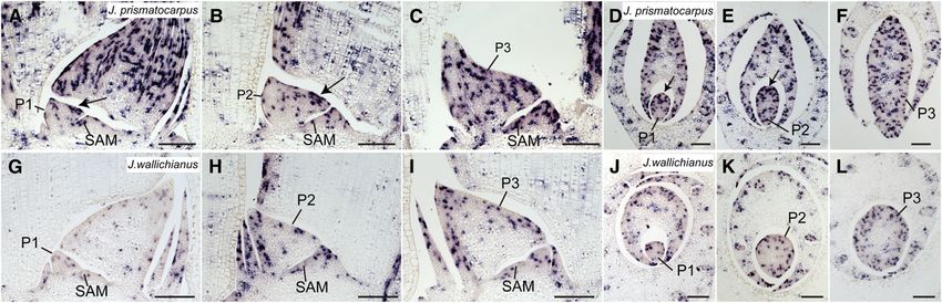
Flattened Unifacial Leaf Blade Formation          5 of 15

Figure 4. Differential Laminar Outgrowth in Unifacial Leaves of J. prismatocarpus and J. wallichianus.

(A) Seedling of J. wallichianus 4 weeks after germination.
(B) Lateral view of a leaf in J. wallichianus.
(C) and (D) Transverse sections of leaf blade (C) and leaf sheath (D) of J. wallichianus. Note that the internal air spaces are formed by cell death as in
J. prismatocarpus, and the internal region of the young leaf primordium is occupied by dividing cells, as seen in (J).
(E) to (G) Transverse sections of shoot apices of J. prismatocarpus through P1 (E), P2 (F), and P3 (G) leaf blades showing directional laminar outgrowth
along the median plane.
(H) to (J) Transverse sections of shoot apices of J. wallichianus through P1 (H), P2 (I), and P3 (J) leaf blades showing no directional laminar outgrowth.
Plastochron numbers of leaf blades are indicated (P1, P2, and P3). Bar in (A) and (B) = 1 cm; bar in (C) to (J) = 200 mm.

Byrne et al., 2000), and studied their expression patterns. We iden-           the P1 to P2 stages (Figures 6A to 6C and 6E). This is similar to
tified two genes that had expression patterns that significantly               the expression pattern of DL in rice leaves. The temporal pattern
differed between J. prismatocarpus and J. wallichianus. One is an              of Jp DL expression was correlated with the stage during which
ortholog of the DL gene, and the other is a homolog of the PRS gene.           laminar outgrowth occurs. After the P3 stage, DL ceased to be
    DL is a member of the YABBY gene family (see Supplemental                  expressed in mesophyll tissues and exhibited residual expres-
Figure 5A and Supplemental Data Set 4 online) and has a unique                 sion around the central large vascular bundle. At this stage, we
function in monocot bifacial leaves, such as those of rice                     found that DL expression also became detectable around the
(Yamaguchi et al., 2004; Ishikawa et al., 2009). In rice, DL is                large vascular bundles located nearest to the secondary central
expressed at the center of leaves, where it regulates the forma-               domain of the flattened leaf blade (Figure 6D; discussed later).
tion of the leaf midrib, a rigid and thickened structure at the                By contrast, in J. wallichianus, a DL ortholog (Jw DL) was only
center of the leaf, through a function to promote cell proliferation           weakly expressed around the central large vascular bundle, and
of leaf primordia toward the SAM side (Yamaguchi et al., 2004). In             no expression was observed in proliferating mesophyll tissue
J. prismatocarpus, a DL ortholog (Jp DL) was strongly expressed                throughout leaf development (Figures 6F to 6J). Therefore,
in the central domain of leaf primordia, extending from the central            expression patterns and levels of these DL orthologs correlated
large vascular bundle to the leaf surface at the SAM side during               with the degree of laminar outgrowth. Given that rice DL plays a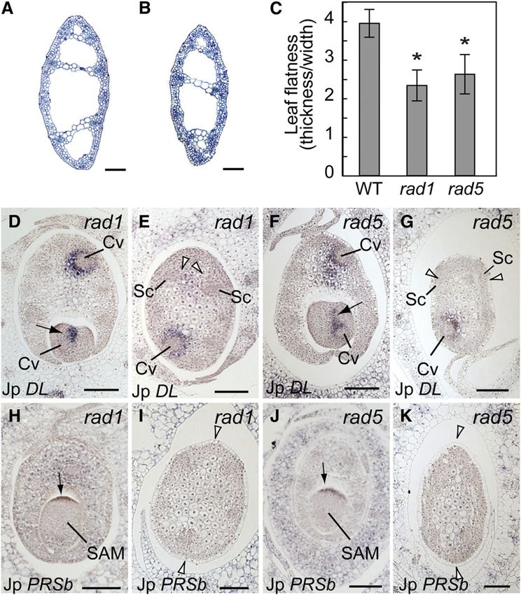
6 of 15     The Plant Cell

Figure 5. Cell Cycle Activity during Leaf Development in J. prismatocarpus and J. wallichianus.
(A) to (C) In situ localization of HistoneH4 transcripts in median longitudinal sections of J. prismatocarpus shoot apices. Leaf primordia development
proceeds from (A) to (C).
(D) to (F) In situ localization of HistoneH4 transcripts in transverse sections through P1 (D), P2 (E), and P3 (F) leaf blades in J. prismatocarpus.
(G) to (I) In situ localization of HistoneH4 transcripts in median longitudinal sections of J. wallichianus shoot apices. Leaf primordia development
proceeds from (G) to (I).
(J) to (L) In situ localization of HistoneH4 transcripts in transverse sections through P1 (J), P2 (K), and P3 (L) leaf blades in J. wallichianus.
Arrows in (A), (B), (D), and (E) indicate a concentration of HistoneH4-expressing cells at the SAM side of J. prismatocarpus leaf blades. Plastochron
numbers of leaf blades are indicated (P1, P2, and P3). Bars = 200 mm.

role in promoting cell proliferation of leaf primordia toward the            throughout leaf development (Figures 7K and 7L). Thus, PRSb
SAM side, it is possible that leaf blade flattening in J. prismato-          was expressed in margin-like regions in flattened leaf blades of
carpus is regulated by a similar DL function.                                J. prismatocarpus but not in cylindrical leaf blades of J. wallichi-
                                                                             anus. These results suggest that PRSb may also regulate the
PRSb Is Expressed Only in the Flattened Leaf Primordia                       flattening of unifacial leaf blades by promoting marginal growth.

PRS is a member of the WOX (for WUSCHEL related homeobox)
gene family (Haecker et al., 2004) and is necessary for the                  Genetic Analysis of Leaf Blade Flatness Using
establishment of marginal domains in bifacial leaves via spe-
                                                                             Interspecific Hybrids
cific expression in leaf margins (Matsumoto and Okada, 2001;
Vandenbussche et al., 2009), with loss of function in maize resulting        To reveal whether DL, PRSb, or both are responsible for the
in a narrow leaf phenotype (Nardmann et al., 2004). Phylogenetic             differences in laminar outgrowth between J. prismatocarpus and
analysis revealed that, in Juncaceae and Poaceae, the PRS                    J. wallichianus, we performed genetic analysis by generating the
genes consist of two subclasses, which we have designated                    interspecific hybrids between the two species. We found that the
PRSa and PRSb (see Supplemental Figure 6 and Supplemental                    two species could be hybridized and the F1 hybrids produced
Data Set 5 online). Expression patterns of PRSa were similar                 fertile seeds. We evaluated leaf flatness by calculating the ratio of
between J. prismatocarpus and J. wallichianus. In both species,              leaf thickness to leaf width in transverse leaf sections (Figure 8A).
PRSa was specifically expressed in the leaf margins of develop-                 We first analyzed leaf flatness in the F1 and F2 generations of
ing leaf sheaths but not in the unifacial leaf blade (Figures 7A to          interspecific hybrids between J. prismatocarpus and J. walli-
7F). These results demonstrate that unifacial leaf blades do not             chianus to understand the inheritance pattern of leaf flatness. In
differentiate a normal leaf margin identity, which further supports          the F1 generation, leaf blades were somewhat flattened (Figure
the abaxialization of unifacial leaf blades and indicates that PRSa          8B; see Supplemental Table 1 online). In the F2 generation, the
is not involved in leaf blade flattening in unifacial leaves.                distribution of leaf flatness was broader and more continuous
   On the other hand, expression patterns of PRSb differed                   (Figure 8B; see Supplemental Table 1 online). These results
between the two species. In both species, PRSb was expressed                 indicate that the difference in leaf flatness between J. prismato-
in the presumptive leaf marginal domains before the initiation of            carpus and J. wallichianus is a polygenic trait and is regulated by
leaf primordia (P0 stage; Figures 7G and 7J). In J. prismatocar-             at least two loci, including dominant or semidominant factors,
pus, PRSb (Jp PRSb) expression was not initially observed in leaf            which promote laminar outgrowth in J. prismatocarpus.
primordia during the P1 to P2 stages (Figure 7H) but became                     Next, we analyzed the genetic linkage between leaf flatness
detectable in the margin-like regions of flattened leaf blades at            and DL or PRSb genotypes in 284 siblings of the F2 generation.
the P3 stage (Figure 7I). By contrast, expression of J. wallichianus         We found that differences in the DL genotype corresponded
PRSb (Jw PRSb) was not observed in the cylindrical leaf blades               with significant differences in leaf flatness (Figure 8C; see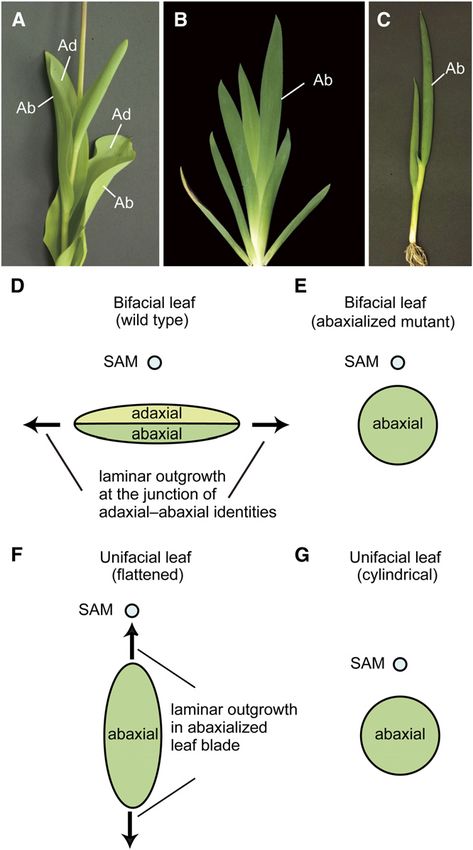
Flattened Unifacial Leaf Blade Formation           7 of 15

Figure 6. Expression Pattern of DL in Leaf Primordia of J. prismatocarpus and J. wallichianus.
(A) to (E) In situ localization of Jp DL transcripts in J. prismatocarpus shoot apices.
(A) to (C) Transverse sections of shoot apices through the SAM (A), a P1 leaf blade (B), and a P2 leaf blade (C), showing strong Jp DL expression in the
central domain of leaf primordia (arrows).
(D) Transverse section through a P3 leaf blade, showing Jp DL expression in the secondary central domain (arrowheads).
(E) Longitudinal section through the SAM showing strong Jp DL expression (arrow).
(F) to (J) In situ localization of Jw DL transcripts in J. wallichianus.
(F) to (I) Transverse sections of shoot apices through the SAM (F), a P1 leaf blade (G), a P2 leaf blade (H), and a P3 leaf blade (I), showing no Jw DL
expression in the mesophyll tissues and weak expression around the central vascular bundle. White arrowheads indicate loss of Jw DL expression in the
secondary central domain.
(J) Longitudinal section through the SAM showing weak Jw DL expression.
Bl, leaf blade; Sh, leaf sheath; Cv, central large vascular bundle. Plastochron numbers of leaf blades are indicated (P1, P2, and P3). Note that the central
large vascular bundle differentiates in a slightly off-center position in Juncus leaves. Bars = 200 mm.

Supplemental Table 1 online). Leaves were more flattened in                     expression analysis (Figure 6). To confirm this speculation, we
homozygous Jp DL plants than in homozygous Jw DL plants.                        used real-time RT-PCR analysis to study the relationship be-
Leaf flatness was intermediate between these phenotypes when                    tween DL expression level and DL genotype in the F2 generation.
DL was heterozygous. On the other hand, leaf flatness in the F2                 We found that the total amounts of DL transcripts increased as
generation was not affected by the PRSb genotype (Figure 8D;                    the copy number of Jp DL increased (Figure 9A). These results
see Supplemental Table 1 online). Combinations of each DL and                   indicate that the Jp DL locus expresses higher amounts of DL
PRSb genotype did not have synergistic effects on leaf flatness                 transcripts than the Jw DL locus in the F2 generation.
because differences in leaf flatness depended only on the DL                       To further clarify if differential DL expression between the Jp
genotypes (Figure 8E). These results indicate that the DL locus or              DL and Jw DL loci was due to cis-regulatory changes at the DL
a locus tightly linked to the DL locus is one of the loci responsible           locus, to differential trans-acting factors linked to the DL locus, or
for the laminar outgrowth difference between the two species                    to differences in leaf shape, we next examined allele-specific DL
and that the allele at this locus of J. prismatocarpus works as a               expression levels in the F1 hybrid using a single nucleotide
semidominant factor that promotes laminar outgrowth. On the                     polymorphism located in the 39 untranslated region of DL cDNA
other hand, PRSb is not directly involved in the difference in leaf             (Figure 9B). In the F1 shoot, the Jp DL allele expressed higher
flatness between J. prismatocarpus and J. wallichianus, with Jp                 amounts of DL transcripts than the Jw DL allele (Figure 9B). As
PRSb and Jw PRSb possessing similar functions.                                  the F1 hybrid contains both Jp DL and Jw DL alleles in an
                                                                                identical trans-acting environment and in an identical leaf shape
Expression Analysis of the DL Locus                                             background, this result indicates that differential DL expression
                                                                                is due to cis-regulatory changes in the DL locus itself and not to
Genetic analysis indicated that the DL locus was a particularly                 trans-acting factors or differences in leaf shape. In rice, DL is also
intriguing candidate for flattened unifacial leaf blade formation               expressed in developing carpel primordia, where it regulates
and suggested that the activity of DL differed between the two                  carpel identity (Yamaguchi et al., 2004). In situ hybridization of
species. As the putative DL protein amino acid sequences of the                 DL in developing flowers of the two Juncus species revealed
two species were identical (see Supplemental Figure 5B online),                 that DL orthologs were also expressed in carpel primordia both
the differential DL activity was possibly the result of differential            in J. prismatocarpus and in J. wallichianus at similar levels
DL expression between the two species, as suggested by in situ                  (see Supplemental Figure 7 online). Allele-specific expression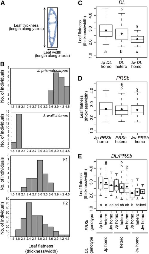
8 of 15     The Plant Cell

                                                                                analysis in the F1 flower showed that almost the same amount
                                                                                of DL mRNA was expressed from both DL alleles (Figure 9B).
                                                                                Thus, differential cis-regulatory activity of DL between the two
                                                                                species was organ specific.

                                                                                Regulation of Leaf Central-Marginal Polarity Differentiation

                                                                                During the P3 stage of leaf blade development in J. prismato-
                                                                                carpus, DL expression also became detectable around the large
                                                                                vascular bundles located nearest to the secondary central do-
                                                                                main of the flattened leaf blade (Figure 6D). Based on this
                                                                                expression of DL, together with the expression of PRSb in the
                                                                                margin-like domains of the flattened unifacial leaf blade of
                                                                                J. prismatocarpus (Figure 7I), but not in the cylindrical leaf blade
                                                                                of J. wallichianus (Figure 7L), we assumed that central-marginal
                                                                                polarity was reorganized to follow the flattened leaf shape in
                                                                                the late stages of leaf development. To test this possibility, we
                                                                                isolated mutants of J. prismatocarpus with a radialized leaf blade
                                                                                phenotype (radial leaf1, rad1; radial leaf5, rad5; Figures 10A to
                                                                                10C) and examined expression patterns of DL and PRSb in these
                                                                                mutants. As in the wild type, DL and PRSb were initially ex-
                                                                                pressed in these mutants in the primary central domain (Figures
                                                                                10D and 10F) and in the presumptive leaf marginal domain
                                                                                (Figures 10H and 10J), respectively. However, unlike the wild
                                                                                type, we did not observe the late expression of DL or PRSb in the
                                                                                secondary central and marginal domains in these mutants (Fig-
                                                                                ures 10E, 10G, 10I, and 10K). As we found no obvious mutation in
                                                                                the DL and PRSb loci of these mutants, loss of expression of
                                                                                these genes in the later stages of mutant leaf development is
                                                                                probably not caused by defects in cis-regulation or mRNA
                                                                                stability. Thus, these observations indicate that leaf blade flat-
                                                                                tening induces DL and PRSb expression in the secondary central
                                                                                and marginal domains, respectively, and suggest that central-
                                                                                marginal polarity can differentiate somewhat autonomously via a
                                                                                leaf flatness–dependent mechanism. Considering also our link-
                                                                                age analysis results, we further suggest that the loss of PRSb
                                                                                expression in the leaf blades of J. wallichianus results from the
Figure 7. Expression Patterns of PRSa and PRSb in Leaf Primordia of             loss of blade flattening in this species and not from differential
J. prismatocarpus and J. wallichianus.                                          PRSb promoter activity.
(A) to (C) In situ localization of Jp PRSa transcripts in transverse sections
of J. prismatocarpus shoot apices.
(D) to (F) In situ localization of Jw PRSa transcripts in transverse sections   DISCUSSION
of J. wallichianus shoot apices.
(A) and (D) Transverse sections through the SAM.
                                                                                We investigated the genetic mechanisms underlying flattened
(B) and (E) Transverse sections through P1 leaf blades.                         leaf blade formation in unifacial leaves. Based on the results, we
(C) and (F) Transverse sections through P3 leaf blades. Arrows in (A) to        propose the following model of laminar outgrowth in unifacial
(F) indicate PRSa expression in leaf margins of the leaf sheath.                leaves (Figure 11). Developmentally, the default shape of the
(G) to (I) In situ localization of Jp PRSb transcripts in transverse sections   unifacial leaf blade is cylindrical, as in J. wallichianus, because of
of J. prismatocarpus shoot apices.                                              abaxialization (Figure 11A, i). However, in monocots, DL func-
(J) to (L) In situ localization of Jw PRSb transcripts in transverse sections   tions to thicken leaf primordia by promoting cell proliferation
of J. wallichianus shoot apices.                                                toward the shoot apex. Such DL function in unifacial leaves may
(G) and (J) Initial PRSb expression in presumptive leaf marginal domains
                                                                                lead to flattened leaf blade formation, as in J. prismatocarpus
(arrowheads).
                                                                                (Figure 11A, ii), while in monocot bifacial leaves it leads to leaf
(H) and (K) Loss of PRSb expression in leaf blades at the P1 stage.
(I) and (L) PRSb expression in P3 stage leaf blades in margin-like
                                                                                midrib formation (Figure 11B). Flattening of the unifacial leaf
domains only in J. prismatocarpus ([I], arrows), but not in J. wallichianus     blade then triggers the differentiation of a gradient of central-
([L], white arrowheads).                                                        marginal polarity corresponding to the flattened leaf shape
Bl, leaf blade; Sh, leaf sheath. Plastochron numbers of leaf blades are         (Figure 11A, iii), which induces DL and PRSb expression in the
indicated in parenthesis. Bars = 200 mm.                                        secondary central and marginal domains, respectively (Figure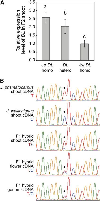
Flattened Unifacial Leaf Blade Formation             9 of 15

                                                                               11A, iv). The developmental and evolutionary mechanisms of leaf
                                                                               blade flattening in unifacial leaves are discussed below.

                                                                               Unifacial Leaf Blades Are Abaxialized at the Gene
                                                                               Expression Level

                                                                               In the leaf blade of J. prismatocarpus, ARF3a is expressed
                                                                               throughout the outer region of the leaf blade, while PHB is only
                                                                               expressed in the presumptive xylem region. These results dem-
                                                                               onstrate that the unifacial leaf blade is abaxialized at the gene
                                                                               expression level. Loss of PRSa expression in the leaf blade also

Figure 8. Genetic Analysis of Leaf Flatness Using Interspecific Hybrids
between J. prismatocarpus and J. wallichianus.

(A) Schematic of leaf flatness analysis. The ratio of leaf thickness to leaf
width was calculated to evaluate leaf flatness, with a larger value
indicating a more flattened leaf.
(B) Histograms showing leaf flatness distribution in each generation.
Generations are indicated at the top right.
(C) to (E) Box plots showing differences in leaf flatness in 284 siblings of
                                                                               Figure 9. Expression Level of DL Depends on the DL Genotype in the
the F2 generation, depending on DL (C) or PRSb (D) genotypes, and in all
                                                                               Interspecific Hybrid.
combinations of the DL and PRSb genotypes (E). Each box encloses
50% of the distribution, with the horizontal line marking the median           (A) Quantitative real-time RT-PCR analysis of DL transcripts in the
and the dot marking the mean. The lines extending from each box                interspecific hybrid F2 generation of each DL genotype. Data (mean 6
indicate the minimum and maximum values that fall within 1.5 times the         SD) are presented as relative expression units after normalization to a
height of the box. Open circles indicate outliers. Genotypes are indicated     TUBULIN gene (n = 12). Different letters (i.e., a to c) above the columns
beneath the plots. Sample numbers are shown in Supplemental Table              indicate significant variations between the genotypes based on one-way
1 online. Different letters (i.e., a to d) below the columns in (C) and (E)    ANOVA and Tukey’s HSD test (a = 0.05, with Bonferroni correction).
indicate significant differences between genotypes. One-way ANOVA              (B) Chromatograms of sequenced RT-PCR products, showing allele-
and Tukey’s HSD test (a = 0.05, with Bonferroni correction) were used for      specific DL expression in the interspecific hybrid F1 generation. Tem-
multiple comparisons.                                                          plates are indicated on the left. The dot indicates the position of a T (red)
[See online article for color version of this figure.]                         or C (blue) single nucleotide polymorphism between Jp DL and Jw DL.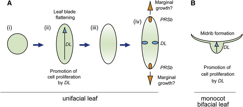
10 of 15      The Plant Cell

Figure 10. DL and PRSb Expression in the Radialized Leaf Blades of J. prismatocarpus rad1 and rad5 Mutants.

(A) and (B) Transverse sections of rad1 (A) and rad5 (B) leaf blades showing the radialized leaf blade phenotype.
(C) Leaf flatness in the wild type, rad1, and rad5. Data are mean 6 SD. Wild type, n = 20, mean = 3.95 6 0.36; rad1, n = 12, mean = 2.34 6 0.40; rad5,
n = 12, mean = 2.63 6 0.5. Asterisks indicate significant difference compared with the wild type (P < 0.05, t test).
(D) to (G) In situ localization of Jp DL transcripts in transverse sections of rad1 ([D] and [E]) and rad5 ([F] and [G]) shoot apices at an early ([D] and [F])
and late ([E] and [G]) developmental stage of leaf primordia. Arrows in (D) and (F) show initial Jp DL expression in the primary central domain.
Arrowheads in (E) and (G) show loss of Jp DL expression in the secondary central domain.
(H) to (K) In situ localization of Jp PRSb transcripts in transverse sections of rad1 ([H] and [I]) and rad5 ([J] and [K]) shoot apices at an early ([H] and [J])
and late ([I] and [K]) developmental stage of leaf primordia. Arrows in (H) and (J) show initial Jp PRSb expression in the presumptive leaf marginal
domain. Arrowheads in (I) and (K) show loss of Jp PRSb expression at a later stage of leaf development.
Cv, central large vascular bundle; Sc, large vascular bundle in the secondary central domain. Bars = 200 mm.

supports the abaxialization of the unifacial leaf blade because                    et al., 2008), which supports the notion of differential sensitivity to
PRSa is expressed at the junction of adaxial and abaxial iden-                     adaxial-abaxial polarity defects between the leaf blade and the
tities in the leaf sheath. Observations of developmental patterns                  leaf sheath in monocots.
of unifacial leaf primordia, together with in situ expression                         It has been suggested that alterations to adaxial-abaxial
analysis, have shown that the distal region of the unifacial leaf                  patterning mechanisms could be major driving forces in modi-
primordia is abaxialized from a very early stage, which leads to                   fying leaf forms (Kim et al., 2003; Gleissberg et al., 2005;
the formation of abaxialized leaf blades. The abaxialization effect                Johnston et al., 2010). The unifacial leaf is one of the most
seems to be somewhat incomplete, as the basal sheath region                        interesting examples in which alterations in leaf adaxial-abaxial
acquires adaxial-abaxial polarity in unifacial leaves, indicating                  polarity have given rise to a novel leaf form. Identification of
that the distal region of monocot leaves may be more sensitive to                  genes responsible for unifacial leaf development is essential for a
the abaxialization effect than the basal region. In maize, the                     better understanding of the developmental and evolutionary
milkweed pod1 mutant, a loss-of-function mutant of a KANADI                        mechanisms underlying unifacial leaf blade formation. The es-
homolog, shows adaxialization only in the leaf sheath (Candela                     tablishment of adaxial-abaxial polarity is regulated by several
Flattened Unifacial Leaf Blade Formation              11 of 15

Figure 11. Genetic Framework of Flattened Leaf Blade Formation in Unifacial Leaves.

(A) Model of laminar outgrowth and autonomous differentiation of central-marginal polarity in flattened unifacial leaves. (i) Cylindrical leaf blade as a
result of abaxialization. (ii) Leaf blade flattening through DL function. (iii) Differentiation of gradient of central–marginal polarity to follow the flattened leaf
shape. (iv) Induction of DL and PRSb expression in the secondary central and marginal domains, respectively.
(B) DL function in monocot bifacial leaves.
Blue arrows indicate the DL function to promote cell proliferation of leaf primordia toward the SAM side. Orange arrows indicate a putative PRSb
function to promote marginal growth in flattened unifacial leaves.

distinct families of transcription factors and small regulatory                      J. prismatocarpus and J. wallichianus. Genetic analysis using
RNAs (Husbands et al., 2009). It is possible that a genetic change                   interspecific hybrids demonstrates that the chromosome region
or changes in one or more of these regulators may have resulted                      containing the DL locus from J. prismatocarpus flattens the
in unifacial leaf development. Further functional studies of each                    unifacial leaf blade. Although further examinations are required
regulator are expected to reveal the genetic mechanisms of leaf                      to finely map the locus, expression analysis demonstrates that
blade abaxialization in unifacial leaves.                                            the expression activities of DL differ between the two species,
                                                                                     probably due to cis-regulatory changes at the DL locus itself,
DL May Promote Flattened Leaf Blade Formation in                                     rather than differences in trans-acting factors of DL. These
J. prismatocarpus                                                                    results suggest that DL is one of the genetic factors that promote
                                                                                     laminar outgrowth in J. prismatocarpus. During unifacial leaf
Observations of developmental patterns of leaf blades in J.                          blade flattening, DL is probably involved in active cell prolifera-
prismatocarpus and J. wallichianus have demonstrated that leaf                       tion on the SAM side of leaf primordia at an early stage of leaf
blade flattening in unifacial leaves is promoted by directional                      development because strong DL expression is observed during
laminar outgrowth along the median plane of the leaf primordia.                      the P1 and P2 stages and DL has a similar function in rice in
At the cellular level, the directional laminar outgrowth appears                     promoting cell proliferation in the leaf primordia toward the SAM
to be initially triggered by active cell proliferation on the SAM                    side. Functional studies, such as those involving the generation
side of the leaf blade, as demonstrated by a concentration of                        of near-isogenic lines carrying the Jp DL allele in a J. wallichianus
HistoneH4-expressing cells in J. prismatocarpus leaf primordia.                      background and vice versa, or the isolation of loss- and/or gain-
Subsequently, laminar outgrowth may be bidirectionally main-                         of-function DL lines by mutation or transgenic approaches,
tained by more diffuse cell proliferation activity. A similar obser-                 would further reveal the exact role of DL in unifacial leaf devel-
vation has been reported in another flattened unifacial-leafed                       opment.
species, Acorus calamus, where unifacial leaf blade flattening is                       Genetic analysis indicated that factors other than DL are also
initially mediated by adaxial (i.e., SAM side) meristematic activity                 involved in the differential laminar outgrowth observed between
and then bidirectionally proceeds by more diffuse meristematic                       J. prismatocarpus and J. wallichianus. Such factors may interact
activity (Kaplan, 1970). Therefore, leaf blade flattening of unifacial               with DL at an early stage or regulate more diffuse cell proliferation
leaves, which probably has a conserved mechanism among                               activity during the later stages of leaf development. Identifying
monocots, could be dissected into two developmental pro-                             such factors by quantitative trait locus mapping using interspe-
cesses: active cell proliferation at the SAM side of leaf primordia                  cific hybrids and by isolating the causative genes for the rad
at an early stage of development and more diffuse cell prolifer-                     mutants in J. prismatocarpus would further clarify the genetic
ation during later stages.                                                           mechanisms underlying leaf blade flattening in unifacial leaves.
   We identified the DL ortholog as a candidate responsible for                         Identification of cis-regulatory differences at the DNA se-
leaf blade flattening in J. prismatocarpus. Expression levels and                    quence level in the DL locus of the two Juncus species would
patterns of DL correlate with the degree of laminar outgrowth in                     further deepen our understanding of the mechanisms that
12 of 15 The Plant Cell
regulate DL expression. As flattened unifacial leaves are wide- in flattened unifacial leaves, although it is not clearly evident in
spread in monocots from the earliest divergent extant family, J. prismatocarpus. Some flattened unifacial-leafed species, such
Acoraceae, to specialized families, such as Iridaceae and as A. calamus or Iris ensata, develop a clear secondary midrib in
Juncaceae (Rudall and Buzgo, 2002; Yamaguchi and Tsukaya, the central domain of the flattened leaf blade, and margin-like
2010), and expression patterns of DL are similar between bifacial tissues often differentiate at both tips of the flattened blade
leaves of rice and flattened unifacial leaves of J. prismatocarpus, (Kaplan, 1970; Yamaguchi and Tsukaya, 2010). In these species,
it is likely that expression activity in leaf primordia is reduced in DL and PRSb homologs may regulate the formation of the
J. wallichianus. Interestingly, the differential cis-regulatory activity secondary midrib and margin-like tissues, respectively. It is also
of DL is organ specific: Jp DL and Jw DL alleles express almost possible that PRSb expression in margin-like regions promotes
the same amount of DL transcripts in carpels of the interspecific flattening of unifacial leaf blades by promoting marginal growth,
hybrid F1 generation. It has been suggested that organ-specific although PRSb is not directly involved in the difference in leaf
cis-regulatory changes of multifunctional genes may provide a flatness between J. prismatocarpus and J. wallichianus. Loss of
mechanism for generating morphological diversity, while pre- PRSb expression in J. wallichianus is probably a secondary
serving their roles in other developmental processes (Carroll, effect due to a lack of leaf blade flattening in this species, and Jw
2000; Shapiro et al., 2004; Prud’homme et al., 2006). In the PRSb would possess a cryptic function equivalent to that of Jp
evolution of unifacial leaves, DL expression may have been PRSb, as indicated by genetic analysis. Isolation of PRSb mu-
modified multiple times to generate diversity in transverse forms tants from J. prismatocarpus would help to reveal the role of this
of unifacial leaf blades. Thus, it will be of great interest to identify gene during flattened unifacial leaf development.
the actual sequences of the cis-regulatory elements of DL, so as
to examine their roles in the evolution of flattened and radialized
Evolution of Leaf Flattening in Unifacial Leaves
unifacial leaves.
Although the independent evolution of similar morphological
Regulation of Central-Marginal Leaf Polarity Differentiation traits is widespread, the underlying mechanisms are not fully
understood (Gould, 2002; West-Eberhard, 2003; Yoon and
The mechanisms that regulate the specification of central- Baum, 2004). Flattened leaves have independently evolved in
marginal leaf polarity are largely unknown even in model species bifacial and unifacial leaves, probably for efficient light capture.
because of a lack of useful mutants. For example, it remains Our research demonstrates that flattening of leaf blades in
unknown whether specification of central-marginal polarity for- unifacial leaves is promoted by DL function, which plays a
mation requires positional cues regarding the relationship to the distinct phenotypic role in bifacial leaves, namely, in leaf midrib
SAM. It is also unclear whether specification of leaf margin formation. However, DL seems to play a similar function at the
identity requires positional information from the junction of leaf cellular level both in the bifacial and unifacial leaf development to
adaxial-abaxial domains, where leaf margins generally develop promote cell proliferation of leaf primordia toward the shoot
in bifacial leaves. In this study, we have shown that leaf blade apex. Such DL function may have been easily co-opted to make
flattening is a key that triggers DL and PRSb expression in the abaxialized leaf blades flatten during the evolution of unifacial
secondary central and marginal domains of the flattened leaf leaves. Thus, a preexisting gene function can be recruited to play
blade, respectively. This finding indicates that central-marginal a distinct phenotypic role in organisms with different body plans,
leaf polarity can somewhat autonomously differentiate depend- without changing the gene’s cellular function, and such recruit-
ing on the flattened leaf shape without information from a source ment can give rise to convergence of similar morphological traits.
outside of the leaf, such as the SAM. In addition, specification of Unifacial leaves have repeatedly evolved in monocots (Rudall
leaf margin identity could be partly independent of adaxial- and Buzgo, 2002; Yamaguchi and Tsukaya, 2010), indicating the
abaxial polarity, although positional information from the junction existence of backgrounds that allow unifacial leaf development
of adaxial-abaxial polarity is necessary for PRSa expression and in monocots. Since DL orthologs function in leaf development in
probably for rigid specification of leaf margin identity. We pro- monocots alone (Bowman and Smyth, 1999; Yamaguchi et al.,
pose that once the leaf blade is flattened, a gradient of central- 2004; Fourquin et al., 2005; Ishikawa et al., 2009; Wang et al.,
marginal polarity is formed by an as yet unidentified factor, which 2009), monocot leaves may possess the unique ability to be-
is distributed in a symmetrical pattern in the flattened leaf blade. come flattened by escaping from a developmental constraint to
The plant hormone auxin is known to act as a gradient signal in be radialized, even when they are abaxialized. Thus, the specific
multiple contexts throughout plant development (De Smet and function of DL in leaf development in monocots may account for
Jurgens, 2007; Bowman and Floyd, 2008). During Arabidopsis one genetic background that has allowed the repeated evolution
leaf development, auxin is symmetrically distributed on either of unifacial leaves in monocots.
side of the midvein (Reinhardt et al., 2003; Zgurski et al., 2005)
and is therefore a potential candidate for this unidentified mol-
METHODS
ecule. Thus, it will be of great interest to study the distribution of
auxin in unifacial leaves and to determine its relationship with
central-marginal polarity differentiation. Plant Materials and Growth Conditions
The functions of DL and PRSb in the secondary central and Juncus prismatocarpus subsp leschenaultii Kirschner and Juncus wall-
marginal domains are also of interest. Morphological differenti- ichianus Laharpe were collected from wild populations in Okazaki, Japan.
ation of the secondary central-marginal polarity is often observed Herbarium specimens were verified by Futoshi Miyamoto at TokyoFlattened Unifacial Leaf Blade Formation 13 of 15
University of Agriculture, Japan. At least six generations of each species Genotyping Interspecific Hybrids
were self-pollinated before use. Seeds of both species were cold-treated
Genomic DNA from the DL and PRSb loci were amplified using PCR, then
for 1 week at 48C and germinated on agar plates containing Murashige
treated with the restriction enzymes NruI and NaeI, respectively, and
and Skoog salts. Seedlings were grown in soil under a 16-h-light/8-h-dark
separated by agarose gel electrophoresis. Primer sequences are listed in
cycle at 228C. For mutagenesis, seeds of J. prismatocarpus were treated
Supplemental Table 2 online.
with 0.3% ethyl methanesulfonate for 14 h at room temperature. Approx-
imately 5000 M2 plants were screened.
Allele-Specific DL Expression
RNA Extractions and Degenerate PCR
Total RNA was isolated from lateral branches that produced four visible
Total RNA was isolated from 4-week-old seedlings of J. prismatocarpus leaves and young inflorescences (5 mm in length) of interspecific F1
and J. wallichianus using Plant RNA Isolation Reagent (Invitrogen), with hybrids using Plant RNA Isolation Reagent, with subsequent treatment
subsequent treatment with DNaseI (Invitrogen). Total RNA (1 mg) was with DNaseI. Total RNA (1 mg) was used for first-strand cDNA synthesis
used for first-strand cDNA synthesis using the SuperScript III first-strand using the SuperScript III first-strand synthesis system, and 0.5 mL of this
synthesis system (Invitrogen), and 0.5 mL of this reaction was used as the reaction was used as the template for PCR amplification. A portion of DL
template for PCR amplification. Degenerate primers were designed using cDNA was amplified by 20 cycles of PCR and then purified and se-
the CODEHOP program (Rose et al., 2003). Amplified DNA fragments quenced directly. The genomic DNA of the F1 hybrid was used as a
were gel-extracted and cloned into pCRII-TOPO (Invitrogen), and at least control. Primer sequences are listed in Supplemental Table 2 online.
16 clones per fragment were sequenced. Primer sequences are listed in
Supplemental Table 2 online. Phylogenetic Analysis
The amino acid sequences of HD-ZIPIII, ARF3/ARF4, YABBY, and WOX
59 and 39 Rapid Amplification of cDNA Ends
proteins were first aligned by ClustalW and readjusted manually. The
To determine 59 and 39 sequences of homologous genes, 59 and 39 rapid phylogenetic trees were generated using MEGA4 (Tamura et al., 2007) by
amplification of cDNA ends was performed. cDNA was generated using a the neighbor-joining method (Saitou and Nei, 1987) with 1000 iterations of
GeneRacer kit (Invitrogen) according to the manufacturer’s protocol, bootstrap analysis. For phylogenetic analysis of Juncus species, the
using 2 mg of total RNA per reaction. Amplified products were cloned into nuclear ribosomal DNA internal transcribed spacer (ITS) region se-
pCRII-TOPO (Invitrogen), and at least eight clones per fragment were quences were aligned by ClustalW and readjusted manually. Maximum
sequenced. Full-length cDNAs were reamplified by RT-PCR. Primer likelihood and maximum parsimony analyses were performed using
sequences are listed in Supplemental Table 2 online. PAUP* version 4.0b10 (Swofford, 2002) as described by Roalson
(2005). Sequences used to generate the phylogeny are presented in
In Situ Hybridization Supplemental Data Sets 1 to 5 online. Accession numbers used in the
phylogenetic analyses are shown in Supplemental Table 3 online.
Shoot apices and inflorescences were fixed in 3% paraformaldehyde and
0.25% glutaraldehyde in 0.1 M sodium phosphate buffer for ;16 h at 48C.
Tissue was then dehydrated in a graded ethanol series, replaced with Accession Numbers
xylene, and embedded in Paraplast Plus (Oxford Labware). Hybridization Sequence data from this article can be found in the GenBank/EMBL data
was performed as previously described (Yamaguchi et al., 2004) on 9-mm libraries under the following accession numbers: AB539879 (Jp PHB),
paraffin sections. A portion of the cDNA sequences were amplified by RT- AB539877 (Jp ARF3a), AB539878 (Jp DL), AB539882 (Jw DL), AB539880
PCR, cloned into pCRII-TOPO (Invitrogen), and used to generate sense (Jp PRSa), AB539881 (Jp PRSb), AB539883 (Jw PRSa), AB539884 (Jw
and antisense probes. Primer sequences are listed in Supplemental Table PRSb), and AB540127 (Jw ITS).
2 online. In all cases, sense probes produced no signal.
Author Contributions
Real-Time PCR
T.Y. conceived the project, designed the study, performed the experi-
Total RNA was isolated from lateral branches that produced four visible
ments, and wrote the article. S.Y. performed statistical analysis. H.T.
leaves using Plant RNA Isolation Reagent, with subsequent treatment
designed and directed the study and wrote the article.
with DNaseI. Accumulation levels of DL transcripts were analyzed using a
7500 Real-Time PCR system (Applied Biosystems) by monitoring ampli-
fication with SYBR Premix Ex Taq II (Takara), as described in the Supplemental Data
manufacturer’s protocol. All data are presented as relative expression
The following materials are available in the online version of this article.
units after normalization to a TUBULIN gene. All data were reproduced in
two or more additional independent experiments. Supplemental Figure 1. Phylogenetic Tree of HD-ZIPIII Proteins.
Supplemental Figure 2. Phylogenetic Tree of ARF3/ARF4 Proteins.
Analysis of Leaf Flatness
Supplemental Figure 3. Serial Longitudinal Sections of J. prismato-
The middle portions of leaf blades that were formed at the final vegetative carpus Shoot Apices.
nodes were sampled after maturation, and cross sections were made by Supplemental Figure 4. Simplified Phylogenetic Tree of Juncus
hand. Images of sections were taken and boxes enclosing the transverse Showing the Relationship between J. prismatocarpus and J. wall-
planes were drawn on digital images. Leaf thickness (length along y axis) ichianus.
and width (length along x axis) were measured using Image J software
Supplemental Figure 5. Phylogeny of YABBY Family Proteins and
(http://rsbweb.nih.gov/ij/). The ratio of leaf thickness to leaf width was
Putative Amino Acid Sequences of J. prismatocarpus and J. wall-
calculated to evaluate leaf flatness. One-way analysis of variance
ichianus DL Proteins.
(ANOVA) and the Tukey’s HSD test (a = 0.05, with Bonferroni correction)
were used for multiple comparisons. Supplemental Figure 6. Phylogenetic Tree of WOX Proteins.You can also read