Downregulation of Filamin a Expression in the Aorta Is Correlated With Aortic Dissection - Frontiers
←
→
Page content transcription
If your browser does not render page correctly, please read the page content below
ORIGINAL RESEARCH
                                                                                                                                            published: 13 August 2021
                                                                                                                                      doi: 10.3389/fcvm.2021.690846

                                            Downregulation of Filamin a
                                            Expression in the Aorta Is Correlated
                                            With Aortic Dissection
                                            Yue Chen 1† , Xiang Wei 1,2,3,4† , Zihao Zhang 1 , Yi He 1 , Bo Huo 1 , Xian Guo 1 , Xin Feng 1 ,
                                            Ze-Min Fang 1 , Ding-Sheng Jiang 1,2,3,4* and Xue-Hai Zhu 1,2,3,4*
                                            1
                                             Division of Cardiothoracic and Vascular Surgery, Sino-Swiss Heart-Lung Transplantation Institute, Tongji Hospital, Tongji
                                            Medical College, Huazhong University of Science and Technology, Wuhan, China, 2 Key Laboratory of Organ Transplantation,
                                            Ministry of Education, Chinese Academy of Medical Sciences, Wuhan, China, 3 NHC Key Laboratory of Organ
                                            Transplantation, Chinese Academy of Medical Sciences, Wuhan, China, 4 Key Laboratory of Organ Transplantation, Chinese
                                            Academy of Medical Sciences, Wuhan, China

                                            Filamins (FLNs) are actin cross-linking proteins, and as scaffolding proteins, FLNs are
                                            closely associated with the stabilization of the cytoskeleton. Nevertheless, the biological
                                            importance of FLNs in aortic dissection (AD) has not been well-elucidated. In this study,
                         Edited by:
                                            we first reanalyzed datasets downloaded from the Gene Expression Omnibus (GEO)
                       Sarah Parker,        database, and we found that in addition to the extracellular matrix, the actin cytoskeleton
         Cedars-Sinai Medical Center,       is a key structure associated with AD. Given that FLNs are involved in remodeling the
                       United States
                                            cytoskeleton to affect cellular functions, we measured their expression levels in the
                          Reviewed by:
                             Dan Rudic,     aortas of patients with Stanford type A AD (TAAD). Our results showed that the mRNA
    Augusta University, United States       and protein levels of FLNA were consistently decreased in dissected aortas of both
                           Louis Saddic,
  University of California, Los Angeles,
                                            humans and mice, while the FLNB protein level was upregulated despite decreased
                           United States    FLNB mRNA levels, and comparable expression levels of FLNC were observed between
                   *Correspondence:         groups. Furthermore, the immunohistochemistry results demonstrated that FLNA was
                     Ding-Sheng Jiang       highly expressed in smooth muscle cells (SMCs) of aorta in non-AD samples, and
                      jds@hust.edu.cn
                          Xue-Hai Zhu       downregulated in the medial layer of the dissected aortas of humans and mice. Moreover,
               13072724207@163.com          we revealed that FOS and JUN, forming a dimeric transcription factor called AP-1
     † These   authors have contributed     (activating protein-1), were positively correlated with the expression of FLNA in aorta.
                    equally to this work    Either overexpression of FOS or JUN alone, or overexpression of FOS and JUN together,
                                            facilitated the expression of FLNA in primary cultured human aortic SMCs. In the present
                    Specialty section:
          This article was submitted to
                                            study, we not only detected the expression pattern of FLNs in aortas of humans and
          Atherosclerosis and Vascular      mice with or without AD, but we also found that the expression of FLNA in the AD
                              Medicine,
                                            samples was significantly reduced and that AP-1 might regulate the expression of FLNA.
                a section of the journal
  Frontiers in Cardiovascular Medicine      Our findings will contribute to the elucidation of the pathological mechanisms of AD and
            Received: 04 April 2021         provide potential therapeutic targets for AD.
            Accepted: 13 July 2021
                                            Keywords: aortic dissection, FLNA, filamins, FOS/JUN, AP-1, bioinformatics
          Published: 13 August 2021
                             Citation:
Chen Y, Wei X, Zhang Z, He Y, Huo B,
 Guo X, Feng X, Fang Z-M, Jiang D-S
                                            INTRODUCTION
 and Zhu X-H (2021) Downregulation
of Filamin a Expression in the Aorta Is
                                            Aortic dissection (AD) is a life-threatening cardiovascular disorder requiring urgent surgical
    Correlated With Aortic Dissection.      therapy. Despite advances in diagnostic modalities, surgical treatments, and medical devices, the
  Front. Cardiovasc. Med. 8:690846.         mortality of AD remains high, and almost 75% of patients with Stanford type A AD (TAAD) die
      doi: 10.3389/fcvm.2021.690846         within 2 weeks when untreated (1). Hence, emergency surgery is currently the optimal solution for

Frontiers in Cardiovascular Medicine | www.frontiersin.org                        1                                           August 2021 | Volume 8 | Article 690846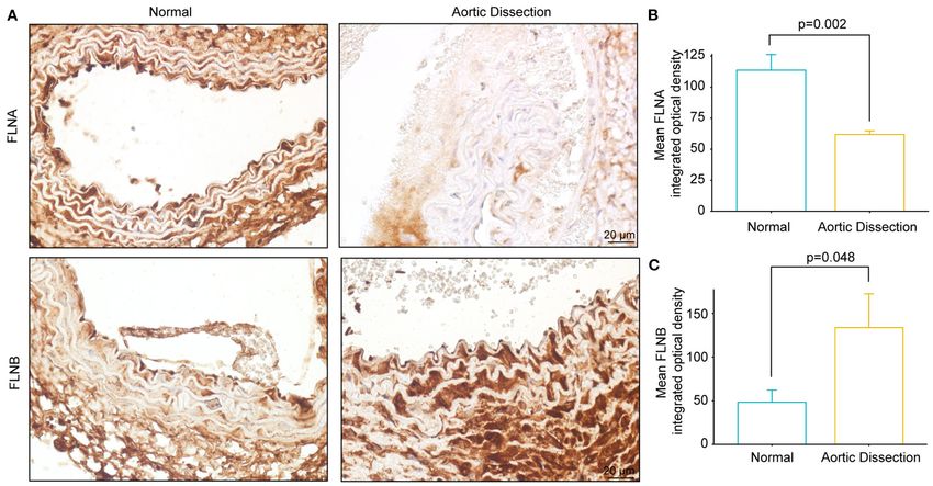
Chen et al.                                                                                              Expression Regulation of FLNA During AD

saving the lives of patients with TAAD. However, the outcome is          with FLNA expression in the aorta of humans. Moreover, both
unpredictable and patients may suffer many complications, such           FOS and JUN overexpression can promote the expression of
as spinal cord ischemia, stroke, mesenteric ischemia/infarction,         FLNA in cultured primary human aorta smooth muscle cells
and acute renal failure during rapid open surgical repair                (HASMCs). Thus, our data indicate that the AP-1/FLNA axis
(2). Therefore, further exploration of the pivotal molecular             might play a potential role in AD formation and may serve as
mechanisms of AD to identify effective therapeutic targets is            a promising therapeutic target for AD.
urgently needed.
    Actins and actin-binding proteins are cytoskeletal proteins
that are indispensable for cellular structure and function (3).          MATERIALS AND METHODS
In the aorta, these structural proteins are closely related to           Human Aortic Samples
SMC contractile force and play a crucial role in vascular                All protocols using human specimens were approved by
tension (4). Thus, the aberrant expression of these molecules            the Human Research Ethics Committees of Tongji Hospital,
can result in remodeling of the aorta and vascular disorder.             Tongji Medical College, Huazhong University of Science and
For example, mutations in ACTA2, which encodes SMC α-actin,              Technology, and informed consent was obtained from patients or
are responsible for 14% of patients with inherited ascending             their family members. Aortic tissues were obtained from patients
thoracic aortic aneurysms and dissections (5). ACTA2 deficiency          with TAAD and patients undergoing heart transplantation
in mice reduces elastin levels with increased collagen deposition,       (controls). All samples were stored in liquid nitrogen or
facilitating angiotensin II (Ang II)-induced TAAD (6). In                paraformaldehyde as soon as possible after aorta excision to avoid
addition to actins, smooth muscle 22 (SM22)-α, an SMC-specific           specimen degradation.
actin-binding protein, is a contractile marker for SMC phenotype
transition (7). The expression of SM22 is decreased in TAAD
tissues and the suppression of SM22 in SMCs significantly
                                                                         Animal Experiments
                                                                         All animal experiments were performed in accordance with the
contributes to proliferation, indicating the transformation from
                                                                         protocols approved by the Animal Care and Use Committees of
contractile to synthetic SMCs (7). Additionally, the depletion
                                                                         Tongji Hospital, Tongji Medical College, Huazhong University
of actin-binding protein Girdin attenuates the proliferation and
                                                                         of Science and Technology. All mice (C57BL/6 background)
migration of SMCs to affect vascular remodeling (8). Thus, these
                                                                         were housed in specific-pathogen-free facilities with a 12-
studies indicated that actins and actin-binding proteins are vital
                                                                         h light/dark cycle and controlled temperature (20–22◦ C). To
for the stabilization of SMC functions and vascular homeostasis.
                                                                         induce AD, the 3-week-old mice were stimulated with 0.6% β-
    Filamins (FLNs) are actin-filament-crosslinking proteins and
                                                                         aminopropionitrile (BAPN, A3134; Sigma-Aldrich) taken orally
consist of three homologous proteins: FLNA, FLNB, and FLNC
                                                                         for 4 weeks. The physical conditions of the mice were observed
(9). As actin-binding proteins, FLNs can stabilize delicate three-
                                                                         every day and autopsy was performed when the mice died.
dimensional actin webs and link them to cellular membranes
to maintain cellular morphology (9). It was reported that FLNs
interact with more than 70 proteins including transmembrane              Western Blotting
receptors and signaling molecules to play a vital role in cell           Western blotting was performed as previously described (14).
motility, adhesion, spreading, and signal transduction (10). For         Human aorta specimens were homogenized with RIPA lysis
SMCs, the degradation of FLNs inhibits differentiation and               buffer containing protease inhibitor complex and phosphatase
migration and interrupts phenotype switching (11). FLNA is               inhibitors, and the protein concentration was assayed using
the most abundant and widely understood actin-binding protein            a BCA protein assay kit (23227, Thermo Fisher Scientific).
among the three FLN isoforms. Kevin Retailleau et al. reported           Twenty micrograms of protein were separated by 10% sodium
that the deletion of FLNA in SMCs might cause a reduction                dodecyl sulfate-polyacrylamide gel electrophoresis and then
in arterial stiffness and a compensatory increase in the conduit         transferred to polyvinylidene fluoride membranes (IPVH00010,
artery diameter of mice (12). Furthermore, FLNA is involved with         Millipore). The membranes were blocked with 5% nonfat milk
a G protein-coupled P2Y2 nucleotide receptor to regulate the             and then incubated with primary antibodies at 4◦ C overnight.
migration of vascular SMCs (13). However, the role of the FLN            Subsequently, the membranes were washed and incubated with
family in AD remains unclear.                                            the corresponding horseradish peroxidase-conjugated secondary
    In the present study, we first analyzed the differentially           antibody. Finally, the membranes were incubated in ECL
expressed genes in aortic samples of normal subjects and patients        reagents prior to visualization using a ChemiDocTM XRS+
with AD, and further focused on the expression of the FLN                system (Bio-Rad). The antibodies used in this study included
family. Compared with non-AD samples, significantly reduced              FLNA (ab76289, Abcam), FLNB (GTX101206, GeneTex), FLNC
FLNA mRNA and protein levels were observed in the aortic                 (ab180941, Abcam), HA (H3663, Sigma), Flag (F1084, Sigma),
samples obtained from AD patients. We next generated a murine            and β-Actin (AC026, ABclonal).
AD model by treating mice with β-aminopropionitrile (BAPN)
for 4 weeks and found a similar expression pattern of FLNA in            Real-Time PCR
normal and AD mice. Further bioinformatics analysis suggested            Real-time PCR was performed using established protocols
that the expression levels of FOS and JUN, which form the                (15). Total RNA from human aorta specimens was extracted
activator protein-1 (AP-1) complex, were positively correlated           using TRIzol reagent (15596026, Ambion) according to the

Frontiers in Cardiovascular Medicine | www.frontiersin.org           2                                    August 2021 | Volume 8 | Article 690846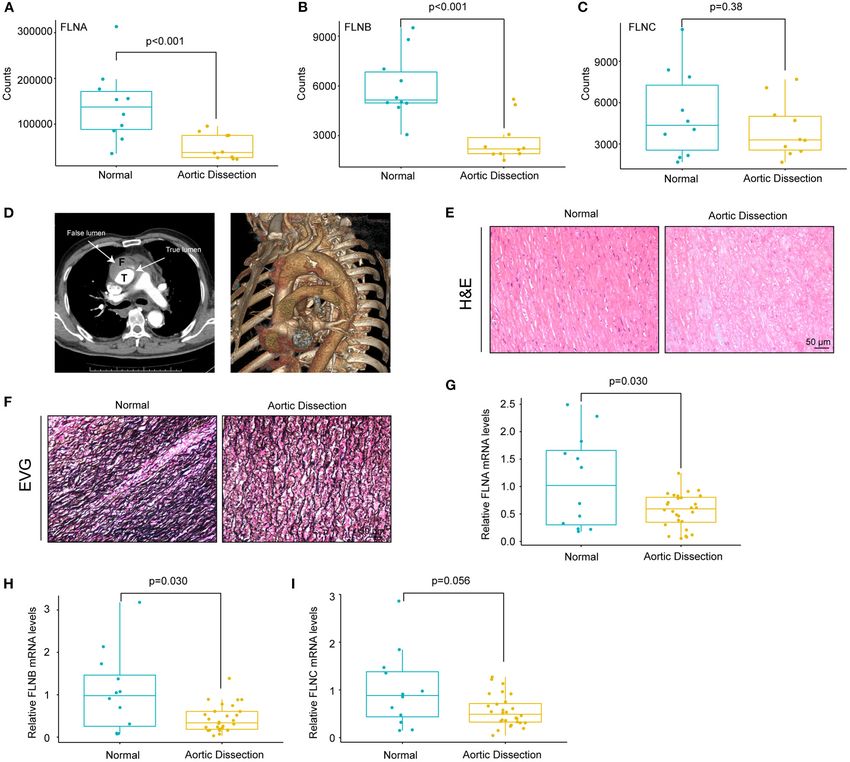
Chen et al.                                                                                             Expression Regulation of FLNA During AD

manufacturer’s instructions. In total, 5 µg of RNA was reverse-         respectively. pHAGE-FOS-HA and pHAGE-JUN-Flag plasmids,
transcribed using the Transcriptor First Strand cDNA synthesis          and the packaging plasmids psPAX2 (12260, Addgene) and
kit. The relative mRNA levels of FLN family members were                pMD2.G (12259, Addgene) were cotransfected into HEK293T
detected by a quantitative real-time PCR system using SYBR              cells in the presence of polyethylenimine (764604, Sigma-
green (11201ES08, Yeasen), and the results were normalized              Aldrich) and incubated for 48 h and the supernatants containing
against 18S expression. Primer sequences are as follows:                lentivirus were harvested and filtered through a 0.22 µm
FLNA forward primer 5′ -CCGCAATGACAATGACACC-3′ ,                        filter (SLGP033RB, Millipore) for cell infection. HASMCs were
FLNA reverse primer 5′ -TGGAGATACTGCCACTGAGA-3′ ,                       infected with the corresponding lentivirus (control, lenti-FOS-
FLNB forward primer 5′ -ACTGTCATGGCCACAGATGG-3′ ,                       HA, lenti-JUN-Flag, lenti-FOS-HA+ lenti-JUN-Flag) for 24 h via
FLNB reverse primer 5′ -AAATCCCAGGCCGTTCATGT-3′ ,                       hexadimethrine bromide (10 µg/mL, H9268, Sigma-Aldrich) and
FLNC forward primer 5′ - CTCCAGCTACAGCTCCATCC-3′ ,                      then cultured with DMEM/F12 medium without FBS for cell
FLNC reverse primer 5′ -CCATGTGCTTCACGTACACC-3′ , and                   synchronization. Finally, the infected cells were maintained in
18S forward primer 5′ -CTCAACACGGGAAACCTCAC-3′ , 18S                    DMEM/F12 medium containing FBS for 24 h and then collected
reverse primer 5′ -CGCTCCACCAACTAAGAACG-3′ .                            for subsequent research.

Histology and Immunohistochemistry
                                                                        Bioinformatic Analysis
Staining                                                                To obtain the mRNA expression profiles of aortas with or
Histology and immunohistochemistry staining were performed              without AD in humans, the keywords “AD” and "Homo
as previously described (16). Human and mouse aortic                    sapiens” were applied to search datasets in the Gene Expression
samples were fixed in 4% paraformaldehyde and embedded                  Omnibus (GEO) database (https://www.ncbi.nlm.nih.gov/geo/).
in paraffin, and then 5-µm-thick serial sections were stained           In these results, we further analyzed each dataset and excluded
with hematoxylin-eosin (H&E) for morphological examination.             all non-mRNA datasets, such as non-coding RNA and DNA
Elastin fibers were visualized using elastic van Gieson (EVG)           methylation profile sets. After assessing all the datasets
staining according to the manufacturer’s instructions and               independently, we included four gene expression datasets
quantified by counting the total breaks over the length of              “GSE153434,” “GSE98770,” “GSE52093,” and “GSE147026,”
the vessel. Immunohistochemistry analyses were performed                which are datasets of expression profile array or mRNA
using a standard protocol. The paraffin-embedded sections               high-throughput sequencing of aorta with or without AD in
were incubated with citrate antigen retrieval solution (pH              humans. To analyze the data, the raw expression values of
6.0, P0083, Beyotime) for antigen retrieval. The sections were          each dataset were normalized by using the “limma” package
incubated with primary antibodies overnight at 4◦ C and                 in R, and log2 conversion was completed. After processing,
then incubated with horseradish peroxidase (HRP)-conjugated             differentially expressed genes (DEGs) were filtered based on the
secondary antibodies for 1 h. Finally, the DAB horseradish              criteria adjust.p.value (FDR) 1.5. The DEGs
peroxidase color development kit (ZLI-9017, ZSGB-BIO) was               obtained in each dataset were further analyzed by the R package
used for color development.                                             “clusterProfiler” for GO, KEGG, and GSE analysis (17). Each
                                                                        term was represented as a circle node, in which color represented
Cell Culture                                                            its adjusted p-value. In addition, the node sizes indicated gene
Primary HASMCs were isolated from the aortas of patients who
                                                                        ratios and the mean proportions of genes enriched in the GO or
underwent heart transplantation as previously described (14).
                                                                        KEGG categories.
Briefly, aortas were stored in DME/F12 medium (SH30023.01;
                                                                            To explore the transcription factors regulating FLNA, we
HyClone) at 4◦ C, and then the intima and adventitia were
                                                                        queried the database of the website signaling pathways project
stripped under microscope. Furthermore, the medial layer was
                                                                        (https://www.signalingpathways.org/index.jsf) and combined the
peeled as thin as possible and minced in a sterile culture flask.
                                                                        results with the GSE153434 database to analyze the correlation
Small pieces were placed in the wall of a culture flask without
                                                                        between the expression of transcription factors and FLNA. The
culture medium for 30 min to enable their adherence to the flask.
                                                                        Spearman correlation coefficient was used in the correlation
Next, DME/F12 medium containing 10% fetal bovine serum
                                                                        analysis, and t-tests were carried out to determine the
(FBS, SH30084.03; HyClone) and 1% penicillin-streptomycin
                                                                        significant differences.
(15140-122; Thermo Fisher Scientific) was slowly added to the
culture flask, which was maintained in a humidified environment
at 37◦ C with 5% CO2 . A week later, HASMCs were removed                Statistical Analysis
from tissues and then transferred to new culture dishes as first        The data are presented as the mean ± standard deviation (SD).
generation. After two passages of growth, the third generation          All statistical analyses in this study were performed by using SPSS
SMCs were used for the cell experiments.                                software (version 23.0). Comparisons of the means between 2
                                                                        groups were performed by independent sample t-test and one-
Plasmid Constructs and Cell Treatments                                  way ANOVA was performed for the difference assessments of
The full-length coding sequences of the human FOS and JUN               more than two groups. Correlation analyses were performed
genes were amplified from cDNA and then cloned into the                 using Pearson’s correlation analysis. A value of p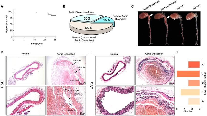
Chen et al. Expression Regulation of FLNA During AD
RESULTS Figures 4A–C, there was no remarkable correlation between the
FLN mRNA levels and the diameters of aorta, including the
Bioinformatics Analysis of mRNA ascending, the thoracic and abdominal aorta, or aortic arch.
Expression Profiles in Human Aortas In contrast, the mRNA expression of FLNA and FLNB was
To explore the differential gene expression in normal controls positively correlated in the aorta (r = 0.6, p < 0.001), but only
and patients with TAAD, we reanalyzed four gene expression a weak correlation was found between FLNA and FLNC and
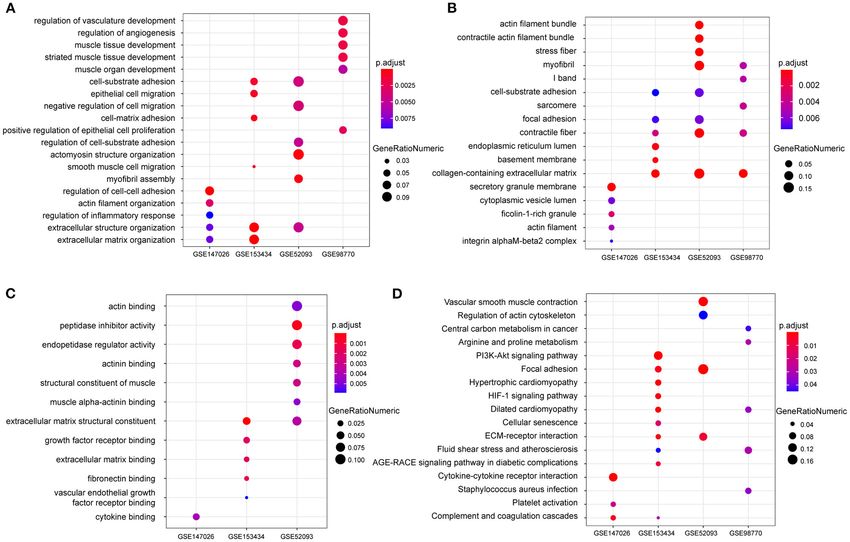
datasets—GSE147026, GSE153434, GSE52093, and GSE98770— between FLNB and FLNC (Figure 4D). Thus, considering the
downloaded from the GEO database. In terms of biological similar expression level of FLNC between the non-AD and AD
process, extracellular structure organization was enriched in samples, FLNC might not be involved in the process of AD and
these four datasets (Figure 1A). Similarly, cellular component should not be further explored in future research. Together, these
and molecular function analysis revealed that extracellular results suggested that FLNA and FLNB might be involved in the
matrix (ECM) was the most highly enriched GO term development of AD.
(Figures 1B,C), consistent with previous investigations (18).
In addition to the ECM, actin associated elements were also
key factors used to elucidate the pathological process of AD The Protein Levels of FLNA and FLNB in
(Figures 1A–C), such as actin filaments and actin-binding TAAD Patients
proteins. Furthermore, the KEGG pathway enrichment analysis We further investigated whether the protein level of FLNA was
displayed conspicuous differences in actin cytoskeleton pathways altered during the process of AD. Total protein was extracted
between control and TAAD samples (Figure 1D). Notably, from aorta with or without TAAD, and the protein levels of
the inflammatory response, which participates in SMC death FLNA and FLNB were detected by Western blotting. The results
and ECM degradation, was identified as a common TAAD showed that consistent with changes in mRNA levels, the protein
associated cause in the four datasets according to gene set level of FLNA also obviously decreased in the TAAD samples
enrichment analysis (GSEA) (Figures 2A–D). Moreover, the compared with that in the controls (Figures 5A,B). Similarly,
actin cytoskeleton was enriched again by using the C5 collection the results of immunohistochemistry showed that FLNA was
(ontology gene sets) used for the GSEA (Figures 2E–H). highly expressed in the normal aorta but significantly reduced
Thus, these results indicated that abnormalities in the actin in the aorta of patients with TAAD (Figures 5D,E). However,
cytoskeleton may be involved in the development of TAAD in contrast to the decrease in mRNA level, the protein level of
in humans. FLNB significantly increased in the aorta of patients with TAAD,
and the results of immunohistochemistry further verified this
The mRNA Expression of FLNA and FLNB result (Figures 5A,C,D,F). Thus, these results further indicated
Is Downregulated in Patients With TAAD that FLNA is a candidate gene affecting AD development.
Given that the actin cytoskeleton was enriched and because FLN
family members are actin-binding proteins, we hypothesized that
FLN family members may be involved in the pathogenesis of
The Mouse AD Model Was Successfully
AD. Therefore, we first analyzed the expression levels of FLNs Established With Young Mice Through the
in the GSE153434 dataset. The results showed that FLNA and Administration of BAPN
FLNB expression was decreased in the aorta of the AD patients, To examine the roles of the FLN family in vivo, BAPN was used
whereas FLNC displayed no difference between the two groups to generate AD models in young mice (19). First, we treated 3-
(Figures 3A–C). To validate the results of the dataset analysis, week-old male mice with 0.6% BAPN per os for 4 weeks and
we collected aortic tissues from TAAD patients and non-AD monitored the condition of the mice daily. We found that after
aortas from patients who underwent heart transplantation. In 2 weeks of BAPN treatment, the mice began to die because of
this research, all enrolled TAAD patients were diagnosed through dissection rupture. The mortality of BAPN-treated mice after 4
CT angiography (CTA), and true/false lumens were clearly visible weeks was 15% (Figure 6A) and the morbidity of AD was 45%
in the CTA images (Figure 3D). Significant destructive ECM according to macroscopic autopsy (Figures 6B,C). Dissection
remodeling of the ascending aorta in AD identified according mainly occurred in the ascending aorta, aorta arch, and thoracic
to its morphology (Figure 3E). Compared with normal aorta aorta (Figure 6C). Furthermore, the aorta of the dissected mice
tissues, TAAD aorta tissues showed disorganized elastin fibers had pathological changes similar to those of the human aorta,
with increased fragmentation as indicated by elastin van Gieson including destructive ECM remodeling and disruption of elastic
(EVG) staining (Figure 3F). We further detected the mRNA fibers (Figures 6D,E). Consistently, H&E staining showed an
levels of FLNs in the aorta of non-AD and TAAD patients by intimal tear between the true and false lumens, and displayed
real-time PCR and found lower mRNA levels of FLNA and FLNB inflammatory cell infiltration in the false lumen (Figure 6D).
in patients with TAAD than in control subjects (Figures 3G,H). According to the degree of elastic fiber fragmentation in the
However, comparable mRNA levels of FLNC were observed aorta (0, 0–25%, 25–50%, 50–75%, and 75–100%), we divided the
between the two groups (Figure 3I). medial degeneration of the aorta into 5 levels (0, 1, 2, 3, and 4)
Since dilatation of the aorta is one of the characteristics of (20). After 4 weeks of BAPN treatment, aortas of 3 mice were
AD patients, we next investigated the relationship between the almost normal, and aortas of the remaining mice exhibited elastic
mRNA expression of FLNs and aortic diameters. As shown in fiber breakage to varying degrees (Figure 6F). In conclusion, we
Frontiers in Cardiovascular Medicine | www.frontiersin.org 4 August 2021 | Volume 8 | Article 690846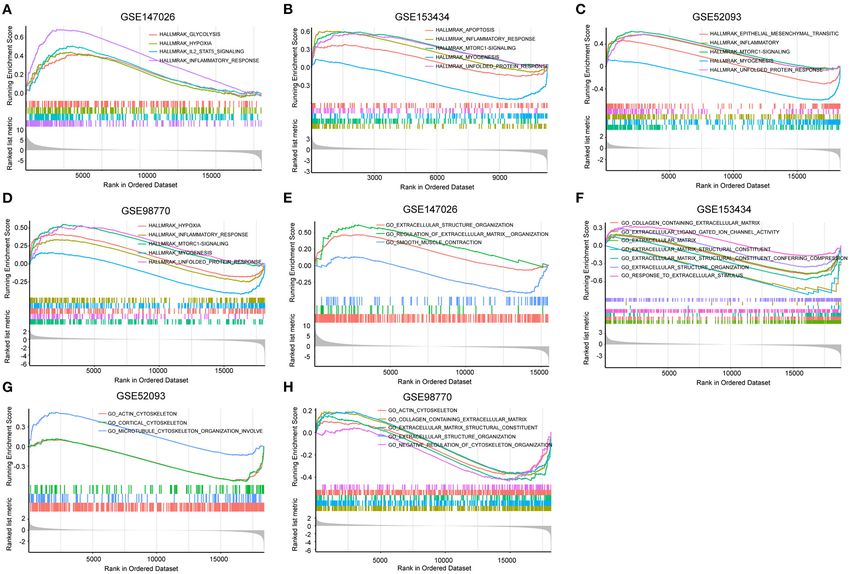
Chen et al.                                                                                                             Expression Regulation of FLNA During AD

 FIGURE 1 | The enrichment analysis shows the differentially expressed genes between the AD and normal groups. (A–C) GO analysis showing the differences of
 mRNA expression in the aorta between AD and normal groups on biological processes (A) cellular component (B) or molecular function (C) among the four GEO
 datasets GSE147026, GSE153434, GSE52093, and GSE98770. (D) KEGG enrichment analysis showing the dramatically altered pathways in AD among the
 indicated datasets.

successfully constructed a mouse AD model that can simulate the                    assay-sequence (ChIP-seq) data obtained from The Signaling
occurrence of human AD by stimulating young mice with BAPN.                        Pathways Project (https://www.signalingpathways.org). We then
                                                                                   screened these transcription factors in the GSE153434 dataset
FLNA Was Downregulated in the Aortas of                                            to identify differentially expressed transcription factors that
Mice Treated With BAPN                                                             potentially regulate FLNA expression. Our screening results
Using a mouse model of AD, we further examined the FLNA and                        showed that five transcription factors, FOS, JUN, E2F3, LMO3,
FLNB protein levels via immunohistochemistry. Consistent with                      and PGR, were differentially expressed in the GSE153434
our observations in human samples, FLNA was highly expressed                       dataset. Among these factors, FOS, JUN, LOM3, and PGR was
in normal aortae, while FLNB was expressed at low levels                           downregulated in patients with AD and positively correlated
(Figure 7A). Furthermore, the protein level of FLNA in the aorta                   with FLNA, while E2F3 expression was upregulated (Figure 8A).
of BAPN-treated mice was significantly decreased compared                          Notably, FOS and JUN are both subunits of the transcription
with its expression in controls (Figures 7A,B), while an obvious                   factor AP-1, which is essential for the function of VSMCs and
increase in FLNB was detected in the aorta of mice after treatment                 associated with AD (21). Therefore, we were very interested
with BAPN (Figures 7A,C). Collectively, the expression patterns                    to know whether AP-1 is a transcription factor that regulates
of FLNA and FLNB in human AD tissue were confirmed in the                          FLNA expression. Therefore, we overexpressed FOS and JUN in
mice, which suggested that they might be indispensable for the                     HASMCs via lentivirus infection (Figure 8B). The results showed
pathogenesis of AD.                                                                that either FOS or JUN overexpression in HASMCs promoted
                                                                                   FLNA expression, which was enhanced to a greater extent by
AP-1 Facilitates FLNA Expression in                                                simultaneous overexpression of FOS and JUN (Figures 8C,D).
HASMCs                                                                             However, the expression of FLNB was not regulated by FOS or
To further explore the upstream mechanism that inhibited                           JUN, as evidenced by the comparable expression level of FLNB
FLNA expression during TAAD development, we collected                              between the indicated groups of HASMCs (Figure 8C). These
transcription factors that were reported to be able to bind to the                 results indicated that AP-1 might be the transcription factor
FLNA promoter, as indicated by chromatin immunoprecipitation                       regulating FLNA expression during AD occurrence.

Frontiers in Cardiovascular Medicine | www.frontiersin.org                     5                                          August 2021 | Volume 8 | Article 690846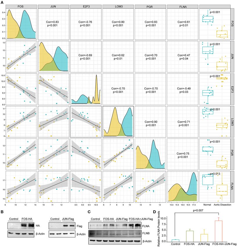
Chen et al.                                                                                                            Expression Regulation of FLNA During AD

 FIGURE 2 | Gene set enrichment analysis (GSEA) was performed to identify gene networks associated with AD. (A–D) Visualization of remarkable enrichment
 hallmark terms among the four GEO datasets GSE147026, GSE153434, GSE52093, and GSE98770. (E–H) Visualization of remarkablely enriched C5 collection
 terms among the four GEO datasets GSE147026, GSE153434, GSE52093, and GSE98770.

DISCUSSION                                                                        Yu et al. showed that P2Y2R regulated the proliferation and
                                                                                  migration of vascular SMCs by binding FLNA (13). It is well-
In this study, by reanalyzing datasets in the GEO database, we                    known that SMCs exhibit two different phenotypes, acting as
identified the actin cytoskeleton as a key component in AD                        contractile or synthetic SMCs, and cells with the contractile
development. We further investigated the expression pattern of                    phenotype, the predominant form in normal aorta, exhibit
the FLN family during AD and found that the mRNA and protein                      relatively low migration and proliferation capacity and high
expression levels of FLNA were significantly downregulated in                     contractile capability (26). Several previous studies confirmed
the aortic wall of AD patients compared with those in the control                 that phenotypic transformation of SMCs from the contractile to
aorta. Furthermore, a similar scenario was observed in the mouse                  synthetic type is one of the typical events in the development of
AD model. Thus, it is highly plausible that FLNA participates                     AD (27, 28). The increase of migration ability is the embodiment
in the process of AD. Finally, we found that the transcription                    of the synthetic type, accompanied by the secretion of matrix
complex FOS/JUN can promote the expression of FLNA, which                         metalloproteinases and a decline in contractile capability (29,
indicated the regulatory mechanism of FLNA in AD.                                 30). Thus, downregulated FLNA expression in AD patients may
    FLNs are important actin crosslinking proteins and their                      be involved in the regulation of migration ability, phenotype
biological function is largely limited to cell migration (9). Heike               transformation, and ECM secretion by VSMCs.
Roth et al. showed that FLNA can facilitate efficient migration                      Other studies found that some functions of FLNA in addition
of human neutrophil-like HL-60 cells via reducing activation                      to migration might be associated with AD. In the patients with
of myosin-II (22). It was reported that neutrophils egress from                   Ehlers-Danlos syndrome and periventricular heterotopia, caused
marrow and infiltrate into the aortic adventitia in BAPN/Ang                      by a loss-of-function mutation in the FLNA gene, was detected
II-treated mice and then release matrix metalloproteinases, such                  and found to contribute the development of aortic dilatation
as MMP8 and MMP9, resulting in the degradation of the                             in early adulthood (31). Indeed, the C-terminal fragment of
ECM and the progression of AD (23–25). In addition, Ningpu                        FLNA was previously proposed as a new biomarker of arterial

Frontiers in Cardiovascular Medicine | www.frontiersin.org                    6                                         August 2021 | Volume 8 | Article 690846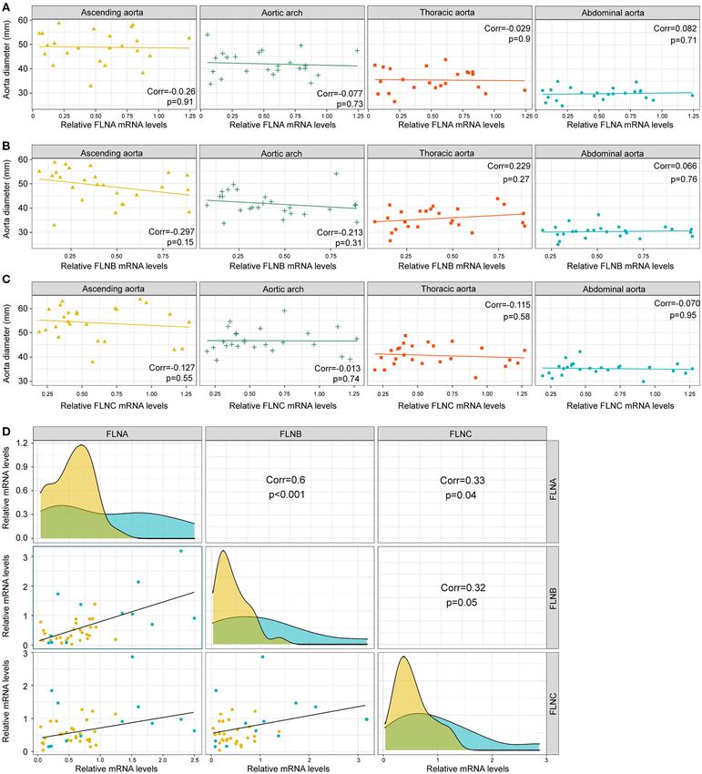
Chen et al.                                                                                                             Expression Regulation of FLNA During AD

 FIGURE 3 | The mRNA levels of FLNA and FLNB were downregulated in human TAAD tissues. (A–C) The expression of FLNA (A) FLNB (B) and FLNC (C) in non-AD
 and dissected human aortas based on GSE153434 dataset. (D) Representative iterative reconstruction CTA images of TAAD patients, T, true lumen; F, false lumen.
 (E,F) Representative images of H&E-stained (E) and EVG-stained (F) aortic sections from non-AD and dissected human aortas. (G–I) The relative mRNA levels of
 FLNA (G) FLNB (H) and FLNC (I) in the aortas of humans from normal donors (n = 14) and AD (n = 33) patients.

wall remodeling in hypertension (32), suggesting that FLNA                         rupture (34). Consistent with aortic dilation as one of the most
is of crucial importance for vascular structure. Feng et al.                       important risk factors for AD, our results in the present study
demonstrated that FLNA is necessary for cell junctions in                          showed that FLNA is decreased in the aortas of AD patients.
vascular development, and FLNA-null mice displayed failure of                      These findings indicated that reduced FLNA may contribute to
vascular remodeling, accompanied by coarse and dilated blood                       aortic dilation and remodeling during AD. Further studies are
vessels (33). Moreover, smooth muscle-specific FLNA knockout                       needed to verify this hypothesis, especially using knockout or
in mice only at the adult stage led to prominent vascular                          overexpression approaches in animal models.
abnormalities, including a reduction in arterial stiffness and a                      Similar to FLNA, FLNB also participates in the regulation of
compensatory increase in conduit artery diameter (12). In AD,                      migration in many cells. FLNB mediates the firm adhesion of
arterial stiffness is increased owing to the deposition of collagen,               leukocytes to the endothelium through interaction with ICAM-
resulting in the enhanced aorta susceptibility to dissection and                   1(35), resulting in the transendothelial migration of leukocytes.

Frontiers in Cardiovascular Medicine | www.frontiersin.org                     7                                          August 2021 | Volume 8 | Article 690846
Chen et al.                                                                                                                  Expression Regulation of FLNA During AD

 FIGURE 4 | The expression of FLNA and FLNB had a significantly positive correlation. (A–C) Correlation analysis showing the relationship between FLNA (A) FLNB
 (B) and FLNC (C) expression and the diameters of ascending, thoracic and abdominal aorta, as well as the aortic arch. (D) Correlation analysis showing the internal
 connection of mRNA levels among FLNA, FLNB, and FLNC.

The recruitment of leukocytes to the vessel endothelium results                       secretion of vascular endothelial growth factor (VEGF)-A via the
in a local inflammatory response, tears in the intima, and                            RAS/ERK pathway, resulting in tumor growth and metastasis
the initiation of AD. In addition, S Bandaru et al. showed                            (36). The roles of MMP9 and VEGF have been exhaustively
that FLNB deficiency enhanced the activity of MMP9 and                                studied in patients with AD. MMP9 degrades type IV collagen,

Frontiers in Cardiovascular Medicine | www.frontiersin.org                        8                                            August 2021 | Volume 8 | Article 690846
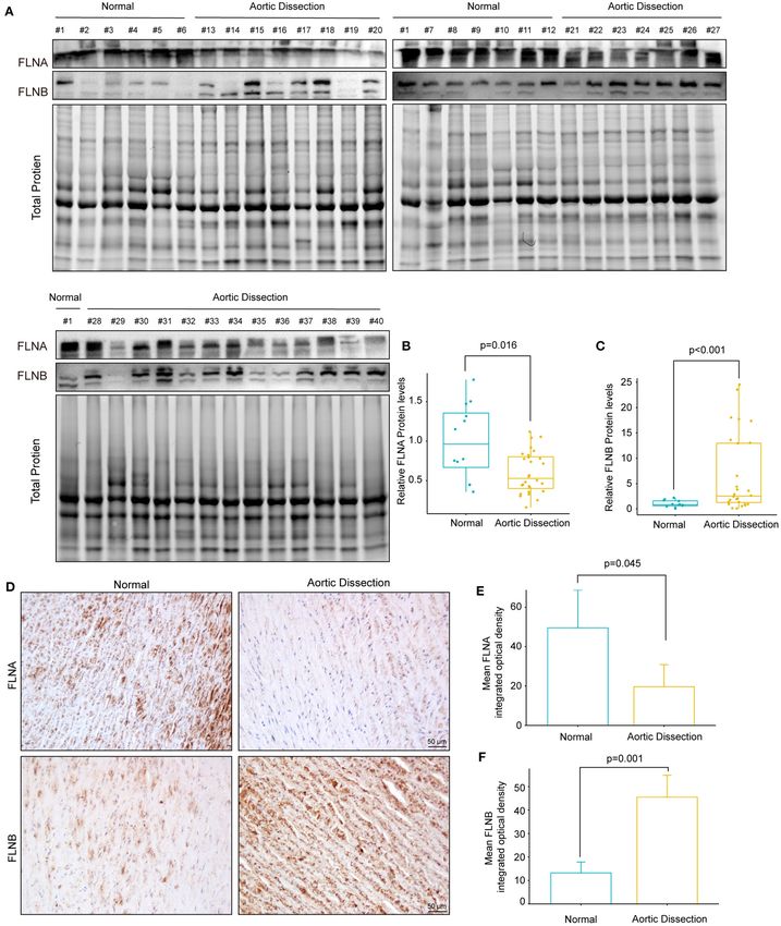
Chen et al.                                                                                                            Expression Regulation of FLNA During AD

 FIGURE 5 | The protein expression of FLNA and FLNB was completely opposite in human TAAD tissues. (A–C) Western blotting and quantitative results of FLNA and
 FLNB in aortic samples obtained from normal donors (n = 12) and AD (n = 28) patients. (D–F) Representative images of immunohistochemical staining and
 quantitative results of FLNA and FLNB in the aortic sections of normal and AD patients (n = 3).

Frontiers in Cardiovascular Medicine | www.frontiersin.org                    9                                          August 2021 | Volume 8 | Article 690846
Chen et al.                                                                                                                    Expression Regulation of FLNA During AD

 FIGURE 6 | The AD model was successfully established with young mice by administering BAPN. (A) Survival curves of the male mice treated with 0.6%
 β-aminopropionitrile (BAPN) when 3 weeks old. (B) The overall incidences of indicated conditions in mice after 4 weeks of BAPN treatment. (C) Representative
 macroscopic images of excised aortas after 4 weeks of BAPN treatment. (D,E) Representative images of H&E-stained (D) and EVG-stained (E) aorta sections from
 non-AD and dissected mouse aortae. The true/false lumens and intimal tears were labeled in the dissected aorta. (F) Distribution of the degree of elastin rupture in
 mice treated with BAPN treatment. n = 20.

elastin, and various basement membrane proteins of SMCs (26),                          attributed to insufficient of degradation in the process of AD,
while VEGF contributes to the proinflammatory actions and                              which should be further investigated.
neoangiogenesis process in aortic wall remodeling (37). It is                             At the mRNA level, we showed that the expression of both
surprising that the mRNA level of FLNB was decreased, whereas                          FLNA and FLNB was downregulated and that a strong positive
the protein expression increased. The inconsistency between the                        correlation between FLNA and FLNB expression was observed
mRNA and protein levels of FLNB might be attributed to the                             in the aortic wall of AD tissues. This result indicates the
following causes. First, mRNA posttranscriptional modification                         possibility that FLNA and FLNB have similar transcriptional
is a familiar form of mRNA stability and translation regulation,                       regulation mechanisms. Filamin family members have a similar
such as N6-methyladenosine (M6 A) modification (38). For                               structure and show 70% amino acid sequence homology (43),
example, ADARB1, an adenosine-to-inosine (A-to-I) RNA-                                 suggesting potential functional compensation between FLNA
editing enzyme, mediates circadian rhythms through mRNA,                               and FLNB. A yeast two-hybrid experiment demonstrated that
and plays a role in the posttranscriptional regulation of FLNB                         the FLNB homodimerization domain can strongly interact with
(39). In addition, the ubiquitin-proteasome degradation pathway                        the corresponding homologous region of FLNA, and then, via
was verified as another mechanism for FLNB post-translational                          immunoprecipitation assays, FLNA-FLNB heterodimers were
regulation. Heuze et al. reported that ASB2 ubiquitin ligase                           verified to exist (44). Previous studies have also demonstrated
activity drives proteasome-mediated degradation of the actin-                          that FLNA and FLNB play overlapping roles in stabilizing the
binding protein FLNB and then inhibits cell spreading on                               actin cytoskeleton and cell function (45). In terms of migration,
fibronectin (40). The E3 ubiquitin ligase specificity subunit                          loss of FLNA or FLNB has no effect on migration, but it
ASB2α regulates cell spreading, migration, and differentiation by                      impairs the initiation of cell migration (46). In chondrocytes,
interacting with the filamin actin-binding domain, which induces                       it was demonstrated that loss of FLNA induces upregulation
FLNB proteasomal degradation (41). Moreover, a novel ASB2                              of FLNB expression, and vice versa (47). Taken together with
isoform, ASB2β, targets FLNB for proteasomal degradation and                           the opposite changes in the protein levels of FLNA and FLNB
impacts myoblast fusion and myotube formation (42). Hence,                             in AD, it is possible that FLNB upregulation may compensate
the ubiquitin proteasome system is a vital pathway for FLNB                            for the decrease in FLNA. Of course, different and even
degradation, and the upregulated FLNB protein level might be                           opposite functions have also been illuminated in other areas

Frontiers in Cardiovascular Medicine | www.frontiersin.org                        10                                            August 2021 | Volume 8 | Article 690846
Chen et al.                                                                                                              Expression Regulation of FLNA During AD

 FIGURE 7 | The expression of FLNA and FLNB in mice with AD is similar to that in human tissue. (A–C) Representative images of immunohistochemical staining and
 quantitative results of FLNA and FLNB in the aorta sections of the normal and BAPN-induced AD mice (n = 3).

of study. For instance, in RAS-induced lung tumorigenesis,                          development of AD, and we suggest that more investigations are
knockout of FLNA significantly reduced the tumor formation                          directed to the AP-1/FLNA axis in AD.
and proliferation of fibroblasts via inactivation of ERK and                           In addition to AD, aortic aneurysm is also a vessel disease
AKT (48). Nevertheless, in contrast to FLNA, FLNB deficiency                        caused by the medial degeneration. However, there are many
enhanced RAS-induced tumor growth and metastasis through                            differences between dissection and aneurysm. Indeed, there
the RAS/ERK pathway (49).                                                           might be aorta dilation before AD formation, but it would
   Another important finding reported here is the transcriptional                   not progress to aneurysm. The rupture of aneurysms is
regulation function of the AP-1 complex (FOS/JUN) on FLNA.                          limited, whereas the rupture of AD shows the formation
To date, few studies have reported the regulatory mechanism                         of true/false lumens, accompanied by extension of the tears.
of FLNA. Matthew R. Sarkisian et al. revealed that MEKK4                            In addition, the most common locations of AD are the
suppression contributes to abnormally high FLNA expression                          ascending aorta, aorta arch, and thoracic aorta (55), while
and inhibits neuronal migration (50). In the present study, we                      aneurysms mainly occur on the abdominal aorta (56). Of
revealed that AP-1 might promote FLNA expression in HASMCs.                         course, the formation of AD is accompanied by dilation
AP-1 is a member of the JUN, FOS, Maf, and ATF subfamilies                          of the aorta, and aneurysms would develop into dissection
(51) and well-recognized as a key transcription factor in cell                      and rupture of the aorta. However, it must be emphasized
proliferation, death, and oncogenesis (52). It was reported that                    that dissection and aneurysm are two different diseases, not
IL-18 facilitated AP-1-dependent MMP9 transcription, resulting                      two forms of one disease, which should be distinguished
in increased SMC migration ability (53). Investigation into the                     in both clinical treatment and basic research. Currently,
possible regulation of FLNA by AP-1 suggested that FLNA might                       CT angiography has widely been applied to AD diagnosis,
be critical for AP-1-mediated SMC migration. Moreover, AP-1 is                      and intimal tears and true/false lumens can be clearly
also a pivotal factor for AD and regulates medial degeneration                      distinguished (57).
(54). Zhang et al. reported that the protein expression of AP-1 in                     There are some limitations in this study. First, we detected
the aorta of AD was downregulated compared to that in normal                        the FLN family expression in the aortas of mice and
aortas, and SIRT1 activated AP-1/decorin signaling to alleviate                     patients diagnosed with AD, but functions of FLNs have not
AD (21). Neutralization of AP-1 via decoy oligodeoxynucleotides                     been investigated, especially using knockout or overexpression
repressed aortic elastolysis with reduced fiber breaks and MMP                      approach in animal models. Second, only the BAPN induced
activity in a Marfan syndrome mouse model (54). Therefore,                          mouse AD model was used to analyze FLNs expression. Two or
we suspect that the inhibition of AP-1 in SMCs aggravates the                       more mouse models of AD would make this conclusion more

Frontiers in Cardiovascular Medicine | www.frontiersin.org                     11                                         August 2021 | Volume 8 | Article 690846
Chen et al.                                                                                                             Expression Regulation of FLNA During AD

 FIGURE 8 | FOS/JUN contributes to FLNA expression in HASMCs. (A) The five transcription factors mostly related to FLNA transcriptional regulation. (B) Western
 blotting of FOS and JUN expression in HASMCs infected with lenti-FOS-HA and lenti-JUN-Flag. (C,D) Western blot and quantitative results showing FLNA and FLNB
 expression in HASMCs infected with corresponding lentivirus (Control, FOS-HA, JUN-Flag, FOS-HA+JUN-Flag). β-Actin served as the loading control.

convincing and meaningful. Finally, we found the possibility                          In summary, our study demonstrated that the expression level
of AP-1 on FLNA transcriptional regulation via bioinformatics                      of FLNA was reduced in the aortas of patients with AD, and AP-
and further validated that AP-1 could promote FLNA expression                      1 might be the transcription factor mediating the expression of
in HASMCs. However, further experiments should be done to                          FLNA in HASMCs. These results indicated that the AP-1/FLNA
strengthen this regulation mechanism and to illustrate their                       axis may be essential for the occurrence of AD, and targeting the
functions in AD development.                                                       AP-1/FLNA axis may be a novel therapeutic strategy for AD.

Frontiers in Cardiovascular Medicine | www.frontiersin.org                    12                                          August 2021 | Volume 8 | Article 690846
Chen et al.                                                                                                                           Expression Regulation of FLNA During AD

DATA AVAILABILITY STATEMENT                                                                 AUTHOR CONTRIBUTIONS
The datasets presented in this study can be found in online                                 YC and XW performed the cell and animal experiments
repositories. The names of the repository/repositories                                      of this study and data analysis. ZZ and YH participated
and accession number(s) can be found in the                                                 in bioinformatics analysis. BH and XG cultured the
article/supplementary material.                                                             primary HASMCs and constructed the plasmid. XF and
                                                                                            Z-MF collected the human aorta tissues. X-HZ and
ETHICS STATEMENT                                                                            D-SJ designed the work and finished the manuscript.
                                                                                            All authors contributed to the article and approved the
The studies involving human participants were reviewed and                                  submitted version.
approved by the Human Research Ethics Committees of Tongji
Hospital, Tongji Medical College, Huazhong University of
Science and Technology. The patients/participants provided their                            FUNDING
written informed consent to participate in this study. The animal
study was reviewed and approved by the Animal Care and                                      This work was supported by grants from the National Natural
Use Committees of Tongji Hospital, Tongji Medical College,                                  Science Foundation of China (NOs. 82070488, 81873458,
Huazhong University of Science and Technology.                                              and 81974013).

REFERENCES                                                                                  13. Yu N, Erb L, Shivaji R, Weisman GA, Seye CI. Binding of the P2Y2 nucleotide
                                                                                                receptor to filamin A regulates migration of vascular smooth muscle cells. Circ
 1. Cifani N, Proietta M, Tritapepe L, Di Gioia C, Ferri L, Taurino M, et al.                   Res. (2008) 102:581–8. doi: 10.1161/CIRCRESAHA.107.162271
    Stanford-A acute aortic dissection, inflammation, and metalloproteinases: a             14. Chen TQ, Hu N, Huo B, Masau JF, Yi X, Zhong XX, et al. EHMT2/G9a inhibits
    review. Ann Med. (2015) 47:441–6. doi: 10.3109/07853890.2015.1073346                        aortic smooth muscle cell death by suppressing autophagy activation. Int J Biol
 2. Erbel R, Aboyans V, Boileau C, Bossone E, Bartolomeo RD, Eggebrecht H,                      Sci. (2020) 16:1252–63. doi: 10.7150/ijbs.38835
    et al. 2014 ESC guidelines on the diagnosis and treatment of aortic diseases:           15. Li R, Yi X, Wei X, Huo B, Guo X, Cheng C, et al. EZH2 inhibits autophagic
    document covering acute and chronic aortic diseases of the thoracic and                     cell death of aortic vascular smooth muscle cells to affect aortic dissection. Cell
    abdominal aorta of the adult. The task force for the diagnosis and treatment                Death Dis. (2018) 9:180. doi: 10.1038/s41419-017-0213-2
    of aortic diseases of the European society of cardiology (ESC). Eur Heart J.            16. Jiang DS, Yi X, Li R, Su YS, Wang J, Chen ML, et al. The histone
    (2014) 35:2873–926. doi: 10.1093/eurheartj/ehu281                                           methyltransferase mixed lineage leukemia (MLL) 3 may play a potential
 3. Pollard TD. Actin and actin-binding proteins. Cold Spring Harb Perspect Biol.               role on clinical dilated cardiomyopathy. Mol Med. (2017) 23:196–
    (2016) 8:a018226. doi: 10.1101/cshperspect.a018226                                          203. doi: 10.2119/molmed.2017.00012
 4. Yamin R, Morgan KG. Deciphering actin cytoskeletal function in the                      17. Yu G, Wang L-G, Han Y, He Q-Y. Clusterprofiler: an R package for
    contractile vascular smooth muscle cell. J Physiol. (2012) 590:4145–                        comparing biological themes among gene clusters. OMICS. (2012) 16:284–
    54. doi: 10.1113/jphysiol.2012.232306                                                       7. doi: 10.1089/omi.2011.0118
 5. Guo DC, Pannu H, Tran-Fadulu V, Papke CL, Yu RK, Avidan                                 18. Zhang L, Yu C, Chang Q, Luo X, Qiu J, Liu S. Comparison of gene expression
    N, et al. Mutations in smooth muscle alpha-actin (ACTA2) lead                               profiles in aortic dissection and normal human aortic tissues. Biomed Rep.
    to thoracic aortic aneurysms and dissections. Nat Genet. (2007)                             (2016) 5:421–7. doi: 10.3892/br.2016.740
    39:1488–93. doi: 10.1038/ng.2007.6                                                      19. Jiang DS, Yi X, Zhu XH, Wei X. Experimental in vivo and ex vivo models for
 6. Cheng J, Zhou X, Jiang X, Sun T. Deletion of ACTA2 in mice promotes                         the study of human aortic dissection: promises and challenges. Am J Transl
    angiotensin II induced pathogenesis of thoracic aortic aneurysms and                        Res. (2016) 8:5125–40.
    dissections. J Thorac Dis. (2018) 10:4733–40. doi: 10.21037/jtd.2018.07.75              20. Schulte S, Sun J, Libby P, Macfarlane L, Sun C, Lopez-Ilasaca M, et al.
 7. Han M, Dong LH, Zheng B, Shi JH, Wen JK, Cheng Y. Smooth muscle                             Cystatin C deficiency promotes inflammation in angiotensin II-induced
    22 alpha maintains the differentiated phenotype of vascular smooth muscle                   abdominal aortic aneurisms in atherosclerotic mice. Am J Pathol. (2010)
    cells by inducing filamentous actin bundling. Life Sci. (2009) 84:394–                      177:456–63. doi: 10.2353/ajpath.2010.090381
    401. doi: 10.1016/j.lfs.2008.11.017                                                     21. Zhang K, Pan X, Zheng J, Liu Y, Sun L. SIRT1 protects against aortic dissection
 8. Miyake H, Maeda K, Asai N, Shibata R, Ichimiya H, Isotani-Sakakibara M,                     by regulating AP-1/decorin signaling-mediated PDCD4 activation. Mol Biol
    et al. The actin-binding protein girdin and its akt-mediated phosphorylation                Rep. (2020) 47:2149–59. doi: 10.1007/s11033-020-05314-9
    regulate neointima formation after vascular injury. Circ Res. (2011) 108:1170–          22. Roth H, Samereier M, Begandt D, Pick R, Salvermoser M, Brechtefeld D, et al.
    9. doi: 10.1161/CIRCRESAHA.110.236174                                                       Filamin A promotes efficient migration and phagocytosis of neutrophil-like
 9. Stossel TP, Condeelis J, Cooley L, Hartwig JH, Noegel A, Schleicher M, et al.               HL-60 cells. Eur J Cell Biol. (2017) 96:553–66. doi: 10.1016/j.ejcb.2017.05.004
    Filamins as integrators of cell mechanics and signalling. Nat Rev Mol Cell Biol.        23. Anzai A, Shimoda M, Endo J, Kohno T, Katsumata Y, Matsuhashi T, et al.
    (2001) 2:138–45. doi: 10.1038/35052082                                                      Adventitial CXCL1/G-CSF expression in response to acute aortic dissection
10. Zhou X, Boren J, Akyurek LM. Filamins in cardiovascular development.                        triggers local neutrophil recruitment and activation leading to aortic rupture.
    Trends Cardiovasc Med. (2007) 17:222–9. doi: 10.1016/j.tcm.2007.08.001                      Circ Res. (2015) 116:612–23. doi: 10.1161/CIRCRESAHA.116.304918
11. Zhu G, Chen H, Zhang W. Phenotype switch of vascular smooth muscle                      24. Wilson WR, Schwalbe EC, Jones JL, Bell PR, Thompson MM. Matrix
    cells after siRNA silencing of filamin. Cell Biochem Biophys. (2011) 61:47–                 metalloproteinase 8 (neutrophil collagenase) in the pathogenesis of abdominal
    52. doi: 10.1007/s12013-011-9159-7                                                          aortic aneurysm. Br J Surg. (2005) 92:828–33. doi: 10.1002/bjs.4993
12. Retailleau K, Arhatte M, Demolombe S, Jodar M, Baudrie V, Offermanns                    25. Kurihara T, Shimizu-Hirota R, Shimoda M, Adachi T, Shimizu
    S, et al. Smooth muscle filamin A is a major determinant of conduit artery                  H, Weiss SJ, et al. Neutrophil-derived matrix metalloproteinase
    structure and function at the adult stage. Pflügers Archiv Eur J Physiol. (2016)            9 triggers acute aortic dissection. Circulation. (2012) 126:3070–
    468:1151–60. doi: 10.1007/s00424-016-1813-x                                                 80. doi: 10.1161/CIRCULATIONAHA.112.097097

Frontiers in Cardiovascular Medicine | www.frontiersin.org                             13                                               August 2021 | Volume 8 | Article 690846
Chen et al. Expression Regulation of FLNA During AD
26. Wu D, Shen YH, Russell L, Coselli JS, LeMaire SA. Molecular migration and can physically interact. Hum Mol Genet. (2002) 11:2845–
mechanisms of thoracic aortic dissection. J Surg Res. (2013) 54. doi: 10.1093/hmg/11.23.2845
184:907–24. doi: 10.1016/j.jss.2013.06.007 45. Nakamura F, Stossel TP, Hartwig JH. The filamins: organizers of cell structure
27. Wang L, Zhang J, Fu W, Guo D, Jiang J, Wang Y. Association of and function. Cell Adh Migr. (2011) 5:160–9. doi: 10.4161/cam.5.2.14401
smooth muscle cell phenotypes with extracellular matrix disorders 46. Baldassarre M, Razinia Z, Burande CF, Lamsoul I, Lutz PG, Calderwood DA.
in thoracic aortic dissection. J Vasc Surg. (2012) 56:1698–709:709 Filamins regulate cell spreading and initiation of cell migration. PLoS ONE.
e1. doi: 10.1016/j.jvs.2012.05.084 (2009) 4:e7830. doi: 10.1371/journal.pone.0007830
28. Ailawadi G, Moehle CW, Pei H, Walton SP, Yang Z, Kron IL, et al. Smooth 47. Hu J, Lu J, Goyal A, Wong T, Lian G, Zhang J, et al. Opposing FlnA
muscle phenotypic modulation is an early event in aortic aneurysms. J Thorac and FlnB interactions regulate RhoA activation in guiding dynamic actin
Cardiovasc Surg. (2009) 138:1392–9. doi: 10.1016/j.jtcvs.2009.07.075 stress fiber formation and cell spreading. Hum Mol Genet. (2017) 26:1294–
29. An Z, Liu Y, Song ZG, Tang H, Yuan Y, Xu ZY. Mechanisms of aortic 304. doi: 10.1093/hmg/ddx047
dissection smooth muscle cell phenotype switch. J Thorac Cardiovasc Surg. 48. Nallapalli RK, Ibrahim MX, Zhou AX, Bandaru S, Sunkara SN, Redfors B,
(2017) 154:1511–21 e6. doi: 10.1016/j.jtcvs.2017.05.066 et al. Targeting filamin A reduces K-RAS-induced lung adenocarcinomas
30. Fisher SA. Vascular smooth muscle phenotypic diversity and function. Physiol and endothelial response to tumor growth in mice. Mol Cancer. (2012)
Genom. (2010) 42A:169–87. doi: 10.1152/physiolgenomics.00111.2010 11:50. doi: 10.1186/1476-4598-11-50
31. Sheen VL, Jansen A, Chen MH, Parrini E, Morgan T, 49. Bandaru S, Zhou AX, Rouhi P, Zhang Y, Bergo MO, Cao Y, et al. Targeting
Ravenscroft R, et al. Filamin A mutations cause periventricular filamin B induces tumor growth and metastasis via enhanced activity of
heterotopia with ehlers-danlos syndrome. Neurology. (2005) matrix metalloproteinase-9 and secretion of VEGF-A. Oncogenesis. (2014)
64:254–62. doi: 10.1212/01.WNL.0000149512.79621.DF 3:e119. doi: 10.1038/oncsis.2014.33
32. Delbosc S, Haloui M, Louedec L, Dupuis M, Cubizolles M, Podust VN, 50. Yuan Y, Wang C, Xu J, Tao J, Xu Z, Huang S. BRG1 overexpression in smooth
et al. Proteomic analysis permits the identification of new biomarkers muscle cells promotes the development of thoracic aortic dissection. BMC
of arterial wall remodeling in hypertension. Mol Med. (2008) 14:383– Cardiovasc Disord. (2014) 14:144. doi: 10.1186/1471-2261-14-144
94. doi: 10.2119/2008-00030.Delbosc 51. Shaulian E, Karin M. AP-1 as a regulator of cell life and death. Nat Cell Biol.
33. Feng Y, Chen MH, Moskowitz IP, Mendonza AM, Vidali L, Nakamura F, et al. (2002) 4:E131–6. doi: 10.1038/ncb0502-e131
Filamin A (FLNA) is required for cell-cell contact in vascular development 52. Tewari D, Nabavi SF, Nabavi SM, Sureda A, Farooqi AA, Atanasov AG,
and cardiac morphogenesis. Proc Natl Acad Sci USA. (2006) 103:19836– et al. Targeting activator protein 1 signaling pathway by bioactive natural
41. doi: 10.1073/pnas.0609628104 agents: possible therapeutic strategy for cancer prevention and intervention.
34. Lyle AN, Raaz U. Killing me unsoftly: causes and mechanisms Pharmacol Res. (2018) 128:366–75. doi: 10.1016/j.phrs.2017.09.014
of arterial stiffness. Arterioscler Thromb Vasc Biol. (2017) 53. Chandrasekar B, Mummidi S, Mahimainathan L, Patel DN, Bailey SR,
37:e1–11. doi: 10.1161/ATVBAHA.116.308563 Imam SZ, et al. Interleukin-18-induced human coronary artery smooth
35. Kanters E, van Rijssel J, Hensbergen PJ, Hondius D, Mul FP, Deelder AM, et muscle cell migration is dependent on NF-κB- and AP-1-mediated matrix
al. Filamin B mediates ICAM-1-driven leukocyte transendothelial migration. metalloproteinase-9 expression and is inhibited by atorvastatin. J Biol Chem.
J Biol Chem. (2008) 283:31830–9. doi: 10.1074/jbc.M804888200 (2006) 281:15099–109. doi: 10.1074/jbc.M600200200
36. Zhou X, Tian F, Sandzen J, Cao R, Flaberg E, Szekely L, et al. Filamin 54. Arif R, Zaradzki M, Remes A, Seppelt P, Kunze R, Schroder H, et
B deficiency in mice results in skeletal malformations and impaired al. AP-1 oligodeoxynucleotides reduce aortic elastolysis in a murine
microvascular development. Proc Natl Acad Sci USA. (2007) 104:3919– model of marfan syndrome. Mol Ther Nucleic Acids. (2017) 9:69–
24. doi: 10.1073/pnas.0608360104 79. doi: 10.1016/j.omtn.2017.08.014
37. Del PF, di Gioia C, Tritapepe L, Ferri L, Leopizzi M, Nofroni I, et al. The 55. Golledge J, Eagle KA. Acute aortic dissection. Lancet. (2008) 372:55–
multitasking role of macrophages in stanford type A acute aortic dissection. 66. doi: 10.1016/S0140-6736(08)60994-0
Cardiology. (2014) 127:123–9. doi: 10.1159/000355253 56. Davis FM, Daugherty A, Lu HS. Updates of recent aortic
38. Chen J, Wei X, Yi X, Jiang D-S. RNA modification by m6A aneurysm research. Arterioscler Thromb Vasc Biol. (2019)
methylation in cardiovascular disease. Oxid Med Cell Longev. (2021) 39:e83–90. doi: 10.1161/ATVBAHA.119.312000
2021:8813909. doi: 10.1155/2021/8813909 57. Nienaber CA, Clough RE. Management of acute aortic dissection. LANCET.
39. Terajima H, Yoshitane H, Ozaki H, Suzuki Y, Shimba S, Kuroda S, et al. (2015) 385:800–11. doi: 10.1016/S0140-6736(14)61005-9
ADARB1 catalyzes circadian A-to-I editing and regulates RNA rhythm. Nat
Genet. (2017) 49:146–51. doi: 10.1038/ng.3731 Conflict of Interest: The authors declare that the research was conducted in the
40. Heuze ML, Lamsoul I, Baldassarre M, Lad Y, Leveque S, Razinia Z, et al. absence of any commercial or financial relationships that could be construed as a
ASB2 targets filamins A and B to proteasomal degradation. Blood. (2008) potential conflict of interest.
112:5130–40. doi: 10.1182/blood-2007-12-128744
41. Razinia Z, Baldassarre M, Bouaouina M, Lamsoul I, Lutz PG, Calderwood
Publisher’s Note: All claims expressed in this article are solely those of the authors
DA. The E3 ubiquitin ligase specificity subunit ASB2alpha targets filamins
for proteasomal degradation by interacting with the filamin actin-binding and do not necessarily represent those of their affiliated organizations, or those of
domain. J Cell Sci. (2011) 124:2631–41. doi: 10.1242/jcs.084343 the publisher, the editors and the reviewers. Any product that may be evaluated in
42. Bello NF, Lamsoul I, Heuzé ML, Métais A, Moreaux G, Calderwood this article, or claim that may be made by its manufacturer, is not guaranteed or
DA, et al. The E3 ubiquitin ligase specificity subunit ASB2β is a novel endorsed by the publisher.
regulator of muscle differentiation that targets filamin B to proteasomal
degradation. Cell Death Differ. (2009) 16:921–32. doi: 10.1038/cdd.2 Copyright © 2021 Chen, Wei, Zhang, He, Huo, Guo, Feng, Fang, Jiang and Zhu.
009.27 This is an open-access article distributed under the terms of the Creative Commons
43. van der Flier A, Sonnenberg A. Structural and functional Attribution License (CC BY). The use, distribution or reproduction in other forums
aspects of filamins. Biochim Biophys Acta. (2001) 1538:99– is permitted, provided the original author(s) and the copyright owner(s) are credited
117. doi: 10.1016/S0167-4889(01)00072-6 and that the original publication in this journal is cited, in accordance with accepted
44. Sheen VL, Feng Y, Graham D, Takafuta T, Shapiro SS, Walsh CA. Filamin A academic practice. No use, distribution or reproduction is permitted which does not
and Filamin B are co-expressed within neurons during periods of neuronal comply with these terms.
Frontiers in Cardiovascular Medicine | www.frontiersin.org 14 August 2021 | Volume 8 | Article 690846You can also read