ANTIBACTERIAL COMPOSITE COATINGS OF MGB2 POWDERS EMBEDDED IN PVP MATRIX
←
→
Page content transcription
If your browser does not render page correctly, please read the page content below

www.nature.com/scientificreports
OPEN Antibacterial composite coatings
of MgB2 powders embedded in PVP
matrix
P. Badica1*, N. D. Batalu2, M. Burdusel1, M. A. Grigoroscuta1, G. Aldica1, M. Enculescu1,
G. Gradisteanu Pircalabioru3, M. Popa3, L. G. Marutescu3, B. G. Dumitriu4, L. Olariu4,6,
A. Bicu4, B. Purcareanu4, L. Operti5, V. Bonino5, A. Agostino5, M. Truccato5 & M. C. Chifiriuc3
Three commercial powders of MgB2 were tested in vitro by MTS and LDH cytotoxicity tests on
the HS27 dermal cell line. Depending on powders, the toxicity concentrations were established
in the range of 8.3–33.2 µg/ml. The powder with the lowest toxicity limit was embedded into
polyvinylpyrrolidone (PVP), a biocompatible and biodegradable polymer, for two different
concentrations. The self-replenishing MgB2-PVP composite materials were coated on substrate
materials (plastic foil of the reservoir and silicon tubes) composing a commercial urinary catheter.
The influence of the PVP-reference and MgB2-PVP novel coatings on the bacterial growth of
Staphylococcus aureus ATCC 25923, Enterococcus faecium DMS 13590, Escherichia coli ATCC 25922,
Pseudomonas aeruginosa ATCC 27853, in planktonic and biofilm state was assessed in vitro at 6, 24,
and 48 h of incubation time. The MgB2-PVP coatings are efficient both against planktonic microbes
and microbial biofilms. Results open promising applications for the use of MgB2 in the design of anti-
infective strategies for different biomedical devices and systems.
The opportunistic and nosocomial agents, such as ESCAPE (Enterococcus faecium, Staphylococcus aureus,
Clostridium difficile, Acinetobacter baumannii, Pseudomonas aeruginosa, and Enterobacteriaceae) pathogens
and Candida albicans represent one of the most important global threats for the public health. They have the
ability to adhere and develop biofilms on live tissues and implanted medical devices, consequently producing
biofilm associated infections1. Biofilms exhibit a particular form of resistance, the phenotypic resistance or toler-
ance. They also have recalcitrance, i.e. the ability to survive in the presence of high concentrations of a ntibiotics2.
The multiple negative consequences of biofilms development in the clinical sector underlines the need to
recognize the problems contributing to poor outcomes and high costs. It also highlights the necessity of a mul-
tidisciplinary effort to prevent, combat, or eradicate biofilms. The antimicrobial strategies can be divided in
microbiostatic/microbicidal based agents or on antipathogenic agents. The first ones involve the use of agents
which inhibit or kill microorganisms, while the antipathogenic ones target the expression of virulence factors
(e.g., adherence capacity, toxigenicity) and of their regulators (Quorum Sensing inhibitors)3. The antibiofilm
strategies belong to both categories of the antimicrobial ones.
Nanotechnology and nanomaterials are of much interest for the development of new antimicrobial approaches,
based on either novel biomaterials or on improving the biological properties of the existing ones. Currently, in a
sustainable and eco-friendly driven approach, many studies are directed to design both clinically and environ-
mentally safe nanomaterials (NMs) for antimicrobial applications. The NMs act as antimicrobial and antibiofilm
agents. They can have additive or synergetic effects in combinations with antibiotics or other antimicrobials4–7.
NMs are also useful as drug delivery for targeted release to the site of infection and as components of composites
including stimuli-responsive coatings, or modified hybrid m aterials8–10. Many physico-chemical properties,
such as the type of the nanomaterial, size, morphology, specific surface-area-to-volume ratio, surface charge,
concentration, behavior in biological medium and pH, stability and others are conditioning their antibiofilm
effect. All these factors influence the contact with the biofilm matrix and biofilm embedded cells, affecting the
release of reactive oxygen species, of antimicrobial ions or of the loaded bioactive compounds11. NMs can be
1
National Institute of Materials Physics, Street Atomistilor 405A, 077125 Magurele, Romania. 2University
Politehnica of Bucharest, Splaiul Independentei 313, 060042 Bucharest, Romania. 3Faculty of Biology and The
Research Institute of the University of Bucharest (ICUB), University of Bucharest, Splaiul Independentei 91‑95,
Bucharest, Romania. 4Biotehnos SA, Strada Gorunului, Nr. 3‑5, Otopeni, Județul Ilfov, Romania. 5Physics and
Chemistry Departments, University of Turin, Via P. Giuria 1‑7, 10125 Turin, Italy. 6Academy of Romanian Scientists,
54 Splaiul Independentei, 050094 Bucharest, Romania. *email: badica2003@yahoo.com
Scientific Reports | (2021) 11:9591 | https://doi.org/10.1038/s41598-021-88885-2 1
Vol.:(0123456789)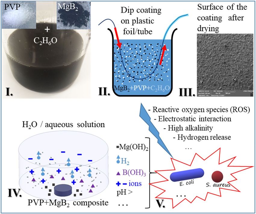
www.nature.com/scientificreports/
modified through functionalization to increase their efficacy and b iocompatibility12. They can simultaneously
attack multiple microbial targets, thereby the risk of emergence of resistance is low. The NMs can interfere with
different stages of biofilm development, i.e., with single-cell adherence, multiplication, and colonization of the
substrate, with biofilm maturation, and with biofilm dispersion. They can interact with the planktonic cells,
inhibiting either the initial adhesion to a substrate and the dispersion, or with the biofilm matrix by facilitating
the biofilm penetration, drug release, or further interaction with the biofilm c ells13.
Metals (silver, copper, gold, chromium), metal oxides (Al2O3, CeO2, Co3O4, Cr2O3, CuO, In2O3, Fe2O3, MgO,
Mn2O3, NiO, Ni2O3, SiO2, TiO2, ZnO, ZrO2, Y2O3, etc.), metal hydroxides, such as Mg(OH)2, and metal halides
nanoparticles (NPs) are among the most studied NMs for antimicrobial effects, due to their intrinsic antimicro-
bial features11,14–18. They exhibit both microbicidal and microbiostatic effects caused by membrane lesions due to
the direct contact with NPs and the release of free metal ions, the proteins inactivation, the nucleic acids damage,
the release of reactive oxygen species (ROS), and stimulation of the host immune system.
The delivery of active NPs inside biofilms is possible by using different delivery systems, including polymers.
Polyvinylpyrrolidone (PVP), also called polyvidone or povidone, is a biocompatible, non-toxic, biodegradable,
hydrophilic polymer with good binding properties and with a stabilizing effect on suspensions and emulsions.
PVP is recognized as safe by the Food and Drug Administration (FDA). Considering also that it has other unique
physical and chemical features, e.g. it is chemically inert, but soluble in water and alcohol, can have different mor-
phologies, it is colorless, temperature-resistant and pH-stable, PVP is largely used for biomedical a pplications19–21.
In this paper, we continue our previous studies on antimicrobial activity of M gB2 powders22,23 and we investi-
gate their potential when embedded in PVP-based polymeric coatings for fabrication of improved plastic medical
devices, more resistant to microbial colonization and thus, less probable to induce biofilm-associated infections.
As substrate materials we use a flexible plastic foil of a urinary catheter reservoir and silicon tubes from the same
commercial device. The challenges in using catheter devices and criteria for their improvement are reviewed
in Refs.24,25. The M gB2 compound is degradable in w ater26,27 and it results that the composite coatings of PVP-
MgB2 are biodegradable and self-replenishing. Other different factors that recommend M gB2 for biomedical
application are: (i) as already mentioned, Mg is a an antimicrobial material, but also boron in the form of boric
acid or sodium salts of boron (borax, disodium tetraborate) is an effective antiseptic, bactericidal, insecticidal,
herbicidal and cleaning agent used in detergents (28 and therein Refs.); (ii) on the other hand, both Mg and
boron are involved in the metabolism of humans from bone growth to wound healing and their amount in the
body is relatively high; boron has positive effects and it is used in treatment of cancer and infections; (iii) MgB2
is a light weight compound with bulk density of 2.63 g/cm3 that is close to that of the bones; (iv) the mechanical
properties of MgB2 are similar to those of ceramic materials and of the bones; (v) MgB2 has metallic conduction
and when decomposes it releases positive (Mg) and negative (B) ions impacting local pH and interaction with
negatively charged cellular wall; (v) there is a family of MgBy (MgB4, MgB7, MgB12, MgB19) phases that may prove
of interest to control the bioprocesses depending on application requirements.
Experimental
Physico‑chemical characteristics of MgB2 raw powders. Commercial raw powders of MgB2 were
produced by LTS Research Laboratories Inc (LTS), Alfa Aesar (AA), and CERAC Inc (CER). The powders have
very different physico-chemical properties. Although results will be presented e lsewhere23, according to X-ray
diffraction measurements and Rietveld analysis, we mention that powders were composed of the main phase
MgB2 and of the secondary phases MgO and MgB4. The highest MgB2 amount was measured in LTS (97 wt. %
MgB2) followed by Alfa Aesar (88 wt. % MgB2), and Cerac (80.3 wt. % MgB2). In the same order, the amount
of residual unreacted metallic Mg (1.2–0 wt. %) decreases, while the amount of secondary phases MgO (1.8–
7.9 wt. %) and MgB4 (0–11.8 wt. %) increases. The crystallite size of the powders is comparable (105–113 nm).
In the LTS and Alfa Aesar powders, the amount of carbon (denoted y) substituting for boron in the crystal
lattice of MgB2 is not much different (y = 0.0011/0.0015). In the Cerac powder y is more than double (0.0039),
considering the chemical formula Mg(B1-yCy)2. The powders have a bimodal particle size distribution. The LTS
powders, followed by Alfa Aesar and Cerac, show high and low intensities of the peaks for the small and large
size fractions, respectively. The large size fraction is composed of stable particle agglomerates. A higher fraction
of small particles explains the lower flowability and a higher rate of pH increase (for powders immersed in water)
towards saturation of LTS followed by Alfa Aesar and Cerac. The pH saturation is within a narrow range of 9.9
and 10.1. The surface of the particles was studied by TEM and it was appreciated that surfaces are relatively clean,
lacking oxides and impurities.
MgB2 raw powders cytocompatibility tests. The cytotoxic (loss of viable cells) effect of MgB2 powders
on fibroblast HS-27 (skin) cell line is assessed by observing the correlated change in cell viability (MTS test29)
and enzymatic activity in the culture medium (LDH test). Since the main function of fibroblasts is to maintain
the structural integrity of connective tissue by synthesizing extracellular matrix components (particularly, type I
collagen), the HS-27 line is a representative standard dermal cell line for testing primarily the potential of M gB2
for topical use and related applications. In this work the quantitative results of this test are used as input guiding
data for the design of the MgB2-PVP coatings compositions.
Cells were incubated with MTS (Kit: CellTiter 96 AQueous One Solution Cell Proliferation Assay, Pro-
mega) colorimetric assay (3-(4,5-dimethylthiazol-2-yl)-2,5-diphenyltetrazolium bromide)30,31. The compound
is reduced by living cells to purple and water-soluble formazan. The conversion of MTS to formazan takes place
under the action of enzymes (dehydrogenases) from metabolically active cells. The absorbance of formazan at
490 nm is measured spectrophotometrically (TriStar Berthold Technologies) directly in the 96-well plates. The
Scientific Reports | (2021) 11:9591 | https://doi.org/10.1038/s41598-021-88885-2 2
Vol:.(1234567890)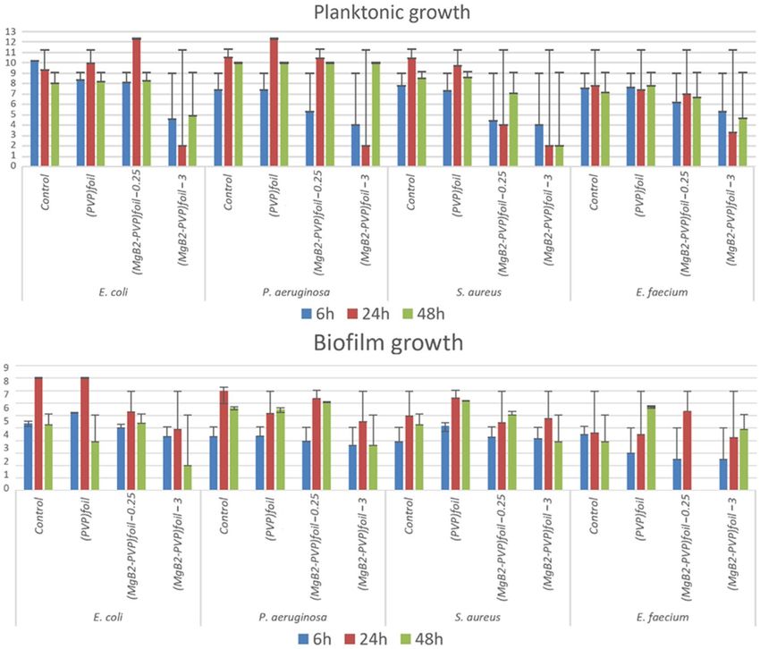
www.nature.com/scientificreports/
Figure 1. Coatings fabrication stages: (a) coatings of PVP with different concentrations (0.5 g of PVP in
3–30 ml of ethanol) on a glass substrate; (b) coatings of M gB2-PVP with different concentrations (0.01–4 g of
MgB2 introduced into a solution of 0.5 g PVP/30 ml ethanol) on a glass substrate (see text); (c–e) coatings on the
polymer foil of the catheter reservoir of P VPfoil (0.5 g PVP/30 ml ethanol), ( MgB2-PVP)foil0.25 (0.25 g MgB2/0.5 g
PVP/30 ml ethanol), ( MgB2-PVP)foil3 (3 g MgB2/0.5 g PVP/30 ml ethanol), respectively; (f) (from left to right)
pristine silicon tube of a catheter and coatings on the catheter silicon tube of P VPtube (0.5 g PVP/30 ml ethanol),
(MgB2-PVP)tube0.25 (0.25 g MgB2/0.5 g PVP/30 ml ethanol), ( MgB2-PVP)tube3 (3 g MgB2/0.5 g PVP/30 ml
ethanol).
amount of produced formazan is quantified by absorbance which is directly proportional to the number of liv-
ing cells in the culture.
Lactate dehydrogenase (LDH) is a cytosolic enzyme from the cytoplasm of any cell. Damage to the cell
membrane causes the leak of LDH into the extracellular fluid. In vitro release of LDH provides an accurate way
to measure cell membrane integrity and, implicitly, cell v iability32. To test the cytotoxic effect of M
gB2 on HS-27
cell culture, cells are exposed to increasing concentrations of M gB2 solutions. The MgB2 solutions used in the
MTS and LDH experiments are the same and their preparation is addressed in the next paragraphs. The release of
LDH in the cell culture supernatant correlates with the cytotoxicity33,34 and it is measured by a test in which two
coupled enzymatic reactions, catalyzed by LDH and diaphorase, take place. The reactions convert a tetrazolium
salt into a red formazan compound. Absorbance at 490 nm is measured spectrophotometrically (TriStar Berthold
Technologies). The kit used in our experiments was CytoTox 96 Non-Radioactive Cytotoxicity Assay (Promega).
For each powder (LTS, Alpha Aesar and Cerac) two stock solutions (8.3 mg/ml and 33.3 mg/ml) in ethanol
(99.8%) were prepared. Decimal dilutions were further used in the test. After 34 h adhesion time in 96-well
culture plates, fibroblasts were treated with M
gB2 solutions for 48 h. The protocol involving an incubator for cell
cultures with a humid atmosphere and 5% CO2 at 37 °C) was applied, where the culture medium was composed
of DMEM, 1% antibiotic/antifungal, 10% fetal bovine serum. The absorbance of the samples was related to that
of the environmental control, the solvent control, and the cell control.
In the MTS and LDH tests, three experiments were performed using samples in triplicate. The absorbance
ratio R of solutions with different concentrations containing treated and untreated cells in the MTS (RMTS) and
LDH (RLDH) tests was plotted vs. concentration of M gB2 solutions. For an increasing concentration, a decrease
of RMTS (equivalent to less formazan produced by fewer living cells) and an increase of RLDH (equivalent to more
LDH as a result of a lower metabolic activity due to more damaged cells) determine the cytotoxicity threshold.
Fabrication of composite MgB2‑PVP coatings. The LTS powder with the highest antimicrobial
activity23 was selected for fabrication of M gB2-PVP composite coatings. Commercial polyvinylpyrrolidone
(PVP) powder (Sigma Aldrich, the monomer N-vinylpyrrolidone, with chemical formula C6H9NO and molar
weight Mw = 1,300,000 g/mol) was dissolved in ethanol at room temperature. The amount of the PVP powder
immersed in ethanol was constant, 0.5 g, and the volume of ethanol was modified from 3 to 30 ml. Droplets of
each solution were placed on a glass substrate (Fig. 1a). A higher uniformity of the coating and less trapped gas
particles were noticed for a lower concentration; hence the lowest tested concentration (0.5 g PVP/30 ml etha-
nol) was selected for further processing stages. The PVP polymer is also soluble in water and this property will
be used to release the active MgB2 particles when in contact with bacteria.
The MgB2 powder was introduced into as-prepared PVP-ethanol solution and mixed ultrasonically for few
minutes, and a colloidal black solution was obtained (Fig. 2I). MgB2 is not soluble in a lcohols35. The amount of
the MgB2 powder varied from 0.01 to 4 g. Droplets of the colloidal solution were placed on a glass substrate and
Scientific Reports | (2021) 11:9591 | https://doi.org/10.1038/s41598-021-88885-2 3
Vol.:(0123456789)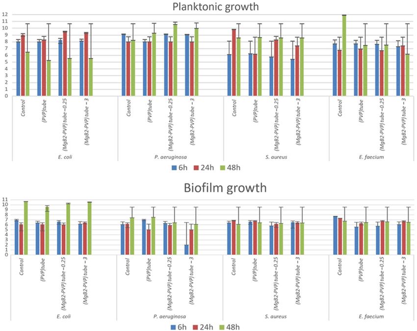
www.nature.com/scientificreports/
Figure 2. Preparation of composite MgB2-PVP coatings (I–III), release of active M
gB2 from the composite
coating and its decomposition in the presence of water/aqueous solution (IV), and the antimicrobial effect
through possible mechanisms on different microbes. SEM image from III is taken on the sample ( MgB2-
PVP)foil0.25.
dried naturally under ambient conditions (Fig. 1b). The uniformity, bubbles size and amount, and adherence of
the coatings were observed. Two concentrations, 0.25 and 3 g per initial solution (0.5 g PVP/30 ml ethanol) were
selected. Considering the density of PVP of 1.2 g/cm3, the selected concentrations of M gB2 in PVP are 60 mg/ml
and 720 mg/ml, respectively. For MgB2 concentrations in PVP greater than 720 mg/ml, coatings are not uniform,
showing a high level of granularity that makes them easily breakable and removable from the substrate.
The plastic foil of a reservoir and the silicon tube, both from a commercial urinary catheter, were used as
substrates for coatings fabrication (Fig. 1c–f). Coatings of bare PVP as reference samples were also deposited.
The composite films on the plastic foils were obtained by dip coating and subsequent drying at room temperature
(Fig. 2II,III). They were cut into samples of 8 mm in diameter (Fig. 1c–e). The PVP-MgB2 composite covered
both sides of the foil substrate. Coloidal solutions of M gB2/PVP/ethanol were poured into a silicon tube and,
after drying, a coating was obtained inside the tube (Fig. 1f). The inner/outer diameter of the tube was 4.5/6 mm.
After coating, the tube was cut into pieces of 1 cm in length.
Coated samples were labeled as: PVPfoil and PVPtube (0.5 g PVP/30 ml ethanol), ( MgB2-PVP)foil0.25 and
(MgB2-PVP)tube0.25 (0.25 g M gB2/0.5 g PVP/30 ml ethanol), ( MgB2-PVP)foil3 and (MgB2-PVP)tube3 (3 g MgB2/0.5 g
PVP/30 ml ethanol).
The microstructure by scanning electron microscopy (SEM, Lyra 3XMU/Tescan) of the composite M gB2-PVP
coatings (covered with Au) on the plastic foil can be visualized in Fig. 3. The surface morphology of the com-
posite film changes with addition of a higher amount of M gB2 powder in the coating. The presence of M gB2
influences drying of the coating and its roughness. While drying improves and roughness decreases for sample
(MgB2-PVP)foil0.25 (Fig. 3c,d) when compared to P VPfoil (Fig. 3a,b), addition of a higher amount of M gB2 as for
sample (MgB2-PVP) 3 (Fig. 3e,f) produces a very rough and granular surface. In the sample (MgB2-PVP)foil3
foil
one can actually clearly distinguish grains with plate like or irregular morphology (Fig. 3e,f) that are gathered
into agglomerates. The morphology of the particles in the composite film is similar to that observed in the raw
LTS MgB2 powder23. Elemental mapping shows the presence of B and Mg, distributed uniformly at the scale
of our observation (Fig. 3g,h). This result and short-time sonication for low energy (200 W) suggests that the
integrity of MgB2 was preserved during its processing in the PVP-ethanol solution for preparation of the com-
posite film. However, it is noteworthy that ultrasonication for 30 min at room temperature of MgB2 in water
produced through exfoliation Mg-deficient hydroxyl-functionalized nanosheets of M gB236, while processing of
MgB2 in acetonitrile mixed with an ion-exchange resin leads to formation of borophane sheets (hydrogenated
borophene, HB)37.
Scientific Reports | (2021) 11:9591 | https://doi.org/10.1038/s41598-021-88885-2 4
Vol:.(1234567890)
www.nature.com/scientificreports/
Figure 3. SEM images at two magnifications (× 500, × 1000) on MgB2-PVP films coated on plastic foil: (a,b)
sample PVPfoil; (c,d) sample (MgB2-PVP)foil0.25; (e,f) sample (MgB2-PVP)foil3. Secondary electron image from (g)
presents a detail at high magnification (× 15,000) of sample (MgB2-PVP)foil3, while (h) is a red–green–blue image
obtained by overlapping the elemental EDS maps of Mg and B measured on image from (g).
In vitro assay of antibacterial activity of MgB2‑PVP coatings. The antimicrobial activity of the
composite MgB2-PVP coatings (on foil and tubes) was tested against bacteria in planktonic and biofilms growth
states (Staphylococcus aureus ATCC 25923, Enterococcus faecium DMS 13590, Escherichia coli ATCC 25922,
Pseudomonas aeruginosa ATCC 27853).
In the case of planktonic bacteria, microbial suspensions of McFarland 0.5 (corresponding to 1.5 × 10⁸ CFU/
ml) were prepared from 24 h cultures in sterile saline solution. The obtained suspensions were further diluted
Scientific Reports | (2021) 11:9591 | https://doi.org/10.1038/s41598-021-88885-2 5
Vol.:(0123456789)www.nature.com/scientificreports/
(a) (b) (c)
0.8 0.8 0.8
AA MTS CER
MTS LDH MTS
0.6 LTS 0.6 0.6
Absorbance ratio R
Absorbance ratio R
Absorbance ratio R
LDH LDH
0.4 0.4 0.4
0.2 0.2 0.2
0 0 0
0 10 20 30 40 50 60 70 80 90 10 20 30 40 50 60 70 80 90 10 20 30 40 50 60 70 80 90
µg/ml µg/ml µg/ml
Figure 4. The absorbance ratio R in MTS and LDH cytotoxicity tests on HS27 cell line for dilutions prepared
from stock solutions of 8.3 mg/ml of three types of MgB2 powders.
1:100 in broth. Each composite MgB2-PVP sample was introduced into a well of a 24-well plate. The plate was
inoculated with the prepared microbial suspension (750 µl). The final inoculum size was of 5 × 10⁵ CFU/ml for
each tested coating. Untreated microbial cultures and sterile broth wells served as positive and negative controls.
The inoculated 24 well-plates were incubated at 37 °C, in aerobic conditions for 6 h, 24 h, and 48 h. After incuba-
tion for the respective time intervals, the density of the microbial broth culture was determined by plating serial
ten-fold dilutions prepared in sterile saline onto PCA (Plate Count Agar) and counting the number of colonies
developed at different dilutions. Results were further used to determine the number of colonies forming units
(CFU/ml).
In the case of biofilms, coatings were placed in contact with an inoculum of 5 × 10⁵ CFU/ml obtained from
each microbial strain and incubated for 6, 24, and 48 h. After incubation, each material was carefully washed
to remove the non-adherent bacteria and sonicated in sterile saline solution to disperse the biofilm, and serial
decimal dilutions were seeded on agar plates to determine the CFU/ml as presented above.
All assays were done in triplicate.
Results
Cytocompatibility of MgB2 powders. All dilutions (66–333 µg/ml) of stock solution with concentration
33.3 mg/ml and for the three investigated MgB2 powders (LTS, Alfa Aesar and Cerac) have shown cytotoxicity
on the H27 cell line. The effect of dilutions with smaller concentrations (0.83–83 µg/ml) prepared from the stock
solution 8.3 mg/ml are presented in Fig. 4. Concentrations of MgB2 solutions below 8.3, 33.2, and 33.2 µg/ml
for the powders LTS, Alfa Aesar, and Cerac are compatible with the metabolism of fibroblasts without a toxic
impact. In other experiments with different cells38 the toxicity limit was in the range of 50–100 µg/ml. The LTS
powder has a lower biocompatibility than Alfa Aesar and Cerac powders.
Higher cytotoxicity of LTS comparative to the other MgB2 investigated powders can be inferred from its low
tendency to form large agglomerates, clean particle surfaces, high amount of M gB2 phase, and high pH-increase
rates23. Different activity of different M
gB2 powders enables the possibility of a time and space-controlled reac-
tion of M gB2 with biological medium, depending on the application requirements, so that the toxic effect is
minimized, and antibacterial impact is maximized.
For design purposes of the M gB2-PVP coatings, apart from the as-evaluated cytotoxicity concentrations,
we considered the minimum inhibitory concentrations for the growth in the presence of LTS, Alfa Aesar and
Cerac MgB2 powders of different reference microbes (Staphylococcus aureus ATCC 25923, Staphylococcus aureus
ATCC 6538, Pseudomonas aeruginosa ATCC 27853, Escherichia coli ATCC 25922, and Candida albicans ATCC
10231) in the planktonic (MIC) and biofilm (MICB) states reported in Ref.23. Namely, the MIC and MBIC values
ranged between 0.31 and 1.25 mg/ml and between 0.039 and 0.62 mg/ml, respectively. The highest antimicrobial
activity was found for LTS and the lowest for Cerac. The M gB2 powders were active also against planktonic cells
and biofilms of 29 methicillin resistant clinical S. aureus isolates and 33 vancomycin resistant E. faecium/faecalis
strains collected from clinical souces. The MIC values of 0.15–2.5 mg/ml were determined for different fungi
collected from the heritage buildings and o bjects38.
Antibacterial activity of MgB2‑PVP composite coatings. The influence of PVP and M
gB2-PVP coat-
ings on the bacterial growth in planktonic state (in the liquid culture medium) and biofilm state (adhered on the
surface of plastic foil or silicon catheter) was assessed at 6, 24, and 48 h of bacterial strains incubation with the
tested samples. Results are presented in Figs. 5 and 6.
The general trend was that the M gB2-PVP samples exhibited an evident inhibitory activity on the planktonic
growth of all tested strains, starting with 6 h of incubation, exacerbating after 24 h, and maintaining it at 48 h
(Figs. 5, 6).
The inhibitory effect of the planktonic growth was more intense in the case of the samples (MgB2-PVP)foil3
containing the highest concentration of M gB2. In the case of the bacterial cells adhered on the surface of the
functionalized plastic samples, the anti-biofilm effect was strain specific, but however, less evident than in the case
of planktonic cells. The E. coli biofilm development was inhibited at all three time points by the ( MgB2-PVP)foil3.
Scientific Reports | (2021) 11:9591 | https://doi.org/10.1038/s41598-021-88885-2 6
Vol:.(1234567890)www.nature.com/scientificreports/
Figure 5. The number of viable microbial cells in log10(CFU/ml) for samples in the form of coatings on the
plastic foil (i.e. samples PVPfoil, (MgB2-PVP)foil0.25, (MgB2-PVP)foil3) tested with microbes in plaktonic and
biofilm growth states.
The same sample inhibited the P. aeruginosa and S. aureus biofilm at 24 h and 48 h, respectively. In the case of
E. faecium, all samples inhibited the initial cells adherence to the plastic foil, quantified after 6 h of incubation,
and the (MgB2-PVP)foil3 exhibited also a slight inhibitory effect against the 24 h biofilm (Fig. 5). The comparative
behaviour of planktonic and adhered cells developed in the presence of MgB2-functionalized plastic-foil samples
suggests that the coatings exhibit their antimicrobial effect mostly by releasing the M gB2 in the active form.
The rate of the bacterial strains growth in the presence of the M
gB2-functionalized silicon tubes was different
from that recorded on the plastic samples, with no significant inhibition of the planktonic growth, except a slight
decrease of the number of the viable cells of S. aureus after 24 h of incubation and of E. coli and E. faecium after
48 h of incubation. No significant differences between the PVP and M gB2-PVP samples (Fig. 6) were noted. In the
gB2-functionalized silicon tubes, the ( MgB2-PVP)tube3
case of the bacterial cells adhered on the surface of the M
inhibited the P. aeruginosa biofilm development, with the anti-biofilm effects intensity decreasing in time from
6 to 48 h. The ( MgB2-PVP)tube0.25 sample has shown a slight tendency for inhibition of the S. aureus biofilm
development, the effect being noticable after 6 h of incubation. All samples slightly inhibited the development
of E. faecium at all three incubation times. As for S. aureus, the effect is stronger at 6 h.
Results indicate that M gB2-containing samples on foils are more active than the coatings on silicon tubes.
To understand it, further research is necessary. Among the possible reasons there could be how the shape of
the sample influences the contact with the cell browth, the release of M gB2 and its interaction with the cells. A
second aspect that could play a role is the fact that adherence of the coatings on the silicon tube is poor when
compared to that on the foil. This is especially problematic for coatings with a high concentration of M gB2, i.e.
for the samples ( MgB2-PVP)tube3.
Discussion
Considering the data presented in “Cytocompatibility of M gB2 powders” it results that selected M
gB2 powder
for coatings fabrication, LTS, has the lowest toxicity limit, corresponding to the highest cytotoxicity. At the same
time, this powder is the most active one from the viewpoint of antimicrobial efficiency, i.e. the MIC and MBIC
values are the lowest23. To understand the context of our antimicrobial assessment from “Antibacterial activity of
Scientific Reports | (2021) 11:9591 | https://doi.org/10.1038/s41598-021-88885-2 7
Vol.:(0123456789)www.nature.com/scientificreports/
Figure 6. The number of viable of microbial cells in log10(CFU/ml) for samples in the form of coatings on the
silicone tube (i.e. samples PVPtube, (MgB2-PVP)tube0.25, (MgB2-PVP)tube3) tested with microbes in planktonic and
biofilm states.
MgB2-PVP composite coatings” and to provide some guiding lines in designing M gB2-PVP coatings for future
different applications, we present some useful details in the following paragraphs.
The thickness of the coatings was estimated from optical measurements at ~ 200 µm. The volume of the
coatings for the foil and tube samples was 0.02 and 0.028 ml, respectively. When using 0.25 g and 3 g of MgB2 to
fabricate the samples, this results into a concentration of 0.6 and 7.2 mg/ml of the powder in the volume of PVP
dissolved in ethanol (density of PVP was taken as 1.2 g/cm3 and density of M gB2 as 2.6 g/cm3). Rescaling for the
foil foil
volume of the coating on the foil, the ( MgB2-PVP) 0.25 and ( MgB2-PVP) 3 samples contain 9.75 and 38.2 mg
of MgB2, respectively. A similar calculation for the ( MgB2-PVP)tube0.25 and ( MgB2-PVP)tube3 samples gives the
values 13.65 and 53.5 mg, respectively.
To obtain a correct intepretation of our data, we should also take into consideration the PVP behavior in
water. To this purpose, a bulk rectangular piece of PVP (0.5 cm × 0.5 cm × 0.1 cm) was prepared by casting a
viscous solution of PVP in ethanol and subsequent drying in the air. The PVP sample with a volume 0.025 ml
was introduced in water (50 ml). It dissolved in 3 h at room temperature, without steering. The dissolution rate
of PVP in water is 0.0083 ml/h.
In the in vitro antimicrobial tests addressed in “Antibacterial activity of M gB2-PVP composite coatings”,
samples were fully immersed in 750 µl of culture medium. The incubation time of 6, 24, and 48 h is higher
than the dissolution time of PVP in water of 3 h. One also observes that the volume of the bulk PVP sample
(0.025 ml) is comparable to that of the coatings (0.02 and 0.028 ml on the foil and on the tube, respectively). This
suggests that the entire amount of MgB2 powder in the coating was available to interact with the bacterial cells
from the broth. Hence, the concentration of M gB2 in the cell broth was as follows: 13, 50.9, 18.2, and 71.3 mg/
ml for samples (MgB2-PVP)foil0.25, (MgB2-PVP)foil3, (MgB2-PVP)tube0.25, and (MgB2-PVP)tube3, respectively. These
values are comparable, or they are one order of magnitude higher that the MIC and MBIC values mentioned
in “Cytocompatibility of MgB2 powders”. They are also about three orders of magnitude larger than the toxicity
limit of the powders. Interaction of M gB2 with water, culture media, or body fluids is complex and needs further
investigations. Some information for Mg, MgO, Mg(OH)2 and MgB2 was reported in Refs.26,27,36,39–41. The involved
processes develop in multistep sequences specific for each phase. Moreover, the reaction products, kinetic factors,
and environmental conditions will ensure, or not, the background for antibacterial activity evolution with time.
For example, the reaction of M gB2 with water leads to formation of Mg(OH)2, while boron in water forms boric
acid. Due to low solubility in water, Mg(OH)2 can passivate the surface of MgB2 decreasing its efficiency. However
Scientific Reports | (2021) 11:9591 | https://doi.org/10.1038/s41598-021-88885-2 8
Vol:.(1234567890)www.nature.com/scientificreports/
in physiologically relevant solutions Mg(OH)2 was found to dissociate32. On the other hand, in the reaction
between MgB2 and water, the H2-gas is released, and it can disrupt the passivating layer and can clean the surface
of the M
gB2 particle. The overlapping of the processes, some of them with possibly opposite contributions, can
explain the non-linear dependence of the bioactivity with time observed in our experiments from “Antibacterial
activity of MgB2-PVP composite coatings”, but one should also consider the decreasing amount of MgB2, which
is consumed in the interaction with cells and environment. Therefore, the current discussion and provided
numbers draw attention on the necessity to properly design the coatings considering each specific application.
The mechanisms by which our composite coatings develop the antimicrobial effect deserves further targeted
studies. In general, for nanomaterials the mechanisms11,42 are related to their influence on production of reactive
oxygen species (ROS) (and in some cases of reactive nitrogen species RNA) through a catalytic action described
by Haber/Weiss- and Fenton- type r eactions13,43, on electrostatic interaction that can impact the disruption/
damage of the membrane integrity and potential so that metabolic functions of the cells are affected, and on
the changes in the local environmental conditions e.g. through modification of aeration and pH. Depending on
materials, cells, and environment there are specific features. For our composite coatings, PVP disolves in water,
MgB2 is released and it becomes active interacting with the aqueous e nvironment44 and the cells (Fig. 2IV). PVP
is inert in respect to its antimicrobial activity45. The possibility of exfoliation of MgB2 with formation of nano
sheet materials such as hydroxyl functionalized Mg-deficient M gB236 or borophane (HB)37 may provide extra
positive specific value in fighting the microbes: the 2D borophene-type materials are shown to have catalytic
properties46 that can impact directly or indirectly also bioprocesses. Furthermore, another 2D material, namely
the graphene oxide emerged as an effective antimicrobial m aterial47 and, through similarities and extrapolation,
one may expect that also the 2D nano boron-type materials derived from MgB2 will play an important role as
antimicrobial materials. However, at present, there are no experiments performed in this direction. In summary,
different ionic species are generated because of the M gB2 presence and they are at the origin of the developing
reactions necessary for the antimicrobial effect (Fig. 2V).
The antimicrobial activity of M gB2 assessed in our work revealed good efficiency against selected bacteria,
e.g. for planktonic growth (Fig. 5) one may observe a decrease for ( MgB2-PVP)foil3 of 7log/24 h against E. coli,
8log/24 h against P. aeruginosa, 8log/24 h against S. aureus, and 5log/24 h against E. faecium. Literature present
other effective composite antibacterial materials, such as AgBr/nPVP45 with a 5log/2 h against E. coli, and a lower
concentration of the active substance (however, no citotoxicity data are available for comparison). In Ref.48 was
reported a decrease of CFU/ml for nitric oxide (NO) of 8 log/24 h against E. coli/A. baumannii/S. aureus. In
Ref.49 for copper (Cu) the decrease of CFU/ml was of 4log/24 h against E. coli/S. aureus. In Ref.50 was shown
for silver (Ag) a decrease of CFU/ml of 5log/1.5 h against E. coli/S. aureus. It is important to note that a direct
comparison between reported data is not entirely possible, as the assessment procedures are slightly different,
the control samples do not start from the same level of cells population, and samples are different in shape and
size and they target different applications.
Conclusion
In vitro MTS and LDH cytotoxicity tests of the MgB2 activity on the HS27 dermal cell line indicated a toxicity
limit in the range of 8.3–33.2 µg/ml depending on the powder type. Three powders were tested and the most
active was LTS. According to literature23 this powder shows the highest amount of MgB2 phase, impurity-free
particle surfaces, the largest fraction of the particle size in the nano-range and the highest rate of pH increase.
The LTS powder was further used for fabrication of M gB2–PVP coatings on plastic elements from a commercial
urinary catheter. The as-prepared self-replenishing and biodegradable M gB2–PVP composite coatings with two
compositions were tested at 6, 24, and 48 h of incubation time against the bacterial growth of Staphylococcus
aureus ATCC 25923, Enterococcus faecium DMS 13590, Escherichia coli ATCC 25922, Pseudomonas aeruginosa
ATCC 27853. Strains were in the planktonic and biofilm growth states. The coatings were efficient both against
planktonic microbes and microbial biofilms when applied on plain, flexible plastic foils. Results are promising
and recommend M gB2 for designing efficient anti-infective solutions for various biomedical devices and systems.
Received: 20 October 2020; Accepted: 7 April 2021
References
1. Beceiro, A., Tomás, M. & Bou, G. Antimicrobial resistance and virulence: A succeful or deterious associasion in the bacterial
world?. Clin. Microbiol. Rev. 26, 185–230. https://doi.org/10.1128/cmr.00059-12 (2013).
2. Lebeaux, D., Ghigo, J.-M. & Beloin, C. Biofilm-related infections: Bridging the gap between clinical management and fundamental
aspects of recalcitrance toward antibiotics. Microbiol. Mol. Biol. Rev. 78, 510–543. https://d
oi.o
rg/1 0.1 128/M MBR.0 0013-1 4 (2014).
3. Kalia, V. C. Quorum sensing inhibitors: An overview. Biotechnol. Adv. 31, 224–245. https://doi.org/10.1016/j.biotechadv.2012.10.
004 (2013).
4. Yetisgin, A. A., Cetinel, S., Zuvin, M., Kosar, A. & Kutlu, O. Therapeutic nanoparticles and their targeted delivery applications.
Molecules 25, 2193. https://doi.org/10.3390/molecules25092193 (2020).
5. Acar, J. F. Antibiotic synergy and antagonism. Med. Clin. North Am. 84, 1391–1406. https://d oi.o
rg/1 0.1 016/S 0025-7 125(05)7 0294-7
(2000).
6. Bollenbach, T. Antimicrobial interactions: Mechanisms and implications for drug discovery and resistance evolution. Curr. Opin.
Microbiol. 27, 1–9. https://doi.org/10.1016/j.mib.2015.05.008 (2015).
7. Doern, C. D. When does 2 plus 2 equal 5? A review of antimicrobial synergy testing. J. Clin. Microbiol. 52, 4124–4128. https://doi.
org/10.1128/JCM.01121-14 (2014).
8. Patra, J. K. et al. Nano based drug delivery systems: Recent developments and future prospects. J. Nanobiotechnol. 16, 71. https://
doi.org/10.1186/s12951-018-0392-8 (2018).
9. Karimi, M. et al. Smart micro/nanoparticles in stimulus-responsive drug/gene delivery systems. Chem. Soc. Rev. 45, 1457–1501.
https://doi.org/10.1039/c5cs00798d (2016).
Scientific Reports | (2021) 11:9591 | https://doi.org/10.1038/s41598-021-88885-2 9
Vol.:(0123456789)www.nature.com/scientificreports/
10. Mohammadi, M. R. et al. Nanomaterials engineering for drug delivery: A hybridization approach. J. Mater. Chem. B 5, 3995–4018.
https://doi.org/10.1039/c6tb03247h (2017).
11. Beyth, N., Houri-Haddad, Y., Domb, A., Khan, W. & Hazan, R. Alternative antimicrobial approach: Nano-antimicrobial materials.
Evid. Based Complement. Altern. Med. 2015(246012), 1–16. https://doi.org/10.1155/2015/246012 (2015).
12. Popescu, R. C., Andronescu, E. & Vasile, B. S. Recent advances in magnetite nanoparticle functionalization for nanomedicine.
Nanomaterials 9, 1791. https://doi.org/10.3390/nano9121791 (2019).
13. Collin, F. Chemical basis of reactive oxygen species reactivity and involvement in neurodegenerative diseases. Int. J. Mol. Sci. 20,
2407. https://doi.org/10.3390/ijms20102407 (2019).
14. Grumezescu, A. M. Nanobiomaterials in Antimicrobial Therapy, Applications of Nanobiomaterials, Vol. 6 (Elsevier, 2016). ISBN:
978-0-323-42864-4.
15. Vasilev, K., Cavallaro, A. & Zilm, P. Special issue: Antibacterial materials and coatings. Molecules 23, 585. https://doi.org/10.3390/
molecules23030585 (2018).
16. Niemeyer, C. M. & Mirkin, C. A. Nanobiotechnology, Concepts, Applications and Perspectives (WILEY-VCH Verlag GmbH & Co.
KGaA, 2004). ISBN: 3-527-30658-7.
17. Pelgrift, R. Y. & Friedman, A. J. Nanotechnology as a therapeutic tool to combat microbial resistance. Adv. Drug Deliv. Rev. 65,
1803–1815. https://doi.org/10.1016/j.addr.2013.07.011 (2013).
18. Blecher, K., Nasir, A. & Friedman, A. The growing role of nanotechnology in combating infectious disease. Virulence 2, 395–401.
https://doi.org/10.4161/viru.2.5.17035 (2011).
19. Bühler, V. Polyvinylpyrrolidone Excipients for Pharmaceuticals: Povidone, Crospovidone and Copovidone 1–254 (Springer, 2005).
https://doi.org/10.1007/b138598.
20. Haaf, F., Sanner, A. & Straub, F. Polymers of N-vinylpyrrolidone: Synthesis, characterization and uses. Polym. J. 17, 143–152. https://
doi.org/10.1295/polymj.17.143 (1985).
21. Franco, P. & De Marco, I. The use of Poly(N-vinyl pyrrolidone) in the delivery of drugs: A review. Polymers (Basel) 12, 1114. https://
doi.org/10.3390/polym12051114 (2020).
22. Batalu, D., Stanciuc, A. M., Moldovan, L., Aldica, G. & Badica, P. Evaluation of pristine and E u2O3-added MgB2 ceramics for medi-
cal applications: Hardness, corrosion resistance, cytotoxicity and antibacterial activity. Mater. Sci. Eng. C 42, 350–361. https://doi.
org/10.1016/j.msec.2014.05.046 (2014).
23. Badica, P. et al. MgB2 powders and bioevaluation of their interaction with planktonic microbes, biofilms, and tumor cells. J. Mater.
Res. Technol. https://doi.org/10.1016/j.jmrt.2021.04.003 (2021).
24. Feneley, R. C. L., Hopley, I. B. & Wells, P. N. T. Urinary catheters: History, current status, adverse events and research agenda. J.
Med. Eng. Technol. 39, 459–470. https://doi.org/10.3109/03091902.2015.1085600 (2015).
25. Lawrence, E. L. & Turner, I. G. Materials for urinary catheters: A review of their history and development in the UK. Med. Eng.
Phys. 27, 443–453. https://doi.org/10.1016/j.medengphy.2004.12.013 (2005).
26. Aswal, D. K. et al. Degradation behavior of MgB2 superconductor. Phys. C 363, 208–214. https://doi.org/10.1016/S0921-4534(01)
00974-1 (2001).
27. Chen, Y. K., An, Z. & Chen, M. Competition mechanism study of Mg+H2O and MgO+H2O reaction. Conf. Ser. Mater. Sci. Eng.
394, 022015. https://doi.org/10.1088/1757-899X/394/2/022015 (2018).
28. Uluisik, I., Karakaya, H. C. & Koc, A. The importance of boron in biological systems. J. Trace Elem. Med. Biol. 45, 156–162. https://
doi.org/10.1016/j.jtemb.2017.10.008 (2018).
29. Malich, G., Markovic, B. & Winder, C. The sensitivity and specificity of the MTS tetrazolium assay for detecting the in vitro cyto-
toxicity of 20 chemicals using human cell lines. Toxicology 124, 179–192. https://doi.org/10.1016/S0300-483X(97)00151-0 (1997).
30. Barltrop, J., Owen, T., Cory, A. H. & Cory, J. G. 5-(3-carboxymethoxyphenyl)-2-(4,5-dimethylthiazoly)-3-(4-sulfophenyl)tetra-
zolium, inner salt (MTS) and related analogs of 3-(4,5-dimethylthiazolyl)-2,5-diphenyltetrazolium bromide (MTT) reducing to
purple water-soluble formazans as cell-viability indicators. Bioorg. Med. Chem. Lett. 1, 611–614. https://doi.org/10.1016/S0960-
894X(01)81162-8 (1991).
31. Riss, T. L. et al. Cell viability assays. in Assay Guidance Manual (2013) (eds Markossian, S. et al.). http://www.ncbi.nlm.nih.gov/
books/NBK144065.
32. Fotakis, G. & Timbrell, J. A. In vitro cytotoxicity assays: Comparison of LDH, neutral red, MTT and protein assay in hepatoma cell
lines following exposure to cadmium chloride. Toxicol. Lett. 160, 171–177. https://doi.org/10.1016/j.toxlet.2005.07.001 (2006).
33. Allen, M., Millett, P., Dawes, E. & Rushton, N. Lactate dehydrogenase activity as a rapid and sensitive test for the quantification of
cell numbers in vitro. Clin. Mater. 16, 189–194. https://doi.org/10.1016/0267-6605(94)90116-3 (1994).
34. Kaja, S., Payne, A. J., Naumchuk, Y. & Koulen, P. Quantification of lactate dehydrogenase for cell viability testing using cell lines
and primary cultured astrocytes. Curr. Protoc. Toxicol. 72, 1–10. https://doi.org/10.1002/cptx.21 (2017).
35. Lim, J. Y. et al. Effects of surface-treated boron powder using chemical solvents on M gB2 bulk superconductors. Prog. Supercond.
Cryogenics 20, 11–14. https://doi.org/10.9714/psac.2018.20.3.011 (2018).
36. Das, S. K., Bedar, A., Kannan, A. & Jasuja, K. Aqueous dispersions of fewlayer-thick chemically modified magnesium diboride
nanosheets by ultrasonication assisted exfoliation. Sci. Rep. 5, 10522. https://doi.org/10.1038/srep10522 (2015).
37. Nishino, H. et al. Formation and characterization of hydrogen boride sheets derived from M gB2 by cation exchange. Am. Chem.
Soc. 139, 13761–13769. https://doi.org/10.1021/jacs.7b06153 (2017).
38. Gheorghe, I. et al. In vitro evaluation of MgB2 powders as novel tools to fight fungal biodeterioration of heritage buildings and
objects. Front. Mater. 7, 601059. https://doi.org/10.3389/fmats.2020.601059 (2021).
39. Lide, D. R. Handbook of Chemistry and Physics 73rd edn (Taylor & Francis, 1992). ISBN 0849304733, 9780849304736.
40. Wetteland, C. L. et al. Dissociation of magnesium oxide and magnesium hydroxide nanoparticles in physiologically relevant fluids.
J. Nanopart. Res. 20, 215. https://doi.org/10.1007/s11051-018-4314-3 (2018).
41. Poinern, G. E. J., Brundavanam, S. & Fawcett, D. Biomedical magnesium alloys: A review of material properties, surface modifica-
tions and potential as a biodegradable orthopaedic implant. Am. J. Biomed. Eng. 2, 218–240. https://doi.org/10.5923/j.ajbe.20120
206.02 (2012).
42. Khatoon, Z., McTiernan, C. D., Suuronen, E. J., Mah, T.-F. & Alarcon, E. I. Bacterial biofilm formation on implantable devices and
approaches to its treatment and prevention. Heliyon 4, e01067. https://doi.org/10.1016/j.heliyon.2018.e01067 (2018).
43. Madkour, L. H. Function of reactive oxygen species (ROS) inside the living organisms and sources of oxidants. Pharm. Sci. Anal.
Res. J. 2, 180023 (2019). https://chembiopublishers.com/PSARJ/PSARJ1800023.pdf.
44. Sadeghi, E., Peighambardoust, N. S., Khatamian, M., Unal, U. & Aydemir, U. Metal doped layered M gB2 nanoparticles as novel
electrocatalysts for water splitting. Sci. Rep. 11, 3337. https://doi.org/10.1038/s41598-021-83066-7 (2021).
45. Sambhy, V., MacBride, M. M., Peterson, B. R. & Sen, A. Silver bromide nanoparticle/polymer composites: Dual action tunable
antimicrobial materials. J. Am. Chem. Soc. 128, 9798–9808. https://doi.org/10.1021/ja061442z (2006).
46. Fujino, A. et al. Hydrogenated borophene shows catalytic activity as solid acid. ACS Omega 4, 14100–14104. https://doi.org/10.
1021/acsomega.9b02020 (2019).
47. Li, P. et al. Synthesis, characterization, and bactericidal evaluation of chitosan/guanidine functionalized graphene oxide composites.
Molecules 22, 12. https://doi.org/10.3390/molecules22010012 (2017).
48. Pegalajar-Jurado, A. et al. Nitric oxide-releasing polysaccharide derivative exhibits 8-log reduction against Escherichiacoli, Acine-
tobacterbaumannii and Staphylococcusaureus. J. Control. Release 220, 617. https://doi.org/10.1016/j.jconrel.2015.09.015 (2015).
Scientific Reports | (2021) 11:9591 | https://doi.org/10.1038/s41598-021-88885-2 10
Vol:.(1234567890)www.nature.com/scientificreports/
49. Esteban-Cubillo, A., Pecharromán, C., Aguilar, E., Santarén, J. & Moya, J. S. Antibacterial activity of copper monodispersed nano-
particles into sepiolite. J. Mater. Sci. 41, 5208. https://doi.org/10.1007/s10853-006-0432-x (2006).
50. Jung, W. K. et al. Antibacterial activity and mechanism of action of the silver ion in Staphylococcusaureus and Escherichiacoli. Appl.
Environ. Microbiol. 74, 2171. https://doi.org/10.1128/AEM.02001-07 (2008).
Acknowledgements
Authors acknowledge Romanian National Authority for Scientific Research and Innovation (UEFISCDI), Italian
Ministry of Education, University and Research (MIUR) and EU for financial support, project 74-COFUND-
M-ERA.NET II—BIOMB. Support from UEFISCDI through the project 5PTE/2020—BIOTEHKER is also
acknowledged. The funding had no role in study, design, data collection, and analysis, decision to publish, or
preparation of the manuscript.
Author contributions
Conceptualization: P.B., N.D.B., M.C.C.; methodology P.B., M.C.C.; validation P.B., M.C.C.; analysis: P.B., N.D.B.,
M.C.C., M.B., M.A.G., G.A., M.E., G.G.P., M.P., L.G.M., B.G.D., L.O., A.B., B.P., L.O., V.B., A.A., M.T.; investiga-
tion; writing—original draft preparation: P.B.; writing—review and editing P.B., N.D.B., M.C.C., G.A., M.T.; visu-
alization: P.B.; supervision P.B.; project administration P.B., N.D.B., M.C.C., M.T.; funding acquisition: M.T., P.B.
Competing interests
The authors declare no competing interests.
Additional information
Correspondence and requests for materials should be addressed to P.B.
Reprints and permissions information is available at www.nature.com/reprints.
Publisher’s note Springer Nature remains neutral with regard to jurisdictional claims in published maps and
institutional affiliations.
Open Access This article is licensed under a Creative Commons Attribution 4.0 International
License, which permits use, sharing, adaptation, distribution and reproduction in any medium or
format, as long as you give appropriate credit to the original author(s) and the source, provide a link to the
Creative Commons licence, and indicate if changes were made. The images or other third party material in this
article are included in the article’s Creative Commons licence, unless indicated otherwise in a credit line to the
material. If material is not included in the article’s Creative Commons licence and your intended use is not
permitted by statutory regulation or exceeds the permitted use, you will need to obtain permission directly from
the copyright holder. To view a copy of this licence, visit http://creativecommons.org/licenses/by/4.0/.
© The Author(s) 2021
Scientific Reports | (2021) 11:9591 | https://doi.org/10.1038/s41598-021-88885-2 11
Vol.:(0123456789)You can also read


