A cis-regulatory change underlying the motor neuron-specific loss of terminal selector gene expression in immotile tunicate larvae - bioRxiv
←
→
Page content transcription
If your browser does not render page correctly, please read the page content below
bioRxiv preprint first posted online Mar. 5, 2019; doi: http://dx.doi.org/10.1101/567719. The copyright holder for this preprint (which
       was not peer-reviewed) is the author/funder, who has granted bioRxiv a license to display the preprint in perpetuity.
                                 It is made available under a CC-BY-NC-ND 4.0 International license.

                                                                                      Lowe et al., March 01 2019 – preprint copy - BioRxiv

      A cis-regulatory change underlying the motor neuron-specific loss of
      terminal selector gene expression in immotile tunicate larvae
      Elijah K. Lowe1,2, Claudia Racioppi2,3, Nadine Peyriéras2,4,5, Filomena Ristoratore2,6, Lionel
      Christiaen2,3, Billie J. Swalla2,7,8, & Alberto Stolfi1,2*
      1. School of Biological Sciences, Georgia Institute of Technology, Atlanta, GA, USA.
      2. Station Biologique de Roscoff, Roscoff, France.
      3. Center for Developmental Genetics, Department of Biology, New York University, New York, NY, USA.
      4. UPS3611 Complex Systems Institute Paris Île-de-France (ISC-PIF), CNRS, Paris, France.
      5. USR3695 BioEmergences, CNRS, Paris-Saclay University, Gif-sur-Yvette, France.
      6. Biology and Evolution of Marine Organisms, Stazione Zoologica Anton Dohrn, Naples, Italy.
      7. Department of Biology, University of Washington, Seattle, WA, USA.
      8. Friday Harbor Laboratories, University of Washington, Friday Harbor, WA, USA.
      * Corresponding author: alberto.stolfi@biosci.gatech.edu

      Abstract

      The evolutionary history of animal body plans cannot be fully reconstructed without considering the roles of both novelties
      and losses. Some of the more remarkable examples of massively parallel evolutionary losses in animals comes from many
      species in the tunicate genus Molgula that have independently lost the swimming larva and instead develop as tail-less,
      immotile larvae that bypass the period of swimming and dispersal observed in other tunicates, marine invertebrate chordates
      that alternate between motile larval and sessile adult life cycle stages. The larvae of Molgula occulta and other tail-less
      species do not fully develop structures that are essential for swimming behavior, including notochord, tail muscles, and
      otolith, and loss-of-function mutations have been identified in various genes required for the differentiation of these tissues.
      However, little is known about the extent of development of the larval nervous system in M. occulta. While differentiated
      neurons might in principle be entirely dispensable to the non-swimming larva, the adult has a fully functional nervous system
      like any other tunicate. To further investigate this conundrum, we studied the specification and patterning of the M. occulta
      Motor Ganglion, which is the key central nervous system compartment that drives the motor movements of swimming
      tunicate larvae. We found that the expression patterns of important regulators of MG neuron subtype specification are highly
      conserved during the development of the non-swimming larvae of M. occulta, suggesting that the gene networks regulating
      their expression are largely intact in this species, despite the loss of swimming ability. However, we identified a M. occulta-
      specific reduction in expression of the important motor neuron terminal selector gene Ebf (Collier/Olf/EBF or COE) in the
      Motor Ganglion. Although M. occulta Ebf is predicted to encode a fully functional protein, its expression was reduced in
      developing motor neurons when compared to species with swimming larvae, which was corroborated by measuring allele-
      specific expression of Ebf in interspecific hybrid embryos produced by crossing M. occulta with the closely related swimming
      species M. oculata. Comparative reporter construct experiments also revealed a specific cis-regulatory sequence change that
      underlies the reduced expression of M. occulta Ebf in motor neurons, but not in other tissues and cell types. This points to a
      potential mechanism for arresting larval motor neuron differentiation in the non-swimming larvae of this species.

                                                                                                                                          1
bioRxiv preprint first posted online Mar. 5, 2019; doi: http://dx.doi.org/10.1101/567719. The copyright holder for this preprint (which
          was not peer-reviewed) is the author/funder, who has granted bioRxiv a license to display the preprint in perpetuity.
                                    It is made available under a CC-BY-NC-ND 4.0 International license.

                                                                                                   Lowe et al., March 01 2019 – preprint copy - BioRxiv

Introduction                                                                    Underlying this radically divergent, non-swimming
                                                                                larval form is the loss of morphogenesis and
The evolution of animal body plans has occurred not                             differentiation of certain cell types that are
only through evolutionary novelties, but also through                           dispensable for non-swimming larva, like the
losses both subtle and catastrophic (Albalat and                                notochord, tail muscles, or pigmented sensory organs
Cañestro, 2016). Although they are defined primarily                            (Jeffery and Swalla, 1991; Swalla and Jeffery, 1990,
by the absence of structures, cell types, or genes, the                         1992). For instance, M. occulta larva muscle cells do
evolutionary loss of these various units of selection can                       not differentiate and the species has even lost certain
help illuminate their functions. For instance,                                  genes encoding proteins specifically required for
identifying traits that are retained in one species but                         muscle function, like muscle actin (Kusakabe et al.,
lost in a closely related species can reveal which are                          1996). Furthermore, M. occulta lack a pigmented
likely to be under purifying selection and provide a                            otolith cell, important for gravity sensing in swimming
measure of their relative adaptive value under certain                          larvae (Jiang et al., 2005; Tsuda et al., 2003), due to the
conditions, e.g. loss of vision in cave-dwelling                                loss of genes encoding functional enzymes required
organisms (Porter et al., 2003).                                                for melanogenesis like Tyrosinase and Tyrosinase-
                                                                                related protein (Tyrp)(Racioppi et al., 2017). Over 17
Some of the most prominent examples of extensive                                species with non-swimming larvae have been
evolutionary losses come from the tunicates (Denoeud                            described in Molgula, and all 31 species in the closely
et al., 2010; Huber et al., 2000), marine filter-feeding                        related molgulid genera Eugyra and Bostrichobranchus
organisms characterized by a protective layer, or                               appear to have non-swimming larvae. The vast
“tunic”, made mostly of cellulose (Sasakura, 2018).                             majority of molgulids (>150 species) have not been
Phylogenomic analyses suggest that vertebrates are                              studied at the larval stage, leaving the possibility that
the sister group to the tunicates (Bourlat et al., 2006),                       many more non-swimming larvae have independently
and most tunicates have a distinct dispersal phase                              evolved within the clade (Maliska et al., 2013; Shenkar
carried out by swimming “tadpole” type larvae that                              et al., 2019).
bear the typical chordate body plan: a rigid notochord
flanked by striated paraxial muscles, controlled by a                           Little is known about the extent of nervous system
dorsal central nervous system. Some tunicate groups,                            development in the larvae of M. occulta and other
such as the pelagic salps and pyrosomes, have                                   species with non-swimming larvae. The typical
completely lost this larval phase, while in other groups                        tunicate larva has a minimal nervous system dedicated
the evolutionary loss of the swimming larva is an                               to controlling its swimming and settlement behavior in
ongoing process, affecting specific aspects of larval                           response to sensory cues such as light, gravity, and
development and behavior. This phenomenon is                                    mechanical stimuli (Jiang et al., 2005; Rudolf et al.,
particularly prominent in molgulid tunicates, in which                          2018; Salas et al., 2018; Zega et al., 2006). The larval
many species have independently lost larval structures                          nervous system of the tunicate Ciona intestinalis has
important for swimming (Hadfield et al., 1995; Huber                            been completely mapped, revealing 177 neurons in a
et al., 2000). The best studied of these is Molgula                             dorsal central nervous system (CNS) and 54 peripheral
occulta, which gives rise to anural (tail-less), non-                           sensory neurons distributed throughout the epidermis
swimming larvae that metamorphose into juveniles                                (Ryan et al., 2016, 2018; Ryan and Meinertzhagen,
without going through the dispersal period of active                            2019). With 231 total neurons, the Ciona larval
swimming observed in urodele (tailed) species (Berrill,                         nervous system is one of the smallest known in all of
1931).                                                                          metazoa, and thus an intriguing model for the study of
                                                                                chordate-specific principles of neuronal function and
                                                                                development (Nishino, 2018).

                                                                                                                                                     2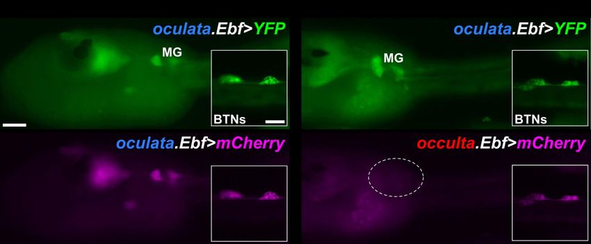
bioRxiv preprint first posted online Mar. 5, 2019; doi: http://dx.doi.org/10.1101/567719. The copyright holder for this preprint (which
          was not peer-reviewed) is the author/funder, who has granted bioRxiv a license to display the preprint in perpetuity.
                                    It is made available under a CC-BY-NC-ND 4.0 International license.

                                                                                                   Lowe et al., March 01 2019 – preprint copy - BioRxiv

Given the evolutionary loss of other structures                                 performed as previously described (Ikuta and Saiga,
important for swimming (notochord, muscles, otolith),                           2007; Stolfi et al., 2014b). To detect cilia, larvae were
we asked whether any neurodevelopmental processes                               incubated with anti-acetylated α-tubulin primary
have been lost during the evolution of M. occulta. To                           antibody (clone 6-11B-1, Sigma) and AlexaFluor-488
this end, we surveyed the development of the nervous                            secondary antibody (ThermoFisher) as previously
system in M. occulta embryos, using species with                                described (Pennati et al., 2015). Cell outlines were
swimming larvae as a basis for comparison. Using in                             counterstained with phalloidin AlexFluor-546
situ hybridization, RNAseq data, and cross-species                              conjugate (ThermoFisher) incubated 1:50 for at least 2
transgenic assays, we report that neurodevelopmental                            hours prior to final washes, and nuclei were labeled
gene expression and patterning is unexpectedly                                  with DAPI during the final wash. Embryos were imaged
conserved in M. occulta larvae. Most notably, the                               on upright and inverted epifluorescence microscopes
Motor Ganglion (MG), the CNS structure that controls                            (Leica), or TCS SP5 AOBS and TCS SP8 X scanning point
the swimming movements of tailed larvae, is also                                confocal systems (Leica).
found in tailless larvae (Nishino et al., 2010). However,
we uncovered specific cis-regulatory mutations that                             Hybrid embryo allele-specific differential expression
might underlie the reduced transcriptional activity of                          analyses
the key cholinergic neuron terminal selector gene Ebf                           M. occulta and M. oculata genomes (Stolfi et al.,
(Kratsios et al., 2012) in larval motor neurons.                                2014b) were improved using Redundans (Pryszcz and
However, unlike Tyrosinase or Tyrp, the Ebf gene has                            Gabaldón, 2016), reads from the initial assemblies
not become pseudogenized in M. occulta, likely due to                           (PRJNA253689) were used to improve scaffolding,
its requirement for the specification of other neurons                          which had been done to improve the M. occidentalis
and cell types that are still important for the complete                        genome and gene models we previously published
life cycle of this species.                                                     (Lowe and Stolfi, 2018). Additionally, the M. oculata
                                                                                genome was used as a reference to increase the
Methods                                                                         scaffolding of the M. occulta genome. To build gene
                                                                                models, previously sequenced RNAseq reads (Lowe et
M. occulta and M. oculata embryo collection                                     al., 2014) were mapped using hisat2 (v2.1.0)(Kim et al.,
Gonads were dissected from gravid M. occulta and M.                             2015), converted to BAM and sorted using SAMTools
oculata adults were collected in August (the only time                          (v1.5) (Li et al., 2009) and merged with picard tools
of the year when gravid individuals can be found there)                         (v2.0.1)       (http://broadinstitute.github.io/picard/).
at Station Biologique in Roscoff, France. Molgula                               Merged bam files were then assembled using Trinity
occidentalis adults were collected and shipped by Gulf                          genome-guide command (v2.6.6)(Grabherr et al.,
Specimen Marine Lab (Panacea, FL). Eggs from                                    2011). Assembled transcripts were then translated and
dissected gonads were fertilized in vitro,                                      filtered               using              TransDecoder
dechorionated, and fixed as previously described                                (https://github.com/TransDecoder/TransDecoder/wiki)
(Stolfi et al., 2014b).                                                         removing any open-reading frames less than 100
                                                                                amino acids. To improve on contiguity, transcripts
mRNA probe synthesis, in situ hybridization, and                                were BLASTed against Ciona robusta predicted
immunostaining                                                                  proteins from ANISEED (Brozovic et al., 2018), then
Templates for mRNA in situ hybridization probes were                            scaffolded using TransPS (Liu et al., 2014). Default
cloned by PCR or SMARTer 3’/5’-RACE (Clontech) from                             parameters were used for all steps and scripts can be
cDNA or genomic DNA (see Supplemental File 1 for                                found at https://github.com/elijahlowe/tailed .
details). In vitro transcription of labeled probes was                          Genomes and transcriptomes used in this study can be
and two-color fluorescent in situ hybridization were                            found at https://osf.io/mj3r7/ . For differential

                                                                                                                                                     3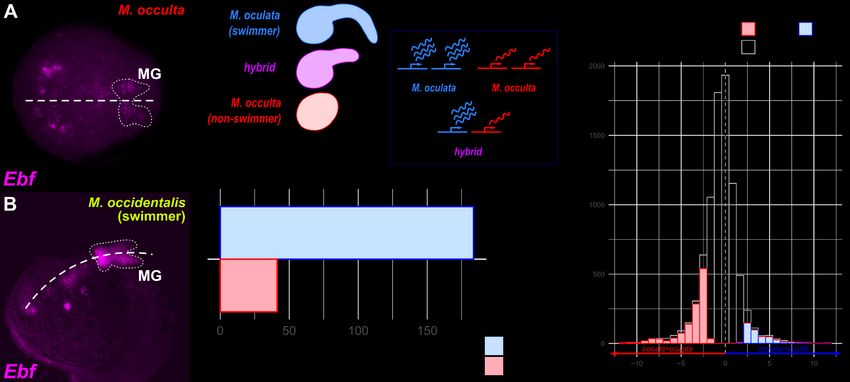
bioRxiv preprint first posted online Mar. 5, 2019; doi: http://dx.doi.org/10.1101/567719. The copyright holder for this preprint (which
          was not peer-reviewed) is the author/funder, who has granted bioRxiv a license to display the preprint in perpetuity.
                                    It is made available under a CC-BY-NC-ND 4.0 International license.

                                                                                                   Lowe et al., March 01 2019 – preprint copy - BioRxiv

expression analysis, previously sequenced RNAseq                                revealed ciliated cells lining the neuropore and the
reads from M. occulta, M. oculata and M. occulta × M.                           shortened caudal neural tube (Figure 1E). In the
oculata hybrid embryos from three different stages                              epidermis, we found that only 4 cells at the very
(Fodor et al. in preparation; Lowe et al., in                                   posterior tip of the tail bore long cilia typical of
preparation)(Lowe et al., 2014) were mapped to the                              tunicate epidermal sensory cells (Figure 1F). Swimming
M. occulta or M. oculata Ebf gene models using                                  larvae of tunicate species in several different families
Salmon v0.6.0 (Patro et al., 2015).                                             have an extended caudal nerve cored lined with
                                                                                numerous ciliated ependymal cells down the length of
Reporter construct cloning and mutagenesis                                      the tail, and bear numerous sensory neurons with
Ebf upstream cis-regulatory sequences were cloned                               elongated cilia, scattered throughout the epidermis of
from M. occulta and M. oculata genomic DNA (see                                 the head and tail (Figure 1G)(Pasini et al., 2006; Ryan
Supplemental File 1 for sequences). Mutations to                                et al., 2018; Torrence and Cloney, 1982; Vorontsova et
convert occulta sequences to oculata-like sequences                             al., 1997). Taken together, these data indicate that
and vice-versa were generated by synthesizing DNA                               both the CNS and peripheral sensory cells of the M.
fragments (Twist Bioscience) and subcloning into the                            occulta larva are only partially formed in comparison
full-length reporter plasmids (see Supplemental File 1                          to typical solitary tunicate larvae, consistent with the
for sequences).                                                                 hypothesis that a fully functional larval nervous system
                                                                                may be dispensable for this species.
Ciona robusta electroporation
Ciona robusta (i.e. intestinalis Type A) adults were                            To survey CNS development in M. occulta embryos, we
collected and shipped by M-REP (San Diego, CA). Eggs                            performed whole-mount mRNA in situ hybridizations
were obtained from dissected adults and                                         for genes that have been well characterized during
dechorionated, fertilized, electroporated, and fixed as                         CNS development in other tunicates with swimming
previously described (Christiaen et al., 2009a, b; Stolfi                       larvae, notably Ciona spp., Halocynthia roretzi, and
et al., 2014b).                                                                 Molgula occidentalis. The first candidates we analyzed
                                                                                were Celf3/4/5 (also known as ETR-1) Onecut, and
Results and discussion                                                          Neurogenin, all relatively broad markers of neuronal
                                                                                fate in swimming tunicate larvae (Figure 2A-
The nervous system of M. occulta larvae                                         C)(D'Aniello et al., 2011; Imai et al., 2009; Lowe and
M. occulta (Figure 1A) and M. oculata (Figure 1B) are                           Stolfi, 2018; Satou et al., 2001; Yagi and Makabe,
closely related species that occur sympatrically off the                        2001). Their mRNA in situ patterns clearly revealed the
coast of Brittany, France. In spite of their close genetic                      developing embryonic neural tube around the tailbud
similarity and their ability to form interspecific hybrids                      stage (~7.5-9.5 hours post-fertilization, hpf). Although
(Swalla and Jeffery, 1990), the larvae of M. occulta are                        M. occulta embryos are more difficult to orient given
tail-less and non-swimming (Figure 1C). Given its                               their nearly perfect spherical shape, the expression
inability to swim, we asked whether the M. occulta                              patterns formed the basic outline of the brain and MG,
larva has a functional nervous system or whether it has                         appearing quite conserved relative to the orthologous
been partially lost in evolution like its vestigial                             expression patterns in swimming species, especially M.
notochord, muscles, and pigment cells. Confocal                                 occidentalis (Lowe and Stolfi, 2018).
imaging of phalloidin-stained hatching M. occulta
larvae revealed that they possess a dorsal hollow                               Unfortunately, we were unable to visualize M. occulta
neural tube, though it is shortened along the anterior-                         neurons in greater detail using fluorescent reporter
posterior (A-P) axis due to the lack of an extended tail                        plasmids, as we have done in Ciona and M. occidentalis
(Figure 1D). Acetylated tubulin immunohistochemistry                            (Lowe and Stolfi, 2018; Stolfi and Levine, 2011). This is

                                                                                                                                                     4
bioRxiv preprint first posted online Mar. 5, 2019; doi: http://dx.doi.org/10.1101/567719. The copyright holder for this preprint (which
was not peer-reviewed) is the author/funder, who has granted bioRxiv a license to display the preprint in perpetuity.
It is made available under a CC-BY-NC-ND 4.0 International license.
Lowe et al., March 01 2019 – preprint copy - BioRxiv
FIGURE 1 – Development and morphology of the non-swimming larvae of Molgula occulta
A) Hatching larva of non-swimming species Molgula occulta, squeezing out of the chorion (pink dotted outline). B) Hatched larva of swimming species
Molgula oculata. C) Hatching M. occulta larva showing twenty notochord cells (purple) that fail to undergo convergent extension. D) M. occulta larva
stained with phalloidin conjugate (phall, red), anti-acetylated alpha tubulin antibody (AcTub, green), and DAPI (blue), showing presence of acetylated
alpha tubulin-rich cilia lining the neuropore/stomodeum and neural tube lumen, and tail tip epidermal cells. E) Inverted monochrome image of
acetylated alpha tubulin stain in D). F) M. occulta larva stained with anti-acetylated alpha tubulin antibody (AcTub, green) and phalloidin conjugate
(magenta). Inset in F’ shows short cilia of epidermal cells covering the majority of the larva. Inset in F” shows longer cilia (presumably sensory) of four
tail tip cells. G) Swimming larva of Ciona robusta stained with same anti-acetylated alpha tubulin antibody (AcTub, green), showing abundance of
ciliated epidermal sensory cells and neural tube ependymal cells along the entire length of the larva. Scale bars in A-D: 25 µm. Scale bars in F: 5 µm.
Scale bar in G: 75 µm.
because M. occulta eggs are extremely difficult to fully that form the bulk of the synaptic connections within
dechorionate and electroporate. Although we the MG, as well as the majority of neuromuscular
previously reported obtaining a rare electroporated M. synapses (Ryan et al., 2016). From now on, we will
occulta embryo (Stolfi et al., 2014b), our low success refer to the core MG neurons on only one side of the
rate and the limited geographic range and spawning larva, with the implicit understanding that our
season of M. occulta/oculata means that we have yet discussion encompasses each left/right pair. We have
to perform routine transfection of these species. previously characterized the core cell lineages of the
Under these conditions, we proceeded with our MG in Ciona and M. occidentalis, documenting the
analysis using mainly in situ hybridization and next- highly conserved, invariant specification of each MG
generation sequencing in M. occulta embryos, and neuron subtype and their diagnostic marker gene
heterologous reporter construct assays in C. robusta expression patterns (Imai et al., 2009; Lowe and Stolfi,
embryos. 2018; Stolfi and Levine, 2011; Stolfi et al., 2011). If the
major function of the MG in tunicate larvae is to simply
Gene expression patterns in the M. occidentalis Motor drive swimming behavior, it is likely of little adaptive
Ganglion value and therefore likely to be lost in a non-swimming
In Ciona, the swimming and escape-response larva like that of M. occulta. On the other hand, MG
behaviors of the larva are controlled by the MG, a progenitors might also contribute to the adult nervous
central pattern generator comprised of 22 total system, or differentiated MG neurons might be
neurons (Nishino et al., 2010; Ryan et al., 2016). Within carrying out other functions beyond swimming, such
the MG, there is a core of 8 left/right pairs of neurons as triggering metamorphosis. We therefore sought to
5bioRxiv preprint first posted online Mar. 5, 2019; doi: http://dx.doi.org/10.1101/567719. The copyright holder for this preprint (which
was not peer-reviewed) is the author/funder, who has granted bioRxiv a license to display the preprint in perpetuity.
It is made available under a CC-BY-NC-ND 4.0 International license.
Lowe et al., March 01 2019 – preprint copy - BioRxiv
characterize the expression patterns of candidate
regulators of MG patterning in M. occulta embryos.
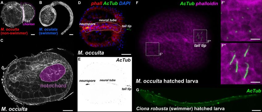
Motor neurons MN1 and MN2
To ask whether larval motor neurons are specified in
M. occulta, we performed in situ hybridization with
Mnx, a conserved regulator of motor neuron fate
(Ferrier et al., 2001) which in Ciona and M. occidentalis
labels the two major motor neurons of the MG, MN1
and MN2 (Ryan et al., 2016). To our surprise, we
detected exactly two left/right pairs of cells expressing
Mnx in M. occulta tailbud embryos (Figure 2D). This
staining appeared identical to Mnx expression in Ciona
and M. occidentalis, with a gap between the two cells
on either side where Interneuron 2 (IN2) should be.
This suggests that M. occulta embryos specify both
major motor neurons found in swimming species.
To possibly distinguish the specification of the two
motor neuron subtypes, we did in situ hybridization for
Nk6 and Islet, markers specifically for MN1 and MN2
FIGURE 2 – Expression patterns in the developing CNS of Molgula occulta
respectively. Nk6 strongly labeled one or two cells on A) Fluorescent whole mount in situ hybridization (ISH) in tailbud embryos
either side of the MG (Figure 2E), similar to its for transcripts of neural marker gene Celf3/4/5, also known as ETR-1,
expression in M. occidentalis (Lowe and Stolfi, 2018). showing the developing nervous system including brain, motor ganglion
and palps. Embryonic dorsal midline indicated by white dashed line.
These two cells likely correspond to MN1 and IN2, Embryo outline indicated by green dashed line. B) ISH for neural
based on their positions and the M. occidentalis regulatory gene Onecut, showing brain and motor ganglion (MG)-specific
pattern. In contrast, we were unable to clearly identify expression. Onecut-expressing MG cells outlined by dotted line. C) ISH
for proneural bHLH gene Neurogenin, showing brain and motor ganglion
MN2 based on Islet expression, as this gene was (MG)-specific expression. Neurogenin-expressing MG cells outlined by
expressed in a mass of cells in the middle of the dotted line. D) ISH for motor neuron-specific marker Mnx, showing
embryo, confounding any hope of distinguishing MN2 expression marking putative Motor Neuron 1 (MN1) and Motor Neuron
2 (MN2) bilateral pairs. E) ISH for Nk6, showing MN1-specific expression
(Figure 2F). This mass of cells undoubtedly and potentially weak expression in MG Interneuron 2 (IN2) on one side.
corresponds to notochord cells, which do not F) ISH for Islet, showing expression in disorganized notochord cells, and
intercalate in M. occulta (Figure 1C). In Ciona and M. potentially in one MN2 cell. G) ISH for MG interneuron marker gene Vsx,
showing expression in MG Interneuron 1 (IN1) and MG interneuron 2
occidentalis, Islet is also strongly expressed in (IN2) bilateral pairs. H) ISH for descending decussating neuron (ddN)-
notochord cells, but because these converge and specific marker Dmbx. I) Cartoon diagram of MG neuron subtype
extend in a very orderly manner, MN2 is clearly organization in M. occulta based on ISH data and comparisons to the MG
of swimming species, namely Ciona robusta and Molgula occidentalis.
distinguishable as an Islet-expressing cell in the Dorsal midline in B-E and G-H indicated by dashed line. Scale bars: 25 µm
developing MG just dorsal to the notochord (Lowe and
Stolfi, 2018; Stolfi and Levine, 2011). We were not able
MG Interneurons 1 and 2 (MGIN1 and MGIN2)
to visualize such clear Islet expression in MN2, but we
In the Ciona MG connectome, three pairs of
conclude that MN1 and MN2 are indeed specified,
descending interneurons are predicted to play an
based on the combined Nk6 and Mnx expression
important role in both the rhythmicity of swimming
patterns.
movements and their modulation by inputs from the
6bioRxiv preprint first posted online Mar. 5, 2019; doi: http://dx.doi.org/10.1101/567719. The copyright holder for this preprint (which
was not peer-reviewed) is the author/funder, who has granted bioRxiv a license to display the preprint in perpetuity.
It is made available under a CC-BY-NC-ND 4.0 International license.
Lowe et al., March 01 2019 – preprint copy - BioRxiv
sensory vesicle (Kourakis et al., 2019; Ryan et al., 2016; (Figure 2H), likely corresponding to the ddNs or their
Salas et al., 2018). Of these, the best studied are immediate progenitors, the A11.120 cells. We
MGIN1 and MGIN2 (referred to from now on as IN1 conclude that the M. occulta larva specifies ddNs
and IN2 respectively), which flank MN1. Both are despite being physically incapable of engaging in
presumed excitatory interneurons that arise from the escape response maneuvers. Taken together, these
A9.30 lineage (Cole and Meinertzhagen, 2004). In data reveal that neuronal subtypes are arrayed in the
Ciona and M. occidentalis, they are marked by M. occulta MG in a manner that is identical to that
expression of Vsx (Imai et al., 2009; Stolfi and Levine, previously described in swimming species such as M.
2011), the ortholog of conserved spinal cord occidentalis and Ciona spp. (Figure 2I). This suggests
interneuron regulator Vsx2/Chx10 (Altun-Gultekin et that, in spite of the non-swimming nature of the M.
al., 2001; Kimura et al., 2006; Liu et al., 1994). occulta larva, MG neuron specification and patterning
have not been evolutionarily lost.
We found that in M. occulta, Vsx labels two cells on
either side of the neural tube at the tailbud stage Reduced expression of Ebf as a potential mechanism of
(Figure 2G). This is similar to the orthologous impaired MG neuron differentiation
expression patterns in Ciona and M. occidentalis. In Although developmental patterning of the MG
Ciona, Vsx is activated in IN1 and IN2 at two different appears to be conserved in the non-swimming larva of
time points (Stolfi and Levine, 2011). IN2 is the first M. occulta, we further pursued the hypothesis that
interneuron to differentiate, and activates Vsx early, perhaps the differentiation of specific MG neurons
before IN1 is even born Later, after IN1 is born and may not occur in M. occulta, similar to how the
specified, Vsx is activated in this cell. In M. occidentalis, differentiation (but not the specification) of
we showed that Vsx expression appears concurrently notochord, tail muscle, and otolith cells have been lost.
in IN2 and in a progenitor cell that will give rise to IN1, To address this possibility, we analyzed other genes
indicating a temporal shift towards precocious known to be involved in motor neuron differentiation.
activation of Vsx in the IN1 lineage (Lowe and Stolfi, One of these is the transcription factor Ebf (also known
2018). Here, we were unable to ascertain the relative as Collier/Olf/EBF or COE), a conserved “terminal
timing of Vsx expression and the identities of the Vsx- selector” (Hobert, 2008) for cholinergic motor neurons
expressing cells but can conclude that IN1 and IN2 (Kratsios et al., 2012). In Ciona, Ebf is required for MN2
specification is conserved in M. occulta. differentiation (Stolfi et al., 2014a) and is sufficient to
activate cholinergic gene expression when ectopically
Descending decussating neurons expressed in other cell types (Kratsios et al., 2012).
The descending decussating neuron (ddN) is a highly When we looked at Ebf expression in M. occulta, we
unique neuron at the periphery of the core MG in both noticed that it appeared substantially weaker in the
Ciona and M. occidentalis. It is the only descending MG (but not in other cells) compared to its
neuron that decussates, or projects its axon orthologous expression in M. occidentalis, although
contralaterally (as the name implies). Due to its this assay was not quantitative (Figure 3A,B). Since Ebf
position and connectivity within the Ciona MG, it was regulates key steps between specification and
proposed to be the homolog of vertebrate Mauthner differentiation in motor neurons, we hypothesized
cells and to function in a homologous escape response that Ebf expression might have been evolutionarily lost
pathway (Ryan et al., 2017; Takamura et al., 2010). In in the vestigial MG of M. occulta. To test this further,
Ciona and M. occidentalis, the ddN is marked by we turned to a more quantitative interspecies
expression of Dmbx (Lowe and Stolfi, 2018; Takahashi comparison of gene expression.
and Holland, 2004). In M. occulta tailbud embryos,
Dmbx was found to be expressed in a single pair of cells
7bioRxiv preprint first posted online Mar. 5, 2019; doi: http://dx.doi.org/10.1101/567719. The copyright holder for this preprint (which
was not peer-reviewed) is the author/funder, who has granted bioRxiv a license to display the preprint in perpetuity.
It is made available under a CC-BY-NC-ND 4.0 International license.
Lowe et al., March 01 2019 – preprint copy - BioRxiv
FIGURE 3 – Loss of MG-specific transcriptional activity of the terminal selector gene Ebf in M. occulta
A) Fluorescent whole mount in situ hybridization (ISH) for the Ebf gene in tailbud-stage M. occulta embryo. MG outlined by dotted line, showing
relatively weak expression of Ebf, compared to its expression in other cells in the same embryo, and to its expression in swimming species (see panel
B). B) ISH for Ebf in tailbud stage embryo of the swimming species Molgula occidentalis, showing strong expression in the MG (dotted outline). C)
Diagram of RNAseq analysis of hybrid embryos, produced by fertilizing eggs of non-swimmer M. occulta with sperm from swimmer M. oculata (left). If
reduced transcriptional activity of Ebf in M. occulta is due to evolutionary changes in cis, the hybrid should show differential allele-specific Ebf
expression that recapitulates the differential expression of Ebf observed between the parental species. D) RNAseq read counts from libraries prepared
from pooled hybrid embryos at 6 hours post-fertilization (hpf), showing greater effective read count representing the M. oculata (swimmer) Ebf allele
relative to the M. occulta (non-swimmer) allele, for a Log2FC of 3.6 (p = 0.0001). Embryonic dorsal midline in A and B indicated by dashed line. E)
Distribution of genes that are differentially expressed in hybrid embryos at 6 hpf (Log2FC>1.5, pbioRxiv preprint first posted online Mar. 5, 2019; doi: http://dx.doi.org/10.1101/567719. The copyright holder for this preprint (which
was not peer-reviewed) is the author/funder, who has granted bioRxiv a license to display the preprint in perpetuity.
It is made available under a CC-BY-NC-ND 4.0 International license.
Lowe et al., March 01 2019 – preprint copy - BioRxiv
FIGURE 4 – Differential activity of Ebf reporter plasmids in Ciona robusta electroporation assay
A) Ciona robusta larva electroporated with a mixture of M. oculata Ebf>YFP (green, top) and M. oculata Ebf>mCherry (magenta, bottom) plasmids,
showing perfect co-expression in the motor ganglion (MG), and in other neurons including Bipolar Tail Neurons (BTNs, insets). B) Ciona robusta larva
electroporated with a mixture of M. oculata Ebf>YFP (green, top) and M. occulta Ebf>mCherry (magenta, bottom) plasmids, showing absence of
mCherry expression specifically in the MG (region indicated by dashed circle). In contrast, co-expression of YFP and mCherry in BTNs (insets) shows
that reporter plasmid activity in other neurons is conserved. Scale bars in A: 25 µm. All panels and insets are at the same respective scales.
robusta, which can be routinely electroporated in the in MG neurons, without affecting its expression in
laboratory with plasmid DNA (Zeller, 2018). When we other neurons.
electroporated M. oculata Ebf reporter constructs
(oculata.Ebf -3654/+24>XFP) into Ciona embryos, we To identify the potential cis-regulatory changes
observed reporter gene expression in MG neurons, underlying this difference in Ebf activation, we aligned
Bipolar Tail Neurons (BTNs), and some brain neurons 735 bp of non-coding sequence immediately 5’ of M.
(Figure 4A). Although there is no non-coding sequence oculata Ebf start codon to the orthologous sequence
conservation between Molgula and Ciona, and acute in M. occulta (Figure 5A). The aligned sequences share
developmental system drift has rendered many >90% identity, consistent with the close genetic
Molgula cis-regulatory sequences inactive in Ciona relationship between these two species. This
(Stolfi et al., 2014b), in this case there was enough alignment revealed an unusually divergent sequence
conservation of the underlying regulatory logic to motif ~470 bp upstream of the Ebf translation start
recapitulate the expression of Ebf in these various codon (Figure 5B). This sequence was of particular
Ciona neuronal subtypes. interest to us because these changes were observed
adjacent to a conserved E-box site that might be a
When we electroporated the corresponding M. binding site for Neurogenin. In C. robusta, Ebf is
occulta constructs (occulta.Ebf -3659/+24>XFP), activated in the MG by the proneural bHLH factor
reporter gene expression was observed in all the same Neurogenin (Imai et al., 2009). We identified a
neurons except MG neurons (Figure 4B) Thus, our conserved E-box site in experimentally validated
heterologous reporter plasmid assays in Ciona upstream Ebf cis-regulatory sequences isolated from
indicated that M. occulta-specific cis-regulatory C. robusta, C. savignyi, and M. occidentalis
changes might underlie lower levels of Ebf expression (Supplemental File 1), even though these sequences
show poor overall conservation with the M.
9bioRxiv preprint first posted online Mar. 5, 2019; doi: http://dx.doi.org/10.1101/567719. The copyright holder for this preprint (which
was not peer-reviewed) is the author/funder, who has granted bioRxiv a license to display the preprint in perpetuity.
It is made available under a CC-BY-NC-ND 4.0 International license.
Lowe et al., March 01 2019 – preprint copy - BioRxiv
FIGURE 5 – A cis-regulatory change underlying MG-specific loss of Ebf expression in M. occulta
A) Alignment of sequences 5’ upstream of Ebf in M. oculata and M. occulta. B) Sequence from boxed area in A, showing highly conserved sequences
including a candidate E-box site potentially bound by Neurogenin, adjacent to a highly divergent motif (purple font). C) Mutated sequences in
Mocu→Mocc>GFP and Mocc→Mocu>GFP constructs in which the highly divergent motif has been swapped between M. oculata and M. occulta Ebf
reporter constructs. D) Results of scoring Ciona robusta larvae electroporated with the wild-type parental Ebf reporter plasmids and the mutated
“swapped” constructs. Changing the M. oculata divergent motif sequence to the M. occulta sequence results in loss of GFP expression specifically in
the motor ganglion, but not in brain or bipolar tail neurons (BTNs). The converse mutation, changing the M. occulta motif to resemble the M. oculata
sequence was not sufficient to restore GFP expression in the motor ganglion. n = 50 embryos for each construct.
oculata/occulta sequences. The presence of an E-box transcriptional activities of swimming vs. non-
within ~800 bp of the start codon in all tunicate species swimming Molgula Ebf loci.
surveyed suggested this region might be part of a
functionally conserved minimal enhancer for MG- To directly test whether M. occulta-specific sequence
specific Ebf regulation. When we compared all 3 changes are responsible for the loss of M. occulta Ebf
Molgula sequences, we found that this E-box-adjacent transcription in the MG, we mutated the E-box-
motif (AGATGGC) was conserved in both M. oculata adjacent sequence in oculata.Ebf -3654/+24>GFP to
and the more distantly related M. occidentalis, but not match the M. occulta sequence (CGAGTGAGATG to
in M. occulta. Because the larvae of M. occidentalis are TCAGCAAAACGAT)(Figure 5C). When we
also swimming, and because this M. occidentalis Ebf electroporated this mutated reporter plasmid (Mocu--
cis-regulatory sequence also drives reporter gene >Mocc>GFP) in Ciona, we found that its transcriptional
expression in C. robusta MG neurons (Stolfi et al., activity was almost completely abolished specifically in
2014b), we hypothesized that the E-box-adjacent the MG, but had little effect on reporter gene
motif may be crucial for MG-specific expression. expression in other neurons (Figure 5D). These data
Therefore, we decided to test whether the loss of the suggest that the mutated motif is necessary for Ebf
E-box-adjacent motif might explain the different expression in MG neurons in M. oculata. Alternatively,
the M. occulta-specific sequence might have created a
10bioRxiv preprint first posted online Mar. 5, 2019; doi: http://dx.doi.org/10.1101/567719. The copyright holder for this preprint (which
was not peer-reviewed) is the author/funder, who has granted bioRxiv a license to display the preprint in perpetuity.
It is made available under a CC-BY-NC-ND 4.0 International license.
Lowe et al., March 01 2019 – preprint copy - BioRxiv
novel binding site for a MG-specific repressor, but the Using cis-regulatory mutations and heterologous
presence of the E-box-adjacent in M. occidentalis reporter plasmid assays, we demonstrate specific
supports the former explanation. Although we changes to conserved cis-regulatory sequences
attempted to “resurrect” the putative ancestral upstream of M. occulta Ebf have affected its
sequence by mutating the E-box-adjacent site in expression in MG neurons, but not in other neurons or
occulta.Ebf -3659/+24>GFP to resemble the M. cell types. If these results hold, this would represent a
oculata sequence (Mocc-->Mocu>GFP), this was not distinct example of tissue-specific gene expression loss
sufficient to restore activity, hinting at the in this species. Previously, it has been shown that
accumulation of multiple loss-of-function (or evolutionary loss of functional larval structures in M.
compensatory) mutations in other parts of the M. occulta correlates with loss-of-function non-coding
occulta Ebf cis-regulatory sequence. mutations resulting in the loss of crucial proteins such
as muscle actin for tail muscle activity (Kusakabe et al.,
Conclusions 1996) or Tyrosinase for melanin pigment synthesis in
the gravity-sensing otolith organ (Racioppi et al.,
We have investigated the molecular signatures of MG 2017). However, these proteins are only expressed in
neuron specification and differentiation in a non- larval structures involved in larval motility (tail muscle
swimming molgulid tunicate larva, finding a surprising cells and otolith, respectively), being apparently
conservation of cell-specific expression patterns of key dispensable for the development of other larval and
developmental regulators during MG development. adult tissues. In contrast, M. occulta Ebf is not a
This could indicate that at least some MG neurons pseudogene and is predicted to encode a fully
might be specified and might differentiate in M. functional protein. This difference is likely because Ebf
occulta in spite of the evolutionary loss of the larval tail is a regulator that is required for the development of
and swimming behavior. Unfortunately, we were myriad cell types throughout the life cycle of M.
unable to investigate neuronal morphology using occulta, especially in the sessile adults of this species,
reporter plasmids, due to our inability to routinely which are morphologically similar to adults of other
transfect M. occulta embryos. Once this method is species, swimming and non-swimming alike. Thus,
established, it will be interesting to revisit this question even if Ebf expression is not needed in MG motor
to fully document the extent of MG development in neurons (in the absence of functional larval muscles),
this species. Nonetheless, MG neuron function is likely its requirement in other tissues may have resulted
seriously impaired in M. occulta, given the reduced instead in the evolutionary loss of tissue-specific cis-
levels of Ebf expression in M. occulta and of the M. regulatory elements, as our data indicate. It may be
occulta Ebf allele in interspecific hybrids. In addition to that the mechanisms we show here underlying cell-
activating the expression of MG neuron-specific specific loss of Ebf expression in M. occulta are
regulators like Islet (Stolfi et al., 2014a), Ebf has been representative of constraints acting on traits that are
shown to act as a conserved “terminal selector” that lost from a particular developmental stage, but that
regulates the terminal differentiation of cholinergic are still retained in a different stage of the same
motor neurons throughout Metazoa, including in the organism’s life cycle. Studying these losses and the
tunicate C. robusta (Kratsios et al., 2012). We mechanisms underlying such losses will complement
therefore expect that reduced Ebf expression in the M. the principles learned from studies of traits that are
occulta MG would likely result in similarly reduced or dispensable to the entire life cycle of a particular
abrogated expression of cholinergic effectors and species, such as the loss of vision in cave-dwelling
other cellular machinery required for motor neuron organisms.
function.
11bioRxiv preprint first posted online Mar. 5, 2019; doi: http://dx.doi.org/10.1101/567719. The copyright holder for this preprint (which
was not peer-reviewed) is the author/funder, who has granted bioRxiv a license to display the preprint in perpetuity.
It is made available under a CC-BY-NC-ND 4.0 International license.
Lowe et al., March 01 2019 – preprint copy - BioRxiv
Is the evolutionary loss of MG development an HD084814 to AS, by start-up package from the College
ongoing process in M. occulta? Might these processes or Arts and Science at New York University and NIH
break down further, as has happened with the loss of award R01 GM096032 to LC, by France BioImaging
genes encoding melanogenesis or muscle structural infrastructure ANR-10-INBS-04 to NP, and by a
proteins in this same species? Or are there special University of Washington Royalty Research Fund
constraints that have maintained the neuron subtype- award (A118261) to BJS. The collaborative MoEvoDevo
specific marker gene expression we see in the MG? Has network is supported by ASSEMBLE (Association of
MG neuron subtype-specific gene expression persisted European Marine Biological Laboratories) MARINE. CR
because the same regulatory logic is required for was supported by a long-term fellowship ALTF 1608-
expression in adult neurons or other tissues? 2014 from EMBO and by a travel grant from the
Alternatively, we know that some MG precursors Boehringer Ingelheim Fonds. This material is also
remain undifferentiated in the larva but can contribute based in part upon collaborative work by BJS and EKL
to the adult nervous system (Horie et al., 2011). and C. Titus Brown, supported by the National Science
Perhaps the cell-specific transcription factor Foundation under Cooperative Agreement No. DBI-
expression we observe in the M. occulta MG reflects 0939454 BEACON, A Center for the Study of Evolution
shared mechanisms of patterning both differentiated in Action. Any opinions, findings, and conclusions or
larval neurons and undifferentiated adult neural recommendations expressed in this material are those
precursors. Another intriguing possibility is that the of the author(s) and do not necessarily reflect the
tunicate MG is required for other larval behaviors views of the National Science Foundation.
beyond swimming. For instance, perhaps MG neurons
are required for triggering developmental processes References
during the metamorphosis of the larva into a juvenile.
In this scenario, specific MG neuron subtypes might Albalat, R., Cañestro, C.J.N.R.G., 2016. Evolution by gene loss. 17, 379.
Altun-Gultekin, Z., Andachi, Y., Tsalik, E.L., Pilgrim, D., Kohara, Y., Hobert, O., 2001.
still be necessary for the proper metamorphosis of M. A regulatory cascade of three homeobox genes, ceh-10, ttx-3 and ceh-23,
occulta larvae, even if Ebf-dependent activation of controls cell fate specification of a defined interneuron class in C. elegans.
Development 128, 1951-1969.
motor neuron effectors in the MG is not required and
Berrill, N.J., 1931. Studies in tunicate development. Part II. Abbreviation of
might have even been selected against. A deeper development in the Molgulidae. Philosophical Transactions of the Royal
understanding of the mechanisms underlying both Society of London. Series B, Containing Papers of a Biological Character 219,
281-346.
larval and adult neurogenesis in tunicates, and the role
Bourlat, S.J., Juliusdottir, T., Lowe, C.J., Freeman, R., Aronowicz, J., Kirschner, M.,
of neural activity in the transition to the adult phase, Lander, E.S., Thorndyke, M., Nakano, H., Kohn, A.B., Heyland, A., Moroz, L.L.,
will be needed to answer these and other questions Copley, R.R., Telford, M.J., 2006. Deuterostome phylogeny reveals
monophyletic chordates and the new phylum Xenoturbellida. Nature 444,
related to the loss of larval-specific structures in non- 85.
swimming species. Brozovic, M., Dantec, C., Dardaillon, J., Dauga, D., Faure, E., Gineste, M., Louis, A.,
Naville, M., Nitta, K.R., Piette, J., Reeves, W., Scornavacca, C., Simion, P.,
Vincentelli, R., Bellec, M., Aicha, S.B., Fagotto, M., Guéroult-Bellone, M.,
Acknowledgments Haeussler, M., Jacox, E., Lowe, E.K., Mendez, M., Roberge, A., Stolfi, A.,
Yokomori, R., Brown, C T., Cambillau, C., Christiaen, L., Delsuc, F., Douzery,
E., Dumollard, R., Kusakabe, T., Nakai, K., Nishida, H., Satou, Y., Swalla, B.,
We dedicate this manuscript to the memory of Veeman, M., Volff, J.-N., Lemaire, P., 2018. ANISEED 2017: extending the
integrated ascidian database to the exploration and evolutionary
Alexander “Zander” Fodor. We thank C. Titus Brown comparison of genome-scale datasets. Nucleic Acids Research 46, D718-
for his encouragement and continued support. We D725.
thank Stéphane Hourdez, Sophie Booker, and Xavier Christiaen, L., Wagner, E., Shi, W., Levine, M., 2009a. Electroporation of transgenic
DNAs in the sea squirt Ciona. Cold Spring Harbor Protocols 2009, pdb.
Bailly of the Station Biologique for help in carrying out prot5345.
experiments at Roscoff. We thank Susanne Gibboney Christiaen, L., Wagner, E., Shi, W., Levine, M., 2009b. Isolation of sea squirt (Ciona)
gametes, fertilization, dechorionation, and development. Cold Spring Harbor
for assistance with cloning Molgula Ebf reporter Protocols 2009, pdb. prot5344.
plasmids. This work was supported by NIH award R00
12bioRxiv preprint first posted online Mar. 5, 2019; doi: http://dx.doi.org/10.1101/567719. The copyright holder for this preprint (which
was not peer-reviewed) is the author/funder, who has granted bioRxiv a license to display the preprint in perpetuity.
It is made available under a CC-BY-NC-ND 4.0 International license.
Lowe et al., March 01 2019 – preprint copy - BioRxiv
Cole, A.G., Meinertzhagen, I.A., 2004. The central nervous system of the ascidian Li, H., Handsaker, B., Wysoker, A., Fennell, T., Ruan, J., Homer, N., Marth, G.,
larva: mitotic history of cells forming the neural tube in late embryonic Ciona Abecasis, G., Durbin, R., 2009. The sequence alignment/map format and
intestinalis. Developmental biology 271, 239-262. SAMtools. Bioinformatics 25, 2078-2079.
D'Aniello, E., Pezzotti, M.R., Locascio, A., Branno, M., 2011. Onecut is a direct Liu, I.S., Ploder, L., Vidgen, D., van der Kooy, D., Kalnins, V.I., Mclnnes, R.R., 1994.
neural-specific transcriptional activator of Rx in Ciona intestinalis. Developmental expression of a novel murine homeobox gene (Chx10):
Developmental biology 355, 358-371. evidence for roles in determination of the neuroretina and inner nuclear
Denoeud, F., Henriet, S., Mungpakdee, S., Aury, J.-M., Da Silva, C., Brinkmann, H., layer. Neuron 13, 377-393.
Mikhaleva, J., Olsen, L.C., Jubin, C., Cañestro, C.J.S., 2010. Plasticity of animal Liu, M., Adelman, Z.N., Myles, K.M., Zhang, L., 2014. A Transcriptome Post-
genome architecture unmasked by rapid evolution of a pelagic tunicate. Scaffolding Method for Assembling High Quality Contigs. Computational
330, 1381-1385. Biology Journal 2014, 4.
Ferrier, D.E., Brooke, N.M., Panopoulou, G., Holland, P.W., 2001. The Mnx Lowe, E.K., Stolfi, A., 2018. Developmental system drift in motor ganglion
homeobox gene class defined by HB9, MNR2 and amphioxus AmphiMnx. patterning between distantly related tunicates. EvoDevo 9, 18.
Development Genes & Evolution 211. Lowe, E.K., Swalla, B.J., Brown, C.T., 2014. Evaluating a lightweight transcriptome
Grabherr, M.G., Haas, B.J., Yassour, M., Levin, J.Z., Thompson, D.A., Amit, I., assembly pipeline on two closely related ascidian species. PeerJ PrePrints.
Adiconis, X., Fan, L., Raychowdhury, R., Zeng, Q., Chen, Z., Mauceli, E., Maliska, M.E., Pennell, M.W., Swalla, B.J., 2013. Developmental mode influences
Hacohen, N., Gnirke, A., Rhind, N., di Palma, F., Birren, B.W., Nusbaum, C., diversification in ascidians. Biology letters 9, 20130068.
Lindblad-Toh, K., Friedman, N., Regev, A., 2011. Full-length transcriptome
assembly from RNA-Seq data without a reference genome. Nature Nishino, A., 2018. Morphology and Physiology of the Ascidian Nervous Systems
biotechnology 29, 644-652. and the Effectors, Transgenic Ascidians. Springer, pp. 179-196.
Hadfield, K.A., Swalla, B.J., Jeffery, W.R., 1995. Multiple origins of anural Nishino, A., Okamura, Y., Piscopo, S., Brown, E.R., 2010. A glycine receptor is
development in ascidians inferred from rDNA sequences. Journal of involved in the organization of swimming movements in an invertebrate
molecular evolution 40, 413-427. chordate. BMC neuroscience 11, 6.
Hobert, O., 2008. Regulatory logic of neuronal diversity: terminal selector genes Pasini, A., Amiel, A., Rothbächer, U., Roure, A., Lemaire, P., Darras, S., 2006.
and selector motifs. Proceedings of the National Academy of Sciences 105, Formation of the ascidian epidermal sensory neurons: insights into the origin
20067-20071. of the chordate peripheral nervous system. PLoS biology 4, e225.
Horie, T., Shinki, R., Ogura, Y., Kusakabe, T.G., Satoh, N., Sasakura, Y.J.N., 2011. Patro, R., Duggal, G., Kingsford, C., 2015. Salmon: accurate, versatile and ultrafast
Ependymal cells of chordate larvae are stem-like cells that form the adult quantification from RNA-seq data using lightweight-alignment. Biorxiv,
nervous system. 469, 525. 021592.
Huber, J.L., da Silva, K.B., Bates, W.R., Swalla, B.J., 2000. The evolution of anural Pennati, R., Ficetola, G.F., Brunetti, R., Caicci, F., Gasparini, F., Griggio, F., Sato, A.,
larvae in molgulid ascidians. Seminars in Cell & Developmental Biology 11, Stach, T., Kaul-Strehlow, S., Gissi, C., Manni, L., 2015. Morphological
419-426. Differences between Larvae of the Ciona intestinalis Species Complex: Hints
for a Valid Taxonomic Definition of Distinct Species. PLOS ONE 10, e0122879.
Ikuta, T., Saiga, H., 2007. Dynamic change in the expression of developmental
genes in the ascidian central nervous system: revisit to the tripartite model Porter, M.L., Crandall, K.A.J.T.i.E., Evolution, 2003. Lost along the way: the
and the origin of the midbrain-hindbrain boundary region. Dev Biol 312. significance of evolution in reverse. 18, 541-547.
Imai, K.S., Stolfi, A., Levine, M., Satou, Y., 2009. Gene regulatory networks Pryszcz, L.P., Gabaldón, T., 2016. Redundans: an assembly pipeline for highly
underlying the compartmentalization of the Ciona central nervous system. heterozygous genomes. Nucleic acids research 44, e113-e113.
Development 136, 285-293. Racioppi, C., Valoroso, M.C., Coppola, U., Lowe, E.K., Brown, C.T., Swalla, B.J.,
Jeffery, W.R., Swalla, B.J., 1991. An evolutionary change in the muscle lineage of Christiaen, L., Stolfi, A., Ristoratore, F., 2017. Evolutionary loss of
an anural ascidian embryo is restored by interspecific hybridization with a melanogenesis in the tunicate Molgula occulta. EvoDevo 8, 11.
urodele ascidian. Developmental biology 145, 328-337. Rudolf, J., Dondorp, D., Canon, L., Tieo, S., Chatzigeorgiou, M., 2018. Quantitative
Jiang, D., Tresser, J.W., Horie, T., Tsuda, M., Smith, W.C., 2005. Pigmentation in analysis reveals the basic behavioural repertoire of the urochordate Ciona
the sensory organs of the ascidian larva is essential for normal behavior. intestinalis. bioRxiv, 382465.
Journal of experimental biology 208, 433-438. Ryan, K., Lu, Z., Meinertzhagen, I.A., 2016. The CNS connectome of a tadpole larva
Kim, D., Langmead, B., Salzberg, S.L., 2015. HISAT: a fast spliced aligner with low of Ciona intestinalis (L.) highlights sidedness in the brain of a chordate
memory requirements. Nature Methods 12, 357. sibling. Elife 5.
Kimura, Y., Okamura, Y., Higashijima, S.-i., 2006. alx, a zebrafish homolog of Chx10, Ryan, K., Lu, Z., Meinertzhagen, I.A., 2017. Circuit homology between decussating
marks ipsilateral descending excitatory interneurons that participate in the pathways in the Ciona larval CNS and the vertebrate startle-response
regulation of spinal locomotor circuits. Journal of Neuroscience 26, 5684- pathway. Current Biology 27, 721-728.
5697. Ryan, K., Lu, Z., Meinertzhagen, I.A., 2018. The peripheral nervous system of the
Kourakis, M., Borba, C., Zhang, A., Newman-Smith, E., Salas, P., Manjunath, B., ascidian tadpole larva: Types of neurons and their synaptic networks. Journal
Smith, W., 2019. Parallel Visual Circuitry in a Basal Chordate. bioRxiv, of Comparative Neurology 526, 583-608.
514422. Ryan, K., Meinertzhagen, I.A., 2019. Neuronal identity: the neuron types of a
Kratsios, P., Stolfi, A., Levine, M., Hobert, O., 2012. Coordinated regulation of simple chordate sibling, the tadpole larva of Ciona intestinalis. Current
cholinergic motor neuron traits through a conserved terminal selector gene. opinion in neurobiology 56, 47-60.
Nature neuroscience 15, 205. Salas, P., Vinaithirthan, V., Newman-Smith, E., Kourakis, M.J., Smith, W.C., 2018.
Kusakabe, T., Swalla, B.J., Satoh, N., Jeffery, W.R., 1996. Mechanism of an Photoreceptor specialization and the visuomotor repertoire of the primitive
evolutionary change in muscle cell differentiation in ascidians with different chordate Ciona. Journal of Experimental Biology, jeb. 177972.
modes of development. Developmental biology 174, 379-392. Sasakura, Y., 2018. Cellulose production and the evolution of the sessile lifestyle
in ascidians. Sessile Organisms 35, 21-29.
13bioRxiv preprint first posted online Mar. 5, 2019; doi: http://dx.doi.org/10.1101/567719. The copyright holder for this preprint (which
was not peer-reviewed) is the author/funder, who has granted bioRxiv a license to display the preprint in perpetuity.
It is made available under a CC-BY-NC-ND 4.0 International license.
Lowe et al., March 01 2019 – preprint copy - BioRxiv
Satou, Y., Takatori, N., Yamada, L., Mochizuki, Y., Hamaguchi, M., Ishikawa, H.,
Chiba, S., Imai, K., Kano, S., Murakami, S.D., 2001. Gene expression profiles
in Ciona intestinalis tailbud embryos. Development 128, 2893-2904.
Shenkar, N., Gittenberger, A., Lambert, G., Rius, M., Moreira Da Rocha, R., Swalla,
B.J., Turon, X., 2019. Ascidiacea World Database. Molgula Forbes, 1848.
Accessed through: World Register of Marine Species.
Stolfi, A., Gandhi, S., Salek, F., Christiaen, L., 2014a. Tissue-specific genome editing
in Ciona embryos by CRISPR/Cas9. Development 141, 4115-4120.
Stolfi, A., Levine, M., 2011. Neuronal subtype specification in the spinal cord of a
protovertebrate. Development 138, 995-1004.
Stolfi, A., Lowe, E.K., Racioppi, C., Ristoratore, F., Brown, C.T., Swalla, B.J.,
Christiaen, L., 2014b. Divergent mechanisms regulate conserved
cardiopharyngeal development and gene expression in distantly related
ascidians. Elife 3.
Stolfi, A., Wagner, E., Taliaferro, J.M., Chou, S., Levine, M., 2011. Neural tube
patterning by Ephrin, FGF and Notch signaling relays. Development 138,
5429-5439.
Swalla, B.J., Jeffery, W.R., 1990. Interspecific hybridization between an anural and
urodele ascidian: differential expression of urodele features suggests
multiple mechanisms control anural development. Developmental biology
142, 319-334.
Swalla, B.J., Jeffery, W.R., 1992. Vestigial brain melanocyte development during
embryogenesis of an anural ascidian. Development, growth & differentiation
34, 17-25.
Takahashi, T., Holland, P.W., 2004. Amphioxus and ascidian Dmbx homeobox
genes give clues to the vertebrate origins of midbrain development.
Development 131, 3285-3294.
Takamura, K., Minamida, N., Okabe, S., 2010. Neural map of the larval central
nervous system in the ascidian Ciona intestinalis. Zoological science 27, 191-
203.
Torrence, S.A., Cloney, R.A.J.Z., 1982. Nervous system of ascidian larvae: caudal
primary sensory neurons. 99, 103-115.
Tsuda, M., Sakurai, D., Goda, M., 2003. Direct evidence for the role of pigment
cells in the brain of ascidian larvae by laser ablation. Journal of Experimental
Biology 206, 1409-1417.
Vorontsova, M., Nezlin, L., Meinertzhagen, I.J.A.Z., 1997. Nervous system of the
larva of the ascidian Molgula citrina (Alder and Hancock, 1848). 78, 177-185.
Yagi, K., Makabe, K., 2001. Isolation of an early neural maker gene abundantly
expressed in the nervous system of the ascidian, Halocynthia roretzi.
Development genes and evolution 211, 49-53.
Zega, G., Thorndyke, M.C., Brown, E.R., 2006. Development of swimming
behaviour in the larva of the ascidian Ciona intestinalis. Journal of
experimental biology 209, 3405-3412.
Zeller, R.W., 2018. Electroporation in Ascidians: History, Theory and Protocols,
Transgenic Ascidians. Springer, pp. 37-48.
14You can also read