Convergent Canonical Pathways in Autism Spectrum Disorder from Proteomic, Transcriptomic and DNA Methylation Data
←
→
Page content transcription
If your browser does not render page correctly, please read the page content below
Preprints (www.preprints.org) | NOT PEER-REVIEWED | Posted: 6 September 2021                   doi:10.20944/preprints202109.0109.v1

     Review

     Convergent Canonical Pathways in Autism Spectrum Disorder
     from Proteomic, Transcriptomic and DNA Methylation Data
     Caitlyn Mahony1 and Colleen O’ Ryan1*

                            1   Molecular & Cell Biology, University of Cape Town, Private Bag, Rondebosch 7700, Cape Town, South
                            Africa
                            * Corresponding author: colleen.oryan@uct.ac.za

                                     Abstract: Autism Spectrum Disorder (ASD) is a complex neurodevelopmental disorder
                                     with extensive genetic and aetiological heterogeneity. While the underlying molecular
                                     mechanisms involved remain unclear, significant progress has been facilitated by recent
                                     advances in high-throughput transcriptomic, epigenomic and proteomic technologies.
                                     Here, we review recently published ASD proteomic data and compare proteomic func-
                                     tional enrichment signatures to those of transcriptomic and epigenomic data. We identify
                                     canonical pathways that are consistently implicated in ASD molecular data and find an
                                     enrichment of pathways involved in mitochondrial metabolism and neurogenesis. We
                                     identify a subset of differentially expressed proteins that are supported by ASD tran-
                                     scriptomic and DNA methylation data. Furthermore, these differentially expressed pro-
                                     teins are enriched for disease phenotype pathways associated with ASD aetiology. These
                                     proteins converge on protein-protein interaction networks that regulate cell proliferation
                                     and differentiation, metabolism and inflammation which demonstrates a link between ca-
                                     nonical pathways, biological processes and the ASD phenotype. This review highlights
                                     how proteomics can uncover potential molecular mechanisms to explain a link between
                                     mitochondrial dysfunction and neurodevelopmental pathology.

                                     Keywords: proteomics; transcriptomics; DNA methylation; mitochondria; metabolism;
                                     OXPHOS; ASD; neurogenesis; gliosis; neurodevelopment

           © 2021 by the author(s). Distributed under a Creative Commons CC BY license.
Preprints (www.preprints.org) | NOT PEER-REVIEWED | Posted: 6 September 2021                doi:10.20944/preprints202109.0109.v1

         Graphical Abstract

         1.   Introduction

         Autism Spectrum Disorder (ASD) is a lifelong neurodevelopmental condition that is characterized by heterogene-
         ous genetic origins and a poorly understood aetiology [1]. Although the heritability of ASD is undisputed, the ge-
         netic contribution of ASD is only 64.6% [2] to 80% [3]. The genetic architecture of ASD is comprised of highly
         penetrant, rare variants, as well as more common variants which have smaller effect sizes [4]. However, no single
         genetic variant accounts for more than 1% of ASD cases [5], with more than 90% of ASD cases being idiopathic [6].
         Recent technological advances in genomics, coupled with systems biology approaches that use functional genomic
         datasets, have facilitated remarkable progress in understanding the functional convergence of the genetic loci asso-
         ciated with ASD [7]. However, a challenge of ASD molecular research is being able to “translate gene discovery
         into an actionable understanding of ASD pathology” [8]. Transcriptomic and epigenomic approaches are widely
         used to study gene regulation and expression in ASD, and these have provided some insight into the functional
         changes at the molecular level. However, there have been fewer proteomic studies in ASD, due in part to the nu-
         merous challenges of this approach. Proteomic methodological approaches vary widely with respect to sample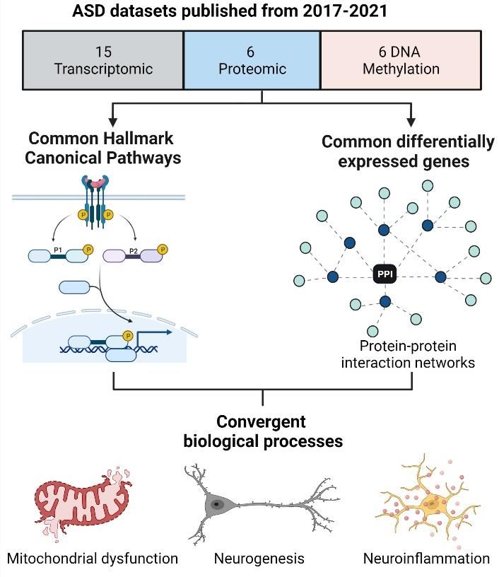
Preprints (www.preprints.org) | NOT PEER-REVIEWED | Posted: 6 September 2021                 doi:10.20944/preprints202109.0109.v1

         preparation, instrumentation and protein quantification methods, all of which culminates in studies with limited
         reproducibility [9]. Additionally, the lack of unified data repositories and standardised protein identifiers make it
         difficult to integrate results between different studies [10]. However, as the “undertakers of biological activities”
         [11], proteins directly reflect the physiological processes underpinning disease aetiology. proteins directly reflect
         the physiological processes underpinning disease aetiology. Therefore, proteomics is an essential tool to charac-
         terise the cellular mechanisms involved in ASD aetiology and to validate the changes observed in the ASD tran-
         scriptome and epigenome.

         We propose that the integration ASD proteomic data with large scale ASD transcriptomic and DNA methylation
         (DNAm) data may yield insight into the molecular mechanisms of ASD. Despite the aetiological and genetic com-
         plexity of ASD, this underlying heterogeneity is thought to converge on a limited number of biochemical pathways
         [12]. Integrative analyses of disparate molecular datasets are well-suited for the identification of common functional
         networks implicated in complex disorders and these networks can provide potential links between disease mecha-
         nisms and phenotypes [13]. In this review, we collate recently published data from independent transcriptomic,
         DNAm and proteomics studies to identify canonical pathways that are consistently implicated in ASD. Importantly,
         the purpose of this review is not to combine these disparate datasets in a bioinformatic analysis. Instead, these
         datasets will be functionally annotated in a uniform manner for comparison. First, we will examine this data to
         identify common shared canonical pathways across different molecular datasets. Subsequently, we will explore
         ASD proteomic datasets to identify differentially expressed proteins that are also implicated in the transcriptomic
         and DNAm data. Finally, these proteins will be characterised with respect to enriched protein-protein interaction
         networks to investigate the link between differentially expressed proteins, enriched canonical pathways and bio-
         logical processes involved in ASD aetiology.

         2.   Methods

         This review integrated data from large-scale quantitative meta-analyses from both transcriptomic and DNAm data,
         as well as from ASD proteomic datasets published over the past five years. Overall, data were collated from 19
         studies and meta-analyses from ASD cohorts published between 2017 and 2021 (Table 1). The publications included
         six proteomic datasets [11,14–18], eight transcriptomic studies [19-26], and five DNAm screens [26–30]. The data
         collated from each study was the published list of genes or proteins found to be significantly associated with ASD
         after data quality control, normalisation, processing, statistical analyses and peer review (Supplementary Table S1).
         The datasets used in this review were treated as inherently and intentionally heterogeneous given that the data
         were: i) were derived from different cohorts and tissue types, ii) generated using different molecular approaches,
         iii) subject to different data processing and quality control workflows, and iv) filtered using different methods to
         determine statistical significance. Rather than combine the raw datasets in a bioinformatic analysis, the final gene
         lists from each dataset were functionally annotated and analysed to identify common canonical pathways.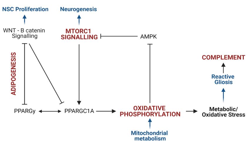
Preprints (www.preprints.org) | NOT PEER-REVIEWED | Posted: 6 September 2021                         doi:10.20944/preprints202109.0109.v1

         Table 1. A description of the 19 datasets included in this review, summarizing the type of study, the cohort size, the tissue
         type, and the definition of the final dataset used from each study. DE = differentially expressed; DM= differentially methyl-
         ated.

                                                                                                                Dataset used in
         Reference            Type of study    Tissue                           Cohort
                                                                                                                analysis
                                                                                626 ASD and 447 controls
         (Tylee et al.                                   Ex vivo blood or                                       90 DE genes in ASD
                                                                                across seven independent
         2017)                                           lymphocytes                                            (p
Preprints (www.preprints.org) | NOT PEER-REVIEWED | Posted: 6 September 2021 doi:10.20944/preprints202109.0109.v1
and 20 age- and gender- secretory proteins;
matched controls. six proteins were
validated using an
ELISA.
146 DE proteins
Brodmann area 19 from BA19 between
(BA19) 9 ASD cases (2-60yrs) and 9 ASD and controls
(Abraham et al. Brain
age- and gender matched (p < 0.05);
2019) tissue
controls (1-60yrs) 191 DE proteins in
Posterior inferior
cerebellum
cerebellum (CB)
(p < 0.05).
Discovery set = 74 males (35
ASD and 39 controls) and 32
females (15 ASD and 17 con- 537 DM genes in
(Mordaunt et al. Umbilical cord trols) in the MARBLES and both discovery and
2020) blood samples EARLI studies. Replication replication sets in
set = 38 males (21 ASD and males
Blood 17 controls) and 8 females (5
ASD and 3 controls).
181 DM genes that
overlap between the
Lymphoblastoid
(Hu et al. 2020) 21 ASD and 21 controls discovery and vali-
cell lines
dation groups
DNA Methyl- males
ation analysis i)Top ranked iASD-
associated DM
Post-mortem brain
probes identified in
tissue from Pre-
(Wong et al. the cross-cortex
frontal cortex, tem- 43 ASD and 38 controls
2019) model incorporat-
Brain poral cortex and
ing both prefrontal
tissue cerebellum
cortex and temporal
cortex data.
56 ASD samples and 41 con- DM genes (Either
(Ramaswami et
Brain tissue trol samples from 33 ASD Promoter or Gene
al. 2020)
and 26 control brains. body, FDRPreprints (www.preprints.org) | NOT PEER-REVIEWED | Posted: 6 September 2021 doi:10.20944/preprints202109.0109.v1
Subsequently, each gene list was subjected to gene set enrichment analysis [GSEA] against the Molecular Signatures
Database (MSigDB) [34], using the open-source GSEA tool developed by UC San Diego and the Broad Institute
(http://www.gsea-msigdb.org/gsea/msigdb/annotate.jsp) [35,36]. Each dataset was annotated with respect to the
top 10 significantly enriched Hallmark Canonical pathways where each pathway represents a well-defined biolog-
ical process in one curated gene set [37]. These enrichment signatures were compared between independent da-
tasets to identify canonical pathways that were consistently dysregulated in ASD at all three molecular levels; pro-
teomic, transcriptomic and epigenomic.
The proteomic datasets were examined to identify a subset of differentially expressed proteins that were also im-
plicated in transcriptomic and DNAm data in ASD. After conversion to NCBI Entrez Gene IDs, this subset of pro-
teins was annotated with respect to significantly enriched Hallmark canonical pathways, ClinVar disease pathways
and Transcription Factor Protein-Protein interactions (TF-PPIs) using the Enrichr gene list analysis tool suite
(https://maayanlab.cloud/Enrichr/) [38,39]. TF-PPIs are defined by a library of datasets corresponding to a list of
transcription factors and the proteins that interact with them. The top 10 significantly enriched TF-PPIs were used
to generate a signalling network based on the SIGnaling Network Open Resource (SIGNOR) v2.0 database [40],
using the Network Analyst web interface (https://www.networkanalyst.ca/NetworkAnalyst/home.xhtml) [41]. This
network was used to explore the relationship between the canonical pathways implicated in ASD, biological pro-
cesses and phenotypic aspects of ASD aetiology.
3. Results
After collating the data from 19 different studies (Table 1), we determined the top 10 significantly enriched Hall-
mark canonical pathways for each of the datasets (Figure S1). These enrichment signatures were compared across
all 19 datasets to identify canonical pathways that were consistently supported by all three molecular approaches
(Table 2). Eight Hallmark canonical pathways were enriched in seven or more different datasets (Figure 1). Of these,
six canonical pathways were supported using all three molecular approaches. There were two pathways that were
most frequently associated with ASD: the oxidative phosphorylation and mTORC1 signalling pathways. These two
pathways were implicated in 10 different datasets and these datasets included proteomic, transcriptomic and
DNAm datasets derived from post mortem brain tissue from individuals with ASD. There were four additional ca-
nonical pathways consistently implicated across molecular datasets: the coagulation, xenobiotic metabolism, p53
and adipogenesis pathways. Notably, three canonical pathways, oxidative phosphorylation, mTORC1 signalling
and xenobiotic metabolism, were each implicated in at least three proteomic, three transcriptomic and two DNA
methylation studies. The canonical pathway most consistently associated with the ASD proteomic data was the
coagulation pathway. This pathway was implicated in five out of six proteomic datasets, as well as three tran-
scriptomic datasets and one DNAm screen.Preprints (www.preprints.org) | NOT PEER-REVIEWED | Posted: 6 September 2021 doi:10.20944/preprints202109.0109.v1
Table 2. A summary of the Hallmark Enrichment Signatures of 19 ASD molecular datasets, highlighting canonical pathways that
were most consistently implicated in ASD proteomic, transcriptomic, and DNAm datasets. Each independent dataset was annotated
with respect to the top ten significantly enriched Hallmark canonical pathways (FDRPreprints (www.preprints.org) | NOT PEER-REVIEWED | Posted: 6 September 2021 doi:10.20944/preprints202109.0109.v1
NO. OF DATASETS
0 1 2 3 4 5 6 7 8 9 10
OXIDATIVE PHOSPHORYLATION
MTORC1 SIGNALING
P53 PATHWAY
COMPLEMENT
COAGULATION
XENOBIOTIC METABOLISM
INTERFERON GAMMA RESPONSE
ADIPOGENESIS
Proteomic Transcriptomic DNAm
Figure 1. Hallmark Canonical Pathways most consistently implicated in ASD Molecular Data. Datasets were collated from 19
studies and meta-analyses on ASD papers published between 2017 and 2021: six proteomic, eight transcriptomic and six DNA meth-
ylation (DNAm) studies. All gene lists were converted into NCBI Entrez Gene IDs. Each dataset was annotated with respect to the
top 10 significantly enriched Hallmark Canonical pathways. The Hallmark Canonical Pathways that were significantly enriched in
seven or more independent ASD proteomic, transcriptomic and DNAm datasets are shown; those shown in bold are those consist-
ently implicated using all three molecular approaches.
We next explored how the proteomic data articulated with the transcriptomic and DNAm data associated with
ASD. We identified a subset of 121 proteins that were implicated in ASD using all three molecular approaches (Fig-
ure S3; Supplementary Table S2). We characterised this dataset with respect to enriched Hallmark canonical path-
ways, disease phenotype pathways, and downstream biological processes in order to explore the functional impli-
cations associated with these proteins. This subset of proteins was significantly enriched for Hallmark canonical
pathways involved in mTOR signalling (this includes mTORC1 signalling and PI3K-AKT-mTOR signaling), metab-
olism (this includes oxidative phosphorylation, glycolysis, fatty acid metabolism and adipogenesis), and immune
responses (this includes complement, allograft rejection and IL6-JAK-STAT3 signalling). The 10 most significantly
enriched Hallmark canonical pathways in this subset of proteins included four canonical pathways that were also
consistently enriched across all 19 datasets; namely, mTORC1 signalling, oxidative phosphorylation, adipogenesis
and the complement response (Figure 2A). Together, the four canonical pathways from both analyses converge on
signalling networks that regulate neural stem cell proliferation, differentiation, metabolism, redox homeostasis and
reactive gliosis during neurodevelopment (Figure 2B).
The subset of 121 proteins that were implicated using all three molecular approaches was also enriched for five
ClinVar disease pathways associated with neurological, immunological and metabolic diseases (Table 3). Notably,
the most significantly enriched disease pathway was for Leigh syndrome, which is a pediatric mitochondrial diseasePreprints (www.preprints.org) | NOT PEER-REVIEWED | Posted: 6 September 2021 doi:10.20944/preprints202109.0109.v1
that manifests with severe neuropathology [42]. Therefore, the above-mentioned proteins converge on three central
components of ASD pathophysiology which may yield insight into the link between canonical pathways and the
dysregulation of biological processes in ASD aetiology. Consequently, we examined the protein-protein interac-
tion networks and downstream signalling pathways associated with this dataset by testing for significant enrich-
ment of TF-PPIs. The top 10 significantly enriched TF-PPIs are key transcriptional regulators of mitochondrial me-
tabolism (CREBP1A, PPARGC1A and HNF1A), lipid metabolism (PPARCG1A and STAT1), adipogenesis (NR3C1),
the p53 pathway (TP53; TP63) and inflammation (NR3C1, STAT1) (Supplementary Table S4). Notably, several of
these transcription factors mediate signalling via the PI3K-AKT-mTOR (HTT, TP53, CREBP1A, PPARGC1A) or
ERK-mTOR (ESR1,STAT1,HNF1A) pathways. This highlights some of the transcription factors that regulate the
canonical pathways implicated in ASD molecular data. The signalling network between the top 10 significantly
enriched transcription factors converged on two SIGNOR stimuli (ROS and DNA damage), as well as six SIGNOR
signalling pathways; namely, polarization, proliferation, apoptosis, cell death, mitochondrial biogenesis and in-
flammation (Figure 3). Therefore, this subset of differentially expressed proteins highlights a link between signal-
ling pathways, biological processes and ClinVar disease phenotypes associated with ASD aetiology. Altogether,
recent proteomic profiling studies, in conjunction with previously published transcriptomic and epigenomic meta-
analyses, consistently implicate canonical pathways involved in neuronal metabolism, differentiation and inflam-
mation.Preprints (www.preprints.org) | NOT PEER-REVIEWED | Posted: 6 September 2021 doi:10.20944/preprints202109.0109.v1
A
B
Figure 2. ASD proteomic profiles converge on four canonical pathways involved in mitochondrial metabolism, neurogenesis and
neuroinflammation. A) The Venn diagram shows the Hallmark canonical pathways implicated in seven or more independent da-
tasets (in blue), the enriched canonical pathways in the 121 differentially expressed proteins implicated in transcriptomic and DNAm
datasets (light grey) and the pathways that overlap (in bold font). B) The four pathways implicated in both analyses (Figure 2A)
converge on the regulation of the following biological processes (in blue): neural stem cell proliferation, neurogenesis, mitochondrial
metabolism and inflammation. The mTORC1 signalling pathway induces oxidative phosphorylation via the activation of camp-re-
sponsive element binding protein 1 (CREB1) and peroxisome proliferator-activated receptor gamma coactivator 1-alpha
(PPARGC1A), which regulates mitochondrial metabolism and the antioxidant response. PPARGC1A is regulated by peroxisome
proliferator-activated receptor gamma (PPARGy), which acts as a master regulator of lipid metabolism and is essential to induce
adipogenesis. Adipogenesis is negatively regulated by WNT signalling, which inhibits PPARGy, PPARGC1A and mTORC1 signal-
ling to regulate stem cell proliferation and differentiation. Mitochondrial metabolism regulates metabolic and redox homeostasis
which governs the inflammatory profile of microglia. Oxidative stress leads to the over proliferation of reactive microglia, which
triggers the pro-inflammatory complement response.Preprints (www.preprints.org) | NOT PEER-REVIEWED | Posted: 6 September 2021 doi:10.20944/preprints202109.0109.v1
Table 3. ClinVar Disease Pathways that are significantly enriched in the 121 differentially expressed proteins that are also impli-
cated in both transcriptomic and DNAm datasets.
ClinVar Disease Pathway Disease Phenotype P value
Leigh Syndrome Neurological Disease 0.0027
Familial Partial Lipodystrophy 0.0299
Pyruvate Dehydrogenase Complex Deficiency Metabolic Disease 0.0299
Mitochondrial DNA Deletion Syndromes 0.0474
Autoimmune Lymphoproliferative Syndrome Autoimmune Disease 0.0358
Biological Process
Stimulus
Seed
Figure 3. Signalling network between the top 10 Transcription Factor Protein-Protein Interactions (PPI) enriched in the set of
121 proteins that are supported by ASD proteomic, transcriptomic and epigenomic datasets. This PPI network was annotated
with respect to signalling pathways in the SIGnaling Network Open Resource (SIGNOR) v2.0 database of manually-annotated
causal relationships between proteins that participate in signal transduction.Preprints (www.preprints.org) | NOT PEER-REVIEWED | Posted: 6 September 2021 doi:10.20944/preprints202109.0109.v1
4. Discussion
Molecular research of ASD requires a “multi-omics” approach to comprehensively characterise the convergent
biological processes associated with its complex genetic architecture. Proteomic, transcriptomic and epigenomic
approaches are well-established as ways to investigate changes in gene regulation, expression and function in
ASD. DNA methylation is widely recognized as an epigenetic regulator of gene expression that contributes to
ASD aetiology [43]. Transcriptomic studies facilitate an understanding of the link between the genetic mutations
associated with ASD and changes in gene expression and function [11]. Recent advances in high-throughput pro-
teomic technologies have facilitated the progress of ASD proteomic profiling studies using both peripheral and
brain tissue to provide insight into the cellular mechanisms involved in ASD aetiology. Proteomic approaches are
uniquely able to detect different protein isoforms or post-translational modifications and have the potential to
identify disease biomarkers and drug targets for the diagnosis and treatment of ASD [9,11]. Furthermore, prote-
omics yields insight into the dysregulation of biologically relevant protein interaction networks and signalling
path- ways [18]. This review integrated the functional annotations of ASD proteomic, transcriptomic and DNAm
datasets to identify common canonical pathways associated with ASD.
We found that the canonical pathways involved in metabolism, redox homeostasis, inflammation and prolifera-
tion are consistently supported by proteomic, transcriptomic and DNAm data in ASD (Figure 1, Figure 2). This
review explored the link between canonical pathways, biological processes and disease phenotype pathways in
ASD by functionally annotating a subset of differentially expressed proteins that were also supported by tran-
scriptomic and DNAm data. This subset of proteins was enriched for four canonical pathways that were also
implicated in seven or more independent datasets. These were the pathways for oxidative phosphorylation,
mTORC1 signalling, adipogenesis and the complement response (Figure 2A). The ten most significantly enriched
TF-PPIs in this subset of proteins highlighted key transcriptional regulators of these canonical pathways. These
transcriptional regulators form a signalling network that highlights how these canonical pathways regulate bio-
logical processes involved in neuropathology (Figure 3). This network converges on the SIGNOR pathways for
mitochondrial biogenesis and inflammation, and this is consistent with prior evidence linking mitochondrial dys-
function [44-49] and neuroinflammation [50-52] to ASD. The polarization and proliferation pathways are essential
regulators of neural stem cell self renewal and differentiation [53–55], while the apoptosis and cell death signalling
pathways are implicated as mechanisms whereby microglia drive synaptic pruning, and the maturation and mi-
gration of neuronal progenitors [56]. In addition, this subset of proteins was significantly enriched for five ClinVar
disease pathways associated with neuropathology, mitochondrial dysfunction or auto-immune disease (Table 3).
Therefore, the differentially expressed proteins highlighted in this review are linked to both molecular and phe-
notypic facets of ASD aetiology.
The common canonical pathways highlighted above are each established as molecular mechanisms contributing
to ASD aetiology. Additionally, when these pathways are considered together in the context of neurodevelop-
ment, they converge on a link between mitochondrial dysfunction, neurogenesis and inflammation in
ASD. Firstly, a role for mTORC1 signalling is well described in ASD [57,58]. Mutations in tuberous sclerosis
complex (TSC) and Phosphatase and tensin homolog (PTEN), which are both central regulators of mTORC1 sig-
nalling, are associated with autistic behaviours [59-62]. Aberrant mTOR signalling is associated with altered syn-
aptogenesis and ASD-like neurophysiology in animal and organoid model systems [63-68]. Moreover, the mTOR
signalling pathway is a promising therapeutic target in ASD [69].Preprints (www.preprints.org) | NOT PEER-REVIEWED | Posted: 6 September 2021 doi:10.20944/preprints202109.0109.v1
The canonical pathways for mTORC1 signalling and oxidative phosphorylation were the two most commonly
implicated pathways in our analyses which is consistent with evidence showing that mitochondrial dysfunction
is involved in ASD aetiology [44-49]. Mitochondrial function has recently been proposed as a central driver of
neuronal differentiation [70-73] and the tight coregulation of mTORC1 signalling and mitochondrial metabolism
is essential to control neurogenesis. The transition from undifferentiated neuronal stem cells (NSCs) to mature
neurons relies on a metabolic shift from aerobic glycolysis to mitochondrial respiration. The latter fuels neuronal
migration, dendrite formation and synaptogenesis [74]. The mTOR signalling pathway plays a central role in
driving this metabolic switch, which is essential for neuronal survival and differentiation [72]. Neurogenic factors
induce AKT-MTORC1 signalling, which upregulates peroxisome proliferator-activated receptor-gamma coacti-
vator (PPARGC1A) signalling to induce mitochondrial respiration and biogenesis [71,75]. Conversely, cellular
metabolism regulates mTOR signalling via the AMP-dependent protein kinase (AMPK), which is inhibited by a
decrease in the AMP:ATP ratio [76]. The dysregulation of signalling between mTORC1 and mitochondrial metab-
olism during development has profound consequences for NSC commitment and differentiation, and the devel-
opment and maintenance of mature neuronal networks.
We also observed a consistent enrichment of the adipogenesis pathway in ASD molecular data, which implicates
the same networks that regulate stem cell metabolism and differentiation. Adipogenesis is regulated by the “op‐
posite interplay” between WNT/B-catenin signalling and peroxisome proliferator-activated receptor gamma
(PPARGy), each of which negatively regulates the other [77]. WNT signalling is directly involved in regulating
NSC maintenance, proliferation and differentiation [78,79] and WNT responsive genes maintain aerobic glycolysis
in NSCs [72]. This highlights that the WNT pathway is at the intersection between stem cell metabolism and de-
velopment. Cross-talk between WNT signalling and mTORC1 signalling is linked to the “Warburg effect” that
induces aerobic glycolysis and over-proliferation in cancer cells [77]. A dysregulation of these same pathways
during neurodevelopment could disrupt NSC commitment and differentiation. In fact, chronic activation of WNT
signalling altered mTOR signalling in human organoids, leading to increased NSC proliferation, impaired neu-
ronal differentiation and disrupted radial glial organization [80]. Moreover, in vivo downregulation of WNT sig-
nalling leads to premature neurogenesis and atypical behaviours [81]. The WNT/B catenin pathway has also been
implicated in ASD genetic and transcriptomic data [82-86].
On the other hand, PPARGy acts as a master regulator of lipid metabolism and regulates target genes necessary
for differentiation, fatty acid transport, carbohydrate metabolism, and energy homeostasis [87]. Mitochondrial
fatty acid oxidation (FAO) is essential for the self-renewal of NSCs, but fatty acid metabolism shifts towards lipo-
genesis during neurogenesis [88-91]. In vitro studies show that this shift in fatty acid metabolism is regulated by
the AMPK-PPARGC1A axis [91]. The coregulation between PPARGy and PPARGC1A means that glucose and
fatty acid metabolism are intrinsically linked during neurodevelopment. Disrupting FAO inhibits stem cell self-
renewal [89,91] and impairs NSC differentiation in mouse models [92]. Recent reviews of ASD proteomic data
also find that proteins involved in lipid metabolism are differentially expressed in ASD [93,94]. A role for PPARGy
is supported by increasing evidence of mitochondrial FAO deficiencies in ASD, and PPARGy agonists have been
proposed as therapeutic agents in ASD [95,96]. This highlights how PPARGy is implicated in neurodevelopment
and ASD, and how the canonical pathways for mTORC1 signalling, oxidative phosphorylation and adipogenesis
converge on the signalling between neuronal metabolism and differentiation.
A dysregulation of the signalling between mitochondrial metabolism and neurogenesis has significant implica-
tions for neuronal development and function. Firstly, mitochondrial oxidative phosphorylation is one of thePreprints (www.preprints.org) | NOT PEER-REVIEWED | Posted: 6 September 2021 doi:10.20944/preprints202109.0109.v1
primary producers of intracellular reactive oxygen species (ROS) and disruptions to mitochondrial metabolism
can lead to oxidative stress [97]. Increased oxidative stress is a well-documented aspect of ASD pathophysiology;
the evidence for markers of oxidative stress associated with glutathione metabolism, lipid peroxidation, protein
oxidation, DNA oxidation and antioxidant enzyme activity has been comprehensively reviewed in recent years
[45,98-103]. In the context of neurodevelopment, this has significant implications for the redox regulation of neu-
rogenesis. Undifferentiated NSCs are characterized by high levels of endogenous ROS, which plays a functional
role in stem cell maintenance [105] and oxidative stress is known to promote NSC self-renewal and inhibit down-
stream neurogenesis [97,105]. Studies on in vitro and in vivo model systems consistently demonstrate that increas-
ing oxidative stress [106 - 108] or disrupting mitochondrial homeostasis and function [109-112] leads to an inhibi-
tion of neurogenesis and a shift towards gliosis; the latter is associated with neuroinflammation and is established
as a hallmark of ASD aetiology. Clinical studies also report that gliosis is one of the neuropathological manifesta-
tions of mitochondrial disease [43,113-116].
Many of the canonical pathways highlighted in this review are implicated as mechanisms involved in the response
to oxidative stress and gliosis during neurogenesis. Clinical data has reported mTOR signalling as a mechanism
that connects mitochondrial disease to gliosis [117], with both in vitro - [118] and in vitro- studies [119] showing
that gliosis can be induced by targeted disruptions to mTOR signalling. Cross-talk between the PPARGy and
WNT canonical pathways is also associated with increased oxidative stress and chronic inflammation in cancer
[119], and WNT/B-catenin-PPARGy signalling can be targeted to reduce reactive gliosis in models of neurodegen-
erative disease [120-122]. In addition, the p53 pathway and xenobiotic metabolism were each implicated in eight
or more independent datasets in our analysis and these pathways were supported by all three types of molecular
data (Figure 1). The p53 signalling pathway is a central regulator of inflammation, oxidative stress and apoptosis
[124], while the canonical pathway for xenobiotic metabolism is comprised of genes that respond to inflammation,
metabolic stress and ROS [125]. Notably, both mTOR and WNT signalling regulate p53 degradation [74,126,127].
The p53 signalling pathway also plays an important role in neural precursor cell self-renewal, neuronal commit-
ment and reactive gliosis in response to mitochondrial dysfunction [111,112,128].
Gliosis is characterized by highly reactive microglia and astrocytes, leading to an overproduction of glial-specific
fibrillary acidic protein (GFAP) and inflammatory cytokines [129]. This review highlights the consistent enrich-
ment of inflammatory canonical pathways in ASD transcriptomic and proteomic data. The canonical pathways
involved in inflammatory immune response pathways were those most frequently associated with the tran-
scriptomic enrichment signatures: six of ten hallmark canonical pathways that were implicated in at least five
independent transcriptomic datasets were related to immune responses. This is consistent with a substantial body
of work implicating neuroinflammation and gliosis as mechanisms of pathology of ASD, which has been thor-
oughly reviewed elsewhere [50-52,130-133]. A role for inflammation, astrocyte function and microglial activation
is well-supported by transcriptomic and proteomic studies in ASD brain tissue [134-136]. This is also consistent
with immunohistochemistry, positron emission tomography and morphological data in ASD [130,137,138]. Nota-
bly, microglial phagocytosis is responsible for the degradation of proliferating NSCs during neurodevelopment
[57]. This is essential for synaptogenesis and post-natal synaptic pruning, both of which are thought to be dysreg-
ulated in ASD [130,139,140]. Importantly, microglial metabolism is closely linked to neuroinflammatory state
[141,142], and both glucose and lipid metabolism regulate microglial activation [143-146]. The dysregulation of
glial and microglial metabolism, proliferation and function can alter neuronal differentiation, impairPreprints (www.preprints.org) | NOT PEER-REVIEWED | Posted: 6 September 2021 doi:10.20944/preprints202109.0109.v1
synaptogenesis and pruning, and change neuroinflammatory state, with profound implications for neural archi-
tecture and connectivity [147].
Our analysis of recent ASD proteomic profiles further supports a role for neuroinflammation in ASD. The com-
plement and coagulation cascades were implicated in three and five out of six ASD proteomic studies respectively,
which is supported in independent reviews of ASD proteomic data [13,93,94]. Both cascades form part of the in-
nate immune system and interactions between them via mannose-binding lectin associated serine proteases are
well documented [148-153]. Both cascades are involved in the inflammatory response by reactive microglia
[154,155]. Coagulation proteins affect the morphology, proliferation and function of astrocytes [154], while the
complement system is implicated as a mechanism by which microglia regulate neurogenesis, neuronal migration
and synaptic pruning [155-158]. Complement proteins play a role in microglial activation, which can influence
neuroinflammatory signalling and neurodevelopment [159]. Animal models show that genetic knockdown of
complement proteins impairs neuronal migration, while activation of the complement system rescues this deficit
[156]. There is mounting evidence for complement system dysfunction in neurodevelopmental disorders [157],
which has been proposed as one of the mechanisms behind the synaptic pruning deficits, increased dendritic
spine density, cortical hyperconnectivity and resultant behavioural phenotypes in ASD [158]. Therefore, the im-
mune response cascades enriched in ASD proteomic profiles function as a link between gliosis, neurogenesis and
synaptogenesis, highlighting a point of convergence between these different mechanisms in ASD pathology.
Collectively, the canonical pathways highlighted in this review are consistent with known pathways that to con-
tribute to ASD aetiology, as well as pathways that have been implicated in independent reviews of ASD molecular
data. Importantly, our review also considers the interactions between these canonical pathways, particularly in
the context of neurodevelopment. The latter is often overlooked when the relevance of each canonical pathway is
evaluated in isolation. Previous reviews have comprehensively described the role of mTORC1 signalling in pro-
tein synthesis, neuronal differentiation, migration and patterning [159], but these reviews have focused less on
the role of mTORC1 as a regulator of mitochondrial metabolism. The same is true for reviews on the role played
by WNT signalling in neurogenesis and ASD [160,161], which describe the central role played by WNT signalling
in NSC proliferation and differentiation. However, our review emphasises how WNT-PPARGy signalling inter-
sects with PPARGC1A, anti-inflammatory and antioxidant responses, and the regulation of mitochondrial bio-
genesis and metabolism. Given that mitochondrial dysfunction is an established component of ASD aetiology,
and that metabolic state plays a central role in neuronal differentiation, our review highlights a potentially im-
portant connection between mTORC1, oxidative phosphorylation and neuropathology in ASD. Finally, our re-
view integrates all of the above with the thoroughly reviewed evidence for neuroinflammation and oxidative
stress in ASD [49-52,98-103] by considering the role of mitochondrial metabolism in gliosis and microglial activa-
tion, and the downstream consequences for synaptogenesis, neuronal connectivity and behavioural phenotypes.
Therefore, our review is not only consistent with current molecular mechanisms implicated in ASD, but also high-
lights an under-explored link between mitochondrial dysfunction, neuroinflammation and neurodevelopmental
pathology.Preprints (www.preprints.org) | NOT PEER-REVIEWED | Posted: 6 September 2021 doi:10.20944/preprints202109.0109.v1
5. Conclusion
The genetic and phenotypic complexity of ASD makes it challenging to identify the molecular mechanisms that
contribute to its aetiology. Proteomic approaches can yield novel insight into the dysregulation of biological pro-
cesses and signalling networks in ASD. Despite the power of proteomics, there are currently limitations to this
approach in terms of scaling up in sample size and efficiency. In light of these caveats, we integrated recently pub-
lished ASD proteomic profiles with large-scale meta-analyses of whole-genome transcriptomic and DNAm data to
compare the functional enrichment signatures of disparate molecular datasets. Our analysis demonstrates that ASD
proteomic, transcriptomic and DNAm data consistently support the dysregulation of mTORC1 signalling, oxidative
phosphorylation, adipogenesis and the response to inflammation and oxidative stress. The mTORC1-, WNT- and
PPARGy- signalling pathways are each well-established regulators of neurodevelopment that are also implicated
in ASD and neurodevelopmental pathology. The mTORC1 signalling pathway regulates oxidative phosphorylation
via PPARGC1A, which operates in concert with PPARGy to regulate mitochondrial respiration and fatty acid oxi-
dation. These pathways play an essential role in the tight coupling between stem cell proliferation, differentiation
and mitochondrial metabolism during neurogenesis. If this coupling is disrupted, this can induce metabolic and
oxidative stress in NSCs. Both metabolic and oxidative stress are known to disrupt neurogenesis, promote gliosis
and induce microglial activation. This proposes a link between mitochondrial dysfunction, oxidative stress and
gliosis, each of which are well-established features of ASD aetiology. Given the central role played by glial networks
in neuronal migration and synaptogenesis, gliosis is implicated as a mechanism involved in neuroinflammation
and the neuropathophysiology associated with ASD. In summary, this review demonstrates that ASD molecular
data converges on canonical pathways involved in mitochondrial function, neurogenesis and neuroinflammation,
highlighting how these three key aspects of ASD aetiology interact, and the relevance of these interactions in the
context of neurodevelopment.
Supplementary Materials: The following are available online at www.mdpi.com/xxx/s1, Supplementary Figure 1: The top
10 significantly enriched Hallmark canonical pathways in 19 datasets of ASD molecular data; the heatmap represents the FDR
value of enrichment; Supplementary Figure 2: Summary of Hallmark Enrichment Signatures across 19 datasets of ASD pro-
teomic, transcriptomic and DNAm datasets. The vertical axis represents the number of datasets for which each Hallmark ca-
nonical Pathway is among the top 10 significantly enriched Hallmark Pathways in that dataset; Supplementary Figure 3. The
121 differentially expressed proteins previously implicated in both transcriptomic and DNA methylation ASD datasets. The
Venn Diagram represents 15 075 unique gene IDs implicated across the 19 datasets included in this review. This includes 12 037
unique genes reported across 11 transcriptomic studies, 5 573 genes across six genome-wide DNA methylation screens and 750
proteins across five proteomic profiling studies. We identified a subset of 121 proteins that had been implicated in ASD using
Transcriptomic, Epigenomic and Proteomic methods; Supplementary Table 1: Full datasets collated from the ASD proteomic,
transcriptomic and DNA methylation studies described in Table 1. Each dataset represents the list of genes or proteins found to
be significantly associated in ASD after data processing and statistical analyses, converted into NCBI Entrez Gene IDs; Supple-
mentary Table 2: The 121 proteins identified in this review that have been implicated in Transcriptomic, Epigenomic and Pro-
teomic data in ASD; Supplementary Table 3: Top 10 Hallmark canonical pathways enriched in the set of 121 proteins identified
in this review that have been shown to be altered on the Transcriptomic, Epigenomic and Proteomic level in ASD; Supplemen-
tary Table 4: The top 10 significantly enriched Transcription Factor Protein-Protein Interactions in the dataset of 121 differen-
tially expressed proteins that are also implicated in both Transcriptomic and DNAm data.Preprints (www.preprints.org) | NOT PEER-REVIEWED | Posted: 6 September 2021 doi:10.20944/preprints202109.0109.v1
Author Contributions: Conceptualization, methodology, analysis, writing—original draft, preparation, writing—review and
editing and visualization: C. Mahony and C. O’Ryan; Project supervision administration and funding acquisition: C. O’Ryan.
All authors have read and agreed to the published version of the manuscript.
Funding: This research was funded by National Research Foundation, South Africa (Grant number 118524).
Institutional Review Board Statement: Not applicable.
Informed Consent Statement: Not applicable.
Data Availability Statement: The data presented in this study are available within the article and supplementary material here.
Conflicts of Interest: The authors declare no conflict of interest.
6. References
1. Wiśniowiecka-Kowalnik, B. & Nowakowska, B. A. Genetics and epigenetics of autism spectrum disorder—
current evidence in the field. Journal of Applied Genetics 60, 37–47 (2019).
2. Xie, S. et al. The Familial Risk of Autism Spectrum Disorder with and without Intellectual Disability. Au-
tism Res. 13, 2242–2250 (2020).
3. Bai, D. et al. Association of Genetic and Environmental Factors With Autism in a 5-Country Cohort. JAMA
Psychiatry 76, 1035–1043 (2019).
4. Pintacuda, G., Martín, J. M. & Eggan, K. C. Mind the translational gap: using iPS cell models to bridge
from genetic discoveries to perturbed pathways and therapeutic targets. Molecular Autism 12, 10 (2021).
5. Torre-Ubieta, L. de la, Won, H., Stein, J. L. & Geschwind, D. H. Advancing the understanding of autism
disease mechanisms through genetics. Nat. Med. 2016 224 22, 345–361 (2016).
6. Duffney, L. J. et al. Epigenetics and autism spectrum disorder: A report of an autism case with mutation in
H1 linker histone HIST1H1E and literature review. Am. J. Med. Genet. Part B Neuropsychiatr. Genet. 177,
426–433 (2018).
7. Choi, L. & An, J.-Y. Genetic architecture of autism spectrum disorder: Lessons from large-scale genomic
studies. Neurosci. Biobehav. Rev. 128, 244–257 (2021).
8. Manoli, D. S. & State, M. W. Autism spectrum disorder genetics and the search for pathological mecha-
nisms. American Journal of Psychiatry 178, 30–38 (2021).
9. Comes, A. L. et al. Proteomics for blood biomarker exploration of severe mental illness: pitfalls of the past
and potential for the future. Transl. Psychiatry 2018 81 8, 1–15 (2018).
10. Huang, H., Shukla, H., Wu, C. & Saxena, S. Challenges and Solutions in Proteomics. Curr. Genomics 8, 21–
28 (2007).
11. Shen, L. et al. Proteomics Study of Peripheral Blood Mononuclear Cells (PBMCs) in Autistic Children.
Front. Cell. Neurosci. 13, 105 (2019).
12. Ruggeri, B., Sarkans, U., Schumann, G. & Persico, A. M. Biomarkers in autism spectrum disorder: the old
and the new. Psychopharmacol. 2013 2316 231, 1201–1216 (2013).
13. Higdon, R. et al. The promise of multi-omics and clinical data integration to identify and target personal-
ized healthcare approaches in autism spectrum disorders. OMICS A Journal of Integrative Biology 19, 197–
208 (2015).Preprints (www.preprints.org) | NOT PEER-REVIEWED | Posted: 6 September 2021 doi:10.20944/preprints202109.0109.v1
14. Hewitson, L. et al. Blood biomarker discovery for autism spectrum disorder: A proteomic analysis. PLoS
One 16, e0246581 (2021).
15. Shen, L. et al. iTRAQ-Based Proteomic Analysis Reveals Protein Profile in Plasma from Children with Au-
tism. PROTEOMICS - Clin. Appl. 12, 1700085 (2018).
16. Yang, J. et al. Peptidome Analysis Reveals Novel Serum Biomarkers for Children with Autism Spectrum
Disorder in China. PROTEOMICS - Clin. Appl. 12, 1700164 (2018).
17. Yao, F. et al. Protein Biomarkers of Autism Spectrum Disorder Identified by Computational and Experi-
mental Methods. 12, 1–13 (2021).
18. Abraham, J. R. et al. Proteomic Investigations of Autism Brain Identify Known and Novel Pathogenetic
Processes. Sci. Rep. 9, 1–11 (2019).
19. Tylee, D. S. et al. Blood transcriptomic comparison of individuals with and without autism spectrum disor-
der: A combined-samples mega-analysis. Am. J. Med. Genet. Part B Neuropsychiatr. Genet. 174, 181–201
(2017).
20. Mordaunt, C. E. et al. A meta-analysis of two high-risk prospective cohort studies reveals autism-specific
transcriptional changes to chromatin, autoimmune, and environmental response genes in umbilical cord
blood. Mol. Autism 10, 1–21 (2019).
21. Gao, H. et al. Integrated Systems Analysis Explores Dysfunctional Molecular Modules and Regulatory Fac-
tors in Children with Autism Spectrum Disorder. J. Mol. Neurosci. 1–11 (2020). doi:10.1007/s12031-020-
01658-w
22. He, Y., Zhou, Y., Ma, W. & Wang, J. An integrated transcriptomic analysis of autism spectrum disorder.
Sci. Rep. 9, 1–9 (2019).
23. Forés-Martos, J. et al. Transcriptomic metaanalyses of autistic brains reveals shared gene expression and
biological pathway abnormalities with cancer. Mol. Autism 10, 17 (2019).
24. Rahman, M. R. et al. Comprehensive Analysis of RNA-Seq Gene Expression Profiling of Brain Transcrip-
tomes Reveals Novel Genes, Regulators, and Pathways in Autism Spectrum Disorder. Brain Sci. 10, 747
(2020).
25. Wright, C. et al. Altered expression of histamine signaling genes in autism spectrum disorder. Transl. Psy-
chiatry 7, e1126 (2017).
26. Ramaswami, G. et al. Integrative genomics identifies a convergent molecular subtype that links epigenomic
with transcriptomic differences in autism. Nat. Commun. 11, 1–14 (2020).
27. Mordaunt, C. E. et al. Cord blood DNA methylome in newborns later diagnosed with autism spectrum
disorder reflects early dysregulation of neurodevelopmental and X-linked genes. Genome Med. 12, 1–25
(2020).
28. Hu, V. W., Hong, Y., Xu, M. & Shu, H. T. Altered DNA methylation in a severe subtype of idiopathic au-
tism: Evidence for sex differences in affected metabolic pathways. Autism 136236132097108 (2020).
doi:10.1177/1362361320971085
29. Wong, C. C. Y. et al. Genome-wide DNA methylation profiling identifies convergent molecular signatures
associated with idiopathic and syndromic autism in post-mortem human brain tissue. Hum. Mol. Genet. 28,
2201–2211 (2019).
30. Stathopoulos, S. et al. DNA Methylation Associated with Mitochondrial Dysfunction in a South African
Autism Spectrum Disorder Cohort. Autism Res. 13, 1079–1093 (2020).
31. Huang, D. W., Sherman, B. T. & Lempicki, R. A. Systematic and integrative analysis of large gene lists us-
ing DAVID bioinformatics resources. Nat. Protoc. 4, 44–57 (2009).Preprints (www.preprints.org) | NOT PEER-REVIEWED | Posted: 6 September 2021 doi:10.20944/preprints202109.0109.v1
32. Huang, D. W., Sherman, B. T. & Lempicki, R. A. Bioinformatics enrichment tools: Paths toward the com-
prehensive functional analysis of large gene lists. Nucleic Acids Res. 37, 1–13 (2009).
33. Raudvere, U. et al. g:Profiler: a web server for functional enrichment analysis and conversions of gene lists
(2019 update). Nucleic Acids Res. (2019). doi:10.1093/nar/gkz369
34. Liberzon, A. et al. Molecular signatures database (MSigDB) 3.0. Bioinformatics 27, 1739–1740 (2011).
35. Mootha, V. K. et al. PGC-1α-responsive genes involved in oxidative phosphorylation are coordinately
downregulated in human diabetes. Nat. Genet. 34, 267–273 (2003).
36. Subramanian, A. et al. Gene set enrichment analysis: A knowledge-based approach for interpreting ge-
nome-wide expression profiles. Proc. Natl. Acad. Sci. U. S. A. 102, 15545–15550 (2005).
37. Liberzon, A. et al. The Molecular Signatures Database Hallmark Gene Set Collection. Cell Syst. 1, 417–425
(2015).
38. Chen, E. Y. et al. Enrichr: Interactive and collaborative HTML5 gene list enrichment analysis tool. BMC Bio-
informatics 14, (2013).
39. Kuleshov, M. V. et al. Enrichr: a comprehensive gene set enrichment analysis web server 2016 update. Nu-
cleic Acids Res. 44, W90–W97 (2016).
40. Licata, L. et al. SIGNOR 2.0, the SIGnaling Network Open Resource 2.0: 2019 update. Nucleic Acids Res. 48,
D504–D510 (2020).
41. Xia, J., Gill, E. E. & Hancock, R. E. W. NetworkAnalyst for statistical, visual and network-based meta-anal-
ysis of gene expression data. Nat. Protoc. 10, 823–844 (2015).
42. Lake, N. J., Bird, M. J., Isohanni, P. & Paetau, A. Leigh Syndrome Neuropathology and Pathogenesis. J.
Neuropathol. Exp. Neurol. 74, 482–492 (2015).
43. Tremblay, M. W. & Jiang, Y. DNA Methylation and Susceptibility to Autism Spectrum Disorder.
https://doi.org/10.1146/annurev-med-120417-091431 70, 151–166 (2019).
44. Siddiqui, M. F., Elwell, C. & Johnson, M. H. Mitochondrial Dysfunction in Autism Spectrum Disorders.
Autism. Open. Access (2016). doi:10.4172/2165-7890.1000190
45. Griffiths, K. K. & Levy, R. J. Evidence of Mitochondrial Dysfunction in Autism: Biochemical Links, Ge-
netic-Based Associations, and Non-Energy-Related Mechanisms. Oxid. Med. Cell. Longev. 2017, 4314025
(2017).
46. Rose, S. et al. Clinical and Molecular Characteristics of Mitochondrial Dysfunction in Autism Spectrum
Disorder. Molecular Diagnosis and Therapy (2018). doi:10.1007/s40291-018-0352-x
47. Castora, F. J. Mitochondrial function and abnormalities implicated in the pathogenesis of ASD. Prog.
Neuro-Psychopharmacology Biol. Psychiatry 92, 83–108 (2019).
48. Citrigno, L. et al. The Mitochondrial Dysfunction Hypothesis in Autism Spectrum Disorders: Current Sta-
tus and Future Perspectives. Int. J. Mol. Sci. 21, 5785 (2020).
49. Thorsen, M. Oxidative stress, metabolic and mitochondrial abnormalities associated with autism spectrum
disorder. in Progress in Molecular Biology and Translational Science 173, 331–354 (Elsevier B.V., 2020).
50. Gładysz, D., Krzywdzińska, A. & Hozyasz, K. K. Immune Abnormalities in Autism Spectrum Disorder—
Could They Hold Promise for Causative Treatment? Molecular Neurobiology 55, 6387–6435 (2018).
51. Liao, X., Liu, Y., Fu, X. & Li, Y. Postmortem Studies of Neuroinflammation in Autism Spectrum Disorder: a
Systematic Review. Molecular Neurobiology 57, 3424–3438 (2020).
52. Pangrazzi, L., Balasco, L. & Bozzi, Y. Oxidative Stress and Immune System Dysfunction in Autism Spec-
trum Disorders. Int. J. Mol. Sci. 2020, Vol. 21, Page 3293 21, 3293 (2020).Preprints (www.preprints.org) | NOT PEER-REVIEWED | Posted: 6 September 2021 doi:10.20944/preprints202109.0109.v1
53. Götz, M. & Huttner, W. B. The cell biology of neurogenesis. Nat. Rev. Mol. Cell Biol. 2005 610 6, 777–788
(2005).
54. Vieira, M. S. et al. Neural stem cell differentiation into mature neurons: Mechanisms of regulation and bio-
technological applications. Biotechnol. Adv. 36, 1946–1970 (2018).
55. Castro-Torres, R. D. et al. Involvement of JNK1 in Neuronal Polarization During Brain Development. Cells
2020, Vol. 9, Page 1897 9, 1897 (2020).
56. Sierra, A. et al. Microglia shape adult hippocampal neurogenesis through apoptosis-coupled phagocytosis.
Cell Stem Cell 7, 483–495 (2010).
57. Ganesan, H. et al. mTOR signalling pathway - A root cause for idiopathic autism? BMB Rep. 52, 424 (2019).
58. Boksha, I. S., Prokhorova, T. A., Tereshkina, E. B., Savushkina, O. K. & Burbaeva, G. S. Protein Phosphory-
lation Signaling Cascades in Autism: The Role of mTOR Pathway. Biochem. 2021 865 86, 577–596 (2021).
59. Geschwind, D. Genetics of autism spectrum disorders. Trends Cogn. Sci. 15, 409–416 (2011).
60. Zhou, J. & Parada, L. F. PTEN signaling in autism spectrum disorders. Curr. Opin. Neurobiol. 22, 873–879
(2012).
61. Wiśniowiecka-Kowalnik, B. & Nowakowska, B. A. Genetics and epigenetics of autism spectrum disorder—
current evidence in the field. Journal of Applied Genetics 60, 37–47 (2019).
62. Masini, E. et al. An Overview of the Main Genetic, Epigenetic and Environmental Factors Involved in Au-
tism Spectrum Disorder Focusing on Synaptic Activity. Int. J. Mol. Sci. 21, 8290 (2020).
63. Kwon, C. H. et al. Pten Regulates Neuronal Arborization and Social Interaction in Mice. Neuron 50, 377–388
(2006).
64. Poduri, A. et al. Somatic Activation of AKT3 Causes Hemispheric Developmental Brain Malformations.
Neuron 74, 41–48 (2012).
65. Tsai, P. T. et al. Autistic-like behaviour and cerebellar dysfunction in Purkinje cell Tsc1 mutant mice. Nature
488, 647–651 (2012).
66. Tang, G. et al. Loss of mTOR-Dependent Macroautophagy Causes Autistic-like Synaptic Pruning Deficits.
Neuron 83, 1131–1143 (2014).
67. Lieberman, O. J. et al. mTOR Suppresses Macroautophagy During Striatal Postnatal Development and Is
Hyperactive in Mouse Models of Autism Spectrum Disorders. Front. Cell. Neurosci. 0, 70 (2020).
68. Zhang, W. et al. Cerebral organoid and mouse models reveal a RAB39b–PI3K–mTOR pathway-dependent
dysregulation of cortical development leading to macrocephaly/autism phenotypes. Genes Dev. 34, 580–597
(2020).
69. Basilico, B., Morandell, J. & Novarino, G. Molecular mechanisms for targeted ASD treatments. Current
Opinion in Genetics and Development 65, 126–137 (2020).
70. Khacho, M., Harris, R. & Slack, R. S. Mitochondria as central regulators of neural stem cell fate and cogni-
tive function. Nat. Rev. Neurosci. 20, 34–48 (2019).
71. Beckervordersandforth, R. Mitochondrial Metabolism-Mediated Regulation of Adult Neurogenesis. Brain
Plast. 3, 73–87 (2017).
72. Zheng, X. et al. Metabolic reprogramming during neuronal differentiation from aerobic glycolysis to neu-
ronal oxidative phosphorylation. Elife 5, (2016).
73. Maffezzini, C., Calvo-Garrido, J., Wredenberg, A. & Freyer, C. Metabolic regulation of neurodifferentiation
in the adult brain. Cell. Mol. Life Sci. (2020). doi:10.1007/s00018-019-03430-9
74. Licausi, F. & Hartman, N. W. Role of mTOR complexes in neurogenesis. International Journal of Molecular
Sciences 19, (2018).Preprints (www.preprints.org) | NOT PEER-REVIEWED | Posted: 6 September 2021 doi:10.20944/preprints202109.0109.v1
75. Cunningham, J. T. et al. mTOR controls mitochondrial oxidative function through a YY1-PGC-1α transcrip‐
tional complex. Nature 450, 736–740 (2007).
76. Vadlakonda, L., Pasupuleti, M. & Pallu, R. Role of PI3K-AKT-mTOR and Wnt signaling pathways in tran-
sition of G1-S phase of cell cycle in cancer cells. Front. Oncol. 3 APR, 85 (2013).
77. Vallée, A., Lecarpentier, Y., Guillevin, R. & Vallée, J. Opposite Interplay Between the Canonical WNT/β-
Catenin Pathway and PPAR Gamma: A Potential Therapeutic Target in Gliomas. Neurosci. Bull. 34, 573–588
(2018).
78. Faigle, R. & Song, H. Signaling mechanisms regulating adult neural stem cells and neurogenesis. Biochimica
et Biophysica Acta - General Subjects 1830, 2435–2448 (2013).
79. Rao, T. P. & Kühl, M. An Updated Overview on Wnt Signaling Pathways. Circ. Res. 106, 1798–1806 (2010).
80. López-Tobón, A. et al. Human Cortical Organoids Expose a Differential Function of GSK3 on Cortical Neu-
rogenesis. Stem Cell Reports 13, 847–861 (2019).
81. Hussaini, S. M. Q. et al. Wnt signaling in neuropsychiatric disorders: Ties with adult hippocampal neuro-
genesis and behavior. Neurosci. Biobehav. Rev. 47, 369–383 (2014).
82. Caglayan, A. O. Genetic causes of syndromic and non-syndromic autism. Dev. Med. Child Neurol. 52, 130–
138 (2010).
83. Mulligan, K. A. & Cheyette, B. N. R. Neurodevelopmental Perspectives on Wnt Signaling in Psychiatry.
Mol. Neuropsychiatry 2, 219–246 (2016).
84. Iakoucheva, L. M., Muotri, A. R. & Sebat, J. Getting to the Cores of Autism. Cell 178, 1287–1298 (2019).
85. Caracci, M. O., Avila, M. E. & De Ferrari, G. V. Synaptic Wnt/GSK3 β Signaling Hub in Autism. Neural
Plast. 2016, (2016).
86. Kwan, V., Unda, B. K. & Singh, K. K. Wnt signaling networks in autism spectrum disorder and intellectual
disability. J. Neurodev. Disord. 8, (2016).
87. Perera, R. J. et al. Identification of novel PPARγ target genes in primary human adipocytes. Gene 369, 90–99
(2006).
88. Panov, A., Orynbayeva, Z., Vavilin, V. & Lyakhovich, V. Fatty acids in energy metabolism of the central
nervous system. BioMed Research International 2014, (2014).
89. Ito, K. et al. Self-renewal of a purified Tie2+ hematopoietic stem cell population relies on mitochondrial
clearance. Science (80-. ). 354, 1156–1160 (2016).
90. Longo, N., Frigeni, M. & Pasquali, M. Carnitine transport and fatty acid oxidation. Biochim. Biophys. Acta -
Mol. Cell Res. 1863, 2422–2435 (2016).
91. Knobloch, M. et al. A Fatty Acid Oxidation-Dependent Metabolic Shift Regulates Adult Neural Stem Cell
Activity. Cell Rep. 20, 2144–2155 (2017).
92. Xie, Z., Jones, A., Deeney, J. T., Hur, S. K. & Bankaitis, V. A. Inborn Errors of Long-Chain Fatty Acid β-Oxi-
dation Link Neural Stem Cell Self-Renewal to Autism. Cell Rep. 14, 991–999 (2016).
93. Ristori, M. V. et al. Proteomics and Metabolomics Approaches towards a Functional Insight onto AUTISM
Spectrum Disorders: Phenotype Stratification and Biomarker Discovery. Int. J. Mol. Sci. 2020, Vol. 21, Page
6274 21, 6274 (2020).
94. Shen, L. et al. Biomarkers in autism spectrum disorders: Current progress. Clin. Chim. Acta 502, 41–54
(2020).
95. Barone, R. et al. Nuclear Peroxisome Proliferator-Activated Receptors (PPARs) as therapeutic targets of
resveratrol for autism spectrum disorder. Int. J. Mol. Sci. 20, (2019).You can also read