Astrocytic atrophy as a pathological feature of Parkinson's disease with LRRK2 mutation
←
→
Page content transcription
If your browser does not render page correctly, please read the page content below
www.nature.com/npjparkd

                  ARTICLE                 OPEN

                  Astrocytic atrophy as a pathological feature of Parkinson’s
                  disease with LRRK2 mutation
                  Paula Ramos-Gonzalez1,2, Susana Mato1,2,3,4, Juan Carlos Chara1,2,3, Alexei Verkhratsky                       2,5,6
                                                                                                                                        , Carlos Matute    1,2,3
                                                                                                                                                                   and
                  Fabio Cavaliere1,2,3 ✉

                     The principal hallmark of Parkinson’s disease (PD) is the selective neurodegeneration of dopaminergic neurones. Mounting
                     evidence suggests that astrocytes may contribute to dopaminergic neurodegeneration through decreased homoeostatic support
                     and deficient neuroprotection. In this study, we generated induced pluripotent stem cells (iPSC)-derived astrocytes from PD
                     patients with LRRK2(G2019S) mutation and healthy donors of the similar age. In cell lines derived from PD patients, astrocytes were
                     characterised by a significant decrease in S100B and GFAP-positive astrocytic profiles associated with marked decrease in astrocyte
                     complexity. In addition, PD-derived astrocytes demonstrated aberrant mitochondrial morphology, decreased mitochondrial activity
                     and ATP production along with an increase of glycolysis and increased production of reactive oxygen species. Taken together, our
                     data indicate that astrocytic asthenia observed in patient-derived cultures with LRRK2(G2019S) mutation may contribute to neuronal
                     death through decreased homoeostatic support, elevated oxidative stress and failed neuroprotection.
                     npj Parkinson’s Disease (2021)7:31 ; https://doi.org/10.1038/s41531-021-00175-w
1234567890():,;

                  INTRODUCTION                                                                          assembly, organisation, rearrangement, and maintenance, sug-
                  Parkinson’s disease is the second most common neurodegenera-                          gesting that the biological function of LRRK2 is linked to
                  tive disorder with unknown aetiology1. Age is the principal risk                      cytoskeletal dynamics16. The same study demonstrated that
                  factor for PD, which affects around 1% of people older than 65                        LRRK2 binds to F-actin and modulates F-actin assembly in mouse
                  years2. The progressive death of dopaminergic neurones in the                         primary dopaminergic neurones in vitro. This suggests that
                  substantia nigra pars compacta (SNpc) and the appearance of                           morphological changes and abnormalities in neurites outgrowth
                  protein deposits in a form of Lewy bodies (LB) mainly composed                        and branching may be consequences of LRRK2-modulation of
                  by α-synuclein (α-syn) represent two major histopathological                          cytoskeletal dynamics.
                  hallmarks of PD3–5. Although the disease is mostly idiopathic, 10%                       Thus, analysis of PD pathogenesis has been mostly focused on
                  of the cases appear related to specific mutations in different                         the mechanisms underlying ventral midbrain dopaminergic
                  genes. The G2019S mutation in Leucine Rich Repeat Kinase 2                            neurones (vmDAn) degeneration and death. Neuronal survival,
                  (LRRK2) gene is the most common cause of the familial PD6. This                       however, is defined by multiple neuroprotective mechanisms
                  mutation leads to an idiopathic phenotype of the disease albeit, in                   expressed in astrocytes, the principal homoeostatic and defensive
                  certain cases, with the absence of LB7. The G2019S mutation is the                    cells of the central nervous system17–19. Astrocytes density in the
                  most frequent pathogenetic mutation in the overall LRRK2-PD                           SN is relatively low20, which may strain their ability to adequately
                  population8. This mutation occurs in the kinase domain of LRRK2,                      support and protect neurones. In PD, in contrast to other α-
                  leading to an increase in the activity of the enzyme9, which has                      synucleopathies, astrocytes do not mount reactive astrogliosis21,
                  been shown to affect mitochondrial functionality, cytoskeletal                        an evolutionary conserved defensive response; rather, astrocytes
                  dynamics, response to reactive oxygen species (ROS) production,                       become dysfunctional and lose their protective capabilities22.
                  and autophagy10,11.                                                                   Astroglial atrophy, asthenia and loss of homoeostatic and
                     Fibroblasts from PD patients carrying the G2019S mutation                          protective function contribute to several neurodegenerative and
                  showed abnormal mitochondrial morphology12. Similarly, over-                          psychiatric diseases23. A recent study demonstrated that the
                  expression of wild-type LRRK2 in SH-SY5Y neuroblastoma cells                          treatment of LRRK2G2019S transgenic mice with α-syn increases the
                  caused mitochondrial fragmentation, which was further enhanced                        expression of endoplasmic reticulum (ER) stress proteins in
                  when the R1441C and G2019S mutations were expressed13.                                astrocytes thus affecting neurites length and neuronal viability,
                  Overexpression of LRRK2G2019S mutation in SH-SY5Y cells causes                        supporting the idea that ER stress in PD astrocytes can aggravate
                  mitochondrial uncoupling, leading to membrane depolarisation                          neuronal damage24.
                  and increased oxygen consumption14. The LRRK2G2019S mutation                             In this study, we have generated and characterised human iPS-
                  also delays the digestion of dysfunctional mitochondria and the                       derived astrocytes (hiA) from PD patients carrying LRRK2G2019S
                  initiation of mitophagy15.                                                            mutation. These PD astrocytes display an atrophic morphology
                     Numerous studies have established a connection between                             with decreased complexity, as well as altered mitochondrial
                  LRRK2 and both microtubules (MTs) and filamentous actin                                functionality that results in higher basal protein oxidation. As a
                  (F-actin). A high-throughput screening performed to reveal LRRK2                      consequence, PD astrocytes show reduced mitochondrial meta-
                  interactome identified proteins involved in actin filament                              bolism and increased glycolytic activity. Overall, we suggest that

                  1
                   Department of Neurosciences, University of the Basque Country UPV/EHU, Leioa, Spain. 2Achucarro Basque Center for Neuroscience, Leioa, Spain. 3Centro de Investigación
                  Biomédica en Red sobre Enfermedades Neurodegenerativas (CIBERNED), Madrid, Spain. 4Biocruces, Bizkaia, Barakaldo, Spain. 5Faculty of Biology, Medicine and Health, The
                  University of Manchester, Manchester M13 9PT, UK. 6Sechenov First Moscow State Medical University, Moscow, Russia. ✉email: fabio.cavaliere@ehu.eus

                  Published in partnership with the Parkinson’s Foundation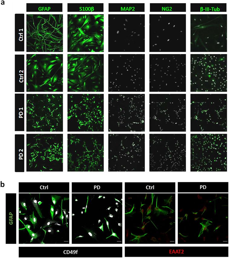
P. Ramos-Gonzalez et al.
                  2
                      LRRK2G2019S mutation in astrocytes induces mitochondrial unba-            (Tuj1 for ectoderm), smooth muscle actin (SMA for mesoderm)
                      lance, leading to cell autonomous and non-autonomous damage               and alpha-Fetoprotein (AFP for endoderm) (Supplementary Fig. 2).
                      that ultimately translates to or exacerbates neurodegeneration.           To induce the differentiation to NSC, neural rosettes were
                      Our results highlight an improvement of astroglial functionality as       cultured with a 50%:50% mixture of laminin 211 (LN211) and
                      a relevant therapeutic target.                                            laminin 111 (LN111) coating. Unlike LN521, these two laminins
                                                                                                are mostly expressed in extra-embryonic membranes and
                                                                                                promote cell differentiation. In our culture conditions, NSCs were
                      RESULTS
                                                                                                differentiated to astrocyte progenitor cells in 21 days. Subse-
                      Generation of patient-derived astrocytes from dermal                      quently, astroglial precursors were further differentiated into
                      fibroblasts
                                                                                                mature astrocytes (see Material and Methods) while maintaining
                      Skin fibroblasts from two patients with LRRK2G2019S mutation and           the coating with LN211/LN111 (50%:50%). After 60–75 days of
                      two healthy donors (Supplementary Table 1) were reprogrammed              maturation, cells were fixed and stained with the astrocyte marker
                      and differentiated to mature astrocytes. Fibroblast were repro-           GFAP. Maturation efficiency was evaluated by cytofluorimetry
                      grammed using the episomal Sendai viral vector bearing the
                                                                                                assay (Supplementary Fig. 3) demonstrating 95%–98% of astro-
                      Yamanaka factors Klf4-Oct3/4-Sox2 (KOS), L-Myc and Klf4 (see
                                                                                                cyte differentiation. Astrocyte differentiation was also confirmed
                      Supplementary Fig. 1 for protocol details). Fibroblasts were
                      expanded in Geltrex until they formed colonies positive for the           by immunofluorescence with antibodies to GFAP and S100B,
                      pluripotent markers Sox2, Oct4 and Nanog (Supplementary Fig. 2).          whereas expression of MAP2 and β-III tubulin (for neurones) and
                      To further potentiate the formation of iPSC, colonies were picked         NG2 (for non-astrocyte glia) was absent or minimal (Fig. 1a). The
                      and expanded for 2–4 days in human recombinant laminin-521                hiA also expressed the functional markers EAAT2 (glutamate
                      (LN521). LN521 is normally expressed in the human embryo at the           transporter) and CD49f, with undetectable differences between
                      inner cell mass and replicates the human stem cell niche in vitro         healthy subjects and PD donors (Fig. 1b). All generated lines from
                      stabilising pluripotent gene expression. At this stage and before         the four donors (healthy and PD) displayed neither genetic nor
                      neural induction, iPSC can differentiate to the three germ layer as       structural variations in somatic and sex chromosomes as
                      evidenced by the expression of Neurone-specific class III β-tubulin        demonstrated in Supplementary Fig. 4.
1234567890():,;

                      Fig. 1 Astrocytic marker expression in hiA. a hiA were maturated for 60 days in LN211/LN111 and fixed for immunofluorescence. All cell
                      cultures, from healthy (Ctrl1-2) and patient (PD1-2) donors, were positive for GFAP and S100β expression, whereas neuronal (MAP2 and β-III-
                      Tub) and non-astrocyte-glial (NG2) markers were nearly absent. White staining shows nuclei labelling by DAPI. b hiA co-immunostaining of
                      GFAP with CD49f and EAAT2 in healthy (Ctrl) and patient (PD) donors. The picture is representative of two Ctrls and two PD cell lines. Scale bar
                      is 25 μm. Photographs are representative of at least five experiments.

                      npj Parkinson’s Disease (2021) 31                                                        Published in partnership with the Parkinson’s Foundation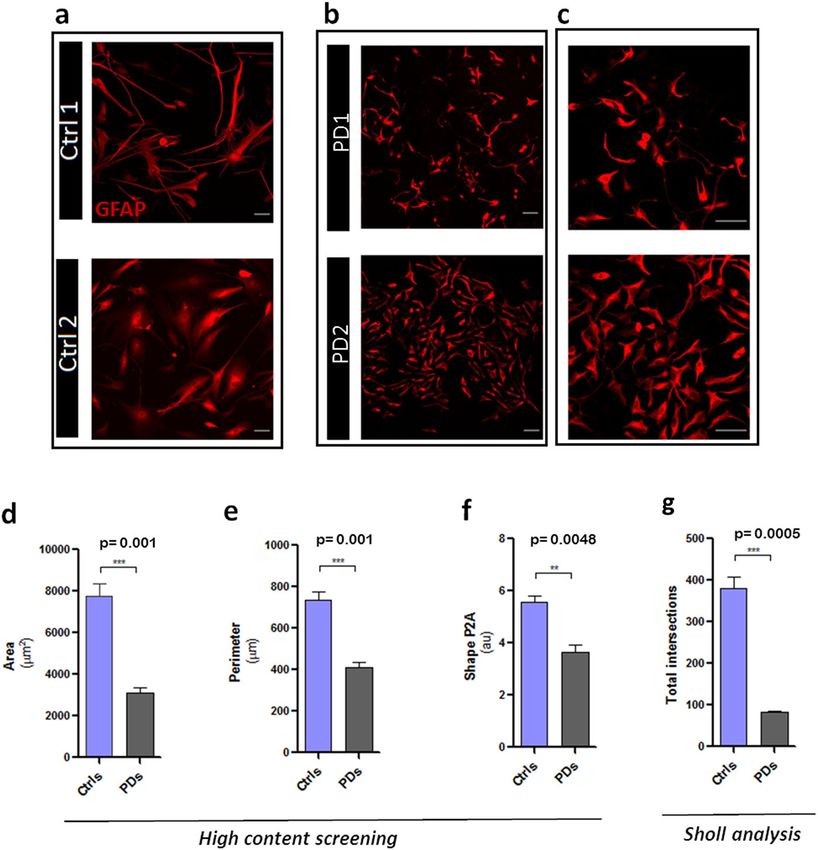
P. Ramos-Gonzalez et al.
                                                                                                                                                  3

Fig. 2 Cell morphology analysis of PD astroglia. Cells from healthy (Ctrl1-2 in a) and patients (PD1-2 in b, c) donors were fixed after 60 days
of maturation and stained with GFAP antibodies. Images in c illustrate a higher magnification of a subfield in b. Morphological analysis was
performed by high-content screening (d–f) and Sholl analysis (g) as described in Materials and Methods. d–f histograms showing astrocyte area
(as a means of squared μm in d), perimeter (as a means of linear μm in e) and complexity (as a means of arbitrary units-au- of shape P2A in f).
In g is expressed the total number of intersections with concentric rings of Sholl grid (5 μm apart) after GFAP immunostaining of controls and
PD astrocytes. Data are presented as mean values ± SEM with n = 4.

Morphology of PD astroglia                                               cytosolic and mitochondrial Ca2+ concentrations controlling
We observed a striking difference in the morphology between              bioenergetics; abnormal astrocytic Ca2+ signalling is increasingly
healthy and PD astrocytes (Fig. 2a–c). The surface area and              recognised as a key process in neurodegenerative conditions25–27.
perimeter of GFAP-positive profiles of PD-derived astrocytes were,        Thus, we analysed Ca2+ dynamics in PD astroglia. Neither healthy
respectively, 60% and 45% smaller when compared to healthy               nor PD astrocytes generated spontaneous Ca2+ transients under
cells, as measured by high-content screening (Fig. 2d–e). Similar        our experimental conditions and we found no difference in resting
data (decrease in surface area and perimeter by 69% and 50%;             cytoplasmic Ca2+ concentration ([Ca2+]i) between healthy and PD
data not shown) were obtained by manual measurements using               hiA (Fig. 3a–b). Application of 100 μM ATP (an archetypal activator
the image software Fiji. Astrocytes from PD patients showed a            of astroglial Ca2+ signalling) evoked transient [Ca2+]i elevation
lower complexity with significant reduction in number or                  (Fig. 3c–d) confirming the presence of functional purinergic
complete absence of primary and secondary processes (Fig. 2f),           receptors coupled to astrocytic Ca2+ signalling machinery. In PD
as evidenced by high-content screening analysis (35% lower than          astrocytes we observed a tendency (which did not reach the level
healthy astrocytes), suggesting a decreased structural capacity for      of significance) of reduction in amplitude and integral of Ca2+
supporting neurones. Decreased complexity of PD-derived astro-           transients in response to ATP (Fig. 3c–d). It has to be noted
cytes was also confirmed by Sholl analysis. As shown in Fig. 2g,          however, that control hiA lines tested in this study displayed
astrocytes derived from PD donors exhibit 61% less intersections.        marked differences in the amplitude of ATP-induced Ca2+
Morphological atrophy in PD cultures was not detected in                 transients. We further tested mitochondrial membrane polarisa-
fibroblasts; moreover, we did not detect differences during the           tion by imaging the quenching of the mitochondrial membrane
iPSC colony formation, but only after astrocyte differentiation          potential probe Rhodamine 123 in the presence of FCCP, which
(data not shown), suggesting a specificity for the astrocytic             revealed significant differences between control and PD hiA lines
phenotype.                                                               (p = 0.0365); (Fig. 3e–f). Taken together, these finding confirm that
                                                                         hiA, with differences between healthy and PD astrocytes, express
Functional characterisation of PD astrocytes                             functional receptors for ATP, typical astrocytic Ca2+ signalling
We next characterised functional properties of hiA. Astroglial           machinery; the PD-derived astrocytes also demonstrated signs of
function and reactivity are tightly integrated with the dynamics of      mitochondrial malfunction.

Published in partnership with the Parkinson’s Foundation                                                   npj Parkinson’s Disease (2021) 31
P. Ramos-Gonzalez et al.
4
Fig. 3 Cytosolic Ca2+ responses to ATP and FCCP in human astrocytes. a–b Time-courses show basal cytosolic Ca2+ responses in control
and PD hiA loaded with fura-2 (n = x-y cells). c Ca2+ responses evoked by ATP (100 μM) in hiA (n = x-y cells). d Comparison of the area under
the curve (AUC) calculated for each experimental condition (n = 3 cultures). e–f Measurement of mitochondrial membrane potential in hiA.
*p = 0.0365. One-way ANOVA followed by Newman–Keuls tests. Ctrls, control. PDs Parkinson´s disease. Data are presented as mean values ±
SEM with n = 3.
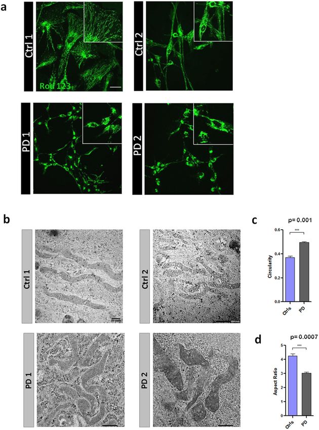
Mitochondrial impairment in PD astrocytes astrocytes. Collectively these results indicate that astrocytes in PD
It is well known that LRRK2 protein interacts with mitochondrial switch from oxidative phosphorylation to the aerobic glycolytic
membranes and affects mitochondrial respiration13,28. We, thus, respiration.
analysed mitochondrial metabolism in healthy and in PD Mitochondrial malfunction is frequently associated with aber-
astrocytes using Seahorse technology. We first measured the rant morphology29 and, therefore, we compared intracellular
mitochondrial oxygen consumption rate (OCRs) of hiA in a live-cell distribution and the ultrastructure of mitochondria in healthy and
metabolic assay (Fig. 4a). PD astrocytes showed lower OCRs in PD astrocytes. Mitochondrial distribution and gross morphology
was visualised by staining with Rhodamin 123. In healthy
both basal (Fig. 4b) and maximal (Fig. 4c) respiration paradigms,
astrocytes, mitochondria were elongated and interconnected,
when compared to healthy cells. PD astrocytes also produced less forming a homogenous network distributed throughout the entire
ATP (Fig. 4d). We did not, however, observe significant differences cytoplasm, being present in the soma and in the principal
between healthy and PDs astrocytes in terms of spare respiratory processes (Fig. 6a). In contrast, PD astrocytes had fewer
capacity or proton leak (Fig. 4e–f). Consistent with a mitochondrial mitochondria, which were apparently more fragmented and
respiration deficit, PD astrocytes displayed increased glycolytic mainly concentrated in the perinuclear region; in addition
capacity as determined by changes in the extracellular acidifica- mitochondria were absent from short processes (Fig. 6a). The
tion rate (ECAR) (Fig. 5a), both in basal (Fig. 5b) and in very same distribution pattern was observed after staining with
compensatory glycolysis (Fig. 5c). Similarly, basal proton efflux Mitotracker (Supplementary Fig. 5), which demonstrates evident
rate (PER, the measure of extracellular acidification, Fig. 5d), but perinuclear concentration of mitochondria in PD astrocytes.
not the PER derived from glycolysis (Fig. 5e), was increased in PD Ultrastructural analysis of mitochondria (Fig. 6b), revealed further
npj Parkinson’s Disease (2021) 31 Published in partnership with the Parkinson’s Foundation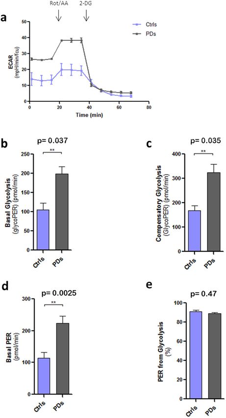
P. Ramos-Gonzalez et al.
                                                                                                                                                      5

Fig. 4 Mitochondrial metabolism and respiration. a Oxygen
consumption rates (OCRs) of Ctrl and PD astrocytes. Oligomycin,
FCCP and rotenone (Rot) were added, respectively, after 20, 40, and
60 min as respiratory chain blockers. OCRs are expressed in b–f as
pmol por minute after cell viability normalisation with calcein        Fig. 5 Glycolytic activity. a Extracellular acidification rate (ECAR) of
staining. b Basal respiration, c maximal respiration and d ATP         Ctrl and PD astrocytes. b Basal glycolysis, expressed as pmol/min of
production are reduced in PD astrocytes compared to the controls.      glycolytic proton efflux rate (PER), c Compensatory glycolysis and
e Spare respiratory capacity and f H+ Leak do not show statistically   d Basal PER, expressed as pmol/min of PER, are increased in PD
significant changes (n = 4). Statistical analysis was performed using   astrocytes compared to the controls. e PER from glycolysis is used as
one-way ANOVA. Data are presented as mean values ± SEM.                an internal control and it is similar in the four lines (n = 3). Statistical
                                                                       analysis was performed using one-way ANOVA. Data are presented
                                                                       as mean values ± SEM.
differences between the healthy and PD astrocytes. The measure-        astrocytes, with impaired mitochondrial respiration, cellular
ment of the circularity, usually used as an index of ROS               localisation and mitochondrial ultrastructure.
production, demonstrated that mitochondria in PD astrocytes
were more rounded than in the control cells (Fig. 6c). Accordingly,
the Aspect Ratio (the major axis divided the minor axis of the         DISCUSSION
mitochondria) was higher in healthy astrocytes indicating the
                                                                       To study human diseases, the “humanised” experimental prepara-
presence of more elongated mitochondria.
                                                                       tions are essential; even the most sophisticated animal models of
   According to the hypothesis by which fragmented mitochon-
dria are associated with higher levels of ROS29,30, we investigated    human pathologies are not faithful replicas31,32. In this paper, we
astrocytic metabolic profile. Using Oxyblot analysis, we measured       analysed morphological characteristics and metabolic profile of
the carbonyl groups of total proteins extracted from healthy and       astrocytes derived from iPSCs generated from PD patients bearing
diseased lines as a readout of the oxidative status of the proteins.   the LRRK2G2019S mutation. Using different combinations of several
We found higher amount of oxidised proteins (32%) in PD                laminins coating, we obtained almost homogeneous cultures of
astrocytes when compared to the controls (Fig. 7), suggesting a        human astrocytes (95%–98%). The purity of cultures was
basal oxidative status of astrocytes in PD higher than in healthy      confirmed by citofluorimetry analysis (Supplementary Fig. 3). We
astrocytes. We may conclude, therefore, that LRRK2G2019S muta-         simulated the physiological conditions occurring during the
tion corresponds to a general mitochondrial dysfunction in             embryonic development by mixing laminins LN521 and LN511.

Published in partnership with the Parkinson’s Foundation                                                   npj Parkinson’s Disease (2021) 31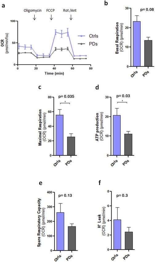
P. Ramos-Gonzalez et al.
6

    Fig. 6 Analysis of mitochondrial morphology. a Mitochondrial staining with Rhodamin 123 of hiA cultures from healthy (Ctrl1-2) and
    patients (PD1-2) donors . Images were taken with the confocal microscope Leica TCS STED CW SP8. Squared inlets represent a higher
    magnification of the field. Scale bar 20 μm. b Representative images of mitochondrial ultrastructure in Ctrl and PD astrocytes. c Circularity is
    measured considering 1 as the perfect circle and d aspect ratio (ratio of circularity vs. elongation) reveal a more rounded shape in PD astrocyte
    mitochondria compared to the control. More than 100 mitochondria were analysed for each line. Statistical analysis was performed using one-
    way ANOVA. Data are presented as mean values ± SEM.

    Both these laminins are expressed in the inner cell mass and                  Nonetheless, early in vitro experiments have clearly demon-
    support survival and self-renewal of the pluripotent stem cells            strated that astrocytes protect and support survival of dopami-
    through the interaction with α6β1 integrin and PI3/Akt activa-             nergic neurones45. Subsequent studies revealed that functional
    tion33,34. In contrast, mature astrocytes express LN111 and                exhaustion and loss of astroglial homoeostatic support are
    LN21135,36; activation of these two laminins supports cell                 dominant glial contribution to the PD, and the special definition
    differentiation and specialisation, such as, for example, the              of “dysfunctional” astrocytes has been introduced22,46. Further-
    maintenance of the blood-brain barrier integrity36.                        more, analysis of post-mortem samples of susbtantia nigra
       The role of astrocytes in pathological progress of PD is yet to be      obtained from PD patients demonstrated significant decrease in
    fully characterised. Recent works conducted on some inflamma-               expression of astroglial markers compared to healthy controls47,48;
                                                                               these findings being in general agreement with our concept of
    tory experimental paradigms have suggested two subtypes of
                                                                               astroglial atrophy linked to the disease. Astroglial asthenia,
    astrocytes, A1 and A2 with neurotoxic and neuroprotective
                                                                               atrophy and loss of homoeostatic and neuroprotective capacities
    profiles37. The A1/A2 dichotomy has been based on correlative               were noted in aging40 and in various neurodegenerative and
    analysis of limited number of genes detected for specific                   neuropsychiatric diseases23,49; astroglial atrophy thus represents a
    conditions in the in vitro settings. This binary polarisation has          defined class of astrogliopathies50.
    not been confirmed38–43 and, similarly to once popular, but now                Obtaining an almost pure population of iPSC-derived astrocytes
    discarded, M1/M2 microglial polarisation concept, has been                 allowed us to study human astrocytes bearing pathophysiological
    repudiated by neuroglial community44.                                      signature. We have found a prominent aberrant morphology of

    npj Parkinson’s Disease (2021) 31                                                         Published in partnership with the Parkinson’s Foundation
P. Ramos-Gonzalez et al.
7
as an exacerbating factor in neurodegenerative process; chroni-
cally malfunctional astroglia was suggested to contribute to death
of dopaminergic neurones60. It is early to conclude that astrocytic
atrophy is the hallmark of PD until the same astrocytic atrophy is
characterised in situ in patient’s brain tissues or in astrocytes
derived from other mutations. Detailed analysis of astrocyte
morphology should be performed in other regions of the brain
that are related with dopaminergic degeneration (e.g., subtha-
lamic nucleus or globus pallidum) and not only in degenerating
regions where astrocytes are pathologically remodelled.
Mutation in the LRRK2 gene may be specifically responsible for
both aberrant morphology and mitochondrial dysfunction
observed in LRRK2G2019S hiA. Abnormalities in neurites outgrowth
and branching are among the earliest pathological phenotypes
observed in LRRK2 mutations61,62. It has been initially proposed
Fig. 7 Detection of oxidised proteins in total astrocyte protein. that the origin of these morphological changes could be related to
Total proteins from hiA cultures were extracted after 60 days of an apoptotic process62; however, further studies provided
in vitro maturation. Oxidised proteins are visualised after Western evidence for an association of LRRK2 with tubulin/actin, thus
blot analysis as the conversion of the 2,4-dinitrophenol (DNP) to 2,4- suggesting that morphological changes may be consequences of
dinitrophenylhydrazine (DNPH). Each sample is loaded as a negative
control (Neg) with non-derivatised procedure. DNPH levels were
LRRK2-modulation of cytoskeletal dynamics63. Several lines of
normalised with total proteins stained with Red Ponceau. evidence suggest the relationship of LRRK2 protein with the
cytoskeleton: (i) The GTPase domain of LRRK2 protein can pull-
down α/β tubulin from cell lysates of mouse fibroblasts and
astrocytes derived from PD LRRK2G2019S patients. Differentiated human embryonic kidney64; (ii) LRRK2 co-precipitates with β
astrocytic cultures, obtained from both healthy and PD subjects, tubulin from wild-type mouse brain and (iii) Recombinant LRRK2
expressed classical astrocyte markers (GFAP, S100B, CD49f and can phosphorylate β tubulin in vitro65. High-throughput screening
EAAT2). The PD astrocytes, however, were characterised by of LRRK2 interactome revealed proteins of the actin family and of
substantially smaller area and perimeter; they also show the actin-regulatory network as interactors of LRRK2 in actin
diminished complexity of primary and secondary processes as polymerisation in vitro16. We presume therefore, that the atrophy
evidenced by high-content screening and Sholl analysis. These observed in PD LRRK2G2019S astrocytes could be a consequence of
morphological changes do not represent culture artefact because the mutated LRRK2 protein breakdown that becomes unable to
this atrophy was observed only in fully differentiated astrocytes properly modulate cytoskeletal dynamics. Similarly, LRRK2 muta-
and not at preceding derivation stages. Previously published study tion can be responsible for mitochondrial dysfunction and
of iPSCs-derived astrocytes with LRRK2G2019S mutation51 did not fragmentation, as already observed in fibroblasts, neural stem
found conspicuous morphological changes, although astrocytic cells or neuroblastoma cell lines13,66–68. Multiple studies demon-
appearance was not analysed in detail. We assume that the use of strated that LRRK2 loss of function, associated with G2019S and
specific feeder layers (laminins) and smaller number of cell R1441G mutations impair mitochondrial oxidative state increasing
passages in our protocol (differentiation to astrocyte proceeds the neuronal susceptibility to oxidative stress damage69–71. One
with weekly passages) diminishes cell reactivity thus reliably possible explanation might be a mitochondrial DNA damage
revealing cell morphology. Our findings of pronounced morpho- induced by the LRRK2 mutations, which was observed in midbrain
logical atrophy in human iPSCs parallel recent demonstration of cultures and PD patient-derived lymphoblastoid cell lines72.
similar morphological atrophy in iPSC-derived astrocytes gener- The observations that LRRK2 mutation may be responsible for
ated from early familial and late sporadic AD patients52. morphological atrophy and mitochondrial malfunction, indicate
Morphological atrophy of astrocytes is arguably associated with possible mechanism associated with reduced neuroprotection in
neuronal damage (due to failed homoeostatic support) and this mutation carriers. A recent observation demonstrated that
aberrant synaptic connectivity manifest in neurodegenerative G2019S mutation in hiA alters the astrocyte-to-neurone commu-
and psychiatric diseases (for a review see ref. 50). In many cases, nication mediated by extracellular vesicles73. In this work, the
astrocytic atrophy precedes cell death and neuronal degeneration. LRRK2 mutation in astrocytes was claimed to affect morphology
For example, in acute excitotoxic neurodegeneration and ALS, and the content of extracellular vesicles and multivesicular bodies
morphological aberrations are accompanied with the down- (MVB). The authors found that neurones incorporated astrocyte
regulation of glutamate transporters and increased excitotoxi- MVB with an abnormal accumulation of key PD-related proteins
city18. Morphological atrophy in AD has been described in animal such as LRRK2 and phospho-S129 α-Syn. Dopaminergic neurones
models54–56, in human iPSC-derived astrocytes from patients53, in incorporating the dysfunctional MVB released by the LRRK2G2019S
deprenil-based brain imaging in patients57, and in post-mortem astrocytes showed an aberrant morphology73.
brain at late stages of the disease (Rodriguez and Verkhratsky, In this study, we propose an hallmark for PD with LRRK2G2019S
unpublished results). In our culture conditions atrophic astrocytes mutation. Our hypothesis postulates that astrocytes with this
from PD patients showed normal viability, as demonstrated by mutation fail to support neurones because of loss of homoeostatic
expression of classical markers (Fig. 1) and by physiological [Ca2+]i support resulting from substantial morphological atrophy and loss
dynamics (Fig. 3). At the same time PD astrocytes demonstrate of complexity; in addition, astrocytes demonstrated mitochondrial
reduced mitochondrial functionality (Figs 4–6). Mitochondrial dysfunction that also affects their neuroprotective capabilities.
aberrations and morphological atrophy may explain why astro-
cytes in PD with LRRK2G2019S mutation fail to support and protect
neurones. This loss of function became even more evident in METHODS
specific brain regions, specifically for substantia nigra pars Human samples
compacta and striatum, where astrocytic density is lower Human fibroblasts were obtained from two healthy donors (Ctrl1 was
compared to other regions20. Furthermore, astrocytes from purchased from AXOL and Ctrl 2 from the Coriell stem cell bank) and two
substantia nigra seem to be unusually vulnerable to ischaemic Parkinson´s disease patients with LRRK2G2019S mutation (PD1 from the
attack58 and oxidative stress59. Loss of astroglial support may act Coriell stem cell bank and PD 2 provided by the BioDonostia Hospital, San
Published in partnership with the Parkinson’s Foundation npj Parkinson’s Disease (2021) 31P. Ramos-Gonzalez et al.
8
Sebastian, Spain) (Supplementary Table 1). Control patients who matched digital black/white CCD camera (ORCA, Hamamatsu Photonics Iberica,
PD donors in age and gender did not show any neurological symptoms. All Barcelona, Spain) and images were acquired every 5 s. Image acquisition
procedures with human cells were approved by the National and local and data analysis were performed using the AquaCosmos software
ethical committees (with code M30_2018_030) according to the National programme (Hamamatsu Photonics Iberica). Intracellular Ca2+ measure-
law 14/2007 on Biomedical research. ments were expressed as the ratio of F340/F380 and normalised to baseline
values. Results for statistical comparison were calculated as area under the
curve (AUC) of the response for each cell from the start of each compound
Generation of human induced astrocytes (hiA)
application.
Fibroblasts were grown in DMEM F12 (Gibco/ThermoFisher, Spain) and
infected with the CytoTune iPS 2.0 Sendai Reprogramming Kit (Thermo-
fisher, Spain) as described in Supplementary Fig. 1. The commercial Sendai Immunofluorescence
virus expressed the key genetic factors necessary for reprogramming Cell cultures were fixed in 4% para-formaldehyde (Merck/Sigma),
somatic cells into iPSCs (Klf4/Oct3-4/Sox2-KOS, hc-Myc, Klf4). Infection permeabilised with 0.1% Triton (Sigma) and non-specific epitopes were
efficiency was evaluated by co-infection with a EmGFP fluorescent reporter blocked with 5% normal goat serum in PBS. Primary antibodies
plasmid provided by the kit. Seven days later, transduced fibroblasts were (Supplementary Table 2) were incubated overnight and then washed
seeded in Geltrex (ThermoFisher, Spain) in Essential 8 Flex medium (E8, three times with 0.1% Triton in PBS. Secondary conjugated antibodies
Gibco/ThermoFisher, Spain). E8 medium was changed every day for Alexa 488, Alexa 594, Alexa 647 or Alexa 405 (Invitrogen, 1:500), were
21 days until we observed the formation of iPSC colonies. Colonies were incubated for 1 h in the dark at room temperature. After three washes with
manually isolated using a 27G Braun Sterican Needle and replated in 0.1% Triton in PBS, cell nuclei were counter-stained for 1 min with DAPI
laminin-521 (LN521-Biolamina, Sundbyberg Sweden) with E8 medium and (ThermoFisher). Finally, coverslips were mounted with Glycergel (Dako,
ROCK inhibitor (Y-27632; Millipore, Madrid, Spain). The day after, ROCK Barcelona, Spain) and analysed using the confocal microscope Leica TCS
inhibitor was removed and replaced with fresh medium. Colonies were STED CW SP8.
sequentially isolated and re-suspended at single cell level. Embryoid bodies
(EB) were generated (Supplementary Fig. 2) after re-suspending the iPSC
colonies in Essential 6 medium (E6, Gibco) for 2–4 days in the Morphological analysis by high-content screening
AggrewellTM800 plates (StemCell, Grenoble, France). Half-medium in the Cells were seeded in glass bottom Cellvis 24-well plates (Cellvis, Bilbao,
microwells was replaced daily with fresh medium. EBs were then seeded in Spain) coated with LN111/LN211 (Biolamina). After fixation with 4% PFA for
a LN521/LN211 mix (50% each) (Biolamina) and the differentiation to 8 min, cells were immunostained for GFAP expression (Goat anti-GFAP,
neural precursor cells (NPC) as neural rosettes was promoted using the Abcam 53554). Alexa fluor Donkey anti-goat was used as a secondary
STEMdiff Neural Induction Medium (Stemcell). After 7 days, neural rosettes antibody. Images were taken with the CellInsight CX7 high-content
were selected and detached using the STEMdiff Neural Rosette Selection screening system (Thermo Scientific) using a 10x objective. Morphological
Reagent (Stemcell). Cells were incubated for 2 h with this reagent at 37 °C parameters for area (defined as the number of microns squared of the
with 5% CO2 and then, mechanically re-suspended at single cell level and object), perimeter (length of the boundary of the object) and ShapeP2A
seeded in LN211/LN111 (50% each) (Biolamina). Differentiation of NPC to (measure of the ratio of the perimeter squared of the object to four times)
progenitor astrocytes was triggered using the astrocyte differentiation were calculated with High-Content Analysis platform. More than 100 cells
medium (STEMdiff astrocyte differentiation #100-0013, StemCell). To per cell line were analysed.
maintain the appropriate cell density (70% of confluence) cells were
passed every week in the same coating mix during 21 days. Maturation.
Finally, astrocytes progenitor cells were maturated in the Astrocyte
Morphological assessment
Maturation Medium (STEMdiff astrocyte maturation #100-0016, StemCell) Sholl analysis was performed with the public software Fiji75, to measure the
for 60 to 75 days. During the whole protocol, the correct state of the cells complexity of GFAP-positive human astrocytes. A transparent grid with
in each step was evaluated using the EVOS FL microscope (Life concentric circles (every 5 μm from the centre of the cell soma across the
Technologies, AME4300). See Supplementary Fig. 1 for an overview of whole radius) were superimposed onto the cells after immunofluorescence
simplified protocol steps. with GFAP antiserum. Sholl measurements were obtained by quantifying
the number of intersections with each concentric circle.
Cytofluorimetry assay
Cells (500.000) were detached with TryPLE (Sigma, Spain) and fixed as a Electron microscopy
single cell suspension with PFA 4% for 10 min. Cells were washed in Cells were fixed in 4% PFA for 10 min and post-fixed in 3% glutaraldehyde
phosphate-buffered saline (PBS) at 400 x g for 5 min and re-suspended in for 30 min. After a wash in phosphate buffer (PB) samples were osmicated
blocking solution (0.5 g BSA in PBS with 0.01% Triton (PBS-T) with agitation (1% OsO4 in 0.1 M PB; pH 7.4) for 30 min. After 3 x 10 min washes in 0.1 M
overnight at 4 °C. The following day cells were washed and incubated with PB, samples were dehydrated in graded ethanol concentrations (50%
the primary antibody goat anti-GFAP (Abcam, 53554) for 2 h at 4 °C. After to100%) to propylene oxide and embedded in epoxy resin (Sigma-Aldrich)
further wash for 5 min in PBS-T 0.01% cell suspension was incubated with by immersion in decreasing concentration of propylene oxide (1:3 for
the secondary conjugated antibody Alexa fluor 488 donkey anti-goat for 30 min, 1:1 for 1 h and 3:1 for 2 h). Samples were then embedded in fresh
1 h at 4 °C. After a further wash with PBS-T 0.01%, cells were finally re- resin overnight and allowed to polymerise at 60 °C for 2 days. Following
suspended in PBS 1x. Cells were analysed in the BD FACSJazz (USB, inFlux visualisation at the light microscope, selected portions were trimmed and
Compact) analyser using the Blue 488 laser. Unstained cells were gated glued onto epoxy resin capsules. Semi-thin sections (500 nm-thick were cut
and used as a negative control. from epoxy blocks using a Power Tome ultramicrotome (RMC Boeckeler,
Tucson, AZ, USA and stained with 1% toluidine blue. Ultrathin (50–60 nm
thick) sections were then cut with diamond knife (Diatome, Hatfield PA,
Calcium imaging USA), collected on nickel mesh grids and stained with 4% uranyl acetate
Cytosolic calcium (Ca2+) levels were estimated by the 340/380 ratiometric for 30 min and 2.5% lead citrate for electron microscope visualisation. For
microfluorimetry as described previously74. Astrocytes were loaded with Image Acquisition and analysis, electron microscopy images of mitochon-
fura-2 AM (5 μM; ThermoFisher/Invitrogen) for 20 at 37 °C and subse- dria were taken from randomly selected fields with a Jeol JEM 1400 Plus
quently washed in the recording solution containing 137 mM NaCl, 5.3 mM electron microscope at the Service of Analytical and High-Resolution
KCl, 0.4 mM KH2PO4, 0.35 mM Na2HPO4, 20 mM HEPES, 4 mM NaHCO3, Microscopy in Biomedicine of University of the Basque Country UPV/EHU.
10 mM glucose, 1 mM MgCl2, 2 mM CaCl2 (pH 7.4) to allow de- Images were taken at a magnification of 12,000X. Circularity and aspect
esterification. In experiments with FCCP, Ca2+ was omitted from the ratio (ratio of circularity vs. elongation) were measured with Fiji-Software
recording solution. Experiments were performed in a coverslip chamber using a self-made plug-in. More than 100 mitochondria were analysed for
continuously perfused with buffer at 1 ml/min by exposing the cells to ATP each line.
(100 μM) or FCCP (1 μM). The perfusion chamber was mounted on the
stage of a Zeiss (Oberkochen, Germany) inverted epifluorescence
microscope (Axiovert 35), equipped with a 150 W xenon lamp Polychrome Mitochondrial membrane potential (ΔΨm) measurement
IV (T.I.L.L. Photonics, Martinsried, Germany), and a Plan Neofluar 403 oil Mitochondrial membrane potential (ΔΨm) of human astrocytes was
immersion objective (Zeiss). Cells were visualised with a high-resolution assessed by the Rhodamine 123 (Rh123) staining. Briefly, cells were
npj Parkinson’s Disease (2021) 31 Published in partnership with the Parkinson’s FoundationP. Ramos-Gonzalez et al.
9
seeded in 35 mm glass bottom plates (Ibidi GmbH, Germany) at a mean REFERENCES
density confluence of 50–70% and loaded with 10 μM Rh123 at 37 °C and 1. Parkinson, J. An essay on the Shaking Palsy. Arch. Neurol. 20, 441–445 (1969).
5% CO2. After 15 min cells were washed with 900 μl Hanks’ balanced salt 2. Reeve, A., Simcox, E. & Turnbull, D. Ageing and Parkinson’s disease: why is
solution and analysed by time lapse every 15 s for 5 min using the confocal advancing age the biggest risk factor? Ageing Res. Rev. 14, 19–30 (2014).
microscope Leica TCS STED CW SP8. To establish the basal line, cells were 3. Spillantini, M. G. et al. α-synuclein in Lewy bodies. Nature 388, 839–840 (1997).
stimulated with 1 μM FCCP after the first 60 s. Fluorescence intensity after 4. Ross, C. A. & Poirier, M. A. Protein aggregation and neurodegenerative disease.
FCCP treatment was measured with the Leica LASX Software and data Nat. Med. 10, S10–S17 (2004).
were analysed with GraphPad Prism 5 (San Diego, CA, USA). 5. Desplats, P. et al. Inclusion formation and neuronal cell death through neuron-to-
neuron transmission of α-synuclein. Proc. Natl Acad. Sci. 106, 13010–13015
Measurement of mitochondrial function and glycolytic activity (2009).
6. Blauwendraat, C., Nalls, M. A. & Singleton, A. B. The genetic architecture of Par-
The oxygen consumption rate (OCR), as an indicator of mitochondrial kinson’s disease. Lancet Neurol. 19, 170–178 (2019).
respiration, the extracellular acidification rate (ECAR), as indicator of 7. De Wit, T., Baekelandt, V. & Lobbestael, E. LRRK2 phosphorylation: behind the
glycolytic activity, and the proton efflux rate (PER), which correlates with scenes. Neuroscientist 24, 486–500 (2018).
lactate production, were measured with the Seahorse XF96 extracellular 8. Singleton, A. B., Farrer, M. J. & Bonifati, V. The genetics of P arkinson’s disease:
flux analyser. For the analysis of mitochondrial respiration, human
progress and therapeutic implications. Mov. Disord. 28, 14–23 (2013).
astrocytes (30,000 cells/ mm2) were seeded in LN211/LN111 (Biolamina)
9. West, A. B. et al. Parkinson’s disease-associated mutations in LRRK2 link enhanced
precoated wells. The day of the experiment, cell medium was changed to GTP-binding and kinase activities to neuronal toxicity. Hum. Mol. Genet. 16,
Mito XF Medium (XF basal medium with phenol red, 0.001 M piruvic acid, 223–232 (2007).
0.002 M glutamine, glucose 0.01 M, pH 7.4). The OCRs were obtained after 10. Juárez-Flores, D. L. et al. Exhaustion of mitochondrial and autophagic reserve may
the sequential treatment with oligomycin (2 μM), FCCP (1 μM), and contribute to the development of LRRK2 G2019S-Parkinson’s disease. J. Transl.
rotenone combined with antimycin A (0.5 μM). To measure the glycolytic Med. 16, 1–13 (2018).
activity, we used the same protocol with the following modifications. The
11. Häbig, K. et al. LRRK2 guides the actin cytoskeleton at growth cones together
day of the experiment, cell medium was changed to Glico XF Medium
with ARHGEF7 and Tropomyosin 4. Biochim. Biophys. Acta (BBA)-Mol. Basis Dis.
without phenol red (DMEM Base Medium without Phenol Red with 5 mM 1832, 2352–2367 (2013).
HEPES, 10 mM glucose, 1 mM sodium pyruvate, 2 mM glutamine, pH 7.4 at 12. Mortiboys, H., Johansen, K. K., Aasly, J. O. & Bandmann, O. Mitochondrial
37 °C). The ECAR and PER were obtained after the sequential treatment impairment in patients with Parkinson disease with the G2019S mutation in
with rotenone combined with antimycin A (0.5 μM) and 2DG (50 mM), LRRK2. Neurology 75, 2017–2020 (2010).
respectively. Four replicates were performed for each condition or cell type
13. Wang, X. et al. LRRK2 regulates mitochondrial dynamics and function through
for every experiments (n = 3). Data was analysed with the Wave
direct interaction with DLP1. Hum. Mol. Genet. 21, 1931–1944 (2012).
2.4.0 software. 14. Papkovskaia, T. D. et al. G2019S leucine-rich repeat kinase 2 causes uncoupling
protein-mediated mitochondrial depolarization. Hum. Mol. Genet. 21, 4201–4213
Western blot for detection of oxidatively modified proteins (2012).
(oxyblot) 15. Hsieh, C. H. et al. Functional impairment in miro degradation and mitophagy is a
shared feature in familial and sporadic Parkinson’s disease. Cell Stem Cell 19,
Astrocytes (30.000/well) were maturated for 60 days in 24-well plates
709–724 (2016).
coated with LN211/LN111 (Biolamina) and solubilised for 20 min with
16. Meixner, A. et al. A QUICK screen for Lrrk2 interaction partners–leucine-rich
equal volume of 2x Extraction Buffer. Samples were prepared according
repeat kinase 2 is involved in actin cytoskeleton dynamics. Mol. Cell. Proteomics
to manufacturer’s instruction with all the reagents provided in Oxidised
10 M110.001172 (2011)
protein western blot detection kit (ab178020; Abcam). Briefly, the
17. Verkhratsky, A. & Nedergaard, M. Physiology of astroglia. Physiological Rev. 98,
carbonyl groups in the protein side chains were derivatised to 2,4-
239–389 (2018).
dinitrophenylhydrazone (DNP-Hydrazone) by reaction with 2,4-dinitro-
18. Pekny, M. et al. Astrocytes: a central element in neurological diseases. Acta
phenylhydrazine (DNPH). Two aliquots of each sample were prepared to
Neuropathologica 131, 323–345 (2016).
be analysed simultaneously. One aliquot was treated with “derivatisa-
19. Verkhratsky, A., Steardo, L., Parpura, V. & Montana, V. Translational potential of
tion reaction” (DNPH Solution) and the other control aliquot was treated
astrocytes in brain disorders. Prog. Neurobiol. 144, 188–205 (2016).
with “derivatisation control reaction”. Protein concentration was
20. von Bartheld, C. S., Bahney, J. & Herculano-Houzel, S. The search for true numbers
quantified with a detergent-compatible assay reagent (Pierce BCA
of neurons and glial cells in the human brain: a review of 150 years of cell
Protein Assay Kit) according to the manufacturer’s instructions (Thermo-
counting. J. Comp. Neurol. 524, 3865–3895 (2016).
Fisher Scientific). Proteins loading was normalised after Red Ponceau
21. Song, Y. J. C. et al. Degeneration in different parkinsonian syndromes relates to
staining. All blots derive from the same experiment and they are
astrocyte type and astrocyte protein expression. J. Neuropathol. Exp. Neurol. 68,
processed in parallel.
1073–1083 (2009).
22. Booth, H. D., Hirst, W. D. & Wade-Martins, R. The role of astrocyte dysfunction in
Statistical analysis Parkinson’s disease pathogenesis. Trends Neurosci. 40, 358–370 (2017).
Results are expressed as mean ± standard error of the mean (S.E.M) with n 23. Verkhratsky, A., Marutle, A., Rodriguez-Arellano, J. J. & Nordberg, A. Glial asthenia
corresponding to the number of cells or cultures tested. Data were and functional paralysis: a new perspective on neurodegeneration and Alzhei-
analysed with Excel (Microsoft, Seattle, WA, USA) and GraphPad Prism mer’s disease. Neuroscientist 21, 552–568 (2015).
software. Statistical significance between datasets was tested using one- 24. Lee, J. H. et al. Parkinson’s disease-associated LRRK2-G2019S mutant acts through
way analysis of variance (ANOVA) followed by Newman–Keuls multiple regulation of SERCA activity to control ER stress in astrocytes. Acta Neuropatho-
comparison test, with a significance threshold of p < 0.05. logica Commun. 7, 1–19 (2019).
25. Shigetomi, E., Saito, K., Sano, F. & Koizumi, S. Aberrant calcium signals in reactive
astrocytes: a key process in neurological disorders. Int. J. Mol. Sci. 20, 996 (2019).
Reporting summary 26. Grolla, A. A. et al. Amyloid-β and Alzheimer’s disease type pathology differentially
Further information on research design is available in the Nature Research affects the calcium signalling toolkit in astrocytes from different brain regions.
Reporting Summary linked to this article. Cell Death Dis. 4, e623 (2013).
27. Kuchibhotla, K. V., Lattarulo, C. R., Hyman, B. T. & Bacskai, B. J. Synchronous
hyperactivity and intercellular calcium waves in astrocytes in Alzheimer mice.
DATA AVAILABILITY Science 323, 1211–1215 (2009).
The data that support the findings of this study are available from the corresponding 28. Stafa, K. et al. Functional interaction of Parkinson’s disease-associated LRRK2 with
author upon reasonable request. members of the dynamin GTPase superfamily. Hum. Mol. Genet. 23, 2055–2077
(2014).
29. Picard, M., Shirihai, O. S., Gentil, B. J. & Burelle, Y. Mitochondrial morphology
Received: 8 October 2020; Accepted: 3 March 2021; transitions and functions: implications for retrograde signaling? Am. J. Physiol.
Regul. Integr. Comp. Physiol. 304, R393–R406 (2013).
Published in partnership with the Parkinson’s Foundation npj Parkinson’s Disease (2021) 31P. Ramos-Gonzalez et al.
10
30. Ježek, J., Cooper, K. F. & Strich, R. Reactive oxygen species and mitochondrial 58. Karunasinghe, R. N., Dean, J. M. & Lipski, J. Acute sensitivity of astrocytes in the
dynamics: the yin and yang of mitochondrial dysfunction and cancer progres- Substantia Nigra to oxygen and glucose deprivation (OGD) compared with hip-
sion. Antioxidants 7, 13 (2018). pocampal astrocytes in brain slices. Neurosci. Lett. 685, 137–143 (2018).
31. Hartung, T. Thoughts on limitations of animal models. Parkinsonism Relat. Disord. 59. Elgayar, S. A., Abdel-Hafez, A. A., Gomaa, A. M. & Elsherif, R. Vulnerability of glia
14, S81–S83 (2008). and vessels of rat substantia nigra in rotenone Parkinson model. Ultrastructural
32. Ransohoff, R. M. All (animal) models (of neurodegeneration) are wrong. Are they Pathol. 42, 181–192 (2018).
also useful? J. Exp. Med. 215, 2955 (2018). 60. Kuter, K., Olech, Ł. & Głowacka, U. Prolonged dysfunction of astrocytes and
33. Domogatskaya, A., Rodin, S. & Tryggvason, K. Functional diversity of laminins. activation of microglia accelerate degeneration of dopaminergic neurons in the
Annu. Rev. Cell Dev. Biol. 28, 523–553 (2012). rat substantia nigra and block compensation of early motor dysfunction induced
34. Rodin, S., Antonsson, L., Hovatta, O. & Tryggvason, K. Monolayer culturing and by 6-OHDA. Mol. Neurobiol. 55, 3049–3066 (2018).
cloning of human pluripotent stem cells on laminin-521–based matrices under 61. Tsika, E. et al. Conditional expression of Parkinson’s disease-related R1441C LRRK2
xeno-free and chemically defined conditions. Nat. Protoc. 9, 2354 (2014). in midbrain dopaminergic neurons of mice causes nuclear abnormalities without
35. Liddelow, S. A. et al. Neurotoxic reactive astrocytes are induced by activated neurodegeneration. Neurobiol. Dis. 71, 345–358 (2014).
microglia. Nature 541, 481–487 (2017). 62. MacLeod, D. et al. The familial Parkinsonism gene LRRK2 regulates neurite pro-
36. Al-Dalahmah, O. et al. Single-nucleus RNA-seq identifies Huntington disease cess morphology. Neuron 52, 587–593 (2006).
astrocyte states. Acta Neuropathol. Commun. 8, 19 (2020). 63. Wallings, R., Manzoni, C. & Bandopadhyay, R. Cellular processes associated with
37. Amini-Bavil-Olyaee, S. et al. Genotype characterization and phylogenetic ana- LRRK 2 function and dysfunction. FEBS J. 282, 2806–2826 (2015).
lysis of hepatitis B virus isolates from Iranian patients. J. Med. Virol. 75, 227–234 64. Gandhi, P. N., Wang, X., Zhu, X., Chen, S. G. & Wilson-Delfosse, A. L. The Roc
(2005). domain of leucine-rich repeat kinase 2 (LRRK2) is sufficient for interaction with
38. Das, S., Li, Z., Noori, A., Hyman, B. T. & Serrano-Pozo, A. Meta-analysis of mouse microtubules. J. Neurosci. Res. 87, 1711–1720 (2008).
transcriptomic studies supports a context-dependent astrocyte reaction in acute 65. Gillardon, F. Leucine-rich repeat kinase 2 phosphorylates brain tubulin-β isoforms
CNS injury versus neurodegeneration. J. Neuroinflammation 17, 227 (2020). and modulates microtubule stability–a point of convergence in Parkinsonian
39. Diaz-Castro, B., Gangwani, M. R., Yu, X., Coppola, G. & Khakh, B. S. Astrocyte neurodegeneration? J. Neurochem. 110, 1514–1522 (2009).
molecular signatures in Huntington’s disease. Sci. Transl. Med. 11, eaaw8546 66. Grünewald, A. et al. Does uncoupling protein 2 expression qualify as marker of
(2019). disease status in LRRK2-associated Parkinson’s disease?. 20, Antioxid. Redox Sig-
40. Grubman, A. et al. A single-cell atlas of entorhinal cortex from individuals with nal. 1955–1960 (2014)
Alzheimer’s disease reveals cell-type-specific gene expression regulation. Nat. 67. Su, Y. C. & Qi, X. Inhibition of excessive mitochondrial fission reduced aberrant
Neurosci. 22, 2087–2097 (2019). autophagy and neuronal damage caused by LRRK2 G2019S mutation. Hum. Mol.
41. Zhou, Y. et al. Human and mouse single-nucleus transcriptomics reveal TREM2- Genet. 22, 4545–4561 (2013).
dependent and TREM2-independent cellular responses in Alzheimer’s disease. 68. Smith, G. A. et al. Fibroblast biomarkers of sporadic Parkinson’s disease and
Nat. Med. 26, 131–142 (2020). LRRK2 kinase inhibition. Mol. Neurobiol. 53, 5161–5177 (2016).
42. Sixt, M. et al. Endothelial cell laminin isoforms, laminins 8 and 10, play decisive 69. Reinhardt, P. et al. Genetic correction of a LRRK2 mutation in human iPSCs links
roles in T cell recruitment across the blood–brain barrier in experimental auto- parkinsonian neurodegeneration to ERK-dependent changes in gene expression.
immune encephalomyelitis. J. Cell Biol. 153, 933–946 (2001). Cell Stem Cell 12, 354–367 (2013).
43. Yao, Y., Chen, Z. L., Norris, E. H. & Strickland, S. Astrocytic laminin regulates 70. Cooper, O. et al. Pharmacological rescue of mitochondrial deficits in iPSC-derived
pericyte differentiation and maintains blood brain barrier integrity. Nat. Commun. neural cells from patients with familial Parkinson’s disease. Sci. Transl. Med. 4,
5, 1–12 (2014). 141ra90–141ra90 (2012).
44. Escartin, A. C. et al. Consensus paper: Reactive astrocyte nomenclature, defini- 71. Nguyen, H. N. et al. LRRK2 mutant iPSC-derived DA neurons demonstrate
tions, and future directions. Nat. Neurosci. 24, 312–325 (2021). increased susceptibility to oxidative stress. Cell Stem Cell 8, 267–280 (2011).
45. Mena, M. A., Casarejos, M. J., Carazo, A., Paino, C. L. & de Yébenes, J. G. Glia 72. Howlett, E. H. et al. LRRK2 G2019S-induced mitochondrial DNA damage is LRRK2
conditioned medium protects fetal rat midbrain neurones in culture from L- kinase dependent and inhibition restores mtDNA integrity in Parkinson’s disease.
DOPA toxicity. Neuroreport 7, 441–445 (1996). Hum. Mol. Genet. 26, 4340–4351 (2017).
46. Mena, M. A. & Garcia De Yebenes, J. Glial cells as players in parkinsonism: the 73. de Rus Jacquet, A. et al. The LRRK2 G2019S mutation alters astrocyte-to-neuron
“good,” the “bad,” and the “mysterious” glia. Neuroscientist 14, 544–560 communication via extracellular vesicles and induces neuron atrophy in a human
(2008). iPSC-derived model of Parkinson’s disease. Preprint at bioRxiv https://doi.org/
47. Tong, J. et al. Low levels of astroglial markers in Parkinson’s disease: relationship 10.1101/2020.07.02.178574 (2020).
to alpha-synuclein accumulation. Neurobiol. Dis. 82, 243–253 (2015). 74. Mato, S., Sánchez-Gómez, M. V., Bernal-Chico, A. & Matute, C. Cytosolic zinc
48. Kano, M. et al. Reduced astrocytic reactivity in human brains and midbrain accumulation contributes to excitotoxic oligodendroglial death. Glia 61, 750–764
organoids with PRKN mutations. NPJ Parkinson’s Dis. 6, 33 (2020). (2013).
49. Verkhratsky, A. et al. Astroglial asthenia and loss of function, rather than reac- 75. Schindelin, J. et al. Fiji: an open-source platform for biological-image analysis. Nat.
tivity, contribute to the ageing of the brain. Pflugers Arch-Eur. J. Physiol. E-pub Methods 9, 676–682 (2012).
ahead of print, https://doi.org/10.1007/s00424-020-02465-3 (2020).
50. Verkhratsky, A., Rodrigues, J. J., Pivoriunas, A., Zorec, R. & Semyanov, A. Astroglial
atrophy in Alzheimer’s disease. Pflügers Arch.-Eur. J. Physiol. 471, 1247–1261
(2019). ACKNOWLEDGEMENTS
51. Verkhratsky, A., Zorec, R. & Parpura, V. Stratification of astrocytes in healthy and This work was supported by BIOEF (BIO17/ND/008 to FC), Euskampus, CIBERNED
diseased brain. Brain Pathol. 27, 629–644 (2017). (CB06/0005/0076 to C.M.), the Ministry of Economy and Competitiveness, Govern-
52. di Domenico, A. et al. Patient-specific iPSC-derived astrocytes contribute to non- ment of Spain (SAF2016-75292-R to C.M. and PID2019-109724RB-I00 to C.M.), FEDER
cell-autonomous neurodegeneration in Parkinson’s disease. Stem Cell Rep. 12, and ISCIII (AES 2018-PI18/00513 to S.M.) and the Basque Government (IT1203-19 to C.
213–229 (2019). M.; PIBA19-0059 to S.M.). P.R.G. was supported by a fellowship from the Basque
53. Jones, V. C., Atkinson-Dell, R., Verkhratsky, A. & Mohamet, L. Aberrant iPSC- Government. This study used fibroblast samples from the NINDS Repository, as well
derived human astrocytes in Alzheimer’s disease. Cell Death Dis. 8, e2696–e2696 as personal but anonymous data. NINDS Repository sample numbers corresponding
(2017). to the sample used are 38530A (healthy control) and PD33879. We deeply thank Dr. L.
54. Olabarria, M., Noristani, H. N., Verkhratsky, A. & Rodríguez, J. J. Concomitant
Escobar for her valuable contribution with cytofluorimetry assays.
astroglial atrophy and astrogliosis in a triple transgenic animal model of Alz-
heimer’s disease. Glia 58, 831–838 (2010).
55. Beauquis, J. et al. Environmental enrichment prevents astroglial pathological
changes in the hippocampus of APP transgenic mice, model of Alzheimer’s AUTHOR CONTRIBUTIONS
disease. Exp. Neurol. 239, 28–37 (2013). P.R.G., design, performing the experiments and writing; S.M., performing experiments
56. Polis, B., Srikanth, K. D., Elliott, E., Gil-Henn, H. & Samson, A. O. L-Norvaline
and comment on the final version of the paper; J.C.C., preparation of samples and
reverses cognitive decline and synaptic loss in a murine model of Alzheimer’s
experiments with electronic microscope; A.V., writing and critical revision of the
disease. Neurotherapeutics 15, 1036–1054 (2018).
manuscript; C.M., design of the experiments and conceptualisation of the study; F.C.,
57. Rodriguez-Vieitez, E. et al. Diverging longitudinal changes in astrocytosis and
amyloid PET in autosomal dominant Alzheimer’s disease. Brain 139, 922–936 principal designer of the experiments, writing and conceptualisation of the
(2016). experiments.
npj Parkinson’s Disease (2021) 31 Published in partnership with the Parkinson’s FoundationYou can also read