Helicobacter pylori - World Gastroenterology Organisation
←
→
Page content transcription
If your browser does not render page correctly, please read the page content below
WGO Global Guidelines   Helicobacter pylori 1

                  World Gastroenterology Organisation Global Guidelines

                              Helicobacter pylori
                                              May 2021

                                  Guideline Update Team
     Peter Katelaris (Co-Chair, Australia), Richard Hunt (Co-Chair, United Kingdom),
      Franco Bazzoli (Italy), Henry Cohen (Uruguay), Kwong Ming Fock (Singapore),
   Manik Gemilyan (Armenia), Peter Malfertheiner (Germany), Francis Mégraud (France),
           Alejandro Piscoya (Peru), Duc Quach (Vietnam), Nimish Vakil (USA),
                 Louis G. Vaz Coelho (Brazil), Anton LeMair (Netherlands)

© World Gastroenterology Organisation, 2021
WGO Global Guidelines             Helicobacter pylori 2

Contents

1      Summary .................................................................................................................................. 4

2      Introduction.............................................................................................................................. 4

3      Natural history, transmission and epidemiology—global aspects ............................................. 5
    3.1    Natural history of infection ........................................................................................................ 5
    3.2    Transmission of infection ........................................................................................................... 6
    3.3    Epidemiology .............................................................................................................................. 6

4      The impact of H. pylori infection and the effect of eradication ................................................. 8
    4.1    H. pylori and peptic ulcer disease .............................................................................................. 8
    4.2    H. pylori and gastric cancer and MALT lymphoma ..................................................................... 9
    4.3    H. pylori–associated dyspepsia ................................................................................................ 10

5      Diagnosis of H. pylori infection ............................................................................................... 11
    5.1    Who to test and treat? ............................................................................................................. 11

6      How to test for H. pylori ......................................................................................................... 11
    6.1    Endoscopic diagnostic tests ..................................................................................................... 11
    6.2    Noninvasive diagnostic tests .................................................................................................... 13
    6.3    Testing to assess the outcome after eradication therapy ........................................................ 14
    6.4    Diagnostic pathways ................................................................................................................ 14
    6.5    Empirical therapy in low-resource regions .............................................................................. 15

7      Treatment of H. pylori infection .............................................................................................. 16

8      Translating treatment principles into therapeutic choices ...................................................... 17
    8.1 Choice of first-line eradication therapy.................................................................................... 17
       8.1.1 PPI, amoxicillin, clarithromycin triple therapy ................................................................ 18
       8.1.2 Bismuth-based quadruple therapies .............................................................................. 19
       8.1.3 Nonbismuth quadruple therapies .................................................................................. 20
       8.1.4 Levofloxacin triple therapy ............................................................................................. 20
    8.2 Choice of second and subsequent eradication therapies ........................................................ 22
       8.2.1 Bismuth-based quadruple therapy and levofloxacin triple therapy ............................... 22
       8.2.2 Other salvage therapies .................................................................................................. 22
    8.3    Treatment choices for patients with penicillin allergy ............................................................. 23
    8.4    Treatment pathways ................................................................................................................ 23
    8.5    The role of culture .................................................................................................................... 26
    8.6    Compliance ............................................................................................................................... 26
    8.7    After treatment ........................................................................................................................ 27

© World Gastroenterology Organisation, 2021
WGO Global Guidelines Helicobacter pylori 3
9 Regional views for best-practice eradication therapy based on local data and resources ....... 27
9.1 Australia ................................................................................................................................... 27
9.2 Pacific region ............................................................................................................................ 27
9.3 Southeast Asia .......................................................................................................................... 28
9.4 Eurasia ...................................................................................................................................... 28
9.5 Western Europe ....................................................................................................................... 28
9.6 Southern Europe ...................................................................................................................... 29
9.7 North America .......................................................................................................................... 29
9.8 South and Central America ...................................................................................................... 30
10 Abbreviations used in this WGO guideline .............................................................................. 30
11 References .............................................................................................................................. 31
List of tables
Table 1 Global burden of cancer in 2020 ..............................................................................9
Table 2 Indications for treatment of H. pylori infection .................................................11
Table 3 Cascades: Diagnostic tests for H. pylori ................................................................12
Table 4 Key principles guiding the choice of H. pylori eradication therapy ..............16
Table 5 Pooled prevalences of primary and secondary antibiotic resistance ..........18
Table 6 Overview of first-line eradication therapies .......................................................21
Table 7 Triple therapies and quadruple-therapy combinations ...................................22
Table 8 Cascades: Treatment considerations for low-resource regions ....................24
List of figures
Fig. 1 Global prevalence of H. pylori ...................................................................................6
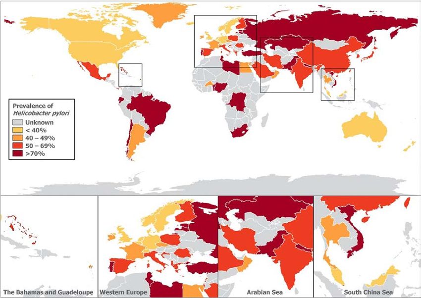
Fig. 2 Prevalence of H. pylori in pediatric patients in Kuala Lumpur .........................7
Fig. 3 Cascades: treatment pathways for low-resource regions ..............................15
Fig. 4 Treatment pathways for H. pylori ..........................................................................25
© World Gastroenterology Organisation, 2021WGO Global Guidelines Helicobacter pylori 4 1 Summary Helicobacter pylori continues to be a major health problem worldwide, causing considerable morbidity and mortality due to peptic ulcer disease and gastric cancer. The burden of disease falls disproportionately on less well-resourced populations. As with most infectious diseases, the greatest impact on reducing this burden comes from improvements in socioeconomic status, which interrupt transmission. This has been observed in many regions of the world, but the prevalence of infection remains high in many regions in which improvements in living standards are slow to occur. Meanwhile, the optimal clinical management and treatment pathways remain unsettled and are evolving with changing antimicrobial resistance patterns. Despite decades of research and clinical practice, major challenges remain. The quest for the most effective, safe, and simple therapy is still a major issue for clinicians. An effective vaccine also still appears to be elusive. Clinical guidelines not infrequently proffer discordant advice. It is very difficult for guidelines to achieve relevance across a variety of populations with varying spectrums of disease, antimicrobial resistance rates, and vastly different resources. As local factors are central to determining the impact and management strategies for H. pylori infection, it is important for pathways to be based on the best available local knowledge, rather than solely extrapolated from guidelines formulated in other regions, which may be less applicable. To this end, this revision of the WGO H. pylori guideline uses a “cascades” approach that seeks to summarize the principles of management and offer advice for pragmatic, relevant, and achievable diagnostic and treatment pathways based on established key treatment principles and using local knowledge and available resources to guide regional practice. 2 Introduction Helicobacter pylori has been recognized as a major pathogen of humankind for nearly four decades. However, despite the impact of treatment of infected individuals and the reduced transmission of infection in communities in which socioeconomic living standards have improved, it continues to be the most common human bacterial pathogen, infecting perhaps half of the world’s population [1]. As a result, it is still a major cause of morbidity and mortality worldwide. H. pylori infection invariably causes active chronic gastritis. In most people, this may be clinically silent throughout life, but in a substantial minority it causes gastroduodenal diseases, most importantly peptic ulcer disease, noncardia gastric cancer, and gastric mucosa- associated lymphoid tissue (MALT) lymphoma. It also increases the risk of gastroduodenal ulceration and bleeding in patients who are taking nonsteroidal anti-inflammatory drugs (NSAIDs) such as aspirin and is responsible for symptoms in a subset of patients with functional dyspepsia. H. pylori has been studied intensively. A literature search reveals more than 45,000 publications. A great deal has been learned about the epidemiology of infection, biology, genetics, pathophysiology, disease expression, diagnosis, and treatment. However, major gaps in our knowledge remain. The precise mode of transmission of infection remains unclear, © World Gastroenterology Organisation, 2021
WGO Global Guidelines   Helicobacter pylori 5

despite many epidemiological studies that identify risk factors for infection. The determinants
of disease expression are still incompletely understood, including many aspects of the host–
pathogen interaction. The pathophysiology of this interaction is complex and has been
reviewed in detail elsewhere [2,3]. The optimal clinical management pathways in different
settings are still a matter of debate, and refinements in diagnostic modalities continue to be
sought. The quest for the most effective, safe, and simple treatment is still a major issue for
clinicians, and the problem of antimicrobial resistance to therapy is a significant challenge.
The best method for surveillance of adverse histological changes in the gastric mucosa has not
been determined, and the quest for an effective vaccine is ongoing.
   There have been many reviews and clinical guidelines on H. pylori [4–12]. As the field is
changing rapidly, there is a need for periodic updating and revision of these position papers.
In addition, it is very difficult for guidelines to achieve relevance across a wide variety of
populations with varying spectrums of disease and often with vastly different resources with
which to deal with it. Guidelines not infrequently proffer discordant advice. As local factors
are central to determining the impact and management strategies for H. pylori infection, this
is not surprising. It is important for clinical advice to be based on the best available local data,
rather than extrapolated from guidelines formulated in other regions, which may be less
applicable. However, in many areas in which the impact of H. pylori infection is greatest, there
is a lack of high-quality data to determine the local best practice. Addressing this gap in
knowledge is a significant challenge. In the meantime, decisions need to be based on the best
available local evidence, extrapolation from higher-quality data from elsewhere, and expert
opinion.
   The purpose of this update to the WGO guideline is to summarize and review the evidence
from a number of new guidelines that outline best practice and to suggest how these
principles may be applied around the world using the “cascades” approach. This approach
recognizes variations in the regional prevalence and impact of infection and the vast
differences in health resources available to address the problem, which require pragmatic,
tailored local approaches. The burden of disease wrought by H. pylori falls disproportionately
on less well-resourced regions, which are insufficiently represented in epidemiological surveys
and are often not the focus of clinical guidelines.
Key statement
It is a major challenge for guidelines to achieve relevance across a wide variety of
populations with varying spectrums of disease and with vastly different resources with
which to deal with it.

3         Natural history, transmission and epidemiology—global aspects

3.1       Natural history of infection
H. pylori infection usually persists for life, unless it is treated with antibiotics or
autoeradication occurs when long-standing infection causes widespread gastric mucosal
atrophy and metaplasia with achlorhydria. Transient infection may occur in some infants.
Reinfection after treatment in adults is uncommon in both higher-prevalence and lower-
prevalence regions. Reinfection may be confused with recrudescence, when infection is

© World Gastroenterology Organisation, 2021
WGO Global Guidelines   Helicobacter pylori 6

suppressed transiently, below the threshold of detection by tests, but has not been eradicated
by antibiotics. There are variations in the virulence of different H. pylori strains globally. The
interplay between host and environmental factors may result in differences in the expression
of disease.

3.2       Transmission of infection
Although there are well-described risk factors for infection, and plausible hypotheses, the
precise mode of transmission has not been definitively established. Most infection appears to
occur in early childhood, with a minority of cases developing in adults. There is strong
evidence from epidemiology and genetic studies of person-to-person transmission,
particularly within families. Mothers appear to be particularly important in transmission to
their young children. Ingestion of the organism seems most plausible via the gastro–oral or
oral–oral route. Fecal–oral transmission appears less likely, at least in developed countries.
Whether transmission occurs via water, food, household pets, or flies is still a matter of
speculation.

3.3       Epidemiology
Although half of the world’s population are thought to be infected with H. pylori, there is
widespread variation in the prevalence of infection, between and within countries (Fig. 1). In
addition, the prevalence may vary within a single city and also between subgroups within a
population (Fig. 2) [13]. For example, there may be wide variations in the prevalence between
more affluent urban populations and rural populations.
Fig. 1 Global prevalence of H. pylori. From Hooi et al. 2017 [1].

© World Gastroenterology Organisation, 2021
WGO Global Guidelines   Helicobacter pylori 7

Fig. 2 Prevalence of H. pylori among children and young adults in Kuala Lumpur, Malaysia.
From Goh [13].

                                         H. pylori prevalence %
                                                                                                                      30
                                                                  26.3
                                                                                                                      25
                           20.8
                                                                                                                      20
                   16.7
           13.3                                                                                                       15
                                                         11.8
                                                                                                        10
                                                                                                7.6                   10
                                                  5.1                                    5.9
                                                                                                                      5

                                                                                                                      0
                  Indian                                Chinese                                Malay

                                              0-5 yrs    6-10 yrs        11-17 yrs

   The quality of prevalence data varies. Many studies are not true prevalence studies, but
rather audits of clinical subsets. Other studies may not represent a valid cross-section of the
population. Moreover, there is significant variability in the quality of reports. In some regions,
diagnostic methods may be less reliable, while some countries are poorly represented as they
lack any reliable data at all. For all these reasons, a single figure cannot be taken to summarize
and represent the prevalence of infection in an entire country and must be applied with
caution. For example, a prevalence study from one city in one region of a populous,
multiethnic country with wide variation in socioeconomic standards is unlikely to represent
the true prevalence across the entire country and cannot reflect high-risk and low-risk subsets.
However, countries and regions can usually be characterized as high-prevalence, mid-
prevalence, and low-prevalence locations [1].
   The major determinant of the prevalence of infection is socioeconomic status in childhood.
Socioeconomic factors reflect levels of hygiene, sanitation, density of living, and educational
level.
  A strong inverse relationship has been consistently reported. Thus, as expected, the
prevalence of infection is generally higher in developing countries, and infection is almost
ubiquitous in some of the most resource-poor subsets of these populations. Migrants from
such regions are recognized as being a high-risk group in more developed, low-prevalence
countries.
Key statement
The major determinant of the prevalence of infection is socioeconomic status in childhood.
   The prevalence of H. pylori infection increases with age. This is mostly due to the cohort
effect, in which the risk of acquiring infection was greater during the childhood of those born
longer ago in comparison with more recently, rather than reflecting ongoing adult acquisition.
Ethnicity has been described as a risk factor, but is most likely closely correlated with

© World Gastroenterology Organisation, 2021
WGO Global Guidelines Helicobacter pylori 8 socioeconomic status or practices that may increase the risk of transmission, rather than having a genetic basis. A striking observation has been the change in the prevalence of infection over time in some countries. Reports of rapidly falling infection rates, most marked in children and younger adults, are common from developed countries, and from countries that have undergone rapid economic development that has led to raised socioeconomic standards. In these countries, the prevalence of infection is now low. A gradual fall in the prevalence of peptic ulcer disease and noncardia gastric cancer is predicted by this observation, since in general the prevalence of peptic ulcer disease and gastric cancer reflects the prevalence of H. pylori in a population. Indeed, the prevalence of ulcer disease and gastric cancer have been falling for decades in developed countries. The fall in disease expression lags behind the fall in infection rates for many years. The declining prevalence of infection and disease occurred long before H. pylori was recognized and treatments were developed. As with most endemic infectious diseases, a decline in prevalence has more to do with improvements in population hygiene and sanitation than with individual, case-by-case treatment, since in most countries, only a minority of infected individuals will ever receive therapy. Notable exceptions are well-resourced high-prevalence countries such as Japan, where screening and treatment is now done systematically in early adulthood. The prevalence of infection appears to be stable in countries in which standards have not improved or have deteriorated, and it is unlikely to fall substantially until improvements do occur. Peptic ulcer disease is still rampant in many of these countries. The burden of gastric cancer also falls disproportionately on these populations. Key statement As with most endemic infectious diseases, a decline in prevalence has more to do with improvements in population hygiene and sanitation than with individual, case-by-case treatment, since in most countries, only a minority of infected individuals will ever receive therapy. 4 The impact of H. pylori infection and the effect of eradication 4.1 H. pylori and peptic ulcer disease The recognition that H. pylori was the cause of most duodenal ulcers and about two-thirds of gastric ulcers was a seminal, Nobel Prize–winning medical breakthrough [14]. In many developed countries with a decreasing prevalence of infection and cure of ulcer patients, the proportion of all peptic ulcers due to H. pylori is falling. In less developed countries, where the prevalence of infection remains high and fewer ulcer sufferers receive curative treatment, peptic ulcer disease (PUD) continues to be a very common and important condition. H. pylori infection has been estimated to confer an individual lifetime risk of peptic ulcer disease of 15– 20%. Untreated, PUD is a chronic relapsing and remitting disease that causes major mortality and morbidity due to pain, bleeding, and perforation. It also results in economic losses. Eradication of H. pylori heals most active peptic ulcers and prevents further relapses, thus © World Gastroenterology Organisation, 2021
WGO Global Guidelines   Helicobacter pylori 9

effecting a cure. Eradication of H. pylori in patients with a history of ulcer disease prevents
subsequent relapses.
   NSAIDs and aspirin cause most other peptic ulcers. H. pylori and NSAIDs act
synergistically to increase the risk of ulcers and bleeding. Eradication of H. pylori reduces this
risk before the start of chronic NSAID therapy.

4.2       H. pylori and gastric cancer and MALT lymphoma
In susceptible infected hosts, long-standing active chronic gastritis may result in gastric
mucosal atrophy with intestinal metaplasia. In a minority, these premalignant mucosal
changes progress to dysplasia and clinically silent, early cancer, followed by advanced gastric
cancer. Gastric cancer often presents at an advanced, symptomatic stage and it has a generally
poor prognosis. H. pylori has been estimated to confer an individual lifetime risk of gastric
cancer of 1.5–2.0% in infected individuals. Despite the relatively low individual risk, as the
global number of people infected is estimated in the billions, there is a global burden of gastric
cancer of over one million per year, with a high fatality rate (Table 1) [15]. This burden is not
distributed evenly. East Asia—Japan, Korea, and eastern China—has the highest prevalence of
disease. China suffers 40% of world cases of gastric cancer. Most, but not all, gastric cancers
are related to H. pylori. The risk of progression to gastric cancer varies and is related to host
and pathogen factors. Host cofactors include smoking and diet. High salt intake, the
consumption of pickled foods, and diets low in antioxidants are dietary cofactors. Genetic risk
factors in the host that are associated with increased risk include the presence of
polymorphisms in genes that determine the expression of interleukin-1 (IL-1;
proinflammatory cytokines) and pathogen recognition receptors. Genotyping of strains of
H. pylori has revealed differences in virulence factors that promote inflammation and are
associated with an increased risk of cancer.

Table 1 Global burden of cancer in 2020

Most common cancers globally
● Breast (2.26 million cases)
● Lung (2.21 million cases)
● Colon and rectum (1.93 million cases)
● Prostate (1.41 million cases)
● Skin (nonmelanoma) (1.20 million cases)
● Stomach (1.09 million cases)

Most common causes of cancer deaths are cancers of the:
● Lung (1.80 million deaths)
● Colon and rectum (935,000 deaths)
● Liver (830,000 deaths)
● Stomach (769 000 deaths);
● Breast (685 000 deaths)

Source: World Health Organization [15].

© World Gastroenterology Organisation, 2021
WGO Global Guidelines   Helicobacter pylori 10

   Eradication of H. pylori before the occurrence of adverse, precancerous histological changes
has been shown to prevent gastric cancer and is the rationale for mass test-and-treat screening
programs in young adults in countries with a high burden of disease and with sufficient
resources to devote to this endeavor. In less well-resourced regions with a high burden of
gastric cancer, such a strategy remains aspirational rather than feasible, given cost constraints,
logistical difficulties, and competing health-care needs.
   Eradicating H. pylori after mucosal atrophy and/or intestinal metaplasia have developed
may reduce the risk of gastric cancer, but does not eliminate it [16]. In any individual, the
residual risk is related to the extent and severity of the mucosal changes, as well as other host
risk factors. Endoscopic surveillance of intestinal metaplasia may be appropriate in some
settings.
  Gastric mucosa-associated lymphoid tissue (MALT) lymphoma is rare. Most cases are a
consequence of H. pylori infection, and eradication of H. pylori when the lymphoma is at a
low-grade stage results in regression and cure. Late recurrences after eradication have
occasionally been reported.
Key statement
Eradication of H. pylori before the occurrence of adverse, precancerous histological changes
has been shown to prevent gastric cancer and is the rationale for mass test-and-treat
screening programs in young adults in countries with a high burden of disease and with
sufficient resources to devote to this endeavor.

4.3       H. pylori–associated dyspepsia
Most H. pylori gastritis is asymptomatic, but it is commonly associated with upper gut
symptoms in the absence of ulcer disease. However, only about one-third or less of infected
patients with “functional dyspepsia” experience sustained relief of symptoms after eradication
therapy. This is because functional dyspepsia is a heterogeneous condition that may be caused
by different mechanisms. H. pylori may be causal in some patients with symptoms and may be
present incidentally in others. However, the proportion of infected patients who improve after
eradication therapy is greater than those who are given empirical acid-suppressive therapy. In
addition, patients may benefit from a reduced lifetime risk of ulcer disease and cancer,
especially if they are treated before adverse histological changes have developed in the gastric
mucosa.
   A recent revised classification of gastritis has recognized H. pylori–associated dyspepsia as a
distinct entity, and it has been incorporated into the 11th revision of the International
Classification of Diseases (ICD-11) [11]. The classification also highlights the significance of
H. pylori gastritis as the precursor lesion that leads to peptic ulcer disease and gastric cancer,
irrespective of whether symptoms are present.
  H. pylori infection has been associated with a variety of other conditions. In most cases, the
association has not been shown to be causal, and common conditions will inevitably coexist in
some patients. There is modest evidence linking H. pylori to immune thrombocytopenic
purpura, and eradication therapy has been tried, with variable results.

© World Gastroenterology Organisation, 2021
WGO Global Guidelines   Helicobacter pylori 11

5         Diagnosis of H. pylori infection

5.1       Who to test and treat?
The decision on whether or not to treat H. pylori must be an active one that takes into account
the individual patient’s circumstances and risks. The decision to test for H. pylori should
therefore only be made with therapeutic intent.
Good practice point
The decision to test for H. pylori should only be made with therapeutic intent.
   Evidence-based indications for testing for and treating H. pylori are summarized in Table 2
[4,17]. The applicability of each indication in different regions will depend on the prevalence
of infection and disease, resources, competing needs, and individual patient factors. Peptic
ulcer disease is the prime indication in most of the world. The clinical and health-economic
benefits of short-term curative therapy for a common, chronic, important disease have been
amply demonstrated over many years. In resource-poor regions, this indication for therapy
should be prioritized.

Table 2 Indications for treatment of H. pylori infection
•   Past or present duodenal and/or gastric ulcer, with or without complications
•   Gastric mucosa-associated lymphoid tissue (MALT) lymphoma
•   Gastric mucosal atrophy and/or intestinal metaplasia
•   Following resection of gastric cancer
•   Patients who are first-degree relatives of patients with gastric cancer
•   Patients’ wishes (after full consultation with their physician)
•   Functional dyspepsia
•   To reduce the risk of peptic ulcer and upper gastrointestinal bleeding in nonsteroidal anti-
    inflammatory drug-naive users
•   Before starting long-term aspirin therapy for patients at high risk for ulcers and ulcer-related
    complications
•   Patients receiving long-term low-dose aspirin therapy who have a history of upper gastrointestinal
    bleeding and perforation
•   Patients with gastroesophageal reflux disease who require long-term proton-pump inhibitors
•   As a strategy for gastric cancer prevention in communities with a high incidence
•   Unexplained iron-deficiency anemia, or idiopathic thrombocytopenic purpura

Adapted from Fock et al. 2009 [4]. Note: the strength of indications may vary regionally and
individually.

6         How to test for H. pylori

6.1       Endoscopic diagnostic tests
Diagnostic tests for H. pylori infection may be invasive (endoscopic) or noninvasive
(nonendoscopic) (Table 3). Biopsies taken at endoscopy are most commonly for histological
analysis and urease testing. Biopsies for culture are less often used for diagnosis, unless
antimicrobial resistance testing is available and is needed to aid individual clinical decision-
making or determine population resistance rates. A combination of two testing modalities
taken from two topographic locations in the stomach is generally most effective for diagnosis.

© World Gastroenterology Organisation, 2021
WGO Global Guidelines Helicobacter pylori 12
In practice, this usually means biopsies taken from the antrum and body of the stomach for
histology and from the antrum for a urease test. More structured biopsy protocols may be
used when there is an additional need for histological surveillance, as in the Operative Link on
Gastritis Assessment (OLGA) and Operative Link on Gastritis/Intestinal-Metaplasia
Assessment (OLGIM) protocols [18]. Histology is usually costly and very operator-dependent,
and accuracy cannot be assumed except in comparison with other previous testing modalities.
Table 3 Cascades: Diagnostic tests for H. pylori—relative availability according to high,
intermediate, or low levels of health-care resources
High Intermediate Low
resources resources resources
Endoscopic Histology Widely used Usually used Rarely used
tests
Commercial urease tests Widely used Widely used Rarely used
In-house urease tests Widely used Widely used Widely used
Culture Many centers Major centers Rarely used
PCR: diagnosis/culture Major centers Rarely used Rarely used
14
Breath tests C urea Widely used Usually used Major centers
13
C urea Usually used Major centers Rarely used
Stool tests Stool antigen Usually used Usually used Major centers
Stool PCR Major centers Rarely used Rarely used
Serology Venous Widely used Usually used Usually used
Fingerprick at point of care Usually used Rarely used Rarely used
Clinical assessment Symptoms Widely used Widely used Widely used
PCR, polymerase chain reaction.
In resource-limited regions, reliance on urease tests is common. Most commercial urease
tests appear to be accurate to a sensitivity of about 95%. Although they are much less
expensive than histology, these tests may still incur a significant cost burden in resource-poor
regions, especially when the cost is borne by the patient. A commercial test typically costs
US$ 5. In regions where the average daily income for an unskilled worker may be $1–2, this
may not be affordable. Fortunately, there are very inexpensive generic urease tests that have
been available for many years and can be done on site, with a unit cost of about $0.20. These
are usually unbuffered tests that give a very rapid result and have a sensitivity very similar to
that of commercial tests [19]. They are in use in some countries in Africa, Asia, and the Pacific
region.
Culturing H. pylori from biopsies requires specific transport conditions, laboratory skills,
and equipment. Culture success rates may reach 90% in expert centers, but are often lower
than that in less expert centers. Subculturing for antimicrobial testing may also not always be
successful in less expert laboratories, so that results may not always be obtained when
required. There are now commercially available real-time polymerase chain reaction (PCR)
tests that allow the detection of H. pylori with high levels of sensitivity and specificity, and also
of mutations that cause clarithromycin resistance [20–22]. These tests do not require strict
© World Gastroenterology Organisation, 2021WGO Global Guidelines Helicobacter pylori 13 preanalytic conditions and they can be performed in a few hours. The validation and implementation of these rapid, inexpensive kit-based point-of-care antimicrobial resistance tests promises to be a major advance in management. The availability of such tests in regions of high resistance may greatly aid the choice of therapy for individual patients, while also facilitating surveys of population prevalence. Good practice point The validation and implementation of rapid, inexpensive kit-based PCR diagnostic and antimicrobial resistance tests promises to be a major advance in management. Endoscopic diagnosis of duodenal ulcer disease in a higher-prevalence, poorly resourced region, in a patient who is not taking NSAIDs, has an accuracy of 95% for predicting the presence of H. pylori. While a biopsy-based test to confirm infection is desirable, the presence of the duodenal ulcer has a predictive value similar to that of most tests, and so it is reasonable to treat without incurring further costs (unless inexpensive generic urease tests are available). 6.2 Noninvasive diagnostic tests When endoscopy is not required or not available, noninvasive tests may be used. Urea breath tests (UBTs) are very useful and have higher diagnostic accuracy than other noninvasive tests for identifying H. pylori (in patients without a history of gastrectomy). Somewhat surprisingly, these are not widely available in many countries in which H. pylori and peptic ulcer disease are most common. The reasons for this are complex, and may include a lack of expertise or resources to set up and operate breath analysis laboratories, the relatively high cost of commercial kit tests, or overreliance on either empirical therapy or endoscopy. In many cases, valid anxiety about gastric cancer is a major driver of the use of endoscopy (although once they become symptomatic, gastric cancers are rarely curable). The costs of UBTs vary. In higher-resource countries, costs compare very favorably with endoscopy, although in regions in which endoscopy is relatively inexpensive, the cost advantage disappears unless low-cost UBTs are available. The stable isotope C13 UBT test has been validated in detail in multiple locations, and is often preferred in well-resourced regions. The C14 UBT uses a very low dose of radioactive isotope and usually has a shorter collection time, but has not been as extensively validated. It may be somewhat less accurate. The laboratory set-up costs for C13 UBTs are higher, as a mass spectrometer is required, whereas a less expensive scintillation counter is needed for C14 UBTs. The real (rather than commercial) unit cost of the C14 isotope is low, so the test could be provided at a very low cost using a central laboratory “hub and spoke” model for service delivery, with remotely collected breath samples being delivered from throughout a region. Point-of-care commercial kits and analyzers are available. The accuracy varies, and the unit cost of these kits is often high. Stool antigen testing is another option. These tests appear to be almost as accurate as UBTs, but patients and health-care and laboratory workers often have a lower preference for stool- based tests. Cost is an issue in some locations. Stool-based rapid PCR tests are also available [21]. Although these tests face the same acceptance barriers, as well as requiring laboratory equipment and skills, they have the potential to provide rapid diagnosis and antimicrobial resistance testing in a single noninvasive test. Serological antibody tests are commonly available. Although they are useful as seroepidemiological surveys, these tests often lack the sensitivity and specificity required for decision-making in individual patients and are generally not very helpful. They need to be © World Gastroenterology Organisation, 2021
WGO Global Guidelines   Helicobacter pylori 14

validated for specific locations, and the issue of false results due to cross-reactivity has rarely
been addressed. In a community with moderate H. pylori prevalence, the accuracy of these
tests may not exceed 50%.

6.3       Testing to assess the outcome after eradication therapy
As the success of eradication is very variable, outcome assessment should ideally be done in all
patients, although this may not be feasible universally. Priority should be given to those who
remain at highest risk for harm if the infection is ongoing, such as those who are being treated
for complicated ulcer disease (bleeding or perforation).
   Biopsy-based testing may be used to determine the outcome after eradication therapy when
endoscopy is required (to assess gastric ulcer healing and exclude neoplasia, or to survey
adverse histology, for example). Otherwise, noninvasive tests are preferred. UBTs and stool
tests should be done not less than 1 month after the completion of eradication therapy. To
minimize false-negative results, no antibiotics or bismuth compounds should be taken by the
patient for at least a month before testing, and proton-pump inhibitor (PPI) use should be
avoided for at least one and preferably two weeks. Serology is not useful for assessing the
outcome, as antibody levels often persist for years after therapy. Despite the widespread
validation of noninvasive diagnostic tests, and of breath tests in particular, they are still not
available at low cost in many places around the world, and this remains a major unmet clinical
need.

6.4       Diagnostic pathways
The choice of diagnostic test depends to a large extent on the clinical context, availability,
expertise, and cost. If all modalities for diagnosis are available, the key issue is whether
endoscopy is required to investigate symptoms or signs of upper gut disease. In low-
prevalence, more developed countries, assessment for gastroesophageal reflux (GERD),
functional dyspepsia, cardia and esophageal cancer concerns are common indications for
endoscopy, and it is usual to biopsy the stomach for H. pylori at that time. H. pylori is still an
issue in such regions, particularly in higher-risk subgroups such as older patients and those
with lower socioeconomic status, or migrants from high-prevalence regions. In these
countries, a noninvasive “test-and-treat” strategy using UBTs have been validated in younger
patients and are cost-effective, although the use of this strategy may be declining. An
empirical trial of PPI therapy is often done in primary care instead, with recourse to
endoscopy if the symptoms are not relieved. Although popular, this is problematic when the
symptoms are not typical of GERD, and the ideal duration of such a treatment trial is unclear.
It may lead to failure to diagnose H. pylori. Although the organism may be incidental to the
presentation, treatment in younger adults is associated with significant long-term risk
reduction. The cost-effectiveness of management strategies for H. pylori in well-resourced,
lower-prevalence countries varies with local health-care costs.
  In higher-prevalence countries, there is often a distinct preference by both doctor and
patient for prompt endoscopy, due to the fear of gastric cancer—although as noted, it is not
certain whether this improves survival when patients present with symptoms. For individual
decision-making, the pretest probability of infection, the patient’s age, the nature of
symptoms or signs, and the local prevalence of ulcer disease and gastric cancer must be
considered.

© World Gastroenterology Organisation, 2021
WGO Global Guidelines Helicobacter pylori 15
6.5 Empirical therapy in low-resource regions
Where there is very limited access to endoscopic or noninvasive means of diagnosing
H. pylori infection, decision-making must be empirical, based on the clinical setting. Peptic
ulcer disease may be strongly suspected on clinical grounds when there is a clear history of
periodic upper gut pain and/or any earlier or recent history of upper gastrointestinal bleeding.
In regions in which it is known that the prevalence of H. pylori is high and peptic ulcer disease
is common, it is reasonable to use empirical eradication therapy for the presumptive clinical
diagnosis of peptic ulcer disease (Fig. 3). The cohort so treated will include many with peptic
ulcer disease, who will gain major benefit. It will also include some who have H. pylori–
associated gastritis but no active ulcer. In this group, symptom resolution occurs more
frequently than with the use of any other therapy (commonly PPIs), and importantly,
successful therapy reduces lifelong risks of peptic ulcer disease and gastric cancer. Treatment
of both peptic ulcer disease and gastritis has also been shown to be cost-effective.
Fig. 3 Cascades: treatment pathways for upper gastrointestinal symptoms in regions with a high
prevalence of H. pylori and with low health-care resources.
Investigate
Alarm symptoms? YES (endoscopy or other tests)
as indicated and if available
BETTER
Empirical H. pylori Clinical assessment
eradication therapy of outcome
− Avoid clarithromycin and NOT BETTER
YES levofloxacin if prior use
− Ensure full duration and
dose
− Compliance support
Case-by-case
evaluation
Symptoms H. pylori test if available
suggestive YES and affordable
of peptic ulcer (noninvasive or invasive)
disease?
Investigate
as indicated and if available
(endoscopy or other tests)
− Empirical H. pylori eradication
therapy
− H. pylori test
− Other strategies
Case-by-case
NO
Evaluation
Note: Treatment for H. pylori in the context of possible ulcer disease dominates the clinical pathway,
as the clinical and health economic benefits likely exceed those of other strategies.
With empirical symptom-based eradication therapy, there will be a subgroup treated who
are not infected and may have other diagnoses. This group will not benefit from eradication
therapy, and there are costs and the unnecessary use of antibiotics involved, but the likelihood
of major harm is low and the overall benefit to the treated group justifies this approach.
© World Gastroenterology Organisation, 2021WGO Global Guidelines Helicobacter pylori 16
Indeed, the Asia–Pacific Consensus Group on H. pylori has specifically endorsed such an
approach in regions in which H. pylori and peptic ulcer disease are common and many people
have no access to investigations, for either economic or geographic reasons. Empirical use of
PPI therapy is likely to be less beneficial than the initial treatment. Such an approach should
be supported by programs for educating health-care workers to recognize symptoms that are
more likely to be due to ulcer disease and to apply this strategy selectively. In these resource-
poor regions, treating all upper gut symptoms with such an approach is harder to justify.
NSAID use is widespread, and NSAID-related peptic ulcer disease is common and may
coexist with H. pylori infection. In an empirical setting of suspected ulcer disease, when
NSAIDs (including aspirin) are being used, it is reasonable both to treat for H. pylori and to
address the NSAID risk by ceasing the use of these agents and treating the patient with PPIs
for a few weeks after the completion of eradication therapy.
Good practice point
In resource-poor, high-prevalence regions in which diagnostic testing is not available, a
history suggesting chronic ulcer disease—periodic upper gut pain and/or past or present
melena—suggests a high likelihood of H. pylori ulcer disease and justifies empirical
eradication therapy, especially in patients with no history or NSAID or aspirin use.
7 Treatment of H. pylori infection
A vast number of studies have addressed therapy issues, and numerous expert guidelines
recommending choices of therapy are available. However, much of the literature and advice
derives from well-resourced countries, with relatively little coming from the poorly-resourced
countries that bear the major burden of diseases caused by H. pylori. Principles for antibiotic
therapy that apply universally have been established. However, there are key issues that must
be addressed locally in order to determine the best local practice, as antimicrobial resistance
patterns and therefore eradication rates vary regionally [23,24] and other local issues such as
the cost and availability of drugs influence the choice of therapy. The key principles that guide
the choice of eradication therapy are outlined in Table 4.
Table 4 Key principles guiding the choice of H. pylori eradication therapy
1. Randomized controlled treatment trials and meta-analyses provide the highest level of evidence,
but are not available for many regions. Local audits of treatment outcome are useful.
2. Treatment recommendations based on resistance patterns and outcome data from one region
may not be applicable elsewhere, due to variation in resistance rates and other factors.
3. Generating high-quality local data and monitoring antibiotic resistance and treatment outcomes
are priorities.
4. Ad hoc, unproven therapies should be avoided.
5. The main determinant of eradication success is pretreatment antibiotic resistance.
6. Primary resistance to clarithromycin, metronidazole, and levofloxacin varies widely regionally.
7. Major determinants of primary resistance appear to be the magnitude and duration of
community usage of these antibiotics as monotherapy for other indications.
8. Prior personal exposure of a patient to these drugs is likely to result in resistance and increases
the chance of treatment failure.
© World Gastroenterology Organisation, 2021WGO Global Guidelines Helicobacter pylori 17
9. Primary clarithromycin resistance (CR) is reported to have increased in many countries over
relatively few years, while remaining stable in other countries.
10. Primary or secondary resistance to amoxicillin and tetracycline are so rare as to not affect
treatment choices.
11. Since much treatment is given presumptively or after noninvasive H. pylori testing, the choice of
therapy will be based on knowledge of likely local antimicrobial resistance patterns.
12. When endoscopy is carried out, culture is not often done routinely prior to first-line therapy in
most places, but this will vary according to skills, resources, local knowledge of resistance rates,
and outcomes. Ideally, culture should also be used to monitor local resistance trends over time.
13. The availability of rapid, inexpensive, point-of-care PCR antimicrobial resistance testing may
change individual treatment choices and facilitate the surveillance of trends in resistance.
14. Secondary resistance after treatment failure is very common with clarithromycin, metronidazole,
and perhaps levofloxacin.
15. Repeating the same therapy has a low likelihood of success and should be avoided.
16. The choice of second-line and subsequent therapies, if needed, should follow a logical decision
path that involves using the most effective drugs first, avoiding repeating the same therapy, and
using evidence-based choices of subsequent therapies.
17. Culture has a very limited role in determining the choice of salvage therapies.
18. The dosage and duration of therapy will influence outcomes.
19 Treatment should be preceded by an informed consent process that outlines the potential risks
and benefits of therapy to the patient.
20. Compliance is a major modifiable determinant of eradication success and should be supported
with clear verbal and written information.
21. Smoking has an adverse effect on eradication success.
22. Unmodifiable risk factors for treatment failure may include CYP2C19 polymorphisms and the
virulence factors of the organism.
23. The role and value of potassium-competitive acid blockers such as vonoprazan is still emerging.
These drugs are not affected by CYP2C19 polymorphisms and result in more uniform and potent
inhibition of gastric acid secretion.
24. Costs may be minimized by using high-quality generic drugs, especially in resource-poor regions.
25. The drugs required should be on essential drug lists and be widely available.
These key principles must be adapted regionally according to the available resources.
8 Translating treatment principles into therapeutic choices
8.1 Choice of first-line eradication therapy
Application of these principles of therapy will ensure the best outcomes possible. In well-
resourced regions, treatment may be based on high-quality trials and audit and culture data;
in resource-poor regions, reliance on a knowledge of community or personal antibiotic usage
and any local audit of outcomes will influence the use of therapies recommended in guidelines
from elsewhere [4–12].
© World Gastroenterology Organisation, 2021WGO Global Guidelines Helicobacter pylori 18
8.1.1 PPI, amoxicillin, clarithromycin triple therapy
In many parts of the world, triple therapy, comprising a proton-pump inhibitor (PPI) with
amoxicillin and clarithromycin (PPI-AC), is still the most commonly used first-line therapy.
This combination was the first very widely recommended therapy and superseded less
effective triple therapies. It has been very well evaluated over the years. The major
determinant of eradication success with this combination is pretreatment clarithromycin
resistance (CR). The prevalence of antibiotic resistance, particularly CR, varies widely around
the world (Table 5). Where clarithromycin has been and is used commonly as monotherapy
for other infections, the level of CR is often high and increasing. There are views that this
therapy should be abandoned in areas where the primary CR rates are known to be 15–20% or
greater, because of the impact this has on eradication rates. A somewhat arbitrary minimum
eradication rate of 80% on an intention-to-treat basis is often quoted as a benchmark for an
acceptable therapy. This is a common eradication rate for PPI-AC in real-world studies in
areas where CR rates are moderate or low (i.e., below 15–20%). Unacceptably lower
eradication results may occur in countries in which the prevalence of CR is higher.
Table 5 Pooled prevalences of primary and secondary antibiotic resistance relative to
World Health Organization region
WHO region Pooled prevalence of antibiotic resistance, % (95% CI)
Africa Clarithromycin Metronidazole Levofloxacin Cla+Met Amoxicillin Tetracycline
Overall 15 (0–30) 91 (87–94) 14 (12–28) – 38 (32–45) 13 (9–17)
a
Americas Clarithromycin Metronidazole Levofloxacin Cla+Met Amoxicillin Tetracycline
Primary 10 (4–16) 23 (2–44) 15 (5–16) – 10 (2–19) –
Secondary 18 (13–23) 30 (19–41) 22 (3–42) – 7 (1–13) –
b c c
Not specified – – – 3 (0–13) – 4 (1–11)
c c
Overall 14 (9–19) 27 (14–39) 14 (12–28) 3 (0–13) 8 (3–13) 4 (1–11)
Eastern Mediterranean Clarithromycin Metronidazole Levofloxacin Cla+Met Amoxicillin Tetracycline
Primary 33 (23–44) 56 (46–66) 19 (10–29) 19 (0–39) 14 (8–20) 10 (4–15)
c c
Secondary 17 (10–27) 65 (54–74) 30 (14–46) 11 (6–20) 10 (5–18) 17 (8–26)
b
Not specified 25 (17–32) 67 (61–72) – 8 (4–11) 15 (8–22) –
Overall 29 (23–25) 61 (55–67) 23 (14–32) 14 (5–23) 14 (10–18) 10 (8–13)
a a a a
Europe Clarithromycin Metronidazole Levofloxacin Cla+Met Amoxicillin Tetracycline
Primary 18 (16–20) 32 (27–36) 11(9–13) 1 (0–2) 0 (0–0) 0 (0–0)
Secondary 48 (38–57) 48 (38–58) 19 (14–24) 18 (16–20) 0 (0–0) 0 (0–1)
b
Not specified 33 (26–39) 47 (35–39) 14 (10–18) 7 (0–13) 1 (0–2) 1(0–2)
Overall 32 (25–31) 38 (33–42) 14 (12–16) 15 (12–18) 0 (0–0) 0 (0–0)
a a
Southeast Asia Clarithromycin Metronidazole Levofloxacin Cla+Met Amoxicillin Tetracycline
Primary 10 (5–16) 51 (26–76) 30 (14–46) – 2 (0–5) 0 (0–1)
c c
Secondary 15 (8–27) 44 (32–58) 24 (15–37) – – –
b
Not specified 25 (0–55) 80 (57–100) 5 (3–11) 6 (1–10) 28 (0–62) 1 (1–2)
Overall 17 (6–28) 59 (40–78) 25 (13–28) 6 (1–10) 12 (6–17) 0 (0–12)
a a a a
Western Pacific Clarithromycin Metronidazole Levofloxacin Cla+Met Amoxicillin Tetracycline
Primary 34 (30–38) 47 (37–57) 22 (17–28) 8 (6–10) 1 (1–1) 2 (1–2)
© World Gastroenterology Organisation, 2021WGO Global Guidelines Helicobacter pylori 19
WHO region Pooled prevalence of antibiotic resistance, % (95% CI)
Secondary 67 (54–80) 62 (50–71) 30 (20–39) 13 (8–18) 1 (1–2) 0 (0–1)
b
Not specified 25 (21–29) 69 (64–74) 19 (17–21) 14 (11–18) 1 (1 2) 10 (7–14)
Overall 34 (30–38) 55 (51–59) 24 (21–26) 11 (9–13) 1 (1–1) 2 (1–2)
From Savoldi et al. 2018 [23]. Cla+Met, combined resistance to clarithromycin and metronidazole.
a b
Notes: P value for subgroup comparison < 0.05. Not specified: the study did not report the type of
c
resistance. Only one study contributed to the analysis.
Key statement
The major determinant of eradication success with PPI-AC is pretreatment clarithromycin
resistance.
The optimal duration of therapy is a matter of contention. Recent calls for universal 14-day
PPI-AC therapy usually originate from regions with higher CR. Initial studies were mostly for
7 days, although that duration may have been influenced by registration trial design.
Proponents of the longer duration of therapy point to somewhat higher eradication rates in
systematic reviews. However, there are other considerations that influence the duration of
therapy, particularly in resource-poor countries. Adding a second week of therapy may
increase eradication rates, typically by about 10%. This means that the number of patients
needed to treat with an extra week of therapy in order to achieve one more treatment success
is 10. The price of this higher eradication rate, if achieved, includes a doubling of the cost of
treatment, which is a major issue in resource-poor regions. (It should be noted that the cost of
a week of triple therapy in very resource-poor regions may be as much as weekly earnings for
the lowest paid.) The risk of adverse effects increases considerably with protracted antibiotics,
as does the likelihood of noncompliance. An alternative is to give shorter therapy where
compliance is likely to be greater and adverse effects and costs fewer, with the understanding
that 10% more patients may need a second-line salvage therapy. Overall antibiotic use will be
much lower with the second strategy, as long as first-line eradication rates are at least
moderately high. The longer therapy is usually recommended in some well-resourced
countries, but more modeling of shorter courses in resource poor-regions is needed. It must
also be noted that acceptable eradication rates with 1-week PPI-AC therapy have been
reported from several countries, and the incremental benefit of a longer course has not been
studied. The optimal dosage for the PPI (standard or high dose) and clarithromycin (250 mg
or 500 mg twice daily) has not been determined in most locations. In high CR regions, neither
one nor two weeks of this therapy may achieve acceptable eradication rates. In such places, the
choice for first-line therapy varies.
The role and value of potassium-competitive acid blockers such as vonoprazan in place of
PPIs in any eradication therapy is emerging. These drugs are not affected by CYP2C19
polymorphisms and result in more uniform and potent inhibition of gastric acid secretion
[25].
8.1.2 Bismuth-based quadruple therapies
The other core choice for first-line therapy, especially in regions with high primary CR, is still
bismuth-based quadruple therapy. The best-studied regimen involves a PPI, bismuth,
tetracycline, and metronidazole (PPI-BTM). This treatment has stood the test of time, since it
leads to reliable and acceptable eradication rates irrespective of primary metronidazole
© World Gastroenterology Organisation, 2021You can also read