Structure and co occurrence patterns of bacterial communities associated with white faeces disease outbreaks in Pacific white leg shrimp Penaeus ...
←
→
Page content transcription
If your browser does not render page correctly, please read the page content below
www.nature.com/scientificreports
OPEN Structure and co‑occurrence
patterns of bacterial communities
associated with white faeces
disease outbreaks in Pacific
white‑leg shrimp Penaeus
vannamei aquaculture
Yustian Rovi Alfiansah1,2,3*, Sonja Peters1, Jens Harder4, Christiane Hassenrück1,7 &
Astrid Gärdes1,5,6,7
Bacterial diseases cause production failures in shrimp aquacultures. To understand environmental
conditions and bacterial community dynamics contributing to white faeces disease (WFD) events,
we analysed water quality and compared bacterial communities in water as well as in intestines and
faeces of healthy and diseased shrimps, respectively, via 16S rRNA gene sequencing and qPCR of
transmembrane regulatory protein (toxR), thermolabile haemolysin (tlh), and thermostable direct
haemolysin genes of pathogenic Vibrio parahaemolyticus as a proxy for virulence. WFD occurred
when pH decreased to 7.71–7.84, and Alteromonas, Pseudoalteromonas and Vibrio dominated the
aquatic bacterial communities. The disease severity further correlated with increased proportions of
Alteromonas, Photobacterium, Pseudoalteromonas and Vibrio in shrimp faeces. These opportunistic
pathogenic bacteria constituted up to 60% and 80% of the sequences in samples from the early and
advances stages of the disease outbreak, respectively, and exhibited a high degree of co-occurrence.
Furthermore, toxR and tlh were detected in water at the disease event only. Notably, bacterial
community resilience in water occurred when pH was adjusted to 8. Then WFD ceased without a
mortality event. In conclusion, pH was a reliable indicator of the WFD outbreak risk. Dissolved oxygen
and compositions of water and intestinal bacteria may also serve as indicators for better prevention of
WFD events.
Bacterial diseases are a major problem for Penaeus vannamei pond aquaculture in Asia and Latin America.
They have been causing severe annual economic losses reaching approximately USD 1 billion over last decade1,2.
Among reported bacterial diseases, acute hepatopancreatic necrosis disease (AHPND) and white faeces disease
(WFD) are the most infectious and lethal ones3,4. The latter has frequently been occurring in Asian shrimp
aquaculture since 2 0093,5,6, which reduced shrimp survival to 20–30%6.
WFD events are characterized by the presence of white faecal strings which float in the rearing water3,6. They
usually occur after approximately 50 days of culture6, resulting in retarded shrimp growth, unprofitable harvests,
and even mass mortality7. Loss of microvilli and subsequent lysis in hepatopancreas and midgut associated with
1
Leibniz Centre for Tropical Marine Research (ZMT), 28359 Bremen, Germany. 2Research Center for Oceanography
(RCO-LIPI), Jakarta 14430, Indonesia. 3Center for Aquaculture Research (ZAF), Alfred Wegener Institute
(AWI), 27570 Bremerhaven, Germany. 4Department of Molecular Ecology, Max Planck Institute for Marine
Microbiology (MPI-MM), 28359 Bremen, Germany. 5Division Biosciences/Polar Biological Oceanography, Alfred
Wegener Institute (AWI), 27570 Bremerhaven, Germany. 6Hochschule (HS) Bremerhaven, 27568 Bremerhaven,
Germany. 7These authors contributed equally: Christiane Hassenrück and Astrid Gärdes. *email:
yustian.alfiansah@leibniz‑zmt.de
Scientific Reports | (2020) 10:11980 | https://doi.org/10.1038/s41598-020-68891-6 1
Vol.:(0123456789)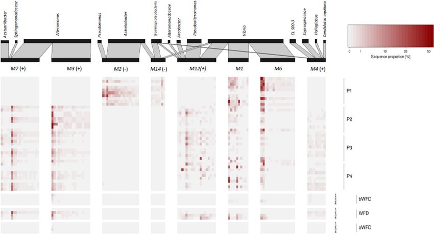
www.nature.com/scientificreports/
WFD indicate a pathological process in shrimp’s gut6. Enterocytozoon hepatopenaei (EHP), vermiform bodies
resembling protozoan gregarines, and certain culturable Vibrio species, such as V. parahaemolyticus, V. fluvialis,
V. mimicus, V. alginolyticus, and Vibrio sp. were reported as potential causative agents of the disease3,6,8,9. Fur-
thermore, deteriorated water quality with oxygen concentrations below 3.0 mg L−1 and alkalinity below 80 ppm
caused peak mortality rates during WFD outbreaks10. However, the aetiology of the WFD in shrimp pond farm-
ing remains i nconclusive6,8,11. For instance, EHP-infected shrimps did not always produce white f aeces11, while
Vibrio such as V. alginolyticus was also reported to be probiotic for P. vannamei12,13.
Often antibiotics and viable beneficial bacterial cells (probiotics) are applied in shrimp farming to enhance
shrimp growth and to avoid disease o utbreaks14–17, without success. Common assessment of culturable hetero-
trophic bacterial numbers, as well as Vibrio counts on thiosulfate-citrate-bile salts-sucrose (TCBS) medium,
failed to predict the pathogenic event. Surprisingly, WFD still happened even though culturable Vibrio counts
were threefold lower than culturable heterotrophic bacteria (pers. comm. with shrimp pond owners). In addi-
tion, the viable plate count method, which selects certain pathogenic b acteria18, was shown to be inadequate to
identify the bacterial population19, which may be associated with the disease. Thus, these practices have been
unsuccessful to predict WFD in P. vannamei aquaculture.
The composition of intestinal bacteria has a strong influence on shrimp health17,20,21. For instance, WFD can
be initiated in healthy shrimps by transplantation of intestinal microbiota of diseased shrimp22. Bacterial com-
munity composition (BCC) in shrimp intestines may dynamically change following shrimp development23 and
diets16,24,25. In addition, shrimp habitat, i.e. the water column and the underlying sediment, may affect intestinal
bacteria (IB) with those of wild shrimps differing from those of domesticated/cultured s hrimps26,27. Yet, there is
little information about the interaction between rearing water parameters, IB, faecal string bacteria (FSB) and
BCC in pond waters before, during, and after disease outbreaks. Also, investigation of bacterial communities in
pond water (WB) during WFD events is still neglected. Thus, more extensive information of bacterial commu-
nity dynamics including pathogenic bacteria in pond water and in association with the shrimps at disease and
non-disease stages is needed to understand and prevent WFD, and to treat diseased shrimps. We further propose
that sudden changes of water quality will affect firstly bacterial communities in pond water (water bacteria/WB)
and subsequently shrimp physiology and their IB.
In the water column, bacteria occur free-living (FL) or particle-associated (PA), or alternating between these
different lifestyles28. As opportunistic pathogenic microorganisms are known to favour a particle-associated
lifestyle, particles, especially larger aggregates, may constitute pathogen hotspots29–31, while simultaneously serv-
ing as an alternative feed for shrimps and fish32,33. During WFD events, faecal strings, containing among others
opportunistic pathogenic bacteria, disintegrate in the rearing water and faecal bacteria are released. They may
then enrich the WB34 resulting in a high load of potential opportunistic pathogenic bacteria predominantly in the
particulate fraction. We therefore hypothesize that the outbreak of the disease can be facilitated by consumption
of contaminated aggregates. Thus, a separate monitoring of FL and PA bacteria is necessary to predict disease
transfer among shrimps in a closed shrimp aquaculture system.
To address the research needs and hypotheses outlined above, we provide a comprehensive overview of the
bacterial dynamics and the water quality in shrimp ponds over the course of a WFD event, by (i) investigating
water quality parameters, (ii) elucidating the BCC in rearing water (WB), separated into FL and PA bacteria, in
the intestines of healthy L. vannamei (IB), and in white faecal strings (FSB), (iii) quantifying pathogenic Vibrio
by their virulence gene copy numbers, and (iv) analysing bacterial co-occurrence patterns in healthy and dis-
eased shrimps. We conducted 16S rRNA gene amplicon sequencing and virulence factor gene quantification via
qPCR of the transmembrane regulatory protein (toxR), thermolabile haemolysin (tlh), and thermostable direct
haemolysin (tdh) of pathogenic V. parahaemolyticus. Furthermore, we discriminated co-occurring bacterial
sub-population characteristics for healthy and diseased shrimps.
Results
The WFD events investigated in this study occurred in shrimp ponds, whose water parameters and WB during
the full rearing cycle at non-disease events have been reported e lsewhere35. The WFD events occurred in ponds
with moderate (P2) and high stocking densities (P3 and P4) at the 52nd, 63th and 67th day of rearing, respec-
tively, suggesting that the disease may happen regardless of the density of the reared shrimps. The WFD event
coincided with a sudden change in pond water parameters, a shift in the WB, and stress in the cultured shrimps,
indicated by a decrease of appetite 2–3 days prior to the onset of the disease.
Biogeochemical characteristics of the shrimp pond water. Ponds with infected shrimps were char-
acterized by lower pH (7.71–7.84), dissolved oxygen/DO (5.57–5.98 mg mL−1), higher turbidity (38.0–41.7 NTU)
and contained more culturable non-sucrose fermenting presumptive Vibrio colonies (4,000–4,700 CFU mL−1). In
contrast, the pond with healthy shrimps (P1) had higher pH (> 8), DO (> 6 mg mL−1), lower turbidity (< 30 NTU),
and fewer CFU-counts of non-sucrose fermenting presumptive pathogenic Vibrio colonies (0–400 CFU mL−1;
Table 1). Considering the low pH during the WFD events, the shrimp pond owners added limestone at night
after they observed the first symptoms of the disease. This treatment was performed until the symptoms of the
disease disappeared. They added approximately 0.4–1.5 tons per pond (approximated water volume 3,500–3,700
m3) for 3 days. This treatment affected the water quality, particularly the pH value, which increased to above 8,
while numbers of non-sucrose fermenting presumptive pathogenic Vibrio decreased 3 to sixfold after the WFD
outbreaks (Table 1).
Environmental parameters in P1 at day 60th and in P2, P3, and P4 at WFD outbreaks were plotted in a PCA
to characterize the shrimp ponds (Fig. 1). The ponds with healthy and diseased shrimps were separated along
PC1, which accounted for 60% of the variation in the data, and was determined mostly by the abundances of
Scientific Reports | (2020) 10:11980 | https://doi.org/10.1038/s41598-020-68891-6 2
Vol:.(1234567890)
www.nature.com/scientificreports/
Parameters Pond 1 Pond 2 Pond 3 Pond 4
Sampling time (day) 50 60 70 50 53 60 60 63 70 60 67 70
pH 8.12 8.40 8.21 8.18 7.79 7.93 8.02 7.84 7.96 7.74 7.71 8.17
Temperature (°C) 30.32 29.91 30.05 31.14 30.94 30.94 29.56 30.61 30.87 30.29 30.23 30.57
DO (mg L−1) 6.10 6.10 6.20 5.60 5.57 5.80 6.32 5.98 6.02 5.93 5.68 6.58
Turbidity (NTU) 19.60 25.60 18.50 19.10 41.70 25.60 25.70 38.50 38.60 30.70 38.00 49.90
Chl a (mg L−1) 88.66 21.19 39.60 28.96 45.76 10.22 70.28 69.76 69.79 31.64 45.97 94.90
Salinity (PSU) 35.53 34.47 33.36 32.92 34.73 34.02 36.12 34.17 34.09 35.64 34.02 33.27
−1)
SPM (mg L 181.17 194.82 151.88 161.49 184.34 168.74 193.03 196.38 186.52 174.94 183.47 175.60
TPPV (CFU mL−1)
suc(−) 0 0 400 400 4,000 600 300 4,700 700 300 4,000 1,100
suc( +) 7,800 6,700 3,000 4,100 1,700 6,400 3,700 1,700 9,700 4,400 2000 8,800
Inorganic nutrients (mg L−1)
Phosphate (PO43−) 0.063 0.886 0.086 0.914 0.795 0.095 0.245 0.793 0.524 0.031 0.770 0.333
Nitrite (NO2−) 0.001 0.219 0.001 0.019 0.199 0.002 0.001 0.217 0.006 0.002 0.205 0.005
Nitrate (NO3−) 0.029 0.076 0.006 0.046 0.061 0.061 0.011 0.078 0.003 0.003 0.087 0.842
Ammonium (NH4+) 0.662 1.017 0.186 0.482 1.088 0.088 0.373 1.060 0.650 0.062 1.041 0.236
Reactive silicate 0.185 0.643 2.775 0.394 0.554 1.554 0.859 0.803 0.494 0.297 0.566 0.197
Table 1. Biogeochemical parameters of the pond with healthy shrimps (P1) and the ponds which experienced
white faeces disease (P2, P3, P4) before, during (in bold) and after disease events. DO dissolved oxygen, SPM
suspended particulate matter, Chl a chlorophyll a, TPPV total culturable presumptive pathogenic Vibrio, suc(−)
non-sucrose fermenting colonies (green colonies), suc(+) sucrose fermenting colonies (yellow colonies).
Figure 1. Principal component analysis (PCA) of observed environmental parameters. Point labels indicate
the pond without white faeces disease (non-WFD pond P1) and ponds with disease events (P2, P3, and P4).
Water parameters presented in the PCA were taken at day 60, 53, 63, and 67 for P1, P2, P3, and P4, respectively.
SPM: suspended particulate matter; suc(+) TPPV: sucrose-fermenting colonies of total presumptive pathogenic
Vibrio (yellow colonies of presumptive pathogenic Vibrio); suc(−) TPPV: non sucrose-fermenting colonies of
presumptive pathogenic Vibrio (green colonies); DO dissolved oxygen; Chl a chlorophyll a concentration; N O2-:
nitrite; NO3−: nitrate; NH4+: ammonium; r-Silicate: reactive silicate; PO43−: phosphate.
culturable presumptive pathogenic Vibrio, ammonium and phosphate concentrations, pH, temperature, and
turbidity. Nitrate and reactive silicate concentrations, and salinity were among the water parameters which
contributed most to PC2.
Bacterial community composition (BCC). A total of 80 samples from pond water, shrimp intestines,
white faecal strings, presumptive pathogenic Vibrio strains and commercial probiotic were sequenced, resulting
Scientific Reports | (2020) 10:11980 | https://doi.org/10.1038/s41598-020-68891-6 3
Vol.:(0123456789)www.nature.com/scientificreports/
Figure 2. Contribution of the most dominant bacterial genera in pond water communities (A) and shrimp
intestines and faeces (B). Samples were collected from a pond with healthy shrimps (P1) and ponds with
diseased shrimps (P2, P3, and P4). Intestine (P1) for intestinal bacteria (IB) and faecal strings (P2, P3, P4) for
faecal string bacteria (FSB) were sampled at rearing day 60, 53, 63, and 67 for P1, P2, P3, and P4, respectively. FL
free-living fraction, PA particle-associated fraction, bWFD before WFD event, WFD during WFD event, aWFD
after WFD event.
in 3,917,111 high quality sequences ranging from 7,892 to 200,098, with a mean of 48,963 sequences per sample.
After merging the operational taxonomic unit (OTU) profiles of the technical replicates that were collected for
the FL and the PA fractions in P2, P3, and P4 at WFD events, the sequencing data set for bacterial community
analyses consisted of 70 samples with on average 58,336 sequences per sample. Cluster analysis and non-metric
multidimensional scaling (NMDS) of these samples showed highly heterogeneous bacterial communities, which
were grouped in seven clusters at a Bray–Curtis dissimilarity threshold of 0.95. Within each cluster average
Bray–Curtis dissimilarities ranged from 0.51 to 0.72. The WB in FL and PA fractions at non-disease events
clustered in two groups, which were distinct from WB at disease events. Sequences generated from the bacterial
strains grown on the TCBS agar from P1 were exclusively affiliated with the genus Vibrio. They clustered together
with IB from P1, which were likewise dominated by Vibrio (Supplementary Information Fig. 1).
Despite the high overall heterogeneity, bacterial communities in pond water (WB) in P1 showed a similar
composition of dominant bacterial taxa at all investigated sampling times. Based on 16S rRNA sequencing, the
WB in P1 was predominantly comprised of the bacterial taxa Salegentibacter (Bacteroidia), Exiguobacterium
(Bacilli), and Halomonas and Psychrobacter (Gammaproteobacteria). These taxa were also found in the WB of the
FL and the PA fractions of P2, P3, and P4 before and after the disease event (Fig. 2A). During the disease event,
the WB of P2, P3, and P4 were altered with Mesoflavibacter (Bacteroidia), Arcobacter (Campylobacteria), and
Alteromonas, Marinomonas, Photobacterium, Pseudoalteromonas and Vibrio (Gammaproteobacteria) dominating
BCC (Fig. 2A). Those genera exhibited only low proportions in the WB of P1 at all sampling points and in both
fractions, with the exception of Vibrio.
Dominant members of intestinal bacteria (IB) were Gammaproteobacteria of the genera Acinetobacter, Pseu-
domonas, and Vibrio, while faecal string bacteria (FSB) samples were dominated by Arcobacter (Campylobacte-
ria) and Gammaproteobacteria of the genera Alteromonas, Marinomonas, Photobacterium, Pseudoalteromonas
Scientific Reports | (2020) 10:11980 | https://doi.org/10.1038/s41598-020-68891-6 4
Vol:.(1234567890)www.nature.com/scientificreports/
BCC MI P1 MFS P2 MFS P3 MFS P4
IB P1 0.63 0.92 0.95 0.93
FSB P2 (0.80, 0.002) 0.65 0.75 0.80
FSB P3 (0.87, 0.002) (0.22, 0.006) 0.70 0.73
FSB P4 (0.75, 0.002) (0.24, 0.002) (-0.01, 0.523) 0.75
Table 2. Pairwise analysis of similarity (ANOSIM) and Bray–Curtis dissimilarity values comparing bacterial
community compositions (BCC) in the intestine of healthy shrimps (IB) and that of white faecal strings (FSB).
P1 pond with healthy shrimps; P2, P3 and P4 ponds with diseased shrimps. Lower triangle: ANOSIM R values
and Benjamini–Hochberg adjusted p values (in parenthesis). Values written in bold indicate strongly separated
communities. Diagonal: average Bray–Curtis dissimilarity within pond (in italics). Upper triangle: average
Bray–Curtis dissimilarity between ponds (underlined). Sample number per pond: N = 10.
Figure 3. Bray–Curtis dissimilarity values of WB in the free-living (A) and particle-associated (B) fractions
compared to the intestinal (IB) or faecal string bacteria (FSB) for samples from P1 and P2-P4, respectively.
bWFD: before WFD event, WFD: during WFD event, aWFD: after WFD event. P1: pond with healthy shrimps;
P2, P3 and P4: ponds with diseased shrimps.
and Vibrio (Fig. 2B). Interestingly, neither Acinetobacter nor Pseudomonas affiliated sequences were found in
FSB. Conversely, Alteromonas, Marinomonas, Photobacterium and Pseudoalteromonas were absent in healthy
shrimp intestines. This clear distinction between IB and FSB was further supported by pairwise ANOSIM test,
which showed that IB differed from the FSB of P2, P3, and P4, while FSB among ponds with infected shrimps
were more similar (Table 2). Especially, in the faecal string (FS) samples from P2, Alteromonas made up more
than 50% of all sequences in seven out of ten samples, while in the remaining three samples, Alteromonas still
constituted up to 40%.
WB of the FL and PA fractions from ponds with infected shrimps at non-disease events were highly dissimi-
lar to FSB. However, during disease events, FSB and WB shared similar bacterial community compositions as
indicated by consistently decreased Bray–Curtis dissimilarity values in all diseased ponds (Fig. 3; Supplementary
Table 1). In contrast, in the pond with healthy shrimps, IB and WB were highly dissimilar at all sampling times
(Fig. 3).
Scientific Reports | (2020) 10:11980 | https://doi.org/10.1038/s41598-020-68891-6 5
Vol.:(0123456789)www.nature.com/scientificreports/
Min–Max
% Q (Nq/N) Q level Mean Q ± SD ANOVA
Sample Pond N toxR, tlh toxR tlh toxR tlh toxR tlh Q Unit (Log copies)
Shrimp
per mL volume of
Intestine P1 10 90 (9/10) 1.2–6.9 0.9–6.9 4.5 ± 1.8 3.9 ± 2.5
intestine
df: 3, 36 df: 3, 36
Fecal string P2 10 90 (9/10) 1.5–5.0 0.9–5.5 3.7 ± 1.0 3.7 ± 1.2 F-value: 0.71 F-value: 0.14
p: 0.55 p: 0.93 per mL volume of faecal
Fecal string P3 10 100 (10/10) 2.1–6.6 1.6–6.6 3.7 ± 1.4 3.5 ± 1.5
string
Fecal string P4 10 90 (9/10) 1.6–6.4 2.1–6.5 3.9 ± 1.5 4.3 ± 1.6
Water
P1 3 0 < LoQ < LoQ < LoQ < LoQ
P2 3 100 (3/3) 13.9–15.9 11.7–13.8 14.9 ± 1.3a 13.0 ± 1.2a
PA
P3 3 100 (3/3) 3.4–6.7 6.1–7.0 4.6 ± 1.9bc 6.7 ± 0.5b
P4 3 100 (3/3) 6.4–6.8 5.8–6.4 6.7 ± 0.2 b
6.0 ± 0.3b df: 5, 12 df: 5, 12
F-value: 49.05 F-value: 126.08 per L of pond water
P1 3 0 < LoQ < LoQ < LoQ < LoQ p: < 0.001 p: < 0.001
P2 3 100 (3/3) 11.7–14.2 14.2–16.1 13.2 ± 1.3a 14.9 ± 1.0a
FL bc
P3 3 100 (3/3) 2.9–4.7 1.5–2.6 5.0 ± 0.8 2.2 ± 0.6c
c
P4 3 100 (3/3) 4.3–5.9 4.0–5.4 3.8 ± 0.9 4.7 ± 0.7b
Table 3. Concentration of toxR and tlh genes in shrimp (intestines of healthy shrimps and faecal strings of
diseased shrimps) and pond water samples. Water samples are separated into free-living (FL) and particle-
associated (PA) fractions. N number of samples for intestine and faecal string, and replicates for water samples;
Q quantified samples; SD standard deviation; PA particle-associated fraction; FL free-living fraction; LoQ limit
of quantification. Different superscript letters after values in PA and FL fractions of water samples indicate that
samples differed significantly according to TukeyHSD post-hoc tests. Copy numbers of toxR and tlh genes were
tested separately.
Virulence gene detection and quantification. Our primer pairs detected toxR, tlh, and tdh genes from
the positive control V. parahaemolyticus DSM 10,143 with a limit of quantification of 26 cells m L−1 (Supplemen-
tary Tables 2 and 3). We targeted these three genes in WB, IB, and FSB samples, but only two virulence genes
(toxR and tlh) could be detected and quantified (Table 3). Concentrations (copy numbers) of the toxR and tlh
gene in intestines and FS did not differ from each other (Table 3). They varied in a range from 3.7 to 4.5 and 3.5
to 4.3 log gene copies, which were equal to 4,926 to 33,665 and 3,140 to 19,907 gene copies per mL volume of
faecal string or intestine for toxR and tlh, respectively.
Concentration of toxR and tlh genes in the pond water differed significantly between FL and PA fractions
(toxR: MANOVA, Pillai2,6 = 0.623, p = 0.05; tlh: MANOVA, Pillai2,6 = 0.854, p < 0.05). The PA and FL fraction from
P2 water contained higher toxR gene copy numbers (14.9 ± 1.3 and 13.2 ± 1.3 log copies L −1, respectively), which
differed from the respective fractions of the two other ponds with diseased shrimps (Table 3). In contrast, no
virulence genes were detected in P1 water at all sampling times as well as in the water of the remaining ponds
(P2, P3, and P4) at non-disease sampling times.
Bacterial co‑occurrence networks. After filtering rare and low sample coverage OTUs, 269 OTUs were
retained from IB and FSB samples for co-occurrence network analysis using sparse inverse covariance estima-
tion for ecological association inference (SPIEC-EASI). Louvain clustering was able to generate 15 bacterial
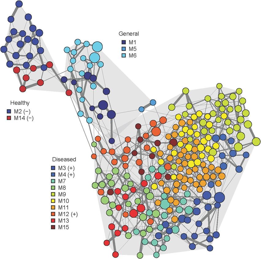
co-occurrence modules (Fig. 4 and Supplementary Table 4). Network modules with highest sequence propor-
tions of their member OTUs in shrimp and PA bacterial community samples were visualized in a heatmap
(Fig. 5). Among 15 modules, two modules (M2 and M14) represented co-occurring OTUs unique to IB samples
of healthy shrimps. They consisted of Acinetobacter, Pseudomonas, as well as two Vibrio OTUs. Interestingly,
these Acinetobacter, Pseudomonas, and Vibrio OTUs were absent in all WB including those from P1. OTUs rep-
resented in modules 1, 5, and 6 occurred in both healthy and diseased shrimps, and were exclusively affiliated
with Vibrio (M1, M6) and Photobacterium (M5). Ten modules (M3, M4, M7, M8, M9, M10, M11, M12 and M15)
comprised of OTUs predominantly found in infected shrimps. Module 3 consisted exclusively of Alteromonas
OTUs, while the remaining modules consisted of more than three genera. Notably, Vibrio OTUs appeared in
both healthy and diseased shrimp samples, and contributed to general network modules (M1, M6), as well as
those characteristic for either healthy (M14) or diseased shrimps (M12), although associated with different other
taxa. For instance, in M12 Vibrio co-occurred with Arcobacter and Pseudoalteromonas, while in M14 Vibrio
OTUs were associated with Acinetobacter. Pairwise random forest models were further used to select network
module best suited to distinguish diseased from healthy shrimp samples based on mean decrease Gini and accu-
racy (Supplementary Table 5). Random forests confirmed M2 and M14 as most characteristic for healthy, and
M3, M4, M12 for diseased shrimp samples.
Scientific Reports | (2020) 10:11980 | https://doi.org/10.1038/s41598-020-68891-6 6
Vol:.(1234567890)www.nature.com/scientificreports/
Figure 4. Bacterial co-occurrence network generated by SPIEC-EASI. Node size corresponds to the average
sequence proportion of operational taxonomic units in intestinal and faecal samples. Network modules detected
by Louvain clustering are shown in different colours, grouped by the samples they predominantly occurred in:
healthy, diseased, both (general). Network modules identified as characteristic for healthy and diseased shrimps
by random forest models are indicated by (−) and (+), respectively. Edge width corresponds to the strength of
the association between OTUs.
Discussion
To better understand WFD in Penaeus vannamei aquaculture, we measured water quality and analysed bacte-
rial community dynamics. Based on the visual estimation of white faecal string (FS) numbers in the ponds, we
discriminated the WFD event into two phases: start of disease (early symptoms), represented by P3 and P4,
with lower numbers of white FS, and early-outbreak (P2), with greater white FS numbers. Because bacterial
communities of fresh shrimp faeces and that of the full intestines of healthy P. vannamei have been shown to be
comparable17,34, we only dissected the intestines of healthy shrimps and analysed them together with the fresh
faecal strings collected from diseased shrimp. In addition, if the shrimp already defecated, it was difficult to
distinguish healthy and infected shrimps since the shrimp intestine was already empty.
Water quality has a large impact on the health status and growth of the shrimps21 as well as on the BCC in
shrimp pond waters36. Regular feed input causes unintended negative effects on water quality, which eventually
limit shrimp growth. Uneaten feed pellets, which are not incorporated by shrimps, together with organic mat-
ter waste (i.e. faeces) stimulate phytoplankton and bacterial growth resulting in bacterioplankton community
instability37. Elevated metabolic activity due to a heterotrophic bacterioplankton bloom exerts an increased oxy-
gen demand, and influences other physical parameters such as the amount of suspended particulate matter and
turbidity38 as well as inorganic nutrient c oncentrations39. Microbial activities including organic matter degrada-
tion, respiration and nitrification process, and accumulation of dissolved carbon dioxide will affect hydrogen ion
concentration in pond water resulting in decrease of pH and a lkalinity38, as was observed in ponds with diseased
shrimps. In contrast, external intervention by regular addition of lime stone which may rich of calcium carbonate
and reactive silicate may buffer pH and alkalinity l evel38, which was the case in the pond with healthy shrimps.
A salinity range of 32.7–34.6 psu in shrimp pond water favoured the dominance of marine heterotrophic
bacteria. At non-disease events, Exiguobacterium, Halomonas, Psychrobacter, Salegentibacter and Sulfitobacter
dominated the bacterial communities in pond water (WB), presumably playing a role in nitrification40–42, organic
matter degradation and sulphite o xidation43. They may also inhibit the growth of potential pathogenic bacteria
in pond water, for example Pseudoalteromonas and Vibrio, as reported in previous studies16,35,44. Furthermore,
Scientific Reports | (2020) 10:11980 | https://doi.org/10.1038/s41598-020-68891-6 7
Vol.:(0123456789)www.nature.com/scientificreports/
Figure 5. Sequence proportion of the members of the most dominant and most distinguishing network
modules between healthy (−) and diseased shrimps (+), as well as their contribution to the particle-associated
fraction from the respective ponds and sampling times. Their taxonomic affiliation is provided on genus level.
Water samples were not used for the network construction.
intestinal bacteria (IB) of the healthy shrimps were dominated by Acinetobacter, Pseudomonas and Vibrio which
correspond to those reported by previous studies15,33,34. Interestingly, toxR and tlh genes belonging to V. para-
haemolyticus were detected in similar concentrations in the intestines of healthy and diseased shrimps. Thus,
we predict that Acinetobacter, Pseudomonas and other Vibrio may inhibit the pathogenicity of V. parahaemo-
lyticus. These seemingly beneficial bacterial taxa are known to drive nitrification processes, accumulate poly-ß-
hydroxybutirate (PHB) which may stimulate the growth of beneficial bacteria, and act as antagonistic bacteria
against pathogens13,26,32,45–48. For instance, they can inactivate acyl-homoserine lactone (AHL), a type of quorum
sensing molecule, which regulates the virulence of pathogenic b acteria48. Furthermore, the IB differed consider-
ably from WB at non-disease events. Since Acinetobacter and Pseudomonas are intolerant to high salinity46,47, we
propose that they cannot persist in the saline shrimp pond water. Therefore, they did not enrich WB, resulting
in the observed high community dissimilarities.
Our study indicates that a pulse d isturbance49, such as a sudden decrease of pH (below 8) and dissolved
oxygen (below 6 mg L−1), and an increase of inorganic nutrients as observed in P2-P4, may affect shrimps and
bacterial communities in shrimp pond waters (WB). The pulse disturbance caused stress in shrimps, which may
in turn have induced changes in the intestinal bacterial communities, resulting in opportunistic pathogenic bac-
teria, such as Alteromonas, Marinomonas, Photobacterium, Pseudoalteromonas and Vibrio, becoming dominant
in the bacterial communities in white faecal strings (FSB). At this stage, we deduce that dysbiosis in the IB, which
was also reported in previous WFD related s tudies22,50, had occurred. We observed a gradual shift from presum-
ably beneficial bacteria-dominated to potential pathogen-dominated FSB, which coincided with the progression
of the disease from the ponds with early symptoms to the pond at early outbreak. This suggests that changes in
intestinal bacterial communities may be closely associated with the severity of the shrimp disease. This hypoth-
esis is supported by a previous studies17, which reported that changes in shrimp intestinal bacteria occurred in
parallel with changes in disease severity, reflecting the transition from a healthy to a diseased state. Among the
potential pathogenic taxa, which dominated FSB communities in our study, Photobacterium, Pseudoalteromonas
and Vibrio corresponded to those previously observed to be associated with the WFD e vents22. However, some
genera such as Aeromonas, Candidatus Bacilloplasma, Phascolarctobacterium and Staphylococcus, which were
reported to be present in previous study20,22, were absent in our samples during the WFD event. It is important to
consider, though, that geographical location, shrimp farm management, and different methodological approaches
may influence the detection of bacterial taxa.
Shifts of WB occurred in both FL and PA fractions during the disease events, which coincided with decreased
pH. We propose that lower pH altered growth rates of heterotrophic bacteria, as also reported previously51 result-
ing in a dominance of opportunistic, potentially pathogenic bacteria such as Alteromonas, Pseudoalteromonas
and Vibrio in WB. Since shrimp faeces easily disintegrate in the pond water (up to 27% within 12 h)34, and could
be unravelled faster due to water movement and mechanical aeration, we suggest that FSB enriched WB, thereby
contributing to the dominance of Alteromonas in FL and PA, as observed in the WB of P2. Disintegration of
faeces will facilitate bacterial dispersion, as well as protein and inorganic nutrient enrichment from f aeces34. The
Scientific Reports | (2020) 10:11980 | https://doi.org/10.1038/s41598-020-68891-6 8
Vol:.(1234567890)www.nature.com/scientificreports/
enrichment of the WB by opportunistic pathogenic bacteria further seemed to correlate with disease severity and
the number of infected shrimps. This is reflected in the significantly higher concentrations of toxR and tlh genes
in pond water samples from the early outbreak phase compared to the ponds with early symptoms. Furthermore,
if greater numbers of pathogenic bacteria are released in the pond water and incorporated into particulate matter,
it will accelerate the spread of the disease among shrimps, since healthy shrimps may consume pathogen-laden
particles and become intoxicated. Thus, in this scenario, FSB not only contribute to bacterial abundance, structure
and function of the WB, but also enforce a detrimental feed-back on shrimp health.
The infection of shrimp tissue is caused by the production of haemolysins by pathogenic bacteria (e.g. V.
parahaemolyticus) upon activation of their virulence factor genes52–54. However, their ability to provoke disease
is dependent on abiotic (e.g. pH, salinity and temperature) and biotic (e.g. bacterial co-occurrence) factors
that support their o utbreak55,56. We explored such biotic interactions using bacterial co-occurrence networks.
Assemblages of co-occurring OTUs of healthy shrimps could be clearly distinguished from those of diseased
shrimps. We propose that Acinetobacter and Pseudomonas composing network module 2, as well as Acineto-
bacter and the two Vibrio OTUs composing network module 14 are part of the indigenous beneficial bacterial
community of the healthy shrimps. The detection of Vibrio OTUs in both healthy and infected shrimps and in
inversely correlated co-occurrence modules suggests the presence of different Vibrio strains with contrasting
interactions. While some Vibrio OTUs might represent opportunistic pathogens, others may even be beneficial
in low p roportions57,58. Alternatively, the co-occurrence with other bacteria such as Acinetobacter may prevent
the activation of virulence factor genes, despite the presence of potentially pathogenic Vibrio in the intestines of
healthy shrimps. Conversely, the change in Vibrio-associated co-occurrence patterns in diseased shrimps from
presumably beneficial to other opportunistic and potentially also pathogenic taxa (network module 12), may
contribute to the disease outbreak.
Considering differences of IB communities of healthy shrimps and WB at non-disease event from those of
WFD samples, as well as co-occurrence patterns in healthy and diseased shrimp samples, we highlight that the
dysbiosis in IB and a shift from halophilic bacteria-dominated to pathogenic bacteria-dominated in pond waters
contribute to the aetiology of the studied WFD outbreak. We emphasize that immediate re-adjustment of water
quality parameters, specifically adjusting pH to above 8, will allow WB to return to its pre-disturbance compo-
sition and terminate the outbreak, followed by recovery from WFD, as indicated by the lack of symptoms and
detectable virulence genes in WB, and no shrimp mortality. This implies a resilience of bacterial communities
in shrimp pond water after short disturbances, as can also be observed in other environments49,59,60. However,
we point out that prolonged exposure to water deterioration and elevated pathogen proportions may increase
disease severity and lead to mass mortality of cultured shrimps as previously o bserved5,61. Our findings on the
application of commercial probiotics to cure WFD in shrimps revealed that probiotic bacteria such as Lactoba-
cillus were absent in WB, IB and FSB, suggesting that such an application was not effective. Lactobacillus was
no longer detectable after they were diluted in the shrimp pond water. Instead of spreading the probiotics into
the pond water, we propose to add them to the feed pellets, which will be eaten by shrimps. With this method,
colonization of probiotic bacteria in the shrimp intestine may occur more effectively.
In conclusion, environmental stressors, specifically a decrease in pH and dissolved oxygen, induced a sub-
stantial community shift in WB and affected shrimp physiology, which in turn resulted in changes of the intes-
tinal bacterial community and subsequently the emergence of WFD. Moreover, we report several opportunistic
bacterial taxa such as Arcobacter, Alteromonas, Marinomonas, Photobacterium and Pseudoalteromonas, which
may contribute to or even cause WFD. To avoid shrimp loss, shrimp farming management should focus on
maintaining sediment/sludge and water quality (i.e. pH, dissolved oxygen, turbidity, inorganic nutrients and
SPM) as well as promoting a stable intestinal bacterial community composition, where beneficial bacteria, even
in low proportions, are able to inhibit the pathogenicity of Vibrio.
Materials and methods
Sample collection and sampling sites. Water samples were collected between 9 and 11 a.m. from one
pond with healthy shrimps (P1, which served as control) and 3 shrimp ponds (P2, P3, and P4) that experienced a
WFD event between 50 to 70 days of rearing in October–November 2016. All ponds were lined with high density
polyethylene (HDPE) plastic and chlorinated 2 weeks before shrimp rearing. Initial population densities were
40 (P2) and 90 post-larvae m−3 (P1, P3, and P4), with the same origin of shrimp fries (PL15, specific pathogen
free, Central Pertiwi Bahari Firm, Rembang, Central Java, Indonesia). Shrimp ponds were located in Rembang
Regency, Central Java, Indonesia (6°37′41.13″ S 111°30′1″ E and 6°42′11.66″ S 111°21′54″ E). Water sampling as
well as measurements of environmental parameters were described in a previous s tudy35. Environmental data
were deposited on PANGAEA (https://doi.pangaea.de/10.1594/PANGAEA.908247).
For bacterial community analysis, ten fresh white faecal strings were collected from feeding trays of each
pond with infected shrimps. Ten healthy shrimps from P1 were collected using the feeding tray and put on ice
in the cold storage immediately. They were then dissected in the laboratory to retrieve their filled intestines with
sterile dissecting tools. Before dissection, shrimps were swabbed with ethanol 70% to sterilize their body and
to avoid contamination from the carapace. All samples were immediately put in Eppendorf tubes, frozen and
stored at − 20 °C until DNA extraction.
Culturable presumptive pathogenic bacterial strain enumeration and identification from pond
water. To obtain culturable presumptive pathogenic bacteria (Vibrio) from all ponds, 100 µL of undiluted to
1 0–4 diluted water sample were plated onto selective thiosulfate citrate bile salts sucrose (TCBS) medium (Roth,
Karlsruhe, Germany), followed by incubation at 35 °C for 24 h. Colonies which grew on the TCBS media were
then counted to determine culturable presumptive pathogenic Vibrio numbers. Strains which grew on TCBS
Scientific Reports | (2020) 10:11980 | https://doi.org/10.1038/s41598-020-68891-6 9
Vol.:(0123456789)www.nature.com/scientificreports/
plates from P1 at 60th day sampling were pooled by swabbing and collected into Eppendorf tubes containing
100 µl sterile sea water, and stored at − 20 °C until DNA extraction and sequencing-based taxonomic analysis. In
total, colonies from 6 TCBS plates were pooled into 1 Eppendorf tube per plate.
Molecular analysis of bacterial communities. 500 mL of water samples were filtered to collect bacterial
cells. To distinguish between free-living (FL) and particle-associated (PA) bacterial communities, a serial filtra-
tion was conducted through 3.0 µm and 0.2 µm polycarbonate filters (ø 47 mm, Whatman, Dassel, Germany) for
the PA and the FL bacterial fractions, respectively. Genomic DNA from water samples was extracted according
to Nercessian et al.62, while bacterial cells from intestines, white faecal strings, and isolates were extracted using
phenol–chloroform methods63. DNA pellets were dissolved in 40 µl TE buffer (10 mM Tris–HCl, 1 mM EDTA,
pH 8.5). DNA concentrations were measured photometrically and checked for purity (ratio of light absorption at
260 to 280 nm) using a nanoquant plate reader (Infinite M200 Pro, Tecan, Germany). Filtration, DNA extraction
as well as genomic DNA concentration measurements were done in triplicates.
16S rRNA gene amplification was performed from genomic DNA extracts. DNA sequences of the V3-V4
hypervariable region of the 16S rRNA gene were obtained from amplicon sequencing with the primer set S-D-
Bact-0314-b-S-17 (5′-CCTACGGGNGGCWGCAG-3′)/S-D-Bact-0785-a-A-21 (5′-GACTACHVGGGTAT
CTAAKCC-3′)64. Sequencing at LGC genomics (Berlin, Germany) was performed on an Illumina MiSeq using
the V3 Chemistry (Illumina) in a 2 × 300 bp paired-end run. Demultiplexing, i.e. grouping of sequences by sam-
ple, and the removal of the primer sequences from the raw paired-end reads were performed by LGC genomics
(Berlin, Germany). Sequences from genomic DNA from water samples before and after the disease period in
P2, P3, and P4, as well as P1 at rearing days 50, 60, and 70 were retrieved from a previous study (PRJEB26390)35.
Sequences were quality-trimmed with a sliding window of four bases and a minimum average quality of
15 with trimmomatic v.03365. Quality trimmed sequences were merged using PEAR v0.9.866. Then, Minimum
Entropy Decomposition (MED) was used to cluster sequences into OTUs67,68. MED applies the principle of
oligotyping67, which uses the Shannon entropy to iteratively partition amplicons at single nucleotide resolution,
thereby providing more accurate descriptions of closely related but distinct t axa69. During MED, we used an
entropy threshold of 0.0965 and a minimum substantive abundance (-M) of 50 to avoid the generation of low
abundant OTUs, decomposing the data set one nucleotide position at a time (-d 1). For each OTU (oligotyping
node), one representative sequence was taxonomically classified with SINA (SILVA Incremental Aligner) v1.2.11
using the SILVA rRNA project reference database (SILVA version 132) at a minimum alignment similarity and
quality of 0.9 and a last common ancestor consensus of 0.770. Unwanted lineages (such as archaea, chloroplasts,
and mitochondria) were removed. In order to obtain results comparable to the previously generated data35 for
WB analysis, OTU profiles from independently sequenced triplicate samples of the FL and PA fractions of P2,
P3, and P4 at the WFD event were merged by taking the sum of the sequence counts per OTU.
Detection and quantification of virulence genes. Three virulence factor genes belonging to Vibrio
which are transcriptional regulator (toxR), thermolabile haemolysin (tlh), and thermostable direct haemolysin
(tdh) were checked in a quantitative PCR machine (CFX Connect Real-time System Bio-Rad, München, Ger-
many) using the primer sets described previously35. qPCR conditions were as follows: a reaction mixture con-
sisted of 10 µL 2X SensiFast SYBR No-ROX (Bioline, Luckenwalde, Germany), 1 µL of 25 mM MgCl2 (Roboklon
EURx, Berlin, Germany), 0.2 µL of 0.5 mM forward and reverse primer (Biomers, Ulm, Germany), 8.8 µL sterile
distilled water, and 2 µl of DNA template (concentration 0.5–10 ng µL−1). The 3-step qPCR amplification was
performed as follows: pre-denaturation at 95 °C for 3 min, followed by 40 elongation cycles consisting of dena-
turation at 95 °C for 10 s, annealing at 60 °C for 15 s, and extension at 72 °C for 20 s, and a dissociation step
after final elongation was added to improve amplification specificity. V. parahaemolyticus DSM 11058 (DSMZ,
Braunschweig, Germany) was used as positive control for toxR, tlh, and tdh genes, while V. vulnificus DSM 10143
(DSMZ, Braunschweig, Germany) served as negative control. A serial dilution of the positive control (known
concentration) was used to estimate gen copy numbers from environmental samples (Supplementary Informa-
tion Table 3). Gene copy numbers for toxR and tlh were determined with the equation y = − 3.554x + 44.891 with
R2: 0.994 and y = − 3.300x + 42.982 with R2: 0.996, respectively.
Data analysis. A principal component analysis (PCA) was conducted to examine the relationship among
environmental parameters and to characterize shrimp ponds during the WFD outbreaks. DNA sequence sam-
ples were categorized into WB (12 PA and 11 FL samples), shrimp bacteria, i.e. IB and FSB (10 and 30 samples,
respectively), culturable Vibrio strains from the pond with healthy shrimp (6 samples), and probiotic bacteria (1
sample). BCC patterns in all samples were visualized by non-metric multidimensional scaling (NMDS) based
on Bray–Curtis dissimilarities, while pairwise ANOSIM tests applying Benjamini–Hochberg p-value correc-
tion were performed to detect separation of bacterial communities between ponds for IB and FSB samples.
Changes in Bray–Curtis dissimilarities between FSB to WB of each diseased pond before, during, and after the
disease event were compared using Kruskal–Wallis rank sum tests, followed by pairwise Wilcoxon tests with
Benjamini–Hochberg p-value correction. Kruskal–Wallis rank sum tests was performed because Bray–Curtis
dissimilarity values were not normally distributed.
Differences in the concentrations of toxR and tlh genes among ponds were tested using ANOVA for shrimp,
and MANOVA for water samples to account for the dependence of observations from FL and PA fractions.
Individual ANOVAs were performed per fraction once MANOVA indicated a difference in gene copy numbers
between the FL and the PA fractions, followed by multiple pairwise comparisons (TukeyHSD post-hoc tests) to
assess difference between ponds.
Scientific Reports | (2020) 10:11980 | https://doi.org/10.1038/s41598-020-68891-6 10
Vol:.(1234567890)www.nature.com/scientificreports/
OTUs from intestine and white faecal string (FS) were analysed to identify sub-populations (modules) of co-
occurring bacteria using SPIEC-EASI (Sparse inverse covariance estimation for ecological association inference)
version 1.0.271. The statistical method SPIEC-EASI comprises two steps, first a transformation for composition-
ality correction of the OTU matrix, and second an estimation of the interaction graph from the transformed
data using sparse inverse covariance selection71. Pre-filtering of OTUs was performed before SPIEC-EASI to
exclude rare and low sample-coverage OTUs, retaining only OTUs which occurred in at least five samples with
a proportion of least 0.1%. Regression coefficients from the SPIEC-EASI output were extracted and used as edge
weights to generate a bacterial co-occurrence network using igraph72. Negative edge weights, which indicated
inverse trends among OTUs were excluded for Louvain clustering, which was then performed to extract network
modules. Modules characteristic for the IB of the healthy pond and the FSB of each of the diseased ponds were
identified using pairwise random forest models based on module eigengenes. Module eigengenes and random
forests models were calculated using the R packages WGCNA73 and r andomForest74, respectively. The sequence
proportions of the members of the modules related to healthy shrimp or the WFD events (based on the highest
mean decrease Gini and accuracy) were visualized in a heatmap.
All statistical analyses, as well as figure visualizations were performed in R (R version 3.4.2, R Core Team,
2017, using R Studio v.0.98.1056) with the packages v egan75, nlme76, gplots77 and packages mentioned previously.
Data availability
DNA sequences generated in this study were deposited on ENA with accession number PRJEB37200 (https://
www.ebi.ac.uk/ena/data/view/PRJEB3 7200) , while biogeochemical parameters and R scripts for statistical analy-
ses were submitted to PANGEA (https://doi.pangaea.de/10.1594/PANGAEA.908247) using the data brokerage
service of the German Federation for Biological Data/GFBio78 in compliance with the Minimal Information
about any (X) Sequence (MIxS) s tandard79.
Received: 2 December 2019; Accepted: 15 June 2020
References
1. De Schryver, P., Defoirdt, T. & Sorgeloos, P. Early mortality syndrome outbreaks: a microbial management issue in shrimp farm-
ing?. PLoS Pathog. 10, e1003919 (2014).
2. Soto-Rodriguez, S. A., Gomez-Gil, B., Lozano-Olvera, R., Betancourt-Lozano, M. & Morales-Covarrubias, M. S. Field and experi-
mental evidence of Vibrio parahaemolyticus as the causative agent of acute hepatopancreatic necrosis disease of cultured shrimp
(Litopenaeus vannamei) in Northwestern Mexico. Appl. Environ. Microbiol. 81, 1689–1699 (2015).
3. Mastan, S. A. Incidences of white feces syndrome (WFS) in farm-reared shrimp, Litopenaeus vannamei, Andhra Pradesh. Indo
Am. J. Pharm. Res. 5, 3044–3047 (2015).
4. Zheng, Y. et al. Bacterial community associated with healthy and diseased Pacific white shrimp (Litopenaeus vannamei) larvae and
rearing water across different growth stages. Front. Microbiol. 8, 1–11 (2017).
5. Durai, V., Gunalan, B., Johnson, P. M., Maheswaran, M. L. & Pravinkumar, M. Effect on white gut and white feces disease in semi
intensive Litopenaeus vannamei shrimp culture system in south Indian state of Tamilnadu. Int. J. Mar. Sci. 5, 1–5 (2015).
6. Sriurairatana, S. et al. White feces syndrome of shrimp arises from transformation, sloughing and aggregation of hepatopancreatic
microvilli into vermiform bodies superficially resembling gregarines. PLoS ONE 9, e99170 (2014).
7. Sung, H. H., Hsu, S. F., Chen, C. K., Ting, Y. Y. & Chao, W. L. Relationships between disease outbreak in cultured tiger shrimp
(Penaeus monodon) and the composition of Vibrio communities in pond water and shrimp hepatopancreas during cultivation.
Aquaculture 192, 101–110 (2001).
8. Tang, K. F. J. et al. Dense populations of the microsporidian Enterocytozoon hepatopenaei (EHP) in feces of Penaeus vannamei
exhibiting white feces syndrome and pathways of their transmission to healthy shrimp. J. Invertebr. Pathol. 140, 1–7 (2016).
9. Piamsomboon, P. et al. Quantification of Enterocytozoon hepatopenaei (EHP) in penaeid shrimps from Southeast Asia and Latin
America using taqman probe-based quantitative PCR. Pathogens 8, 4–9 (2019).
10. Limsuwan, C. White feces disease in Thailand. Boletines nicovita. 2, 1–3 (2010).
11. Tangprasittipap, A. et al. The microsporidian Enterocytozoon hepatopenaei is not the cause of white feces syndrome in whiteleg
shrimp Penaeus (Litopenaeus) vannamei. BMC Vet. Res. 9, 139 (2013).
12. Gomez-Gil, B., Roque, A. & Turnbull, J. F. The use and selection of probiotic bacteria for use in the culture of larval aquatic organ-
isms. Aquaculture 191, 259–270 (2000).
13. Gullian, M., Thompson, F. & Rodriguez, J. Selection of probiotic bacteria and study of their immunostimulatory effect in Penaeus
vannamei. Aquaculture 233, 1–14 (2004).
14. Moriarty, D. J. W. Disease control in shrimp aquaculture with probiotic bacteria. In Microbial Biosystems: New Frontiers. Proceedings
of the 8th International Symposium on Microbial Ecology (eds Bell, C. R. et al.) 1–7 (Atlantic Canada Society for Microbial Ecology,
Halifax, 2013).
15. Franco, R. et al. Evaluation of two probiotics used during farm production of white shrimp Litopenaeus vannamei (Crustacea:
Decapoda). Aquac. Res. 48, 1936–1950 (2017).
16. Zhang, L. et al. Effects of dietary administration of probiotic Halomonas sp. B12 on the intestinal microflora, immunological
parameters, and midgut histological structure of shrimp. Fenneropenaeus chinensis. J. World Aquac. Soc. 40, 58–66 (2009).
17. Xiong, J. et al. Changes in intestinal bacterial communities are closely associated with shrimp disease severity. Appl. Microbiol.
Biotechnol. 99, 6911–6919 (2015).
18. Somboon, M., Purivirojkul, W., Limsuwan, C. & Chuchird, N. Effect of Vibrio spp, in white feces infected shrimp in Chanthaburi,
Thailand. Kasetsart Univ. Fish. Res. Bull. 36, 7–15 (2012).
19. Amann, R. I., Ludwig, W. & Schleifer, K. H. Phylogenetic identification and in situ detection of individual microbial cells without
cultivation. Microbiol. Rev. 59, 143–169 (1995).
20. Hou, D. et al. Intestinal bacterial signatures of white feces syndrome in shrimp. Appl. Microbiol. Biotechnol. 102, 3701–3709 (2018).
21. Xiong, J., Dai, W. & Li, C. Advances, challenges, and directions in shrimp disease control: the guidelines from an ecological per-
spective. Appl. Microbiol. Biotechnol. 100, 6947–6954 (2016).
22. Huang, Z. et al. Microecological Koch’s postulates reveal that intestinal microbiota dysbiosis contributes to shrimp white feces
syndrome. Microbiome 8, 32 (2020).
23. Fan, J. et al. Dynamics of the gut microbiota in developmental stages of Litopenaeus vannamei reveal its association with body
weight. Sci. Rep. 9, 734 (2019).
Scientific Reports | (2020) 10:11980 | https://doi.org/10.1038/s41598-020-68891-6 11
Vol.:(0123456789)www.nature.com/scientificreports/
24. Sha, Y. et al. Bacterial population in intestines of Litopenaeus vannamei fed different probiotics or probiotic supernatant. J. Microbiol.
Biotechnol. 26, 1736–1745 (2016).
25. Vargas-Albores, F. et al. Bacterial biota of shrimp intestine is significantly modified by the use of a probiotic mixture: a high
throughput sequencing approach. Helgol. Mar. Res. 71, 5 (2017).
26. Rungrassamee, W. et al. Characterization of intestinal bacteria in wild and domesticated adult black tiger shrimp (Penaeus mono-
don). PLoS ONE 9, e91853 (2014).
27. Huang, F., Pan, L., Song, M., Tian, C. & Gao, S. Microbiota assemblages of water, sediment, and intestine and their associations
with environmental factors and shrimp physiological health. Appl. Microbiol. Biotechnol. 102, 8585–8598 (2018).
28. Grossart, H. P. Ecological consequences of bacterioplankton lifestyles: changes in concepts are needed. Environ. Microbiol. Rep. 2,
706–714 (2010).
29. Lyons, M. M., Ward, J. E., Smolowitz, R., Uhlinger, K. R. & Gast, R. J. Lethal marine snow: pathogen of bivalve mollusc concealed
in marine aggregates. Limnol. Oceangr. 50, 1983–1988 (2005).
30. Lyons, M. M. et al. Theory of island biogeography on a microscopic scale: organic aggregates as islands for aquatic pathogens.
Aquat. Microb. Ecol. 60, 1–13 (2010).
31. Kramer, A. M., Lyons, M. M., Dobbs, F. C. & Drake, J. M. Bacterial colonization and extinction on marine aggregates: stochastic
model of species presence and abundance. Ecol. Evol. 3, 4300–4309 (2013).
32. Ekasari, J. et al. The size of biofloc determines the nutritional composition and the nitrogen recovery by aquaculture animals.
Aquaculture 426, 105–111 (2014).
33. Hargreaves, J. A. Biofloc production systems for aquaculture. SRAC Publ. 4503, 1–12 (2013).
34. Beardsley, C., Moss, S., Malfatti, F. & Azam, F. Quantitative role of shrimp fecal bacteria in organicmatter fluxes in a recirculating
shrimp aquaculture system. FEMS Microbiol. Ecol. 77, 134–145 (2011).
35. Alfiansah, Y. R. et al. Bacterial abundance and community composition in pond water from shrimp aquaculture systems with
different stocking densities. Front. Microbiol. 9, 2457 (2018).
36. Xiong, J. et al. The temporal scaling of bacterioplankton composition: high turnover and predictability during shrimp cultivation.
Microb. Ecol. 67, 256–264 (2014).
37. Yang, W. et al. Nutrient enrichment during shrimp cultivation alters bacterioplankton assemblies and destroys community stability.
Ecotoxicol. Environ. Saf. 156, 366–374 (2018).
38. Boyd, C. E. & Tucker, C. S. pH in Handbook for Aquaculture Water Quality 95–112 (Craftmaster Printers, Auburn, Alabama, 2002).
39. Zhang, D. et al. Bacterioplankton assemblages as biological indicators of shrimp health status. Ecol. Indic. 38, 218–224 (2014).
40. Chankaew, S., O-Thong, S. & Songnoi, Y. Halomonas sp. SKNB4, a proficient ammonium oxidizing bacterium. In Proceeding of
the 3rd National Meeting on Biodiversity Management in Thailand 4, 186–191 (2016).
41. Chankaew, S., O-Thong, S. & Sangnoi, Y. Nitrogen removal efficiency of salt-tolerant heterotrophic nitrifying bacteria. Chiang Mai
J. Sci 44, 1–10 (2017).
42. Sangnoi, Y., Chankaew, S. & O-Thong, S. Indigenous Halomonas spp., the potential nitrifying bacteria for saline ammonium waste
water treatment. Pak. J. Biol. Sci. 20, 52–58 (2016).
43. Bourne, D. G. et al. Microbial community dynamics in a larval aquaculture system of the tropical rock lobster Panulirus ornatus.
Aquaculture 242, 31–51 (2004).
44. Li, Y. et al. Diversity of cultivable protease-producing bacteria in Laizhou Bay sediments, Bohai Sea, China. Front. Microbiol. 8,
1–10 (2017).
45. Cao, H. et al. Isolation and characterization of a denitrifying Acinetobacter baumannii H1 using NO2–N as nitrogen source from
shrimp farming ponds. Afr. J. Microbiol. Res. 6, 2258–2264 (2012).
46. Vijayan, K. K. et al. A brackishwater isolate of Pseudomonas PS-102, a potential antagonistic bacterium against pathogenic vibrios
in penaeid and non-penaeid rearing systems. Aquaculture 251, 192–200 (2006).
47. Luis-Villaseñor, I. E. et al. Effect of beneficial bacteria on larval culture of Pacific whiteleg shrimp Litopenaeus vannamei. Afr. J.
Microbiol. Res. 7, 3471–3478 (2013).
48. Liu, Y. et al. PHB-degrading bacteria isolated from the gastrointestinal tract of aquatic animals as protective actors against lumi-
nescent vibriosis. FEMS Microbiol. Ecol. 74, 196–204 (2010).
49. Shade, A. et al. Fundamentals of microbial community resistance and resilience. Front. Microbiol. 3, 1–19 (2012).
50. Dai, W., Qiu, Q., Chen, J. & Xiong, J. Gut eukaryotic disease-discriminatory taxa are indicative of Pacific white shrimp (Litopenaeus
vannamei) white feces syndrome. Aquaculture 506, 154–160 (2019).
51. Ventosa, A., Nieto, J. J. & Oren, A. Biology of moderately halophilic aerobic bacteria. Microbiol. Mol. Biol. Rev. 62, 504–544 (1998).
52. DiRita, V. J. Co-ordinate expression of virulence genes by ToxR in Vibrio cholerae. Mol. Microbiol. 6, 451–458 (1992).
53. Williams, S. L., Jensen, R. V., Kuhn, D. D. & Stevens, A. M. Analyzing the metabolic capabilities of a Vibrio parahaemolyticus strain
that causes early mortality syndrome in shrimp. Aquaculture 476, 44–48 (2017).
54. Sirikharin, R. et al. Characterization and PCR detection of binary, pir-like toxins from Vibrio parahaemolyticus isolates that cause
acute hepatopancreatic necrosis disease (AHPND) in shrimp. PLoS ONE 10, e0126987 (2015).
55. Whitaker, W. B. et al. Modulation of responses of Vibrio parahaemolyticus O3:K6 to pH and temperature stresses by growth at
different salt concentrations. Appl. Environ. Microbiol. 76, 4720–4729 (2010).
56. Alonzo, K. H. F., Cadiz, R. E., Traifalgar, R. F. M. & Corre, V. L. Immune responses and susceptibility to Vibrio parahaemolyticus
colonization of juvenile Penaeus vannamei at increased water temperature. AACL Bioflux 10, 1238–1247 (2017).
57. Gomez-Gil, B., Roque, A. & Velasco-Blanco, G. Culture of Vibrio alginolyticus C7b, a potential probiotic bacterium, with the
microalga Chaetoceros muelleri. Aquaculture 211, 43–48 (2002).
58. Zorriehzahra, M. J. et al. Probiotics as beneficial microbes in aquaculture: an update on their multiple modes of action: a review.
Vet Q 36, 228–241 (2016).
59. Allison, S. D. & Martiny, J. B. H. Resistance, resilience, and redundancy in microbial communities. Proc. Natl. Acad. Sci. USA 105,
11512–11519 (2008).
60. Mandakovic, D. et al. Structure and co-occurrence patterns in microbial communities under acute environmental stress reveal
ecological factors fostering resilience. Sci. Rep. 8, 5875 (2018).
61. Anjaini, J., Fadjar, M., Andayani, S., Agustin, I. & Bayu, I. Histopathological in gills, hepatopancreas and gut of white shrimp
(Litopenaeus vannamei) infected white feces disease (WFD). Res. J. Life Sci. 5, 183–194 (2018).
62. Nercessian, O., Noyes, E., Kalyuzhnaya, M. G., Lidstrom, M. E. & Chistoserdova, L. Bacterial populations active in metabolism of
C1 compounds in the sediment of Lake Washington, a freshwater lake. Appl. Environ. Microbiol. 71, 6885–6899 (2005).
63. Green, M. R. & Sambrook, J. Isolation of high-molecular-weight DNA using organic solvents. Cold Spring Harb. Protoc. 10, 356–359
(2017).
64. Klindworth, A. et al. Evaluation of general 16S ribosomal RNA gene PCR primers for classical and next-generation sequencing-
based diversity studies. Nucleic Acids Res. 41, e1 (2013).
65. Bolger, A. M., Lohse, M. & Usadel, B. Trimmomatic: a flexible trimmer for Illumina sequence data. Bioinformatics 30, 2114–2120
(2014).
66. Zhang, J., Kobert, K., Flouri, T. & Stamatakis, A. PEAR: a fast and accurate Illumina Paired-End reAd mergeR. Bioinformatics 30,
614–620 (2014).
Scientific Reports | (2020) 10:11980 | https://doi.org/10.1038/s41598-020-68891-6 12
Vol:.(1234567890)You can also read