Cannabinoids promote embryonic and adult hippocampus neurogenesis and produce anxiolytic- and antidepressant-like effects
←
→
Page content transcription
If your browser does not render page correctly, please read the page content below
Research article

     Cannabinoids promote embryonic and adult
      hippocampus neurogenesis and produce
      anxiolytic- and antidepressant-like effects
                                      Wen Jiang,1,2 Yun Zhang,1 Lan Xiao,1 Jamie Van Cleemput,1
                                             Shao-Ping Ji,1 Guang Bai,3 and Xia Zhang1
                    1NeuropsychiatryResearch Unit, Department of Psychiatry, University of Saskatchewan, Saskatoon, Saskatchewan, Canada.
                            2Department
                                     of Neurology, Xijing Hospital, Fourth Military Medical University, Xi’an, People’s Republic of China.
                 3Department of Biomedical Sciences, Dental School, Program in Neuroscience, University of Maryland, Baltimore, Maryland, USA.

    The hippocampal dentate gyrus in the adult mammalian brain contains neural stem/progenitor cells (NS/PCs)
    capable of generating new neurons, i.e., neurogenesis. Most drugs of abuse examined to date decrease adult
    hippocampal neurogenesis, but the effects of cannabis (marijuana or cannabinoids) on hippocampal neuro-
    genesis remain unknown. This study aimed at investigating the potential regulatory capacity of the potent syn-
    thetic cannabinoid HU210 on hippocampal neurogenesis and its possible correlation with behavioral change.
    We show that both embryonic and adult rat hippocampal NS/PCs are immunoreactive for CB1 cannabinoid
    receptors, indicating that cannabinoids could act on CB1 receptors to regulate neurogenesis. This hypothesis is
    supported by further findings that HU210 promotes proliferation, but not differentiation, of cultured embry-
    onic hippocampal NS/PCs likely via a sequential activation of CB1 receptors, Gi/o proteins, and ERK signaling.
    Chronic, but not acute, HU210 treatment promoted neurogenesis in the hippocampal dentate gyrus of adult
    rats and exerted anxiolytic- and antidepressant-like effects. X-irradiation of the hippocampus blocked both
    the neurogenic and behavioral effects of chronic HU210 treatment, suggesting that chronic HU210 treatment
    produces anxiolytic- and antidepressant-like effects likely via promotion of hippocampal neurogenesis.

Introduction                                                                             producing thousands of new granule cells per day (14). We, and
Cannabis (marijuana, hashish, or cannabinoids) has been used                             others, have shown that these newborn hippocampal neurons are
for medical and recreational purposes for many centuries and                             functionally integrated into the existing neuroanatomical circuitry
is likely the only medicine or illicit drug that has constantly                          (15, 16) and are positively correlated with hippocampus-depen-
evoked tremendous interest or controversy within both the pub-                           dent learning and memory processes (17) and the developmental
lic domain and medical research. Cannabinoids appear to be able                          mechanisms of stress and mood disorders (12). Recent studies have
to modulate pain, nausea, vomiting, epilepsy, ischemic stroke,                           further shown that chronic fluoxetine treatment produced antide-
cerebral trauma, multiple sclerosis, tumors, and other disorders                         pressant and anxiolytic effects (18, 19) and the anxiolytic effects are
in humans and/or animals (1–7). However, marijuana has been                              likely achieved by promoting hippocampal neurogenesis (18).
the most commonly used illicit drug in developed countries,                                Chronic administration of the major drugs of abuse including
producing acute memory impairment and dependence/with-                                   opiates, alcohol, nicotine, and cocaine has been reported to sup-
drawal symptoms in chronic users and animal models (6, 8–10).                            press hippocampal neurogenesis in adult rats (20–23), suggesting
Cannabis acts on 2 types of cannabinoid receptors, the CB1 and                           a potential role of hippocampal neurogenesis in the initiation,
CB2 receptors, which are distributed mainly in the brain and                             maintenance, and treatment of drug addiction (13). The recent
immune system, respectively. In the brain, CB1 receptors are                             finding of prominently decreased hippocampal neurogenesis
also targeted by endogenous cannabinoids (i.e., endocannabi-                             in CB1-knockout mice (24) suggests that CB1 receptor activa-
noids) such as anandamide (AEA), 2-arachidonylglycerol, and                              tion by endogenous, plant-derived, or synthetic cannabinoids
arachidonylethanolamide (1, 6, 10, 11).                                                  may promote hippocampal neurogenesis. However, endogenous
   The recent discovery that the hippocampus is able to generate                         cannabinoids have been reported to inhibit adult hippocampal
new neurons (i.e., neurogenesis) throughout the lifespan of mam-                         neurogenesis (25). Nevertheless, it is possible that exo- and endo-
mals, including humans, has changed the way we think about                               cannabinoids could differentially regulate hippocampal neuro-
the mechanisms of psychiatric disorders (12) and drug addiction                          genesis, as exo- and endocannabinoids act as full or partial ago-
(13). The subgranular zone of the dentate gyrus (SGZ) in the adult                       nists on CB1 receptors, respectively (11).
brain contains neural stem/progenitor cells (NS/PCs) capable of                            The goal of the present study was to test the hypothesis that
                                                                                         the potent synthetic cannabinoid HU210 is able to promote
Nonstandard abbreviations used: AEA, anandamide; FST, forced swimming test;              hippocampal neurogenesis, leading to the anxiolytic and antide-
NeuN, neuronal nuclear antigen; NSF, novelty-suppressed feeding; NS/PC, neural
                                                                                         pressant effects of cannabinoids. We demonstrate here that both
stem/progenitor cell; pERK1/2, phospho-ERK1/2; SGZ, subgranular zone of the
dentate gyrus; TuJ1, b-tubulin III.                                                      HU210 and the endocannabinoid AEA promote proliferation of
Conflict of interest: The authors have declared that no conflict of interest exists.     embryonic hippocampal NS/PCs without significant effects on
Citation for this article: J. Clin. Invest. 115:3104–3116 (2005).                        their differentiation, resulting in more newborn neurons. The
doi:10.1172/JCI25509.                                                                    effects of HU210 on adult hippocampal neurogenesis were quan-

3104                                The Journal of Clinical Investigation    http://www.jci.org    Volume 115    Number 11    November 2005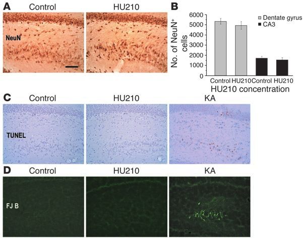
research article
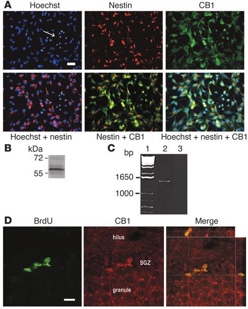
Figure 1
Expression of CB1 receptors in NS/PCs. (A) Coimmunofluorescent
staining of CB1 and nestin in cultured hippocampal NS/PCs derived from
E17 embryos. Hoechst staining was conducted to reveal the total cul-
tured cells. The arrow indicates the glial-like cells, located in the center
of a neurosphere, with CB1 staining and without nestin staining. Scale
bar, 20 µm. (B) Western blot using cultured NS/PC reveals a 60-kDa
protein band corresponding to CB1 receptor. (C) PCR indicates CB1
gene expression in NS/PCs (lane 2) using primers yielding a predicted
product of 1,440 bp (i.e., the full encoding region of CB1 receptor) from
embryonic NS/PCs. Lane 1: molecular weight standards; lane 2: CB1
receptor; lane 3: PCR reaction without sample added. (D) Confocal
microscopic assessments of costaining of BrdU and CB1 receptors in
the SGZ located between the hilus and the granule cell layer (granule)
of the dentate gyrus in an adult rat. Scale bar, 10 µm.
gen. Using PCR, we further identified a band of the predicted
size (1,440 bp) corresponding to the full encoding region of CB1
(Figure 1C), suggesting the presence of CB1 transcripts in NS/PCs.
Similar results, i.e., CB1 protein and gene expression, were seen in
both second and sixth passages of NS/PCs. We then examined adult
naive rats sacrificed 2 hours after receiving a single dose of BrdU
to label dividing cells. We found that about 90% of BrdU-stained
cells in the SGZ were also doubly labeled with CB1 (Figure 1D;
n = 3). These results suggest that both embryonic and adult
hippocampal NS/PCs express CB1 receptors.
Increased proliferation of embryonic NS/PCs by HU210 and AEA. To
examine the effects of HU210 on NS/PC proliferation, cultured
embryonic NS/PCs were incubated with different concentrations
tified in freely moving rats and were correlated with behavioral of HU210. With the WST-8 assay, changes in NS/PC proliferation
testing. We show that 1 month after chronic HU210 treatment, between HU210- and vehicle-treated culture were significant at
rats display increased newborn neurons in the hippocampal den- some concentrations of HU210, as evidenced by significant group
tate gyrus and significantly reduced measures of anxiety- and effects with 1-way ANOVA (F5,18 = 513.129, P < 0.01). Specifically,
depression-like behavior. Thus, cannabinoids appear to be the when 10 nM to 1 µM of HU210 were added to the culture medi-
only illicit drug whose capacity to produce increased hippocampal um containing the mitogenic growth factors bFGF and EGF, the
newborn neurons is positively correlated with its anxiolytic- and WST-8 assay showed a significant increase in NS/PC proliferation
antidepressant-like effects. (Tukey post-hoc tests, P < 0.05); 1 nM of HU210 exerted no signifi-
cant effects (P = 0.072); 10 µM produced profound toxic effects on
Results cultured NS/PCs (Figure 2A). Because HU210 can activate both
Expression of CB1 receptors in embryonic and adult hippocampal NS/PCs. CB1 and CB2 receptors, we next used the selective CB1 receptor
In the mammalian brain, the CB1 receptor is one of the most antagonist AM281 to identify the possible involvement of CB1 in
abundant G protein–coupled receptors, accounting for most, if the action of HU210 on NS/PC proliferation. Although 1 nM to
not all, of the centrally mediated effects of cannabinoids (5). We 1 µM of AM281 alone produced no significant effects on NS/PC
reasoned that if cannabinoids were able to regulate neurogen- proliferation, 10 nM to 1 µM of AM281 blocked the promoting
esis, the NS/PCs capable of producing new neural cells would effects of 10 nM to 1 µM of HU210 on NS/PC proliferation (1-way
contain CB1 receptors. We therefore employed CB1 antibody ANOVA for repeated measures, F2,25.713 = 16.792, P < 0.01; pairwise
immunocytochemistry, Western blotting, and PCR to examine comparisons, HU210-treated cells with or without AM281: P < 0.01)
CB1 protein and gene expression in cultured NS/PCs isolated (Figure 2A), suggesting that HU210 specifically acts on CB1 recep-
from the hippocampus of E17 rat embryos. About 95% of the total tors to promote NS/PC proliferation. While 10 µM of AM281 alone
neurosphere cells labeled with Hoeschst stain were also labeled significantly inhibited NS/PC proliferation (P < 0.01), this con-
with both CB1 and nestin (a marker for NS/PCs) antibodies centration of AM281 did not exert significant effects in prevent-
(Figure 1A). Some Hoechst-labeled cells in the neurospheres ing the lethal effects of 10 µM of HU210 on NS/PCs (Figure 2A),
exhibited the shape of glial cells, with small round nuclei, and were indicating that the lethal effects of 10 µM of HU210 on NS/PC
CB1 immunoreactive but without nestin staining (Figure 1A). cells were caused nonspecifically or by another receptor.
The staining of CB1 antibody appears specific for 2 reasons. To confirm the effects of 10 nM to 1 µM of HU210 on promot-
First, Western blots with the same antibody and cultured NS/PC ing NS/PC proliferation as previously assessed by the WST-8
revealed a strong protein band with the molecular weight of 60 kDa assay, the BrdU incorporation assay was used. It measures cell pro-
(Figure 1B), which corresponds to the CB1 receptor (26). Sec- liferation by detecting dividing cells. Similar to the results of the
ond, we could not detect the positive immunostaining or 60-kDa WST-8 assay, 1-way ANOVA showed significant group effects
protein band using the CB1 antibody preabsorbed with the anti- (F5,18 = 176.004; P < 0.01); Tukey post-hoc tests revealed that 10 nM
The Journal of Clinical Investigation http://www.jci.org Volume 115 Number 11 November 2005 3105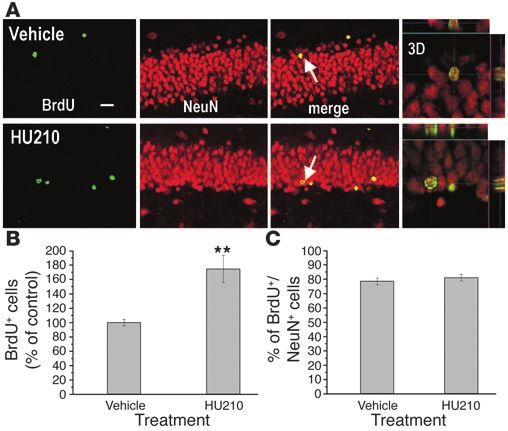
research article

Figure 2
Effects of the cannabinoid HU210 on proliferation of cultured hippocampal NS/PCs. (A) In the WST-8 assay, incubation of NS/PCs with 10 nM
to 1 µM of HU210 for 48 hours significantly promoted NS/PC proliferation, which was blocked by the CB1 receptor antagonist AM281. AM281
alone significantly decreased NS/PC proliferation only with 10 µM, but this concentration of AM281 was not able to block the lethal effects of
10 µM of HU210 on NS/PCs. (B) BrdU incorporation assay confirmed the results obtained with the WST-8 assay shown in A. (C) Incubation
of NS/PCs with 1 µM to 10 µM of AEA for 48 hours significantly promoted NS/PC proliferation in the WST-8 assay. (D) Application of 10 nM to
1 µM of HU210 significantly promoted NS/PC proliferation in both the presence and absence of the growth factors bFGF and EGF in the culture
medium. (E) Pertussis (PTX; 100 ng/ml), a selective blocker for Gi/o protein activation, prevented the effects of 10 nM to 1 µM of HU210 on
promoting NS/PC proliferation. (F) Incubation of NS/PCs with 1 mg/ml of cholera toxin, a selective Gs activator, stimulated a profound increase
in cAMP accumulation in NS/PCs 0.5, 1, 2, and 24 hours after the addition of cholera toxin. (G) Incubation of NS/PCs with 1 mg/ml of cholera
toxin for 0.5, 1, 2, 24, or 48 hours did not induce significant change in NS/PC proliferation. Error bars represent SEM. *P < 0.05 and **P < 0.01
by Tukey post-hoc tests after 1-way ANOVA.

to 1 µM of HU210 significantly increased NS/PC proliferation                     Specifically, 10 nM to 1 µM of HU210 without growth factors pro-
(P < 0.05), which was blocked by 10 nM to 1 µM of the selective CB1              duced significant mitogenic effects on NS/PCs (Tukey post-hoc
receptor antagonist AM281 (1-way ANOVA for repeated measures,                    tests, P < 0.05), whereas 10 µM of HU210 killed the cells. Similar
F2,36 = 19.081, P < 0.01; pairwise comparisons, HU210-treated cells              results were observed in the control culture when different concen-
with or without AM281: P < 0.01) (Figure 2B).                                    trations of HU210 were added to the culture medium containing
  To determine the effects of the endogenous cannabinoid AEA on                  the mitogenic growth factors (F5,30 = 194.429, P < 0.01; Tukey post-
NS/PC proliferation, cultured NS/PCs were incubated with differ-                 hoc tests, P < 0.05) (Figure 2D). Nevertheless, the basal prolifera-
ent concentrations of AEA. The WST-8 assay showed significant                    tion levels with bFGF and EGF were significantly higher than those
group effects with 1-way ANOVA (F5,18 = 61.585, P < 0.01). Tukey                 without bFGF and EGF (1-way ANOVA for repeated measures,
post-hoc tests further showed that 1 µM to 10 µM of AEA signifi-                 F1,30 = 214.703, P < 0.01; pairwise comparisons: P < 0.01) (Figure 2D).
cantly increased NS/PC proliferation (P < 0.05) in the presence of                 Intracellular signaling involved in HU210-induced NS/PC proliferation.
bFGF and EGF; 100 µM produced toxic effects (Figure 2C).                         To investigate the mechanisms underlying the action of HU210
  To explore the possibility of whether HU210 itself is able to                  on NS/PC proliferation, we examined the intracellular signaling
produce mitogenic effects, we further examined NS/PC prolifera-                  pathways. CB1 receptor stimulation activates Gi/o or Gs proteins
tion by adding different concentrations of HU210 to the culture                  (27, 28). To examine whether Gi/o protein mediates the effects of
medium with or without the mitogenic growth factors bFGF and                     HU210, we added pertussis toxin, a selective blocker for Gi/o pro-
EGF. When bFGF and EGF were absent from the culture medium,                      tein activation, to the culture medium 4 hours prior to HU210
a significant overall change in NS/PC proliferation was observed                 treatment. Again, 10 nM to 1 µM of HU210 significantly increased
following HU210 application (F5,30 = 219.076, P < 0.01) (Figure 2D).             NS/PC proliferation (1-way ANOVA, F5,18 = 880.629, P < 0.01; post-

3106                        The Journal of Clinical Investigation    http://www.jci.org    Volume 115    Number 11    November 2005
research article
Figure 3
Effects of the cannabinoid HU210 on PI3K/Akt and ERK signaling in cultured hippocampal NS/PCs. (A) There was no significant change in pAkt or
actin in NS/PCs within the first hour after addition of 100 nM of HU210 to culture medium. (B) Application of 100 nM of HU210 rapidly induced phos-
phorylation of pERK1/2 in NS/PCs in the presence of bFGF and EGF in culture medium. (C) Application of 100 nM of HU210 3 hours after removal
of bFGF and EGF from culture medium also induced phosphorylation of pERK1/2 in NS/PCs. (D) Application of the ERK signaling inhibitor U0126
blocked the promoting effects of 100 nM of HU210 on phosphorylation of pERK1/2 in NS/PCs 5 minutes after addition of HU210 to culture medium.
(E) Addition of U0126 (10 µM) to the culture medium 1 hour before HU210 antagonized the promoting effects of 10 nM to 1 µM of HU210 on NS/PC
proliferation. Error bars represent SEM. *P < 0.05 and **P < 0.01 by Tukey post-hoc tests after 1-way ANOVA. tERK1/2, total ERK1/2.
hoc tests, P < 0.01 between control and each of the 3 concentrations NS/PC proliferation. In contrast, changes in phosphorylation of
of HU210), which was completely blocked by 100 ng/ml of pertus- phospho-ERK1/2 (pERK1/2) during the first 1 hour after HU210
sis (1-way ANOVA for repeated measures, F1,18 = 41.64, P < 0.01; application were dramatic at specific time points, as shown by
pairwise comparisons, HU210-treated cells with or without pertus- 1-way ANOVA (with growth factors, F4,15 = 33.698, P < 0.01; with-
sis: P < 0.01) (Figure 2E). It has been shown that HU210 activates out growth factors, F4,15 = 23.513, P < 0.01). As early as 5 minutes
Gs proteins when Gi/o proteins are inhibited by pertussis toxin (27). after addition of HU210 to culture medium with (Figure 3B) or
Therefore, to determine whether the blockade effects of HU210- without bFGF and EGF (Figure 3C), a 2.5-fold increase in phos-
induced NS/PC proliferation following pertussis treatment is phorylation of pERK1/2 was observed (P < 0.05). At 15 minutes
achieved by activation of Gs proteins, we examined the effects after HU210 application, phosphorylation of pERK1/2 reached
of cholera toxin, a Gs protein activator, on NS/PC proliferation. the peak level, which was about a 4-fold (with growth factors) or
Incubation of NS/PCs with 1 mg/ml of cholera toxin stimulated 7-fold increase (without growth factors) relative to control (P < 0.01).
about 14-, 80-, 90-, and 13-fold increase in cAMP accumulation in By 60 minutes after addition of HU210, phosphorylation of
NS/PCs 0.5, 1, 2, and 24 hours after the addition of cholera toxin; pERK1/2 either significantly decreased (P < 0.05) (Figure 3B) or
cAMP production returned to the basal levels 48 hours after chol- returned to the pretreatment level (Figure 3C). We did not observe
era toxin (1-way ANOVA, F5,18 = 93.341, P < 0.01) (Figure 2F). These any significant changes in the total ERK1/2 during the first 1
results indicate the effective activation of Gs proteins in NS/PCs by hour after HU210 application. Thus, the significant increase in
cholera toxin. However, there was no significant change in NS/PC pERK1/2 in this period suggests an important involvement of
proliferation 0.5, 1, 2, 24, and 48 hours after the addition of chol- ERK signaling pathway in the action of HU210 in promoting
era toxin (1-way ANOVA, F5,18 = 76.562, P = 0.86) (Figure 2G). These NS/PC proliferation. This hypothesis was supported by further
results together suggest the involvement of Gi/o proteins, but not experiments in which U0126, a specific inhibitor of the ERK path-
Gs proteins, in HU210-induced NS/PC proliferation. way, was employed. Figure 3D shows an overall significant differ-
Since Gi/o protein activates PI3K/Akt and ERK signaling (29), ence in pERK1/2 phosphorylation after application of vehicle or
which are well known to play an important role in cell growth 100 nM of HU210 with or without 10 µM of U0126 (F3,8 = 60.769,
and cell death, we studied whether HU210 could activate Akt and P < 0.01). Specifically, HU210 profoundly increased phosphoryla-
ERK1/2. There was no significant change in phosphorylation of tion of pERK1/2 (P < 0.01), which was almost completely blocked
phospho-Akt during the first 1 hour after HU210 application by U0126 (P < 0.01). A parallel experiment demonstrated that
(F4,10 = 1.693, P = 0.228) (Figure 3A), indicating that the PI3K/Akt U0126 blocked the promoting effects of 100 nM of HU210 on
signaling pathway is not involved in the action of HU210 on NS/PC proliferation (1-way ANOVA for repeated measures,
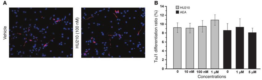
The Journal of Clinical Investigation http://www.jci.org Volume 115 Number 11 November 2005 3107research article Figure 4 Effects of HU210 and AEA on neuronal differentiation of cultured hippocampal NS/PCs. (A) Incubation of NS/PCs with the culture medium con- taining either vehicle or 100 nM of HU210 without growth factors for 8 days produced similar density of neurons (pink cells) stained with TuJ1 antibody. The total cultured cells are labeled deep blue by Hoechst staining. (B) There was no significant difference in the ratio of TuJ1-labeled neurons to total cells following application of HU210 (10 nM to 1 µM) or AEA (1 or 5 µM) to culture medium. F1,17 = 6.356, P < 0.05; pairwise comparisons, HU210-treated cells SEM number of BrdU-positive cells in the SGZ (F3,16 = 11.504, P < 0.001; with or without U0126: P < 0.05) (Figure 3E). n = 5) (Figure 5D). Tukey post-hoc test showed a significant increase HU210 and AEA do not affect neuronal differentiation of cultured NS/PCs. (about 40%) in the number of BrdU-labeled cells following 100 µg/kg To examine the effects of HU210 on neuronal differentiation of HU210 (P < 0.05) but not 25 µg/kg of HU210 (P = 0.979), rela- of cultured NS/PCs, neurospheres were dissociated, plated, and tive to vehicle (Figure 5D). AM281 injection seemingly decreased the cultured in the medium containing bFGF and EGF for 1 day number of BrdU-positive cells in the SGZ, but there was no signifi- and then in another medium containing different concentra- cant difference relative to control (P = 0.099). tions of HU210 without bFGF or EGF for 8 days. After fixation, Increased newborn hippocampal neurons following chronic HU210 treat- immunofluorescence staining was performed using antibodies ment in adult rats. A recent study has demonstrated that newborn against the neuronal marker β-tubulin III (TuJ1), followed by neurons in the dentate granule cell layer that had survived 4 weeks Hoechst staining that detects all the cultured cells. Cell counting were stably integrated into the granule cell layer (30). To examine the revealed no significant difference among the ratios of TuJ1-labeled survival, migration, and differentiation of HU210-induced newborn neurons and Hoechst-labeled total cells following treatment with cells in the SGZ, we injected rats twice daily with HU210 (100 µg/kg, vehicle or 10 nM, 100 nM, or 1 µM of HU210 (1-way ANOVA, i.p.), AM281 (3 mg/kg), or vehicle for 10 days, followed 12 hours F4,20 = 3.307, P = 0.324) (Figure 4), suggesting that HU210 exerts no later by 4 BrdU injections at 12 hours intervals. One month after significant effects on neuronal differentiation of cultured NS/PCs. the last HU210, AM281, or vehicle injection, the majority of Similarly to HU210, AEA (1 and 5 µM) did not produce signifi- BrdU-labeled cells migrated and dispersed into the granule cell cant effects on neuronal differentiation of cultured NS/PCs (1-way layer and showed size and morphology indistinguishable from ANOVA, F2,9 = 0.177, P = 0.840) (Figure 4B). both their neighboring granule neurons and from different treat- Increased hippocampal cell proliferation following HU210 treatment in ment (Figure 6A). The number of BrdU-labeled dentate cells in adult rats. BrdU labeling of dividing cells was used to test the acute HU210-treated rats was significantly higher than that in vehicle- effects of HU210 treatment on cell proliferation in adult hippocam- treated rats (Student’s t test, P < 0.01; n = 5) (Figure 6B), indicat- pus. Adult rats received a single dose of vehicle, AM281 (3 mg/kg, ing that most of chronic HU210–induced newborn cells survived. i.p.), or HU210 (25 or 100 µg/kg, i.p.), followed 2 hours later by BrdU Immunofluorescence staining revealed that HU210- and vehicle- administration and then perfusion 1 day later. BrdU-labeled cells treated rats exhibited a similar proportion of BrdU/neuronal nucle- showed fusiform or irregular shape and were clustered or aggregat- ar antigen (BrdU/NeuN) double-labeling cells to the total BrdU- ed in the SGZ (Figure 5A) throughout the whole hippocampus in all labeled cells (Student’s t test, P = 0.977) (Figure 6C), suggesting that rats examined. Cell counting revealed no significant change in the chronic HU210-induced newborn cells in the SGZ have neuronal number of BrdU-positive cells in the SGZ among rats treated with differentiation ratio similar to that of vehicle-induced newborn vehicle, AM281, or HU210 (1-way ANOVA, F3,16 = 52.784, P = 0.58; cells in the SGZ. Nevertheless, because chronic HU210 treatment n = 5) (Figure 5B). We then examined the effects of chronic HU210 significantly increased the number of BrdU-labeled newborn cells injection on cell proliferation in adult hippocampus. Two hours in the dentate gyrus (Figure 6B), the total number of newborn neu- after receiving the last dose of twice-daily injections of vehicle, rons doubly labeled with BrdU/NeuN in the dentate gyrus also sig- AM281 (3 mg/kg, i.p.), or HU210 (25 or 100 µg/kg, i.p.) for 10 days, nificantly increased following chronic HU210. adult Long-Evans rats received BrdU administration and then were No hippocampal neuronal death following chronic HU210 treat- perfused 1 day later. Immunohistochemical staining showed an ment in adult rats. Ample evidence has illustrated the increased apparent increase in the density of BrdU-labeled cells in the SGZ fol- hippocampal neurogenesis following ischemia, epileptic status, lowing chronic administration of 100 µg/kg of HU210 (Figure 5C). enriched environment, or exercise (15). It is therefore possible that One-way ANOVA revealed a significant overall difference in the mean ± increased hippocampal neurogenesis following chronic HU210 3108 The Journal of Clinical Investigation http://www.jci.org Volume 115 Number 11 November 2005
research article

                                                                                                               hippocampal neurogenesis (18).
                                                                                                               Therefore, we employed the same
                                                                                                               behavioral tests to examine the
                                                                                                               effects of chronic HU210 treat-
                                                                                                               ment on measures of anxiety and
                                                                                                               depression. Rats received twice-
                                                                                                               daily injections of vehicle, AM281
                                                                                                               (3 mg/kg), or HU210 (100 µg/kg)
                                                                                                               for 10 days, followed 12 hours
                                                                                                               later by 4 BrdU injections at 12-
                                                                                                               hour intervals. Rats were subject-
                                                                                                               ed to behavioral testing 1 month
                                                                                                               later, based on the recent find-
                                                                                                               ing that hippocampal newborn
                                                                                                               neurons need 4 weeks to become
                                                                                                               functional (32). In the NSF test,
                                                                                                               1-way ANOVA showed an over-
                                                                                                               all significant difference in the
                                                                                                               latency to eat in the novel envi-
Figure 5                                                                                                       ronment among the 3 groups of
Effects of HU210 treatment on cell proliferation in the dentate gyrus in adult rats (n = 5–7 rats in each rats deprived of food for 48 hours
group). Cell proliferation was assessed by BrdU labeling of dividing cells. (A) Representative microphoto- (F2,20 = 8.187, P < 0.01). As shown
graphs of the dentate gyrus show BrdU-positive cells clustered or aggregated in the SGZ in rats receiving in Figure 8A, relative to vehicle
an acute injection of vehicle or 100 µg/kg of HU210. Scale bar, 60 µm. (B) There was no significant differ-
                                                                                                               treatment, chronic HU210 (but
ence in the average number of BrdU-stained cells in the dentate gyrus per section following 1 dose of acute
vehicle, 100 and 25 µg/kg of HU210, and 3 mg/kg of AM281. (C) Representative microphotographs of the
                                                                                                               not AM281) treatment signifi-
dentate gyrus show that twice-daily injections of 100 µg/kg of HU210 for 10 days apparently increased the cantly reduced the latency to eat
density of BrdU-positive cells in the SGZ relative to chronic vehicle injection. Scale bar, 60 µm. (D) Rela- food in the novel environment
tive to vehicle injection, there was a significant increase in the number of BrdU-immunoreactive cells in the (P < 0.01). However, when returned
dentate gyrus following chronic treatment with 100 µg/kg of HU210, but not 25 µg/kg of HU210 or 3 mg/kg to their home cages immediately
of AM281. Error bars represent SEM. *P < 0.05 by Tukey post-hoc tests after 1-way ANOVA.                       after the test, rats receiving vehi-
                                                                                                               cle, chronic AM281, and chronic
                                                                                                               HU210 showed no significant dif-
treatment in adult rats may result from the toxic effects of chronic ference in the latency to eat food (F2,20 = 0.276, P = 0.762) (Figure 8A)
HU210 treatment on hippocampal neurons. To explore this pos- or the amount of food consumed (F2,20 = 0.839, P = 0.447). In the
sibility, we examine the total number of the dentate granule and FST, there was an overall significant difference in the duration
CA3 pyramidal neurons following twice-daily injections of HU210 of immobility among vehicle-, AM281-, and HU210-treated rats
(100 µg/kg) for 10 days. As depicted in Figure 7A, HU210-treated (F2,19 = 4.441, P < 0.05). Post-hoc test revealed that HU210 (but not
rats did not show detectable loss of NeuN-immunopositive neu- AM281) significantly decreased immobility (P < 0.05) (Figure 8B),
rons in the hippocampus, relative to naive control rats. Stereologi- whereas neither AM281 nor HU210 produced significant effects
cal cell counting confirmed that no significant difference in the on the number of rats climbing in the first 5 minutes in the pretest
total number of the dentate granule cells (F1,4 = 1.443, P = 0.782) sessions of the FST (F2,19 = 7.552, P = 0.887) (Figure 8C). Rats were
and CA3 pyramidal neurons (F1,4 = 5.099, P = 0.553) between naive killed for immunohistochemical staining after behavioral tests.
and HU210-treated rats (Figure 7B). These results, however, do not The majority of BrdU-positive cells in vehicle-, AM281-, or HU210-
exclude the possibility that some of NeuN-stain neurons following treated rats were located in the granule cell layer, suggesting that
chronic HU210 treatment shown in Figure 7A are dying. Accord- they became granule neurons. Cell counting revealed an overall
ingly, we used TUNEL stain and Fluoro-Jade B stain to examine the significant difference in the number of BrdU-stained cells in the
degenerating hippocampal neurons (31) in rats receiving chronic dentate gyrus (F2,19 = 3.896, P < 0.05). Post-hoc test showed results
HU210 treatment, with the naive rats as negative control and kai- similar to those in Figure 5D: namely, relative to vehicle-treated
nic acid–treated rats as positive control (31). We failed to detect rats, HU210-treated rats displayed a significant increase (P < 0.05)
any TUNEL- or Fluoro-Jade B–stained degenerating cells through- in the number of BrdU-positive cells in the dentate gyrus, whereas
out the whole hippocampus in both naive rats and HU210-treated AM281-treated rats exhibited no significant difference (P = 0.165).
rats, whereas kainic acid–injected rats showing epileptic status Thus, these data together suggest that chronic HU210 treatment
exhibited numerous dying cells in the CA3 pyramidal cell layer and promoted hippocampal neurogenesis and exerted anxiolytic- and
even dentate granule cell layer (Figure 7, C and D).                          antidepressant-like effects.
   Anxiolytic and antidepressant effects of chronic HU210. Two recent           Association of hippocampal neurogenesis with anxiolytic- and antide-
studies employing novelty-suppressed feeding (NSF) tests and pressant-like effects of chronic HU210. To determine the relationship
forced swimming test (FSTs) as measures of anxiety and depres- between hippocampal neurogenesis and anxiolytic- and antide-
sion have shown that chronic treatment with the antidepressant pressant-like effects produced by chronic HU210, we examined
fluoxetine produced anxiolytic and antidepressant effects (18, 19), the effects of a selective destruction of the hippocampal neural
and the anxiolytic effects are likely achieved by promoting stem cells on the behavioral effects of chronic HU210. During the

                             The Journal of Clinical Investigation    http://www.jci.org    Volume 115    Number 11    November 2005                3109
research article
Figure 6
Fate and migration of BrdU-labeled cells in the dentate gyrus follow-
ing chronic HU210 treatment. After receiving twice-daily injections of
vehicle or 100 µg/kg of HU210 for 10 days, rats were given 4 BrdU
injections, followed 1 month later by perfusion. (A) Representative
confocal microscopic images show costaining (yellow) of BrdU (green)
and NeuN (red) in the dentate granule cell layer. The majority of BrdU-
stained cells are doubly labeled with the neuronal marker NeuN and
located within the granule cell layer. 3D, 3 dimensional photograph
of doubly stained neurons indicated with arrows. Scale bar, 20 µm.
(B) Chronic HU210 significantly increased the number of BrdU-stained
cells in the dentate gyrus (n = 5 in each group). (C) There was no sig-
nificant difference in the proportion of cells doubly labeled with BrdU
and NeuN to the total cells singly labeled with BrdU. Error bars repre-
sent SEM. **P < 0.01 by Student’s t test.
Discussion
Natural selection has conserved cannabinoid receptors in various
vertebrates and invertebrates that have been evolutionarily sepa-
rate for 500 million years (33), indicating the importance of canna-
binoid receptors to life. A recent study has shown CB1-immunore-
active newborn neurons in the adult rat hippocampus 1 week after
course of receiving chronic HU210 injections, 1 group of Long- BrdU injection (24). Here we have observed that approximately
Evans rats received two 5-Gy doses of x-rays confined to a limited 95% of cultured neurosphere cells were doubly labeled with CB1
brain region including the hippocampus, as previously described and nestin, a marker for NS/PCs. Western blotting and PCR fur-
(18). Four BrdU injections with 12-hour intervals were given after ther showed the expression of CB1 protein and gene in NS/PCs.
the last HU210 injection. Hippocampal irradiation produced a We also detected cells doubly stained with CB1 and BrdU in the
prominent decrease in the number of BrdU-positive cells in the SGZ of adult rats that were sacrificed 2 hours after receiving a
SGZ (F2,12 = 6.011, P < 0.01) (Figure 8D) and a blockade of chron- single dose of BrdU. This time interval allowed us to label mitoti-
ic HU210–induced anxiolytic-like effects (F2,12 = 4.209, P < 0.05) cally active cells (i.e., NS/PCs) in the hippocampal SGZ, as they
(Figure 8E) and antidepressant-like effects (F2,12 = 9.100, P < 0.05) have a doubling time of 11–25 hours (14). Therefore, this study
(Figure 8F) without significant effects on amount of food con- provides the first evidence suggesting that both embryonic and
sumed when rats were returned to their home cages (F2,12 = 2.376 adult hippocampal NS/PCs express CB1 receptors.
P = 0.502) (Figure 8E) and climbing times (F2,12 = 9.113, P = 0.624). Accordingly, we hypothesized that cannabinoids could regulate
Because two 5-Gy doses of x-rays were not found to alter the mor- proliferation of hippocampal NS/PCs by acting on CB1 receptors.
phology and function of mature neurons in the hippocampus, This hypothesis is supported by our subsequent findings that both
hypothalamus, and amygdala (18), our results together suggest the synthetic cannabinoid HU210 and endocannabinoid AEA
that chronic HU210 treatment reduced anxiety and depression, profoundly promoted embryonic hippocampal NS/PC prolifera-
likely via promoting hippocampal neurogenesis. tion, and the effects of HU210 could be blocked by the selective
Figure 7
Effects of chronic HU210 on neuronal survival. (A) Both
naive control rats and rats receiving twice-daily injec-
tions of HU210 (100 µg/kg) for 10 days showed similar
density of NeuN-stained neurons in the dentate granule
cell layer and CA3 pyramidal cell layer. (B) There was no
significant difference in the total number of NeuN-stained
cells in the dentate granule cell layer and CA3 pyramidal
layer between naive and HU210-treated rats (n = 3 for
each group). (C) While naive rats and chronic HU210-
treated rats showed no TUNEL-stained cells in the hip-
pocampus, kainic acid–treated (KA-treated) rats exhibited
numerous TUNEL-positive neurons in the CA3 pyramidal
cell layer and dentate granule cell layer. (D) While naive
rats and chronic HU210-treated rats showed no Fluoro-
Jade B–stained (FJB-stained) cells in the hippocampus,
kainic acid–treated rats exhibited numerous Fluoro-Jade
B–positive neurons in the CA3 pyramidal cell layer (n = 3
for each group). Scale bar, 60 µm.
3110 The Journal of Clinical Investigation http://www.jci.org Volume 115 Number 11 November 2005research article
Figure 8
Effects of chronic HU210 on the NSF test, the FST, and cell proliferation in the dentate gyrus. After receiving chronic vehicle, AM281, or HU210
injections for 10 days, rats were injected with BrdU to label dividing cells, followed 1 month later by behavioral testing and 1 day later by perfusion
(n = 7–8 for each group in A–C; n = 5 for each group in D–F). (A) In the NSF test, rats receiving chronic HU210 (but not AM281) showed sig-
nificantly shortened latency to feed in a novel environment but not in their home cages, suggesting anxiolytic effects produced by HU210. (B) In
the FST, chronic HU210 (but not AM281) significantly shortened the duration of immobility (i.e., antidepressant-like effects). (C) Among the rats
receiving vehicle, AM281, and HU210, there was no significant difference in the number climbing in the first 5 minutes in the pretest sessions of
the FST. (D) Irradiation of the hippocampus prominently reduced cell proliferation in the SGZ. (E) Irradiation of the hippocampus blocked chronic
HU210–induced shortened latency of rats to feed in novel environment but not in their home cages in the NSF test. (F) Irradiation of the hippo-
campus prevented chronic HU210–induced shortened duration of immobility in the FST. Error bars represent SEM. *P < 0.05 and **P < 0.01 by
Tukey post-hoc tests after 1-way ANOVA.
CB1 receptor antagonist AM281. Furthermore, we discovered the both HU210 and AEA did not affect neuronal differentiation
mitogenic effects of HU210 on cultured NS/PCs in the absence of embryonic hippocampal NS/PCs is different from Rueda et
of FGF-2 and EGF in the culture medium, thus excluding the al.’s study showing that endocannabinoids and HU210 inhib-
possibility that HU210 induces NS/PC proliferation via indirect ited neuronal differentiation of cultured embryonic cortical and
action through FGF-2 and EGF and reiterating the direct action human NS/PCs and PC12 cells stably transfected with human
of HU210 on CB1 receptors on the cultured NS/PCs. We next CB1, which was blocked by CB1 receptor antagonist (25). These
observed that HU210 promoted NS/PC proliferation, likely via differing results may be due to the differing effects of cannabi-
Gi/o protein activation and subsequent ERK signaling. Although noids on embryonic cortical and hippocampal NS/PCs. In in vivo
both HU210 and AEA exerted no significant effects on neuronal experiments, Rueda et al. demonstrated that chronic administra-
differentiation of NS/PCs, they significantly increased NS/PC tion of endogenous cannabinoid for 2 weeks increased the num-
proliferation, leading to increased total number of newborn neu- ber of newborn (BrdU-immunopositive), non-neuronal (NeuN-
rons. Similar results were also obtained in freely moving adult rats. immunonegative) cells in the rat dentate gyrus without affecting
That is, chronic, but not acute, HU210 significantly increased the the total number of BrdU-labeled cells (25), which was interpret-
number of newborn hippocampal neurons in adult rats by pro- ed as evidence for a CB1-mediated impairment in neurogenesis.
moting NS/PC proliferation but not differentiation. We also pro- We observed, however, a significant increase in the hippocampal
vided evidence indicating that the promoting effects of chronic newborn neurons following twice-daily HU210 injection for 10
HU210 treatment on adult hippocampal neurogenesis are not days. The differing regulatory effects of endocannabinoid shown
the outcome of hippocampal neuronal death, as we did not detect in Rueda et al.’s study and exocannabinoid HU210 shown in this
neuronal loss or dying hippocampal neurons following chronic study on hippocampal neurogenesis may be produced by the dif-
HU210 injection. Overall, these data support the idea that can- ferent pharmacology of exo- and endocannabinoids in the brain,
nabinoids are able to promote embryonic and adult hippocampal i.e., the full and partial agonist actions of exo- and endocan-
neurogenesis via the CB1 receptors in the NS/PCs. nabinoids on CB1 receptors, respectively (11); or the different
Our findings of cannabinoid-induced increase in hippocampal intracellular signaling pathways induced by exo- and endocan-
neurogenesis are in agreement with the recent observation that nabinoids as speculated by Martin et al. (34); or the opposing
CB1 receptor–knockout mice display profound suppression of effects induced by high and low doses of exocannabinoids (35)
hippocampal neurogenesis (24). However, our observation that and endocannabinoids (36). In fact, some studies have shown
The Journal of Clinical Investigation http://www.jci.org Volume 115 Number 11 November 2005 3111research article that exo- and endocannabinoids have differential or opposing neurogenesis by x-irradiation of a restricted mouse brain region effects in many areas, including nociception (37), the vascular containing the hippocampus blocked anxiolytic effects of several system (38), and epilepsy (39). antidepressants (18). We observed similar results in the present Following the observation that chronic HU210 treatment pro- study that x-irradiation of a brain region containing the hippo- moted neurogenesis in the dentate gyrus, we wondered whether campus blocked both the adult hippocampal neurogenesis and chronic HU210–induced newborn neurons are of functional sig- anxiolytic- and antidepressant-like effects of chronic HU210. nificance. Given the recent findings that chronic fluoxetine treat- Thus, all these lines of evidence support the notion that chronic ment produced antidepressant and anxiolytic effects (18, 19) and HU210 treatment produces anxiolytic- and antidepressant-like the anxiolytic effects are likely achieved by promoting hippocampal effects via promoting hippocampal neurogenesis. neurogenesis (18), we hypothesized that chronic HU210–induced It has been shown that acute, high doses of CB1 agonists or can- hippocampal neurogenesis may also correlate with anxiolytic and nabinoids produced anxiety-like effects in rats (44–49) or depres- antidepressant effects. Our subsequent experiments supported sion-like effects in mice (50, 51). We observed here that chronic this hypothesis. After 1 month of chronic HU210 treatment, administration of high, but not low, doses of HU210 exerts anx- rats deprived of food for 48 hours showed significantly reduced iolytic- and antidepressant-like effects. To make things more latency to eat food in a novel environment, suggesting that chronic complicated, acute, low doses of cannabinoids have been found HU210 treatment exerted anxiolytic effects. These results are con- to induce anxiolytic-like effects in rodents (44, 49, 52, 53). These sistent with a recent study showing that once-daily injections of complicated effects of high and low doses of acute and chronic the cannabinoid receptor agonist CP55,940 for 11 days reduced exposure to cannabinoids may explain the seemingly conflicting anxiety in the elevated plus-maze test performed 30 days after the results observed in clinical studies regarding the effects of can- last CP55,940 injection (40). Chronic HU210–induced shortened nabinoid on anxiety and depression (3, 4, 10). latency to eat food in the novel environment is unlikely due to In summary, since adult hippocampal neurogenesis is sup- the well-known effects of HU210 on appetite (1), because chronic pressed following chronic administration of opiates (20), alcohol HU210 treatment produced no significant effects on the latency to (21), nicotine (22), and cocaine (23), the present study suggests eat food or the amount of food consumed when rats were returned that cannabinoids are the only illicit drug that can promote adult to the familiar environment of their home cages immediately after hippocampal neurogenesis following chronic administration. the test. One week after undergoing NSF testing, the same rats Increased hippocampal neurogenesis appears to underlie the receiving chronic HU210 treatment showed a significantly reduced mechanism of anxiolytic- and antidepressant-like effects produced duration of immobility in the FST, indicating that chronic HU210 by a high dose of chronic HU210 treatment. The opposing effects also exerts antidepressant effects. Because acute cannabinoid treat- of high doses of acute and chronic cannabinoids, together with the ment profoundly affects motor function of humans and animals anxiolytic-like effects caused by a low dose of cannabinoids, may (1, 10), chronic HU210–induced shortened immobility in the FST finally explain discrepancies in the clinical study literature regard- may be produced by its action in changing the motor activity of ing the effects of cannabinoid on anxiety and depression. rats. This is unlikely, however, as we observed no significant dif- ference in the number of rats climbing (41, 42) among HU210-, Methods AM281-, and vehicle-treated groups in the first 5 minutes of the All procedures were in accordance with the guidelines established by the pretest sessions of the FST. The anxiolytic- and antidepressant-like Canadian Council on Animal Care and approved by the University of Sas- behavioral changes in rats 1 month after chronic HU210 treatment katchewan Animal Care Committee. are unlikely to have been produced by the cannabinoid withdraw- Primary NS/PC culture. NS/PCs were isolated and propagated using a al effects, since, as shown in our recent study (8), rodents receiv- neurosphere method developed by Reynolds and Weiss (54) and modified ing chronic cannabinoid would display detectable cannabinoid by Gritti et al. (55). Hippocampi were dissected under a stereomicroscope withdrawal syndrome only after administration of CB1 receptor from E17 Long-Evans rat embryos into HBSS without calcium or magne- antagonists. Finally, the same rats with reduced measures of anxi- sium (Invitrogen Corp.). The tissues were then cut into small pieces, digest- ety and depression following chronic HU210 treatment showed ed with 0.05% trypsin/0.53 mM EDTA (Sigma-Aldrich) for 10 minutes at significantly increased numbers of BrdU-labeled cells within the 37°C and triturated with a fire-polished pipette into individual cells. The dentate granule cell layer. These overall results thus confirmed our cells were collected by centrifugation, resuspended in DMEM/F12 medium above-described novel findings that chronic HU210 treatment sig- (1:1 mixture) (Invitrogen Corp.), and gently forced through a 41-µm nylon nificantly increased newborn neurons in the hippocampal dentate net filter (Millipore) to form a suspension of disaggregated cells. The disag- gyrus (Figure 6). The lack of statistically significant effects pro- gregated cells were seeded into uncoated T25 culture flasks (TPP) at a den- duced by the CB1 antagonist AM281 on both hippocampal neuro- sity of 1 × 10⁵ viable cells/ml in DMEM/F12 medium supplemented with genesis and behavioral testing suggests that daily temporary block- B27 supplement (Invitrogen Corp.), 20 ng/ml FGF-2 (Chemicon Interna- ade of CB1 receptors is not strong enough to affect hippocampal tional), and 20 ng/ml EGF (Invitrogen Corp.). The cells were maintained in neurogenesis and the regulation of anxiety or depression. a humidified incubator at 37°C with 95% atmospheric air/5% CO2, grown Multiple classes of antidepressant drugs increase hippocampal as free-floating aggregates (neurospheres), and passaged every 5–6 days. All neurogenesis in a chronic but not acute time course, which cor- experiments were carried out using the second passage of NS/PCs. responds to the therapeutic time course necessary for effects (12, NS/PC proliferation assay. Single-cell suspensions were prepared from 43). Conversely, cell proliferation is decreased in animal models of neurospheres by enzymatic dissociation with trypsin-EDTA. The cells depression or stress and anxiety paradigms (12, 43). Further evi- were then allowed to pass through a 40-µm nylon net filter and plated in dence supporting the association of hippocampal neurogenesis 96-well microplates at a density of 1 × 10⁵ cells/ml, followed 12 hours later with mood and anxiety disorders comes from a recent study. In by the addition of reagents including 0, 1 nM, 10 nM, 100 nM, 1 µM, or this study the disruption of antidepressant-induced hippocampal 10 µM of HU210 [(6aR)-trans-3-(1,1-dimethylheptyl)-6a,7,10,10a-tetrahy- 3112 The Journal of Clinical Investigation http://www.jci.org Volume 115 Number 11 November 2005
research article

dro-1-hydroxy-6,6-dimethyl-6H-dibenzo[b,d]pyran-9-methanol] (dissolved              FGF-2, and 20 ng/ml EGF for 24 hours. Cells on coverslips were fixed with
in 0.1% [vol/vol] of DMSO; Tocris Bioscience) or AM281 [1-(2,4-dichlo-              4% paraformaldehyde, rinsed in PBS, and blocked with 5% normal goat
rophenyl)-5-(4-iodophenyl)-4-methyl-N-4-morpholinyl-1H-pyrazole-3-                  serum for 60 minutes. Then, cells were incubated in a cocktail solution
carboxamide] (dissolved in 0.1% [vol/vol] of DMSO; Tocris Bioscience),              containing goat anti-CB1 antibody (1:500; Santa Cruz Biotechnology
or 0, 10 nM, 100 nM, 1 µM, 10 µM, or 100 µM of AEA [N-(2-hydroxy-                   Inc.) and mouse anti-nestin antibody (1:2,000; Chemicon International)
ethyl)-5Z,8Z,11Z,14Z-eicosatetraenamide] (dissolved in 0.1% [vol/vol]               at 4°C overnight. The cells were washed in PBS 3 times and incubated in
of ethanol; Sigma-Aldrich). The concentration range of HU210, AM281,                another cocktail solution containing Alexa Fluor 488–conjugated rabbit
and AEA was determined according to our pilot experiments. In separate              anti-mouse IgG (1:400) and Alexa Fluor 568-–conjugated rabbit anti-goat
experiments, 100 ng/ml of pertussis (Sigma-Aldrich) or 10 µM of U0126               IgG (1:400) secondary antibodies (Invitrogen Corp.) at room temperature
[1,4-diamino-2,3-dicyano-1,4-bis(2-aminophenylthio) butadiene] (Cell Sig-           for 3 hours. Cell nuclei were stained in PBS containing 5 µg/ml of Hoechst
naling Technology) were added 4 hours and 1 hour, respectively, prior to            33258 (Sigma-Aldrich) at room temperature for 20 minutes. The cover-
the addition of different concentrations of HU210. The concentrations               slips were washed and mounted with antifade medium (DakoCytomation).
of pertussis and U0126 were chosen based on the previous findings that              Immunohistochemical controls for CB1 antibody were performed using
these concentrations effectively inhibited Gi/o protein and ERK signaling           antiserum preabsorbed with the immunogen (1.0 mg/ml).
in cultured NS/PCs (56, 57). After incubating the microplate at 37°C in                For differentiation experiments, dissociated cells were plated on 12-mm
5% CO2/95% air for 48 hours, cell proliferation was measured using the              glass coverslips coated with 15 µg/ml of poly-l-ornithine within 24-well cul-
WST-8 assay with Cell Counting Kit-8 (Dojindo Molecular Technologies                ture plate at a density of 3 × 10⁴ cells per well and were cultured in DMEM/F12
Inc.) or BrdU incorporation assay with Cell Proliferation ELISA BrdU kit            medium supplemented with B27 supplement, 20 ng/ml FGF-2, and 20 ng/ml
(Roche Diagnostics Corp.). In additional experiments, 1 mg/ml of cholera            EGF for 12 hours. Then, the culture medium was changed to FGF-2– and
toxin (Sigma-Aldrich) was added, and 0, 0.5, 1, 2, 24, or 48 hours later, the       EGF-free DMEM/F12 medium containing B27 supplement and HU210
culture medium containing cholera toxin was replaced with the regular               (10 or 100 nM or 1 µM) or AEA (1 or 5 µM). Cells were cultured further
culture medium. Cell proliferation was measured 48 hours after addition             for 8 days, with complete change of culture medium and drugs once every
of cholera toxin using the WST-8 assay. The concentration of cholera toxin          3 days. After fixation with 4% paraformaldehyde, cells were rinsed in PBS
was determined after a pilot experiment.                                            and incubated in 5% normal goat serum for 60 minutes before incuba-
   WST-8 assay. The number of viable cells was estimated using the WST-8            tion in mouse anti-TuJ1 antibody (1:500; Sigma-Aldrich) at 4°C overnight.
assay, which provides effective and reproducible determination of the pro-          The primary antibody was visualized by incubation of cells in Alexa Fluor
liferative activity of NS/PCs (58). WST-8 [2-(2-methoxy-4-nitrophenyl)-3-           568–conjugated goat anti-mouse secondary antibody (1:400; Invitrogen
(4-nitrophenyl)-5-(2,4-disulfophenyl)-2H-tetrazolium, monosodium salt]              Corp.) at room temperature for 3 hours. Cell nuclei were stained with
is reduced by the mitochondrial enzyme NAD-dependent succinate dehy-                Hoechst 33258 solution for 20 minutes. The number of TuJ1-positive cells
drogenase to form a colored formazan product, which is soluble in the cul-          and Hoechst-stained total cells was counted using a 40× objective lens on
ture medium. The amount of formazan dye generated by the activity of the            an Olympus BX-51 light microscope. Cell counting was conducted in 10
dehydrogenases in cells is known to be directly proportional to the number          randomly chosen fields in each coverslip by an observer who had no knowl-
of living cells. To measure the proliferative activity of NS/PCs in 96-well         edge of the treatment conditions.
microplates, 10 µl of the Cell Counting Kit solution were added into each              Western blotting. NS/PCs were plated on 10-cm culture dishes coated with
well, followed by incubation of the microplates at 37°C in 5% CO2/95% air           15 µg/ml of poly-l-ornithine at 1 × 10⁶ cells/dish for 24 hours. Then, 100 nM
for 5 hours. Absorbance was measured at 450 nm using a microplate reader            of HU210 or vehicle was added to the culture medium, and the cells were
(Molecular Devices) with a reference wavelength of 650 nm.                          collected 0, 5, 15, 30, and 60 minutes later. Total cellular proteins were
   BrdU incorporation assay. The cells were labeled with 10 µM of BrdU and          extracted with lysis buffer (1% Triton X-100, 10% glycerol, 20 mM Tris-HCl,
incubated at 37°C in 5% CO2/95% air for 4 hours. After centrifugation at            pH 7.4, 1 mM EDTA, 1 mM sodium orthovanadate, 1 mM sodium fluo-
300 g for 10 minutes, the labeling medium was removed by aspiration and             ride, and protease inhibitor mix). Equivalent amounts of extracted pro-
the cells were dried. Then, cellular DNA was denatured by adding 200 µl             teins (20 µg) were resolved on 10% SDS-PAGE and electroblotted at 4°C
of FixDenat solution into each well, and the microplates were incubated             for 50 minutes to nitrocellulose membrane (Amersham Biosciences). After
at room temperature for 30 minutes. After removal of the FixDenat solu-             blocking the background staining with 5% (wt/vol) skim milk in PBS, the
tion, 100 µl of anti-BrdU antibody conjugated with peroxidase solution              membranes were incubated in goat anti-CB1 antibody (1:500; Santa Cruz
was added to each well, followed by incubation of microplates at room               Biotechnology Inc.), rabbit anti-pAkt (1:1,000; Alpha Diagnostic Interna-
temperature for 90 minutes. After 3 washes with washing solution, 100 µl            tional), mouse anti-pERK1/2 (1:1,000; Santa Cruz Biotechnology Inc.), and
of substrate solution was added to each well, and the microplates were              rabbit anti–total ERK1/2 (1:1,000; Cell Signaling Technology). Antibody
incubated at room temperature for 40 minutes. The reaction was then                 against β-actin (1:1,000; Sigma-Aldrich) was used as an internal control for
stopped by adding 25 µl of 1 M H2SO4 solution into each well. Absorbance            the concentration of protein loaded. Immunoreactive proteins were detect-
was measured at 450 nm using a microplate reader (Molecular Devices)                ed using HRP-conjugated secondary antibodies and an ECL kit (Amersham
with a reference wavelength of 650 nm.                                              Biosciences) according to the manufacturer’s instructions. Quantification
   Measurement of cAMP levels in cultured NS/PCs. NS/PCs were incubated             of the immunoblots was performed by densitometric analysis of chemi-
with 1 mg/ml of cholera toxin for 0, 0.5, 1, 2, 24, or 48 hours, followed by        luminescence-exposed films, using Image-Pro Plus software (version 4.1;
preparation of samples for the measurement of cAMP production using                 Carsen Group). Immunohistochemical controls were performed by using
the Direct cAMP Enzyme Immunoassay Kit (Sigma-Aldrich) according to                 antiserum preabsorbed with the immunogen (1.0 mg/ml).
manufacturer’s manual.                                                                 PCR. NS/PCs were collected, and rat CB1 full encoding region was ampli-
   Immunocytochemistry for cultured NS/PCs. To detect CB1 receptor in cul-          fied in NS/PCs by PCR with primers (sense: 5′-TGGATCCATGAAGTC-
tured hippocampal NS/PCs, neurospheres were plated on 12-mm glass                   GATCCTAGATGGCC-3′; antisense: 5′-CGAATTCTCACAGAGCCTC-
coverslips coated with 15 µg/ml of poly-l-ornithine and were cultured               GGCGGACGTG-3′). The amplification reaction was carried out in a
in DMEM/F12 medium supplemented with B27 supplement, 20 ng/ml                       PerkinElmer GeneAmp (PCR System 9600) for 30 cycles. Each cycle con-

                               The Journal of Clinical Investigation    http://www.jci.org    Volume 115    Number 11    November 2005                        3113
You can also read


