Mitochondrial redox signalling by p66Shc mediates ALS-like disease through Rac1 inactivation
←
→
Page content transcription
If your browser does not render page correctly, please read the page content below
Human Molecular Genetics, 2011, Vol. 20, No. 21             4196–4208
                                                                 doi:10.1093/hmg/ddr347
                                                                 Advance Access published on August 9, 2011

Mitochondrial redox signalling by p66Shc mediates
ALS-like disease through Rac1 inactivation
Maria Grazia Pesaresi1,5, Ilaria Amori1, Carlotta Giorgi2, Alberto Ferri3, Paolo Fiorenzo1,
Francesca Gabanella3, Anna Maria Salvatore3, Marco Giorgio4, Pier Giuseppe Pelicci4,
Paolo Pinton2, Maria Teresa Carrı̀1,5,{ and Mauro Cozzolino1,∗ , {
1
 Laboratory of Neurochemistry, Fondazione S. Lucia IRCCS, Rome, Italy, 2Section of General Pathology, Department
of Experimental and Diagnostic Medicine, Interdisciplinary Center for the Study of Inflammation (ICSI) and LTTA
Center, University of Ferrara, Ferrara, Italy, 3Institute of Cell Biology and Neurobiology, Rome, Italy, 4IFOM-IEO
Campus, Milan, Italy and 5Department of Biology, University of Rome Tor Vergata, Via della Ricerca Scientifica,

                                                                                                                                             Downloaded from http://hmg.oxfordjournals.org/ at GOT (Consortium) on November 29, 2011
Rome, Italy

Received June 15, 2011; Revised and Accepted August 4, 2011

Increased oxidative stress and mitochondrial damage are among the mechanisms whereby mutant SOD1
(mutSOD1) associated with familial forms of amyotrophic lateral sclerosis (ALS) induces motoneuronal
death. The 66 kDa isoform of the growth factor adapter Shc (p66Shc) is known to be central in the control
of mitochondria-dependent oxidative balance. Here we report that expression of mutSOD1s induces the ac-
tivation of p66Shc in neuronal cells and that the overexpression of inactive p66Shc mutants protects cells
from mutSOD1-induced mitochondrial damage. Most importantly, deletion of p66Shc ameliorates mitochon-
drial function, delays onset, improves motor performance and prolongs survival in transgenic mice model-
ling ALS. We also show that p66Shc activation by mutSOD1 causes a strong decrease in the activity of the
small GTPase Rac1 through a redox-sensitive regulation. Our results provide new insight into the potential
mechanisms of mutSOD1-mediated mitochondrial dysfunction.

INTRODUCTION                                                          chain complexes (9,10), weakened calcium buffering capacity
                                                                      (11) and mitochondria-dependent execution of apoptosis (12 –
Damage to mitochondria is emerging as a central feature that          14). Moreover, recent investigations have drawn attention to
contributes to the degeneration of motor neurons in amyo-             the presence of a generalized energetic imbalance both in
trophic lateral sclerosis (ALS). Recent evidence indicates            patients and in mice, suggesting that ubiquitous defects in
that mitochondria are one of the primary location of damage           mitochondrial physiology might contribute to the disease
inside motor neurons (1), but also astrocytes and muscle              process (15,16).
cells, which both have been involved in the disease, show def-           Compelling evidence has accumulated that uncontrolled
icits in mitochondrial metabolism (2 – 4). Dysfunction of mito-       association of mutSOD1 with mitochondria may be directly
chondria is observed early in patients (and in experimental           responsible for mitochondrial impairment (6,17,18), either
models for ALS) and causes the death of neurons, which                by decreasing protein import selectively in spinal cord
underlies onset of paralysis and death of patients. A large           mitochondria (19) or by binding and inactivating specific
body of studies in cells and mice overexpressing mutSOD1s,            mitochondrial targets such as the voltage-dependent anion
which model many characteristics of the disease, have                 channel 1 (20), the anti-apoptotic protein Bcl-2 (21) or the
addressed different aspects of mitochondrial dysfunction oc-          mitochondrial form of lysyl-tRNA synthetase (22). However,
curring in ALS, ranging from altered morphology (swelling             there is also indication that mitochondrial localization might
and fragmentation) (5 – 8) to impaired activity of respiratory        not be necessary for mutant SOD1 (mutSOD1) to damage

∗
 To whom correspondence should be addressed at: Laboratory of Neurochemistry, Fondazione S. Lucia Istituto di Ricovero e Cura a Carattere
Scientifico, Via del Fosso di Fiorano 64, 00143 Rome, Italy. Tel: +39 06501703071; Fax: +39 06501703323; Email: m.cozzolino@hsantalucia.it
†
 M.T.C. and M.C. are Joint Senior Authors.

# The Author 2011. Published by Oxford University Press. All rights reserved.
For Permissions, please email: journals.permissions@oup.com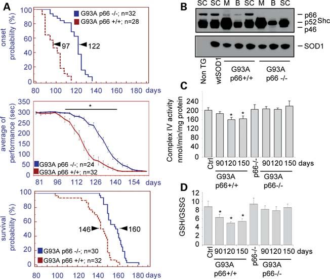
Human Molecular Genetics, 2011, Vol. 20, No. 21                       4197

                                                                                                                                                                    Downloaded from http://hmg.oxfordjournals.org/ at GOT (Consortium) on November 29, 2011
Figure 1. mutSOD1 induces apoptosis in SH-SY5Y cells. (A) SH-SY5Y cells were infected with adenoviruses coding for wild-type SOD1 (wtSOD1) or the
fALS G93A-SOD1 mutant. After 72 h, nuclei of cells were stained with Hoechst 33342, and apoptotic nuclei were quantified as described in Materials and
Methods. (B) Cells were infected as in (A) and analysed after 48 h by indirect immunofluorescence analysis with antibodies against SOD1 (green) or the
active form of caspase-3 (activeC3, red). Cell nuclei are in blue. (C) After 72 h of expression of the indicated SOD1s, nuclear and cytosolic fractions from
cells were isolated. Nuclei were analysed in western blot with antibodies recognizing the cleaved form of PARP1. The nuclear protein Lamin B was analysed
as a standard for equal protein loading. Cytosolic fractions were also controlled for the expression of SOD1. Owing to the high levels of overexpressed exogenous
mutSOD1s, endogenous mouse SOD1 in control cells is not evident in this exposure.

mitochondria (23), suggesting that cytosolic signals, yet to be                   with familial amyotrophic lateral sclerosis (fALS), was transi-
identified, mediate the transfer of the inherent toxic properties                 ently overexpressed in SH-SY5Y human neuroblastoma cells
of mutSOD1 to mitochondria.                                                       by adenoviral infection. In these conditions, G93A-SOD1
   A novel, mitochondria-related signalling mechanism in-                         induces the sequential activation of caspase-3, cleavage of
volving the 66 kDa isoform of the growth factor adapter Shc                       PARP and accumulation of fragmented nuclei, all suggestive
(p66Shc), which is operative in conditions of oxidative                           of an ongoing apoptotic process (Fig. 1). Similar results
stress, has been recently identified. The protein p66Shc is an                    were obtained when SH-SY5Y cells were infected with the
alternatively spliced isoform of a growth factor adapter that                     fALS mutant H80R-SOD1 (Fig. 7 and not shown), indicating
is phosphorylated upon oxidative stress (24). In this form, a                     that the pro-apoptotic effect is common to fALS-linked
fraction of p66Shc localizes to mitochondria, where it binds                      SOD1s. In contrast, infection with adenoviruses expressing
to cytochrome c and acts as an oxidoreductase, generating                         wtSOD1 or a control GFP protein (not shown) has no
ROS and leading to organelle dysfunction and cell death                           evident effects. To determine whether p66Shc is activated
(25). p66Shc2/2 mice exhibit a 30% extended lifespan,                             by mutSOD1 through phosphorylation on Ser36, total lysates
reduced H2O2 levels and an enhanced resistance against oxida-                     from cells were immunoprecipitated with an anti-Shc antibody
tive stress (26), indicating that p66Shc acts as a key molecular                  and analysed by western blotting using an antibody that specif-
sentinel that controls cellular stress responses and mammalian                    ically recognizes phosphorylated p66Shc at Ser36. Expression
lifespan.                                                                         of G93A mutSOD1, but not of wtSOD1, induces p66Shc phos-
   On the basis of these considerations, we have investigated                     phorylation at Ser36 (Fig. 2A), whereas the expression levels
the role of p66Shc in mutSOD1-induced cell toxicity, as                           of all Shc protein isoforms (p46, p52 and p66) are not affected.
well as the functional consequences of p66Shc ablation in
transgenic mice overexpressing G93A-SOD1.
                                                                                  Overexpression of dominant-negative, functionally inactive
                                                                                  p66Shc proteins protects cells against mutSOD1-induced
                                                                                  mitochondrial damage and apoptosis
RESULTS
                                                                                  To investigate the functional relevance of the phosphorylation
Apoptosis induced by mutSOD1 is accompanied by                                    of p66Shc at Ser36 induced by mutSOD1, SH-SY5Y cells
phosphorylation of p66Shc at serine 36                                            stably overexpressing a serine 36-to-alanine (and therefore
p66Shc is phosphorylated on a serine residue in position 36 in                    non-phosphorylatable) mutant of p66Shc (S36A; Fig. 2B)
response to cellular stress induced by various stimuli, and this                  were infected with adenoviruses coding for wild-type SOD1
event is crucial to p66Shc-mediated oxidative stress and apop-                    or G93A mutSOD1 and analysed after 72 h for apoptotic
tosis (26,27). As an initial step to investigate the role of                      markers. As shown in Figure 2B, the number of apoptotic
p66Shc in mutSOD1-induced cell stress, human wild-type or                         cells generated by mutSOD1 is reduced by 90% in cells
G93A-SOD1, the most studied SOD1 mutation associated                              expressing the S36A mutant of p66Shc, and a similar decrease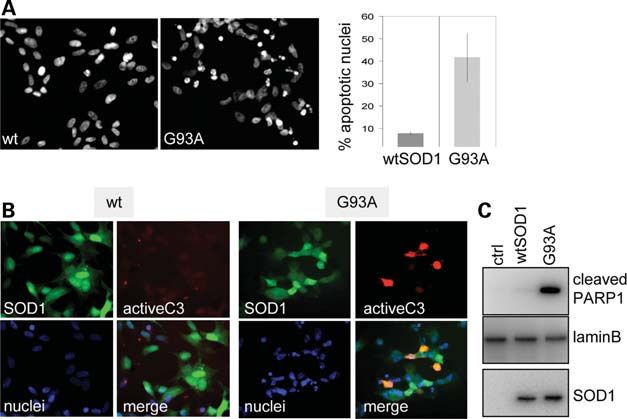
4198       Human Molecular Genetics, 2011, Vol. 20, No. 21

                                                                                                                                                                      Downloaded from http://hmg.oxfordjournals.org/ at GOT (Consortium) on November 29, 2011
Figure 2. Functionally inactive mutant p66Shc protects cells from mutSOD1-induced apoptosis. (A) SH-SY5Y cells were infected with adenoviruses coding for
wtSOD1 or the G93A mutSOD1. After the indicated times, cell lysates were subjected to immunoprecipitation with an anti-Shc antibody. Immunoprecipitates
were analysed in western blot with an antibody recognizing p66Shc phosphorylated on serine 36 (p66P). The blot was re-probed with an anti-Shc antibody to
check for equal protein input. (B) Control (ctrl) SH-SY5Y cells, or cells stably overexpressing the S36A non-phosphorytable p66Shc mutant, were infected as in
(A). After 72 h, cells were stained with Hoechst 33342 and apoptotic nuclei quantified. Values significantly different from relative controls are indicated with an
asterisk when P , 0.01 (n ¼ 3). (C) Cells treated as in (A) were assayed for the activity of caspase-3, expressed in arbitrary units. Two different clones of S36A
p66Shc cells were analysed. Values significantly different from relative controls are indicated with an asterisk when P , 0.01 (n ¼ 3). (D) Control SH-SY5Y
cells, or cells stably overexpressing the S36A, C59S or E132Q/E133Q (EEQQ) mutants of p66Shc, were infected with adenoviruses coding for wtSOD1 or the
G93A mutSOD1. After 72 h, nuclear and cytosolic fractions from cells were isolated. Nuclear fractions were analysed in western blot with antibodies
anti-cleaved PARP1 and anti-lamin B, whereas cytosolic fractions were analysed with anti-SOD1 and anti-Shc.

in caspase-3 activity is observed (Fig. 2C). These data thus in-                   Therefore, using the aequorin technology to monitor mito-
dicate that the phosphorylation of p66Shc plays a role in                          chondrial Ca2+ signalling (29), we analysed the effects of
mutSOD1-induced cell death.                                                        both wild-type and mutSOD1 proteins on SH-SY5Y cells dis-
   The pro-oxidant, pro-apoptotic activities of p66Shc depend                      playing different background for p66Shc, i.e. in wild-type
upon two defined regions of the protein: two glutamic residues                     SH-SY5Y cells, in SH-SY5Y cells overexpressing the
at position 132 and 133, the site where the redox activity of                      mutant S36A-p66Shc, in SH-SY5Y cells overexpressing the
p66Shc has been mapped (25), and a cysteine at position 59,                        mutant EEQQ-p66Shc and in SH-SY5Y cells overexpressing
a regulatory disulphide/thiol site mediating a reversible                          wild-type p66Shc.
dimer – tetramer transition, which has been proposed to                               In SH-SY5Y cells, application of carbachol, an extracellular
control the protein’s apoptosis-inducing activity (28). When                       agonist acting on a Gq-coupled receptor, causes the production
overexpressed in SH-SY5Y cells, both the E132Q/E133Q                               of inositol 1,4,5-trisphosphate and thus the release of Ca2+
(EEQQ) and the C59S mutant p66Shc proteins are able to                             from the endoplasmic reticulum and the transient increase of
inhibit the pro-apoptotic activity of G93A mutSOD1, as mea-                        cytosolic and mitochondrial [Ca2+] (29).
sured by PARP cleavage, similar to what is observed with the                          In wild-type SH-SY5Y cells (Fig. 3A), the overexpression
S36A mutant (Fig. 2D). On the contrary, overexpression of                          of wild-type SOD1 causes an increased mitochondrial
wild-type p66Shc enhances the mutSOD1-induced apoptosis.                           [Ca2+] ([Ca2+]m) response after agonist stimulation. On the
Overall, these observations strongly indicate that p66Shc med-                     contrary, the overexpression of two different mutSOD1s
iates the toxic effects exerted by mutSOD1.                                        causes a drastic reduction in the Ca2+ spike evoked by
   It has been shown that mitochondrial Ca2+ responsiveness,                       agonist stimulation, as an early consequence of mitochondrial
which is a highly sensitive readout of mitochondrial state (27),                   damage as previously reported after p66Shc activation during
is dramatically compromised after p66Shc activation.                               oxidative stress (27).
Human Molecular Genetics, 2011, Vol. 20, No. 21 4199
ATP concentration in SH-SY5Y cells exposed to mutSOD1s
was measured. An [ATP] decrease was evident in cells over-
expressing G93A or H80R mutSOD1 (Fig. 3E); in contrast,
cells co-expressing functionally inactive p66Shc show a
physiological ATP concentration.
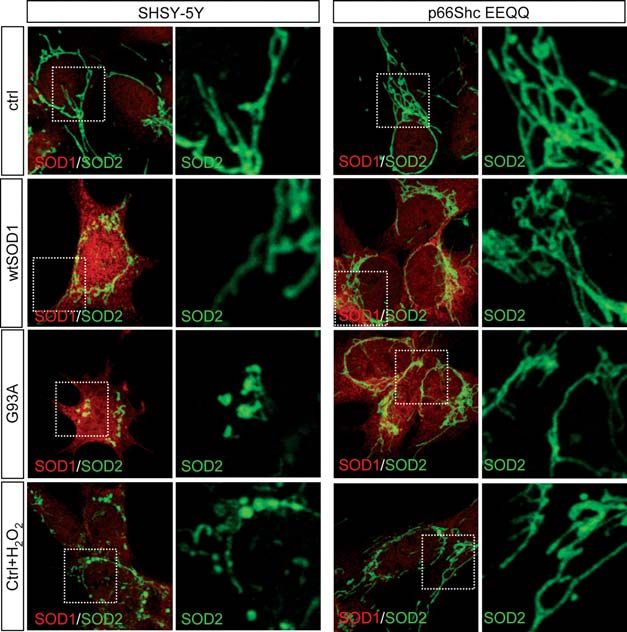
As observed by immunofluorescence analysis of SH-SY5Y
cells using antibodies anti-SOD1 and SOD2, a mitochondrial
matrix protein (Fig. 4, left), cells expressing the G93A
mutSOD1 show a significant alteration of the filamentous
mitochondrial network which characterizes most of the
untransfected or wild-type SOD1-transfected cells, with mito-
chondria appearing fragmented and swollen. Similarly, the
filamentous network is essentially lost when cells are chal-
lenged with H2O2. On the contrary, in cells expressing the
p66Shc EEQQ mutant (Fig. 4, right), the overall filamentous
network is maintained either in the presence of mutSOD1 or
Downloaded from http://hmg.oxfordjournals.org/ at GOT (Consortium) on November 29, 2011
after H2O2 treatment. Similar results were obtained with the
S36A mutant p66Shc (not shown).
On the whole, these data clearly indicate that p66Shc is the
effector downstream of mutSOD1 responsible for the altera-
tions of mitochondrial physiology that eventually lead to cell
death.
Ablation of p66Shc significantly ameliorates ALS
phenotype in mice
To learn whether transgenic G93A-SOD1 mice, an accepted
model for ALS linked to mutSOD1, would be rescued by
genetic removal of p66Shc, we crossed these mice with
p66Shc2/2 mice (26) and determined several behavioural
Figure 3. mutSOD1s cause mitochondrial damage in a p66Shc-dependent and biochemical parameters.
manner. Control SH-SY5Y cells (A), or cells stably overexpressing wild-type As shown in Figure 5B, p66Shc is highly expressed in the
p66Shc (B), the S36A (C) or E132Q/E133Q (EEQQ, D) mutants of p66Shc, spinal cord of control mice, as well as wtSOD1 or
were infected with adenoviruses expressing wtSOD1 and the G93A or
H80R mutSOD1s, together with an adenovirus expressing a mitochondrial tar- G93A-SOD1 transgenic mice, and only to a lower extent in
geted aequorin chimera. After 36 h, cells were challenged with 1 mM charba- the brain and muscle. As expected, ablation of p66Shc does
col. The light signal was collected and calibrated into [Ca2+] values by an not affect the levels of expression of transgenic SOD1 in the
algorithm based on the Ca2+ response curve of aequorin at physiological con- spinal cord. As shown in Figure 5A, G93A-SOD1/
ditions of pH, [Mg2+] and ionic strength. Statistical data are presented as
mean + SEM. (E) SH-SY5Y cells, or cells stably overexpressing S36A or
p66Shc2/2 mice show significantly delayed onset of the
EEQQ mutant p66Shc, were infected with adenoviruses expressing wtSOD1 disease (118.96 + 10.98 versus 98.07 + 8.9 days, P ,
and the G93A or H80R mutSOD1s. After 48 h, total amount of ATP was 0.0001), improved motor performance as measured by
assayed. The ATP concentration was normalized to total cellular protein con- rotarod test and increased survival (141.26 + 14.42 versus
centration and expressed as percent of the relative, uninfected controls. Values 157.50 + 9.11 days, P , 0.0001) with respect to
are reported as mean + SD. Values significantly different from relative con-
trols are indicated with an asterisk when P , 0.05 (n ¼ 3). G93A-SOD1 mice. Such striking effects are paralleled by
improved mitochondrial function specifically in the spinal
cord, where the activity of complex IV is restored (Fig. 5C)
together with the ratio between reduced and oxidized glutathi-
The presence of p66Shc mutant proteins (Fig. 3C and D)
one (GSH/GSSG) in mitochondria, which is an indicator of the
confers mitochondrial insensitiveness to mutSOD1, since the
redox state of the cell (Fig. 5D).
mitochondrial Ca2+ response after agonist stimulation is
almost unaffected by the presence of mutSOD1. On the con-
trary, the alteration of mitochondrial responsiveness induced
by mutSOD1 is maintained in SH-SY5Y cells overexpressing p66Shc mediates the toxicity of mutSOD1 through
wild-type p66Shc (Fig. 3B). Rac1 inactivation
We have recently shown that mutSOD1 induces alterations To learn more on the molecular mechanisms of
in mitochondrial bioenergetics and morphology of SH-SY5Y mutSOD1-induced p66Shc toxicity, and to attempt the dissec-
cells (5). To test whether these effects arise as a consequence tion of such mechanisms in vitro, we focused on Rac1, a
of p66Shc activation, ATP production and mitochondrial member of the Rho family of small GTPases, which controls
morphology were assayed in cells overexpressing wild-type many intracellular processes, including ROS production and
or mutSOD1 together with p66Shc mutant proteins. To cytoskeletal dynamics, and whose activity has been linked to
analyse the mitochondrial capability for energy production, the activity of both SOD1 and p66Shc (30,31).4200 Human Molecular Genetics, 2011, Vol. 20, No. 21
Downloaded from http://hmg.oxfordjournals.org/ at GOT (Consortium) on November 29, 2011
Figure 4. p66Shc inhibition prevents mitochondrial fragmentation induced by mutSOD1. SH-SY5Y control cells or p66Shc/EEQQ overexpressing cells were left
untreated, or infected with adenoviruses coding for the indicated SOD1s. After 48 h, cells were subjected to immunofluorescence analysis with anti-SOD1
(green) and anti-SOD2 (red) antibodies. As a control for mitochondrial fragmentation, SH-SY5Y cells were treated with 500 mM H2O2 for 30 min and processed
for mitochondrial staining as above. Higher magnifications of areas highlighted in insets are shown.
Overexpression of both G93A and H80R mutant proteins p66Shc downregulates Rac1 activity through a
decreases the levels of active, GTP-bound form of Rac1 in redox-dependent mechanism
SH-SY5Y cells, compared with control and wtSOD1-
It has been recently shown that the activity of Rac1 is physio-
expressing cells, as measured by a GST-PAK1 pull-down
logically controlled by the redox environment of the cell,
assay (Fig. 6A and B). The effect is specific for Rac1, since
according to a mechanism where SOD1 itself exerts a
mutSOD1s do not significantly affect the activity of either
RhoA or Cdc42, two small GTPases functionally related to primary role (30). To test whether a redox-dependent mechan-
Rac1 (Fig. 6A and B). Moreover, the overexpression by adeno- ism could account for the inhibition of Rac1 activity following
viral infection of a constitutive active (V12) form of Rac1 p66Shc activation, control SH-SY5Y cells or cells overexpres-
completely protects SH-SY5Y cells from mutSOD1-induced sing a functional inactive mutant of p66Shc (S36A) were
apoptosis, whereas the expression of a dominant-negative, in- treated with H2O2 and the amount of GTP-Rac1 was assayed
active (N17) mutant of Rac1 has no effect (Fig. 7). Significant- accordingly. Indeed, increasing concentration of H2O2
ly, N17 Rac1 is able to induce apoptosis in control cells, and to induces a proportional decrease in Rac1 activity, and this
re-establish the apoptotic phenotype in cells where apoptosis effect is associated with a strong decrease in cell viability
induced by mutSOD1s overexpression has been hampered by (Fig. 10A and B). The expression of an S36A p66Shc
co-expression of functional inactive mutants of p66Shc mutant almost completely prevents H2O2-induced cell death,
(Fig. 8A and B), suggesting that Rac1 acts downstream of and in this condition the decrease in the activity of Rac1 is pre-
p66Shc. More importantly, in cells expressing the dominant- cluded (Fig. 10B and C). These results prompted us to inves-
negative mutant p66Shc-S36A, Rac1 is active even in the tigate whether the activity of Rac1 could be affected by factors
presence of mutSOD1s (Fig. 9A); on the contrary, when a wild- controlling the intracellular pool of GSH, which is the major
type p66Shc protein is overexpressed in cells, Rac1 activity determinant of the cellular redox state (32). As shown in
is dramatically compromised by mutSOD1s (Fig. 9B). Figure 10D, treatment of cells overexpressing mutSOD1
Altogether, these data thus clearly indicate that p66Shc activa- with the cell-permeable ethyl ester form of reduced GSH
tion is an obligatory step for mutSOD1 to inhibit Rac1, which in (GEE), which results in an increase in intracellular GSH,
turn leads to cell death. significantly restores Rac1 activity, whereas depletion ofHuman Molecular Genetics, 2011, Vol. 20, No. 21 4201
Downloaded from http://hmg.oxfordjournals.org/ at GOT (Consortium) on November 29, 2011
Figure 5. Downregulation of p66Shc ameliorates ALS phenotype in mice. (A) Transgenic mice overexpressing G93A-SOD1 were crossed with p66Shc2/2
mice and characterized for onset (grip test, top panel), motor performance (rotarod test, middle panel) and survival (bottom panel). Equal numbers of males
and females were analysed for each genoptype. No sex-dependence was observed and therefore data were pooled. Number of animals tested (n) and the
median onset and survival probabilities (arrowheads) are shown. Values significantly different from relative controls are indicated with an asterisk when
P , 0.01. (B) Western blot analysis of SOD1 and p66Shc in the brain (B), muscle (M) and spinal cord (Sc) from G93A-SOD1 and G93A-SOD1/
p66Shc2/2 mice. As a control, p66Shc expression was analysed in non-transgenic mice and in mice overexpressing wild-type SOD1. (C) Complex IV activity
were determined from mitochondria extracted from spinal cord of G93A-SOD1 and G93A-SOD1/p66Shc2/2 mice. Values are reported as mean + SD. Values
significantly different from relative controls are indicated with an asterisk when P , 0.05 (n ¼ 3). (D) HPLC determination of GSH/GSSG ratio in mitochondria
extracted from spinal cord of G93A-SOD1 and G93A-SOD1/p66Shc2/2 mice. Values are reported as mean + SD. Values significantly different from relative
controls are indicated with an asterisk when P , 0.05 (n ¼ 3).
cytoplasmic GSH with L-buthionine-(S,R)-sulfoximine (BSO) physiological role of this process is currently unknown, al-
has a striking inhibitory effect on the activity of Rac1. though it is clear that it might participate in the control of
intracellular redox-based signal transduction pathways (34).
These mechanisms are indeed relevant for the process of apop-
DISCUSSION tosis, since p66Shc-mediated formation of ROS triggers the
In the present study, we show that inhibition of mitochondrial initiation of the mitochondrial apoptosis pathway, and more
redox signalling by p66Shc is able to rescue viability in cell generally for the regulation of lifespan (26) and energy metab-
models and to improve survival in the mouse model for olism (35,36), which are intimately connected. Moreover,
fALS linked to mutSOD1. p66Shc has a function in various pathological conditions
Indeed, it has been shown, both in models and patients, that where oxidative stress plays a role, such as arteriosclerosis
the pathological phenotypes of ALS well correlate with altera- (37) and endothelial dysfunctions (38).
tions in all the processes related to or controlling mitochon- Although it has been initially suggested that other, non-
drial function, i.e. morphology and bioenergetics, mitochondrial activities of p66Shc might be needed to exert
transportation and clearance, apoptosis and calcium buffering its pro-apoptotic function (25), activation of the p66Shc
(33), thus sustaining a direct role of mitochondrial dysfunction pathway has emerged as a clear readout of mitochondrial
in motor neuron degeneration in ALS pathogenesis. damage in cells (39), and data presented in this work clearly
However, evidence linking mitochondrial abnormalities to point to the intrinsic, mitochondrial redox activity of p66Shc
ALS is yet incomplete. Most of all, it is not entirely clear in mediating the toxicity exerted by mutSOD1. Different
whether, and to what extent, mitochondrial preservation lines of evidence support this conclusion: (i) the accumulation
could be beneficial to the disease. To answer these questions, in cells expressing mutSOD1 of p66Shc species phosphory-
we decided to focus on p66Shc as a central regulator of mito- lated on serine 36, a critical regulatory site for the mitochon-
chondrial ROS metabolism and the mitochondrial apoptosis drial pro-apoptotic activity of p66Shc (27), and the rescuing
pathway (24). p66Shc responds to a variety of stimuli by in- effect on cell viability of the non-phosphorylatable S36A
creasing ROS levels in the mitochondrial intermembrane p66Shc mutant; (ii) the inhibitory effect over the toxic
space through an ROS-producing activity (25). The action of mutSOD1s of the EEQQ and C59S p66Shcs, two4202 Human Molecular Genetics, 2011, Vol. 20, No. 21
Figure 7. A constitutively active (V12) form of Rac1 completely protects
SH-SY5Y cells from mutSOD1-induced apoptosis. SH-SY5Y cells were
infected with adenoviruses expressing wild-type or the G93A (left panel)
and H80R (right panel) mutSOD1s, in the absence or in the presence of ade-
Downloaded from http://hmg.oxfordjournals.org/ at GOT (Consortium) on November 29, 2011
noviruses coding for constitutive active (V12) or inactive (N17) mutants of
Rac1. After 72 h, nuclear fractions from cells were isolated and analysed in
western blot with antibodies anti-cleaved PARP1 and anti-lamin
B. Cytosolic fractions were analysed with anti-SOD1, anti-Rac1 and
anti-b-actin antibodies.
Rac1, a prominent member of Rho family of small
Figure 6. MutSOD1 proteins decrease Rac1-GTP levels in SH-SY5Y cells. GTPases. Rac1 is an intracellular transducer known to regu-
(A) SH-SY5Y cells were infected with adenoviruses expressing wild-type or late multiple signalling pathways that control the organization
the G93A and H80R mutSOD1s. After 72 h, GTP-bound Rac1 and Cdc42
were pulled-down by a GST-PAK protein, whereas GTP-RhoA was pulled-
of cytoskeleton, gene expression and cell proliferation (41).
down with a GST-Rothekin protein, both conjugated to GSH–sepharose. Rac1 and its relatives are also essential regulators of
The GTP-bound (active) as well as the total (input) amounts of GTPases NADPH-dependent membrane oxidase (NOX) that produces
were detected in western blot using the indicated antibodies. Lysates were superoxide anions, in both phagocytic and non-phagocytic
also analysed for SOD1 expression. (B) Quantification of active Rho cells (42), according to a mechanism where SOD1 itself
GTPases in cells treated as in (A). The amounts of active GTPases were nor-
malized to inputs and are expressed as mean + SD of arbitrary densitometric seems to play a pivotal role. Indeed, Harraz et al. (30)
units relative to control, non-infected cells. Values significantly different from have recently shown that when superoxide anions produced
relative controls are indicated with an asterisk when P , 0.01 (n ¼ 4). by microglial NOX exceed a physiological threshold, they
are converted to hydrogen peroxide by SOD1, which normal-
ly binds to Rac1 itself and stimulates its activity. H2O2 then
mutants that are specifically endowed with mitochondrial leads to the oxidative inactivation of Rac1, to the detachment
redox activity (25,28); (iii) the recovery of mitochondrial of SOD1 from Rac1 and eventually to silencing of Rac1-
function (as measured by rescued mitochondrial Ca2+ buffer- dependent activity of NOX.
ing capacity, ATP production and mitochondrial morphology) Similar to Harraz et al. (30), we also observed that Rac1 ac-
and of cell viability obtained by the expression of functionally tivity is strictly controlled, among other mechanisms, by redox
inactive mutant p66Shcs. On the whole, these data clearly conditions. However, we found that active Rac1 and cell via-
support the notion that the p66Shc mitochondrial pathway is bility are directly linked in neuronal cells, and that cell death
a key effector responsible for the alterations of mitochondrial induced by mutSOD1s is achieved through a
physiology evoked by mutSOD1. This conclusion is further p66Shc-dependent inhibition of Rac1, whereas it has been
supported by our data in vivo, in transgenic mice overexpres- clearly shown that the pro-inflammatory activity of
sing mutant G93A-SOD1 but lacking p66Shc, where onset, mutSOD1-expressing microglial cells relies on an uncon-
motor performance and survival are significantly improved, to- trolled, constitutive activation of Rac1 (30). Further, p66Shc
gether with mitochondrial functionality, and is in agreement was proposed to promote Rac1 activation (31), thereby trig-
with recent findings showing that blocking the mitochondrial gering ROS production by NOX, whereas in our cell model,
apoptotic pathway preserves motor neuron viability and func- p66Shc activation is clearly responsible for Rac1 inhibition.
tion in a mouse model of ALS (40). Of note, although a strik- The reasons for these apparent discrepancies can be partially
ing effect of p66Shc removal is obtained on the disease onset explained by differences in the cellular milieu (neurons
in these mice, the outcome on the overall disease progression versus glia) and by the fact that the activity of Rho family
is relatively minor. This would indicate a central role of mito- GTPases is strictly dependent on their subcellular distribution
chondrial dysfunction in mediating disease initiation by and compartmentalization, which again can be profoundly dif-
mutSOD1, and of p66Shc as an important player participating ferent between neuronal and glial cells.
in this process. Nonetheless, different lines of evidence support a role of
To clarify the molecular mechanisms by which p66Shc Rac1 inhibition in the process of neurodegeneration in ALS.
activation is responsible for neuronal cell damage induced Mutations in alsin, which are responsible for a recessive
by overexpression of mutSOD1s, we analysed the functional form of juvenile-onset ALS, are predicted to affect the
relationship between activation of p66Shc by mutSOD1 and guanine nucleotide exchange factor (GEF) activity of thisHuman Molecular Genetics, 2011, Vol. 20, No. 21 4203
Downloaded from http://hmg.oxfordjournals.org/ at GOT (Consortium) on November 29, 2011
Figure 9. Inhibition of p66Shc prevents the downregulation of Rac1 activity
Figure 8. A constitutively inactive (N12) form of Rac1 re-establishes the induced by mutSOD1s. Control SH-SY5Y or SH-SY5Y cells expressing the
apoptotic phenotype in cells overexpressing functional inactive mutants of S36A (A) or the wild-type (B) p66Shc proteins were infected with adeno-
p66Shc. Control SH-SY5Y cells, or SH-SY5Y cells expressing the S36A, viruses expressing wild-type SOD1 or the G93A and H80R mutSOD1s.
C59S (A) or EEQQ (B) p66Shc mutants, were infected with adenoviruses After 72 h, GTP-bound Rac1 was analysed with a GST-PAK pull-down
coding for wild-type or the G93A and H80R mutSOD1s, in the absence or assay. The GTP-bound (active) as well as the total (input) amounts of Rac1
in the presence of adenoviruses expressing constitutive active (V12) or in- GTPase were detected in western blot using an anti-Rac1 antibody. Lysates
active (N17) mutants of Rac1. After 72 h, nuclear and cytosolic fractions were also analysed for p66Shc and SOD1 expression. The numbers represent
from cells were isolated. Nuclear fractions were analysed in western blot fold activity above normalized activity in control uninfected cells (referred to
with antibodies anti-cleaved PARP1 and anti-lamin B, cytosolic fractions as 1.0); densitometric scanning of the films was used to determine relative
with anti-Shc and anti-SOD1. levels of Rac1 activation versus protein amounts. The data are representative
of n ¼ 3 independent experiments.
protein (43,44). Alsin knockdown inhibits axon growth and Cdc42 GTPases. These effects are associated with the inhib-
induces cell death in cultured motoneurons. Notably, these ition of protein geranylgeranylation, a key post-translational
cellular phenotypes are mimicked by expression of a modification for Rho family activity and intracellular localiza-
dominant-negative Rac1 mutant and are completely blocked tion (47). Our results point to a direct role of ROS in Rac1 in-
by expression of a constitutively active Rac1 mutant (45). activation, as suggested by the observation that the decrease of
Overexpression of alsin protects NSC34 cells from Rac1 activity by both hydrogen peroxide or mutSOD1 is pre-
mutSOD1-induced apoptosis, and this neuroprotective activity vented by inhibition of p66Shc-dependent mitochondrial
is completely inhibited by knocking down the endogenous redox signalling or by increased intracellular concentration
Rac1 expression with siRNA for Rac1 (46). On the whole, of GSH. This is in line with recent findings indicating that
these observations clearly point to disruption of Rac1 GTPases may also be directly controlled by redox agents
GTPase function as a causative event of motor neuron degen- (48), a mechanism of regulation that may be particularly rele-
eration. This conclusion is further supported by recent results vant in pathological conditions, such as ALS, where ROS are
obtained in neuronal cells depleted of the 43 kDa TAR DNA- generated and the cellular redox balance altered.
binding protein (TDP-43), a major component of the ubiquiti- In conclusion, our data emphasize that mitochondrial redox
nated inclusions characteristic of ALS and frontotemporal signalling, p66Shc and Rac1 are linked by an intimate connec-
lobar degeneration with ubiquitin-positive inclusions, whose tion in neurons in vitro and in vivo, in adult mice. That such
mutations have been causally linked to familial ALS. The connections are altered in a genetic context mimicking part
knockdown of TDP-43 in differentiated Neuro-2a cells inhi- of ALS patients strongly supports the concept that molecules
bits neurite outgrowth and induces cell death through the in- involved in this signalling play an important role in this
activation of Rho family members RhoA and Rac1, and disease.4204 Human Molecular Genetics, 2011, Vol. 20, No. 21
co-transfected with plasmids coding for different p66Shc pro-
teins in the presence of 1:20 of a plasmid coding for hygromy-
cin resistance, using Lipofectamine Plus reagent (Invitrogen).
After selection with 400 mg/ml hygromycin (Invitrogen),
about 20 clones for each construct were isolated independently
and analysed in western blot with anti-Shc antibodies. At least
three clones for each plasmid were chosen for equivalent ex-
pression of p66Shc proteins and used for further analysis.
All the clones analysed gave consistent results and data from
one clone are shown.
Construction of recombinant adenoviruses expressing wild-
type SOD1, as well as G93A and H80R mutSOD1, was carried
out by inserting cDNAs coding for the different SOD1s into
the pShuttle2 and BD Adeno-X viral DNA (BD Bioscience).
Transient transfections of HEK293 cells were performed
using Lipofectamine 2000 (Invitrogen). Viral titre was deter-
Downloaded from http://hmg.oxfordjournals.org/ at GOT (Consortium) on November 29, 2011
mined by dilution assay using HEK293 cells according to
the manufacturer’s instructions. Adenoviruses were propa-
gated in HEK293 cells as described in Latella et al. (49). In-
fection of SH-SY5Y cells was carried out for 1 h in
OPTIMEM; after removal of the virus, cells were grown for
the indicated period of times before being subjected to
further experimental manipulations. To optimize the protocol
for SH-SY5Y infection, cells were infected with Ad-GFP
and Ad-SOD1s, and expression was measured by immunoblot-
Figure 10. Redox regulation of Rac1 activity by mutSOD1. (A) SH-SY5Y ting and immunofluorescence with the antibody anti-SOD1.
cells were treated with the indicated amounts of H2O2. After 24 h, active Efficiency of infection was 90% at a multiplicity of infec-
Rac1 was measured by a GST-PAK pull-down assay. (B) The viability of tion (m.o.i.) of 1000 after infection for 48 h. V12 and N17
control SH-SY5Y cells or SH-SY5Y cells expressing the S36A mutant Rac1 adenoviruses were described in Cozzolino et al. (50)
p66Shc and treated with 50 mM H2O2 was calculated after 24 h through an
MTS assay. (C) The activity of Rac1 was measured in cells treated as in
and used at an m.o.i. of 50.
(B). (D) SH-SY5Y cells were infected with adenoviruses expressing
wtSOD1 or the H80R mutSOD1, in the absence or in the presence of 5 mM
GEE. Control cells were also treated with 10 mM BSO. After 72 h, cell Nuclei isolation and cleaved PARP1 analysis
lysates were subjected to GST-PAK pull-down assay for the assessment of
Rac1 activity. The numbers represent fold activity above normalized activity On 60 mm Petri dishes, 1.5 × 10⁶ SH-SY5Y cells were plated.
in control uninfected cells (referred to as 1.0). The data are representative of After the indicated treatments, cells were rinsed in ice-cold
n ¼ 3 independent experiments.
PBS and lysed in 150 ml of low-salt buffer (10 mM Hepes,
pH 7.4, 42 mM KCl, 5 mM MgCl2, 0.5 % CHAPS, 1 mM
MATERIALS AND METHODS DTT, 1 mM PMSF, 1 mg/ml leupeptin). After centrifugation
at 2000g, nuclei were resuspended in 50 ml of high-salt
Plasmid construction buffer (50 mM Tris – HCl, pH 7.5, 400 mM NaCl, 1 mM
Human cDNA coding for p66Shc (accession number U73377) EDTA, 1% Triton X-100, 0.5% Nonidet-P40, 10% glycerol,
was cloned by reverse transcription-PCR from human 2 mM DTT, 1 mM PMSF, protease inhibitor cocktail. After
SH-SY5Y neuroblastoma cells cDNA, using the forward 30 min on ice, lysates were centrifuged at 20 000g, superna-
primer 5′ AAA AAG CTT ATG GAT CTC CTG CCC CCC tants were collected and analysed as nuclear fractions in
3′ and the reverse primer 5′ TTT CTC GAG TCA CAG western blot with antibodies anti-cleaved PARP1 (Cell
TTT CCG CTC CAC 3′ . The resulting PCR fragment was Signal) and anti-Lamin B (Santa Cruz Biotechnology).
inserted into HindIII/XhoI restriction sites of pcDNA3 (Invi-
trogen). For the mutagenesis of p66Shc, PCR site-directed mu-
tagenesis was performed using pcDNA3/p66Shc as template, Assessment of apoptosis and cell viability
followed by digestion with Dpn1. All the plasmid construc- Quantification of apoptotic cells was obtained by direct visual
tions were verified by automated sequencing. counting after nuclear staining of 4% paraformaldehyde-fixed
cells with the fluorescent probe Hoechst 33342 (1 mg/ml)
(Sigma-Aldrich). One hundred cells were examined for each
Cell culture, plasmid transfection and adenoviral infection field at a magnification of 200× and eight randomly chosen
Human neuroblastoma cells, SH-SY5Y, were purchased from fields for each experimental condition were counted. Only
the European Collection of Cell Culture and grown in DMEM the cells containing clearly picnotic or fragmented nuclei
(Invitrogen) supplemented with 10% fetal calf serum (FCS, were considered apoptotic. Caspase 3 activity was measured
Euroclone), at 378C in an atmosphere of 5% CO2 in air. For with a TruePoint Caspase 3 assay kit (PerkinElmer), accord-
stable expression of wild-type or mutant p66Shc, cells were ingly to the manufacturer’s instructions.Human Molecular Genetics, 2011, Vol. 20, No. 21 4205
Cell viability was assessed by a colorimetric assay using the sample. The luminescence was measured using a lumines-
3(4,5-dimethylthiazol-2yl)-5-(3-carboxymethoxyphenyl)-2-(4- cence plate reader (Victor3-V, PerkinElmer Life Sciences).
sulfophenyl)-2H-tetrazolium (MTS) assay (CellTiter 96 The ATP concentration was normalized to total cellular
Aqueous One Solution Assay, Promega), according to the protein concentration estimated by Bradford protein assay
manufacturer’s instructions. Absorbance at 490 nm was mea- (Bio-Rad).
sured in a multilabel counter (Victor3-V, PerkinElmer Life
Sciences).
Rho family pull-down assay
The Rac-GTP and Cdc42-GTP pull-down assay was per-
Mitochondrial Ca21 measurements
formed as previously described (50). Briefly, cells were
Cells were seeded before transfection onto 13 mm glass cover lysed in a buffer containing 50 mM Tris, pH 7.2, 100 mM
slips and allowed to grow to 50% confluence. At this stage, the NaCl, 5 mM MgCl2, 1 mM DTT, 10% glycerol, 1%
cells were infected with the adenovirus expressing a mitochon- Nonidet-P40, plus protease inhibitors. One-fiftieth of cell
drial targeted aequorin chimera. Thirty-six hours after infec- lysates were subjected to immunoblotting. Cell lysates were
tion, the cover slips with the cells were incubated with 5 mM mixed with 10 mg of bacterially expressed GST-PAK (rat
coelenterazine for 1 – 2 h in DMEM supplemented with 1% PAK amino acids 1 – 252) bound to GSH– sepharose and incu-
Downloaded from http://hmg.oxfordjournals.org/ at GOT (Consortium) on November 29, 2011
FCS, and then transferred to the perfusion chamber. All bated at 48C with tumbling for 30 min. Beads were collected
aequorin measurements were carried out in KRB (Krebs – by centrifugation and washed twice in lysis buffer before add-
Ringer modified buffer: 125 mM NaCl, 5 mM KCl, 1 mM ition of Laemmli buffer and analysis by western blot with
Na3PO4, 1 mM MgSO4, 5.5 mM glucose, 20 mM HEPES, pH anti-Rac1 and anti-Cdc42 antibodies. For the RhoA-GTP pull-
7.4, 378C) supplemented with 1 mM CaCl2. The agonist, down assay, cell lysis and washes were done in 50 mM Tris–
1 mM carbachol, was then added to the same medium. The Cl, pH 7.2, 500 mM NaCl, 1% (v/v) Triton X-100, 5 mM
experiments were terminated by lysing the cells with 100 mM MgCl2, 1 mM DTT and protease inhibitors. Bacterially
digitonin in a hypotonic Ca2+-rich solution (10 mM CaCl2 in expressed GST-Rhotekin (murine amino acids 7 – 89) bound
H2O), thus discharging the remaining aequorin pool. The to GSH – sepharose was used in place of PAK.
light signal was collected and calibrated into [Ca2+] values
by an algorithm based on the Ca2+ response curve of aequorin
at physiological conditions of pH, [Mg2+] and ionic strength, Immunoprecipitation
as previously described (29). Statistical data are presented as After rinsing the cultures with ice-cold PBS, cell lysis was per-
mean + SEM, significance was calculated by Student’s t-test formed in RIPA buffer (50 mM Tris– HCl, 0.5% Triton X-100,
and correlation analysis was done with the SigmaPlot 5.0 soft- 0.25% Na-deoxycholate, 0.1% SDS, 150 mM NaCl, 1 mM
ware (SPSS, Inc.). EDTA, 5 mM MgCl2) containing 1 mM PMSF and a protease
inhibitor cocktail (Sigma-Aldrich). A clear supernatant was
obtained by centrifugation of lysates at 17 000g for 10 min.
Immunofluorescence analysis Protein content was determined using Bradford protein assay
For immunofluorescence analysis, cells cultured on (Bio-Rad). Equal amounts of lysates were incubated at 48C
poly-L-lysine-coated glass cover slip were washed in PBS for 2 h with a rabbit polyclonal anti-Shc antibody (Cell
and fixed with 4% paraformaldehyde in PBS for 10 min. Signal) and the immunocomplexes were collected by
Fixed cells were washed in PBS followed by permeabilization binding to protein A-Agarose beads (Roche), followed by
with 0.1% Triton X-100 in PBS for 5 min. Cells were blocked three washes with lysis buffer.
for 1 h in 2% horse serum in PBS and incubated for 1 h at
378C with primary antibodies: mouse monoclonal anti-active
casp3 (Cell Signaling), rabbit anti-SOD1 (Stressgen), rabbit Electrophoresis and western blot
anti-SOD2 (Stressgen), mouse monoclonal anti-SOD1 (clone Standard SDS– PAGE was performed as described (10).
SD-G6, Sigma-Aldrich). Cells were washed in blocking Western blot was performed onto nitrocellulose membranes
buffer and incubated for 1 h with an Alexa Fluor 488 goat anti- (Amersham), except for RhoA, Rac1 and Cdc-42 that were
mouse (Invitrogen) and Cy3 goat anti-rabbit (Jackson Immu- blotted onto PVDF membranes (Millipore). After incubation
noResearch Laboratories) antibodies. After rinsing in PBS, in Tris-buffered saline (TBS) solution containing 0.1%
cells were stained with 1 mg/ml Hoechst 33342 Tween 20 and 5% non-fat milk, filters were incubated for
(Sigma-Aldrich) and examined under a Zeiss LSM 510 con- 2 h at room temperature with the indicated antibodies diluted
focal microscopy. Fluorescence images were processed using in a 2% non-fat milk, 0.1% Tween 20/TBS solution. Immunor-
Adobe Photoshop. eactive SOD1 was detected with a rabbit polyclonal
anti-SOD1 antibody (Stressgen). p66Shc proteins were
detected using a mouse monoclonal anti-Shc antibody which
Measurement of cellular ATP recognizes all the three isoforms of Shc (p46, p52 and p66).
Measurement of cellular ATP was performed using the Phosphorylated p66Shc on serine 36 was detected using a
ATPlite Assay (Perkin Elmer-Cetus, Norwalk, CT, USA). In mouse monoclonal specific antibody (Alexis). A goat antibody
brief, cells seeded in 96-well microplates were resuspended (Santa Cruz Biotechnology) was used to detect the cleaved
in 50 ml of lysis buffer and mixed for 10 min. Forty microlitres form of PARP1. Antibodies anti-RhoA and Cdc42 were
of substrate solution (Luciferase/Luciferin) was added to each from Santa Cruz Biotechnology. Anti-Rac1 was from4206 Human Molecular Genetics, 2011, Vol. 20, No. 21
Millipore. b-Actin was detected using a mouse monoclonal Determination of complex IV activity and GSH/GSSG ratio
antibody from Sigma. A goat antibody anti-Lamin B was were carried out on purified mitochondria as previously
from Santa Cruz Biotechnology. described (10).
Following extensive washing in 0.1% Tween 20/TBS solu-
tion, filters were incubated with the appropriated peroxidase-
conjugated secondary antibodies, washed in 0.1% Tween 20/ Statistical analysis
TBS solution and developed using the POD chemilumines- Statistical analysis of rotarod data was performed using
cence detection system (Roche). Image analysis and quantifi- ANOVA. Kaplan – Meier curves were compared using the
cations were performed by Kodak Image Station (KDS log-rank test. Comparisons of protein expression levels,
IS440CF 1.1) with 1D Image Analysis software. complex IV activity and values of GSH/GSSG ratio were per-
formed using two-tailed unpaired Student’s t-test. P-values of
,0.05 were considered significant.
Animals
All animal procedures have been performed according to the ACKNOWLEDGEMENTS
European Guidelines for the use of animals in research (86/
Downloaded from http://hmg.oxfordjournals.org/ at GOT (Consortium) on November 29, 2011
609/CEE) and the requirements of Italian laws (D.L. 116/ We are grateful to Claudia Crosio and Ciro Iaccarino for
92). The ethical procedure has been approved by the Animal helping with construction of adenoviruses, to Cristiana Valle
Welfare Office, Department of Public Health and Veterinary, and Simona Rossi for help with animal motor tests and to
Nutrition and Food Safety, General Management of Animal Silvia Middei for statistical analysis.
Care and Veterinary Drugs of the Italian Ministry of Health.
Conflict of Interest statement. None declared.
At the indicated time, mice were anaesthetized with
500 mg/kg chloral hydrate, sacrificed and dissected for the dif-
ferent experiments. All efforts were made to minimize suffer-
ing. All animals have been raised and crossed in the indoor FUNDING
animal house in a 12 h light/dark cycle in a virus/antigen-free This work was supported by Association Francaise contre les
facility with controlled temperature and humidity and have Myopathies (Project 14354 to M.C.), by Fondation Thierry
been provided with water and food ad libitum. Latran and Neuron EraNet (to M.T.C.) and by the Italian As-
Wild-type SOD1 mice B6.Cg-Tg(SOD1)2Gur/J and sociation for Cancer Research (AIRC), Telethon (GGP09128),
SOD1G93A mice B6.Cg-Tg(SOD1∗ G93A)1Gur/J were pur- the Italian Ministry of Education, University and Research
chased from The Jackson Laboratory and were on C57BL/6J (COFIN) and Italian Ministry of Health (to P.P.).
background. p66Shc2/2 mice were also in the C57BL/6J
background.
Mice compared in this study were all littermates and housed REFERENCES
together to minimize environmental factors. Mice were geno-
1. Cozzolino, M., Ferri, A. and Carri, M.T. (2008) Amyotrophic lateral
typed using PCR protocols from The Jackson Laboratory. sclerosis: from current developments in the laboratory to clinical
Western blot analysis for p66Shc and SOD1 expression was implications. Antioxid. Redox Signal., 10, 405– 443.
carried out as described above on total protein extracts from 2. Cassina, P., Cassina, A., Pehar, M., Castellanos, R., Gandelman, M.,
various tissues obtained through homogenization in RIPA de Leon, A., Robinson, K.M., Mason, R.P., Beckman, J.S., Barbeito, L.
et al. (2008) Mitochondrial dysfunction in SOD1G93A-bearing astrocytes
buffer. promotes motor neuron degeneration: prevention by
mitochondrial-targeted antioxidants. J. Neurosci., 28, 4115–4122.
3. Dobrowolny, G., Aucello, M., Rizzuto, E., Beccafico, S., Mammucari, C.,
Boncompagni, S., Belia, S., Wannenes, F., Nicoletti, C., Del Prete, Z.
Symptom onset, survival and rotarod analysis et al. (2008) Skeletal muscle is a primary target of SOD1G93A-mediated
toxicity. Cell Metab., 8, 425 –436.
Behavioural analysis was performed according to the standard 4. Dupuis, L., di Scala, F., Rene, F., de Tapia, M., Oudart, H., Pradat, P.F.,
operating procedures indicated by Ludolph et al. (51). Briefly, Meininger, V. and Loeffler, J.P. (2003) Up-regulation of mitochondrial
mice were considered terminally paralysed if they were unable uncoupling protein 3 reveals an early muscular metabolic defect in
to right themselves after 10 s of being placed on their side. To amyotrophic lateral sclerosis. FASEB J., 17, 2091–2093.
5. Ferri, A., Fiorenzo, P., Nencini, M., Cozzolino, M., Pesaresi, M.G., Valle,
assess symptom onset, mice were subjected to grip test twice a C., Sepe, S., Moreno, S. and Carri, M.T. (2010) Glutaredoxin 2 prevents
week, starting at 70 days of age. Rotarod testing was per- aggregation of mutant SOD1 in mitochondria and abolishes its toxicity.
formed using the accelerating rotarod apparatus (Ugo Basile Hum. Mol. Genet., 19, 4529– 4542.
7650 model). The rod was accelerated at a constant rate of 6. Cozzolino, M., Pesaresi, M.G., Amori, I., Crosio, C., Ferri, A.,
Nencini, M. and Carri, M.T. (2009) Oligomerization of mutant SOD1 in
4 r.p.m. starting from 3 r.p.m. for a maximum of 5 min. The mitochondria of motoneuronal cells drives mitochondrial damage and cell
time (seconds) at which the animal fell from the bar was toxicity. Antioxid. Redox Signal., 11, 1547–1558.
recorded. Mice were tested twice a week for three trials, 7. Higgins, C.M., Jung, C. and Xu, Z. (2003) ALS-associated mutant
each starting at 80 days of age, until they were unable to SOD1G93A causes mitochondrial vacuolation by expansion of the
remain on the rotarod for at least 20 s. The best trial per day intermembrane space and by involvement of SOD1 aggregation and
peroxisomes. BMC Neurosci., 4, 16.
was recorded and used for analysis. Each time point represents 8. Kong, J. and Xu, Z. (1998) Massive mitochondrial degeneration in motor
the mean + SE of performance of all mice at each data point neurons triggers the onset of amyotrophic lateral sclerosis in mice
as previously described (52). expressing a mutant SOD1. J. Neurosci., 18, 3241– 3250.Human Molecular Genetics, 2011, Vol. 20, No. 21 4207
9. Mattiazzi, M., D’Aurelio, M., Gajewski, C.D., Martushova, K., Kiaei, M., 27. Pinton, P., Rimessi, A., Marchi, S., Orsini, F., Migliaccio, E., Giorgio, M.,
Beal, M.F. and Manfredi, G. (2002) Mutated human SOD1 causes Contursi, C., Minucci, S., Mantovani, F., Wieckowski, M.R. et al. (2007)
dysfunction of oxidative phosphorylation in mitochondria of transgenic Protein kinase C beta and prolyl isomerase 1 regulate mitochondrial
mice. J. Biol. Chem., 277, 29626– 29633. effects of the life-span determinant p66Shc. Science, 315, 659– 663.
10. Ferri, A., Cozzolino, M., Crosio, C., Nencini, M., Casciati, A., 28. Gertz, M., Fischer, F., Wolters, D. and Steegborn, C. (2008) Activation of
Gralla, E.B., Rotilio, G., Valentine, J.S. and Carri, M.T. (2006) Familial the lifespan regulator p66Shc through reversible disulfide bond formation.
ALS-superoxide dismutases associate with mitochondria and shift their Proc. Natl Acad. Sci. USA, 105, 5705–5709.
redox potentials. Proc. Natl Acad. Sci. USA, 103, 13860– 13865. 29. Pinton, P., Rimessi, A., Romagnoli, A., Prandini, A. and Rizzuto, R.
11. Jaiswal, M.K., Zech, W.D., Goos, M., Leutbecher, C., Ferri, A., Zippelius, (2007) Biosensors for the detection of calcium and pH. Methods Cell
A., Carri, M.T., Nau, R. and Keller, B.U. (2009) Impairment of Biol., 80, 297 –325.
mitochondrial calcium handling in a mtSOD1 cell culture model of 30. Harraz, M.M., Marden, J.J., Zhou, W., Zhang, Y., Williams, A., Sharov,
motoneuron disease. BMC Neurosci., 10, 64. V.S., Nelson, K., Luo, M., Paulson, H., Schoneich, C. et al. (2008) SOD1
12. Cozzolino, M., Ferri, A., Ferraro, E., Rotilio, G., Cecconi, F. and Carri, mutations disrupt redox-sensitive Rac regulation of NADPH oxidase in a
M.T. (2006) Apaf1 mediates apoptosis and mitochondrial damage induced familial ALS model. J. Clin. Invest., 118, 659– 670.
by mutant human SOD1s typical of familial amyotrophic lateral sclerosis. 31. Khanday, F.A., Santhanam, L., Kasuno, K., Yamamori, T., Naqvi, A.,
Neurobiol. Dis., 21, 69– 79. Dericco, J., Bugayenko, A., Mattagajasingh, I., Disanza, A., Scita, G.
13. Guegan, C., Vila, M., Rosoklija, G., Hays, A.P. and Przedborski, S. (2001) et al. (2006) Sos-mediated activation of rac1 by p66shc. J. Cell. Biol.,
Recruitment of the mitochondrial-dependent apoptotic pathway in 172, 817–822.
amyotrophic lateral sclerosis. J. Neurosci., 21, 6569– 6576. 32. Schafer, F.Q. and Buettner, G.R. (2001) Redox environment of the cell as
Downloaded from http://hmg.oxfordjournals.org/ at GOT (Consortium) on November 29, 2011
14. Pasinelli, P., Houseweart, M.K., Brown, R.H. Jr and Cleveland, D.W. viewed through the redox state of the glutathione disulfide/glutathione
(2000) Caspase-1 and -3 are sequentially activated in motor neuron death couple. Free Radic. Biol. Med., 30, 1191–1212.
in Cu,Zn superoxide dismutase-mediated familial amyotrophic lateral 33. Shi, P., Gal, J., Kwinter, D.M., Liu, X. and Zhu, H. (2010) Mitochondrial
sclerosis. Proc. Natl Acad. Sci. USA, 97, 13901– 13906. dysfunction in amyotrophic lateral sclerosis. Biochim. Biophys. Acta,
15. Dupuis, L., Oudart, H., Rene, F., Gonzalez de Aguilar, J.L. and Loeffler, 1802, 45–51.
J.P. (2004) Evidence for defective energy homeostasis in amyotrophic 34. Giorgio, M., Trinei, M., Migliaccio, E. and Pelicci, P.G. (2007) Hydrogen
lateral sclerosis: benefit of a high-energy diet in a transgenic mouse peroxide: a metabolic by-product or a common mediator of ageing
model. Proc. Natl Acad. Sci. USA, 101, 11159–11164. signals? Nat. Rev. Mol. Cell. Biol., 8, 722– 728.
16. Dupuis, L., Corcia, P., Fergani, A., Gonzalez De Aguilar, J.L., 35. Berniakovich, I., Trinei, M., Stendardo, M., Migliaccio, E., Minucci, S.,
Bonnefont-Rousselot, D., Bittar, R., Seilhean, D., Hauw, J.J., Lacomblez, Bernardi, P., Pelicci, P.G. and Giorgio, M. (2008) p66Shc-generated
L., Loeffler, J.P. et al. (2008) Dyslipidemia is a protective factor in oxidative signal promotes fat accumulation. J. Biol. Chem., 283, 34283–
amyotrophic lateral sclerosis. Neurology, 70, 1004–1009. 34293.
17. Magrane, J., Hervias, I., Henning, M.S., Damiano, M., Kawamata, H. and 36. Tomilov, A.A., Ramsey, J.J., Hagopian, K., Giorgio, M., Kim, K.M., Lam,
Manfredi, G. (2009) Mutant SOD1 in neuronal mitochondria causes A., Migliaccio, E., Lloyd, K.C., Berniakovich, I., Prolla, T.A. et al. (2011)
toxicity and mitochondrial dynamics abnormalities. Hum. Mol. Genet., 18, The Shc locus regulates insulin signaling and adiposity in mammals.
4552– 4564. Aging Cell, 10, 55– 65.
18. Liu, J., Lillo, C., Jonsson, P.A., Vande Velde, C., Ward, C.M., Miller, 37. Napoli, C., Martin-Padura, I., de Nigris, F., Giorgio, M., Mansueto, G.,
T.M., Subramaniam, J.R., Rothstein, J.D., Marklund, S., Andersen, P.M. Somma, P., Condorelli, M., Sica, G., De Rosa, G. and Pelicci, P. (2003)
et al. (2004) Toxicity of familial ALS-linked SOD1 mutants from Deletion of the p66Shc longevity gene reduces systemic and tissue
selective recruitment to spinal mitochondria. Neuron, 43, 5 –17. oxidative stress, vascular cell apoptosis, and early atherogenesis in mice
19. Li, Q., Vande Velde, C., Israelson, A., Xie, J., Bailey, A.O., Dong, M.Q., fed a high-fat diet. Proc. Natl Acad. Sci. USA, 100, 2112–2116.
Chun, S.J., Roy, T., Winer, L., Yates, J.R. et al. (2010) ALS-linked 38. Camici, G.G., Schiavoni, M., Francia, P., Bachschmid, M.,
mutant superoxide dismutase 1 (SOD1) alters mitochondrial protein Martin-Padura, I., Hersberger, M., Tanner, F.C., Pelicci, P., Volpe, M.,
composition and decreases protein import. Proc. Natl Acad. Sci. USA, Anversa, P. et al. (2007) Genetic deletion of p66(Shc) adaptor protein
107, 21146–21151. prevents hyperglycemia-induced endothelial dysfunction and oxidative
20. Israelson, A., Arbel, N., Da Cruz, S., Ilieva, H., Yamanaka, K., stress. Proc. Natl Acad. Sci. USA, 104, 5217– 5222.
Shoshan-Barmatz, V. and Cleveland, D.W. (2010) Misfolded mutant 39. Pinton, P. and Rizzuto, R. (2008) p66Shc, oxidative stress and aging:
SOD1 directly inhibits VDAC1 conductance in a mouse model of importing a lifespan determinant into mitochondria. Cell Cycle, 7, 304–
inherited ALS. Neuron, 67, 575–587. 308.
21. Pedrini, S., Sau, D., Guareschi, S., Bogush, M., Brown, R.H. Jr, Naniche, 40. Reyes, N.A., Fisher, J.K., Austgen, K., VandenBerg, S., Huang, E.J. and
N., Kia, A., Trotti, D. and Pasinelli, P. (2010) ALS-linked mutant SOD1 Oakes, S.A. (2010) Blocking the mitochondrial apoptotic pathway
damages mitochondria by promoting conformational changes in Bcl-2. preserves motor neuron viability and function in a mouse model of
Hum. Mol. Genet., 19, 2974– 2986. amyotrophic lateral sclerosis. J. Clin. Invest., 120, 3673– 3679.
22. Kawamata, H., Magrane, J., Kunst, C., King, M.P. and Manfredi, G. 41. Etienne-Manneville, S. and Hall, A. (2002) Rho GTPases in cell biology.
(2008) Lysyl-tRNA synthetase is a target for mutant SOD1 toxicity in Nature, 420, 629– 635.
mitochondria. J. Biol. Chem., 283, 28321–28328. 42. Werner, E. (2004) GTPases and reactive oxygen species: switches for
23. Bergemalm, D., Jonsson, P.A., Graffmo, K.S., Andersen, P.M., killing and signaling. J. Cell Sci., 117, 143– 153.
Brannstrom, T., Rehnmark, A. and Marklund, S.L. (2006) Overloading of 43. Topp, J.D., Gray, N.W., Gerard, R.D. and Horazdovsky, B.F. (2004) Alsin
stable and exclusion of unstable human superoxide dismutase-1 variants in is a Rab5 and Rac1 guanine nucleotide exchange factor. J. Biol. Chem.,
mitochondria of murine amyotrophic lateral sclerosis models. 279, 24612– 24623.
J. Neurosci., 26, 4147–4154. 44. Yang, Y., Hentati, A., Deng, H.X., Dabbagh, O., Sasaki, T., Hirano, M.,
24. Gertz, M. and Steegborn, C. (2010) The lifespan-regulator p66Shc in Hung, W.Y., Ouahchi, K., Yan, J., Azim, A.C. et al. (2001) The gene
mitochondria: redox enzyme or redox sensor? Antioxid. Redox Signal., 13, encoding alsin, a protein with three guanine-nucleotide exchange factor
1417– 1428. domains, is mutated in a form of recessive amyotrophic lateral sclerosis.
25. Giorgio, M., Migliaccio, E., Orsini, F., Paolucci, D., Moroni, M., Nat. Genet., 29, 160 –165.
Contursi, C., Pelliccia, G., Luzi, L., Minucci, S., Marcaccio, M. et al. 45. Jacquier, A., Buhler, E., Schafer, M.K., Bohl, D., Blanchard, S., Beclin, C.
(2005) Electron transfer between cytochrome c and p66Shc generates and Haase, G. (2006) Alsin/Rac1 signaling controls survival and growth
reactive oxygen species that trigger mitochondrial apoptosis. Cell, 122, of spinal motoneurons. Ann. Neurol., 60, 105– 117.
221–233. 46. Kanekura, K., Hashimoto, Y., Kita, Y., Sasabe, J., Aiso, S., Nishimoto, I.
26. Migliaccio, E., Giorgio, M., Mele, S., Pelicci, G., Reboldi, P., Pandolfi, and Matsuoka, M. (2005) A Rac1/phosphatidylinositol 3-kinase/Akt3
P.P., Lanfrancone, L. and Pelicci, P.G. (1999) The p66shc adaptor protein anti-apoptotic pathway, triggered by AlsinLF, the product of the ALS2
controls oxidative stress response and life span in mammals. Nature, 402, gene, antagonizes Cu/Zn-superoxide dismutase (SOD1) mutant-induced
309–313. motoneuronal cell death. J. Biol. Chem., 280, 4532– 4543.You can also read