Skeletal Muscle Energetics and Mitochondrial Function Are Impaired Following 10 Days of Bed Rest in Older Adults
←
→
Page content transcription
If your browser does not render page correctly, please read the page content below
Journals of Gerontology: Medical Sciences
                                                                      cite as: J Gerontol A Biol Sci Med Sci, 2020, Vol. 75, No. 9, 1744–1753
                                                                                                                doi:10.1093/gerona/glaa001
                                                                                              Advance Access publication January 7, 2020

Research Article

Skeletal Muscle Energetics and Mitochondrial Function
Are Impaired Following 10 Days of Bed Rest in Older Adults

                                                                                                                                                                  Downloaded from https://academic.oup.com/biomedgerontology/article/75/9/1744/5697344 by guest on 31 December 2020
Robert A. Standley, PhD,1 Giovanna Distefano, PhD,1 Michelle B. Trevino, PhD,2
Emily Chen, PhD,3 Niven R. Narain, PhD,3 Bennett Greenwood, PhD,3
Gramoz Kondakci, PhD,3 Vladimir V. Tolstikov, PhD,3 Michael A. Kiebish, PhD,3
Gongxin Yu, PhD,1 Feng Qi, PhD,2 Daniel P. Kelly, MD,2,4 Rick B. Vega, PhD,1,2
Paul M. Coen, PhD,1,*, and Bret H Goodpaster, PhD1,2
1
 AdventHealth Translational Research Institute, Orlando, Florida. 2Center for Metabolic Origins of Disease, Sanford Burnham Prebys
Medical Discovery Institute, Orlando, Florida. 3BERG LLC, Framingham, Massachusetts. 4Penn Cardiovascular Institute, Perelman School
of Medicine at the University of Pennsylvania, Philadelphia.

*Address correspondence to: Paul M. Coen, PhD, AdventHealth Translational Research Institute for Metabolism and Diabetes, 301 E. Princeton
St., Orlando, FL 32814. E-mail: paul.coen@AdventHealth.com

Received: August 1, 2019; Editorial Decision Date: December 28, 2019

Decision Editor: Jay Magaziner, MSHyg, PhD

Abstract
Background: Older adults exposed to periods of inactivity during hospitalization, illness, or injury lose muscle mass and strength. This, in
turn, predisposes poor recovery of physical function upon reambulation and represents a significant health risk for older adults. Bed rest (BR)
results in altered skeletal muscle fuel metabolism and loss of oxidative capacity that have recently been linked to the muscle atrophy program.
Our primary objective was to explore the effects of BR on mitochondrial energetics in muscle from older adults. A secondary objective was to
examine the effect of β-hydroxy-β-methylbuturate (HMB) supplementation on mitochondrial energetics.
Methods: We studied 20 older adults before and after a 10-day BR intervention, who consumed a complete oral nutritional supplement (ONS)
with HMB (3.0 g/d HMB, n = 11) or without HMB (CON, n = 9). Percutaneous biopsies of the vastus lateralis were obtained to determine
mitochondrial respiration and H2O2 emission in permeabilized muscle fibers along with markers of content. RNA sequencing and lipidomics
analyses were also conducted.
Results: We found a significant up-regulation of collagen synthesis and down-regulation of ribosome, oxidative metabolism and mitochondrial
gene transcripts following BR in the CON group. Alterations to these gene transcripts were significantly blunted in the HMB group.
Mitochondrial respiration and markers of content were both reduced and H2O2 emission was elevated in both groups following BR.
Conclusions: In summary, 10 days of BR in older adults causes a significant deterioration in mitochondrial energetics, while transcriptomic
profiling revealed that some of these negative effects may be attenuated by an ONS containing HMB.

Keywords: Mitochondria, Transcriptomics, Bed rest, Aging, HMB

Skeletal muscle atrophy is a clinically significant problem that occurs                   status and ambulation upon discharge (5), a loss of independence,
during disuse or immobilization due to hospitalization, illness, and                      nursing home placement (6), and an increased risk for falls and frac-
injury, and leads to a loss of muscle strength and physical function                      tures (7).
(1). This is a particular public health problem for older adults who                          Muscle mass is maintained by a balance between protein synthesis
comprise the majority of hospital patients in the United States (2)                       and degradation. Recent evidence from preclinical models indicates
and who may lose more muscle mass during bed rest (BR) (3). Older                         a close link between mitochondrial energetics and control of muscle
adults do not adequately recover following BR without adequate re-                        mass. Mitochondrial oxidative stress has been reported to stimu-
habilitation (4), which likely contributes to their reduced functional                    late muscle protein breakdown by activating lysosome-autophagy

© The Author(s) 2020. Published by Oxford University Press on behalf of The Gerontological Society of America.
                                                                                                                                                         1744
This is an Open Access article distributed under the terms of the Creative Commons Attribution Non-Commercial License (http://creativecommons.org/
licenses/by-nc/4.0/), which permits non-commercial re-use, distribution, and reproduction in any medium, provided the original work is properly cited.
For commercial re-use, please contact journals.permissions@oup.com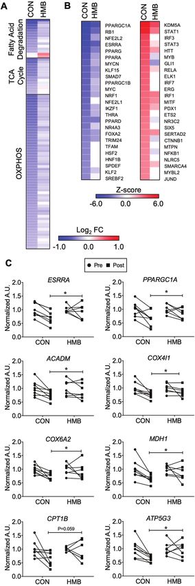
Journals of Gerontology: MEDICAL SCIENCES, 2020, Vol. 75, No. 9                                                                          1745

and proteasome systems (8) and energetic stress due to reduced ATP        Nutrition. Treatment with ONS was initiated 5 days prior to BR and
production, which may also activate the adenosine monophosphate-          continued until the end of BR. For diet stabilization over the 5 days
activated protein kinase (AMPK)-FoxO3 pathways leading to                 Pre-BR Period, subjects were fed a control diet providing the RDA
increased protein degradation (9). Promoting mitochondrial biogen-        for protein intake (0.8 g protein/kg body weight/d). Total caloric
esis by overexpression of PGC-1α protects muscle mass from acute          needs were estimated using the Harris-Benedict equation for resting
atrophy due to immobilization or disuse (10). Considered together,        energy expenditure with an activity factor of 1.375 for the ambula-
this evidence suggests a central role for mitochondrial energetics in     tory Pre-BR and 1.1 for the BR periods. The remaining macronutri-
regulating muscle mass.                                                   ents were distributed according to national dietary guidelines.
    Derangements in metabolism that contribute to muscle atrophy              Following the 5 days diet stabilization (ambulatory period),
include skeletal muscle insulin resistance (11), decreased fatty acid     subjects remained in BR for 10 days. While confined to BR, pre-
oxidation and a shift to glucose oxidation (12), intramyocellular         ventative measures were taken to detect and prevent deep vein

                                                                                                                                                   Downloaded from https://academic.oup.com/biomedgerontology/article/75/9/1744/5697344 by guest on 31 December 2020
lipid accumulation (13), and impaired protein synthesis (14). While       thrombosis (DVT) including passive range of motion exercises, the
mitochondria have been implicated in the etiology of these metabolic      use of Thrombo-Embolism Deterrent hose (TED), and Sequential
dysfunctions in the context of aging per se (15), the role of mito-       Compression Device (SCD) throughout BR. If DVT was suspected,
chondria in human muscle disuse atrophy and loss of function is           subjects received a clinical exam and a bilateral lower extremity
poorly understood, particularly in older adults. Recently, Dirks et al.   venous ultrasound.
found that younger men had a reduction in mitochondrial OXPHOS
content after 10 days of BR; however, markers of oxidative stress         Subjects
were unchanged (16). Kenny et al. showed that reductions in muscle        A total of 21 subjects were randomized to HMB or CON and 1
mitochondrial respiration occur concomitantly with insulin resist-        subject (CON group) dropped from the study prior to BR. The
ance and loss of muscle mass during BR in healthy young men (17).         study was conducted at the AdventHealth Translational Research
Further evidence linking mitochondrial energetics to muscle mass          Institute for Metabolism and Diabetes (TRI-MD) and approved by
and physical function could be critical to help determine whether         the Institutional Review Board of AdventHealth. All study proced-
mitochondria are viable therapeutic targets for muscle health of          ures, risks, and benefits were explained to the subjects before giving
older adults during and following disuse.                                 written consent to participate. The subjects recruited for this inves-
    The primary objective of this study was to examine changes            tigation were a subset of subjects who were part of a larger clinical
in skeletal muscle mitochondrial energetics during a period of            trial (NCT#02090387).
disuse in older men and women. Therapeutic strategies to pre-                 The following inclusion criteria were verified at screening: male
serve muscle health during inactivity and facilitate effective            or female ≥60 to ≤79 years of age; body mass index (BMI) ≥20 but
recovery upon reambulation have included nutritional supplemen-           ≤35 kg/m2; ambulatory with a Short Physical Performance Battery
tation with branched chain amino acids (BCAA), and β-hydroxy-             (SPPB) score of ≥9; LDL cholesterol ≤155 mg/dL, total cholesterol
β-methylbuturate (HMB). We have previously shown that HMB                 ≤250 mg/dL, and total triglycerides ≤250 mg/dL, with or without
supplementation had a positive impact on OXPHOS content in older          statin use or other blood lipid lowering agents; an ankle bra-
adults following BR (18). Our findings are congruent with preclin-        chial index between 1 and 1.4 as defined by the American Heart
ical studies that highlight a potential impact of HMB to promote          Association Task Force on Practice Guidelines; a physical activity
mitochondria and oxidative metabolism beyond its known impact             score between 7.5 and 17.7 metabolic equivalents (METs) or above
on protein metabolism (19,20). Our goal here was to further ex-           by the CHAMPS Physical Activity Questionnaire for older adults;
plore the impact of HMB on mitochondria and oxidative metab-              and handgrip strength ≥29 kg for males and ≥18.5 kg for females.
olism. A secondary objective was to take a comprehensive approach         Exclusion criteria ruled out subjects who have been diagnosed with
to examine the effect of a complete oral nutritional supplement with      Type I or Type II diabetes mellitus; taking drugs known to impact
and without HMB on skeletal muscle mitochondria content, func-            glucose metabolism; fasting blood glucose >115 mg/dL without use
tion, and gene regulatory networks related to mitochondrial bio-          of medications; undergone recent major surgery; has history of ul-
genesis and energetics. We combined both targeted and unbiased            cers, DVT, or other hypercoagulation disorders, significant cardio-
approaches to examine interrogate skeletal muscle transcriptome           vascular events or congestive heart failure, neurological or other
and lipidome, skeletal muscle mitochondrial content, and function         psychiatric disorders, and allergy to any of the ingredients in the
in healthy older adults in response to disuse while consuming a com-      study products; had active malignancy (except basal of squamous
plete oral nutritional supplement.                                        cell skin carcinoma or carcinoma in situ of the uterine cervix); es-
                                                                          timated glomerular filtration rate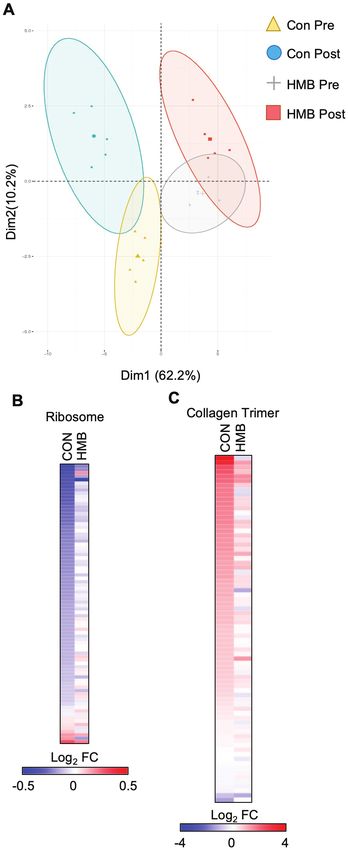
1746                                                                    Journals of Gerontology: MEDICAL SCIENCES, 2020, Vol. 75, No. 9

10–15 cm above the knee under local anesthesia (2% buffered lido-            Alexa Fluor 647-conjugated wheat germ agglutinin (WGA) (50 mg/
caine) with a 5-mm Bergstrom needle with suction, as described               mL for 30 minutes in dark at RT) was used to stain glycoconjugates
previously (21). Approximately 10–15 mg of muscle was placed in              (N-acetylglucosamine and N-acetylneuraminic acid) residues.
biopsy-preserving solution (BIOPS media) for fresh tissue analysis           Digital images (4× magnification) of one section per skeletal muscle
of mitochondrial respiration and H2O2 emission. A separate por-              biopsy were captured using a Nikon eclipse Ti microscope (Nikon
tion of the sample was prepared for immunohistochemistry, and                Technologies, California) and image analysis was performed using
the remaining muscle sample was immediately frozen in liquid ni-             NIS elements software 4.20.01.
trogen (−190°C) until analysis.
                                                                             Western Blot
High-Resolution Respirometry and ROS Emission                                Muscle homogenates were prepared as previously described
Permeabilized fiber bundles (1–3 mg) were prepared immediately               (18,25). Protein was loaded on a 4%–20% gel (Bio-Rad, Mini-

                                                                                                                                                      Downloaded from https://academic.oup.com/biomedgerontology/article/75/9/1744/5697344 by guest on 31 December 2020
following the muscle biopsy, as previously described (22). Briefly,          PROTEAN TGX Precast Gel) and transferred onto a nitrocellulose
individual myofibers were gently teased apart in a petri dish con-           membrane overnight at 30 mA at 4°C. Membranes were blocked
taining ice-cold BIOPS media. The fiber bundles were then perme-             with 5% non-fat milk for 1 hour and then incubated with primary
abilized in saponin (2 mL of 50 µg/mL) for 20 minutes and washed             antibody overnight at 4°C (anti-polyubiquitin, Enzo Life Sciences,
twice (10 minutes each) in Buffer Z (105 mM K-MES, 30 mM                     Farmingdale, NY, 1:1,000; OXPHOS, MitoSciences, Eugene, OR;
KCl, 10 mM KH2PO4, 5 mM MgCl2-6H2O, 5 mg/mL BSA, 1 mM                        α-tubulin, Cell Signaling, Danvers, MA). Membranes were then in-
EGTA, pH 7.4 with KOH). Mitochondrial respiration was evalu-                 cubated with secondary antibody for 1 hour (IRDye 800CW anti-
ated by high-resolution respirometry (Oxygraph-2k, Oroboros                  Rabbit IgG and IRDye 680RD anti-Mouse IgG; Li-Cor Biosciences,
Instruments, Innsbruck, Austria). Measurements were performed                Lincoln, NE). Protein bands were visualized as described above.
at 37°C, in the range of 200–400 nmol O2/mL, in duplicate using              The amount of poly-ub proteins was normalized to tubulin. Gel-
Buffer Z with blebbistatin (25 µm). The assay protocol was as fol-           to-gel variation was controlled for by using a standardized sample
lows: Complex I supported LEAK (LI) respiration was determined               on each gel.
through the addition of glutamate (5 mM) and malate (2 mM).
ADP (4 mM) was added to elicit complex I-supported OXPHOS                    Real-Time Quantitative-PCR
(PI) respiration. Succinate (10 mM) was added to elicit complex I&II         Total RNA was isolated from ~20 to 30 mg of skeletal muscle
-supported OXPHOS (PI+II). Cytochrome c (10 µM) was added to                 tissue using the RNeasy Fibrous Tissue kit (Qiagen, Valencia,
assess the integrity of the outer mitochondrial membrane. Finally,           CA) and transcribed to cDNA using the AffinityScript qPCR
FCCP (1 µM steps) was added to determine maximal complex I&II-               cDNA synthesis kit (Agilent Technologies, Santa Clara, CA).
supported electron transfer system capacity (E) or maximal un-               Specific oligonucleotide primers for target gene sequences are
coupled respiration.                                                         listed in Supplementary Table 1. Gene expression was measured
    Measurement of H2O2 emission by mitochondria was measured                with the Roche LightCycler 480 Real-time PCR system (Roche,
in permeabilized muscle fiber bundles by real-time monitoring of             Indianapolis, IN). Arbitrary units of target mRNA were corrected
amplex red oxidation using a SPEX Fluoromax 4 (Horiba Scientific,            to the expression of expression of 36b4 (Rplp0).
Edison, NJ) spectrofluorometer with temperature control (37°C)
and magnetic stirring as preciously described (23). Briefly, the assay
                                                                             RNA Sequencing and Informatics
was run with buffer Z containing 5,000U/mL CuZn-SOD, 25 µM
                                                                             The quality of total RNA was assessed by the Agilent Bioanalyzer
blebbistatin, 50 µM amplex ultra-red, and 6U/mL horseradish per-
                                                                             Nano chip (Agilent Technologies). One microgram of total RNA
oxidase in the presence of 10 µg/mL oligmycin, 10 mM glutmate,
                                                                             was used as starting material to construct RNA Seq library using
2 mM malate, and 10 mM succinate. The rate of emission of H2O2
                                                                             Illumina’s Truseq Stranded total RNA Library preparation kit. First,
(pmol) was calculated from previously established fluorescence in-
                                                                             the total RNA was Ribo depleted to remove rRNA from total RNA.
tensity standard curves with known concentrations of H2O2, after
                                                                             The remaining non-rRNA was fragmented into small pieces using
correcting for the rate of change in background fluorescence. The
                                                                             divalent cations under elevated temperature. Following fragmenta-
rate of H2O2 emission was expressed as pmol/min/mg dry weight
                                                                             tion, the first-strand cDNA was synthesized using random primers
of fiber bundle.
                                                                             and followed by second strand synthesis using DNA Polymerase
                                                                             I. The cDNA was then ligated with index adapters for each sample
Immunohistochemistry                                                         followed by purification and then enriched with PCR to create the
Histochemical analyses were performed on serial sections using               final library. The quality and quantity of the libraries were detected
methods previously described (24). Briefly, muscle was placed verti-         by Agilent Bioanalyzer and Kapa Biosystems qPCR. Multiplexed li-
cally in mounting medium on cork and frozen in isopentane cooled             braries are pooled and single-end 50-bp sequencing was performed
with liquid nitrogen until thoroughly frozen. Biopsy samples were            on one flow-cell of an Illumina Hiseq 2500. Mapped reads were fil-
sectioned (10 μm) using a cryotome and fixed prior staining. Sections        tered based on the mapping quality. The overall mapping rates were
were incubated in primary antibody cocktail at 4°C overnight (in             about 90%. mRNA quantification and normalization were done
10% goat serum), which includes BA-F8 (type I): IgG2b (1:50); 6H1            using Partek Genomics Suite 6.6, annotation database using RefSeq
(type IIx): IgM (1:50); and sc-71 (type IIa): IgG1 (1:50). All anti-         Transcript 77 July 2016. R package DeSeq (http://bioconductor.
bodies were obtained from the University of Iowa Hybridoma Bank.             org/packages/release/bioc/html/DESeq.html) was used to analyze
Following primary antibody incubation, slides were incubated in              the differential expression of mRNAs. Heat maps were created
secondary antibody cocktail consisting of DyLight 405 (IgG2b) goat           with Morpheus (Broad Institute). The database for annotation,
anti-mouse (1:500), Alexa Fluor 555 (IgM) goat anti-mouse (1:500),           visualization, and integrated discovery (DAVID v6.8) was used for
and Alexa Fluor 488 (IgG1) goat anti-mouse (1:500). In addition,             gene ontology (GO) annotation. Upstream regulator analysis was
Journals of Gerontology: MEDICAL SCIENCES, 2020, Vol. 75, No. 9 1747
performed with Ingenuity Pathway Analysis (Qiagen). The gene ex- differences in height and BMI (Table 1). Due to these baseline differ-
pression data discussed in this publication have been deposited in ences, we used gender and BMI as covariates in the statistical models
NCBI’s Gene Expression Omnibus and are accessible through GEO when examining changes in muscle fiber cross-sectional area (CSA)
Series accession number GSE130722 (https://www.ncbi.nlm.nih. following BR.
gov/geo/query/acc.cgi?acc=GSE130722).
Markers of Skeletal Muscle Atrophy
Lipidomics Skeletal muscle CSA determined by immunohistology are pre-
Muscle tissue was thawed in 10 times diluted PBS and homogenized sented in Figure 1A and B. Type I and type IIa skeletal muscle CSA
in Omni bead tubes with 2.8-mm ceramic beads in the Omni Bead (Figure 1A) and frequency histograms (Figure 1B) indicate a reduc-
Ruptor 24 with Cryo Cooling Unit (Omni International) at 4°C for 2 tion in type IIa muscle fiber CSA following BR (Time Effect: p = .03).
minutes. Protein concentration was determined by the bicinchoninic There were no significant changes in type I muscle fiber CSA fol-
Downloaded from https://academic.oup.com/biomedgerontology/article/75/9/1744/5697344 by guest on 31 December 2020
acid assay. One milligram of protein from each sample was ali- lowing BR. Representative images of immunostained muscle cross
quoted, and a cocktail of deuterium-labeled and odd chain phospho- sections are presented in Supplementary Figure 1. We then meas-
lipid standards from diverse lipid classes was added. Standards were ured the levels of gene transcripts and proteins that govern the skel-
chosen so that they represented each lipid class and were at desig- etal muscle atrophy program. Gene expression levels of atrogin-1
nated concentrations chosen to provide the most accurate quanti- (FBXO32) tended to increase following BR (Time Effect: p = .069).
tation and dynamic range for each lipid species. Four milliliters of There were no significant changes in the level of MuRF1 (TRIM63)
chloroform:methanol (1:1, by volume) were added to each sample, expression (Figure 1C). Total poly-ubiquinated proteins tended to
and lipidomic extractions were performed as previously described increase (Group Effect: p = .078, Interaction: 0.10) following BR in
(26). Lipid extraction was automated using a customized sequence on the HMB group (Figure 1D).
a Hamilton Robotics STARlet system (Hamilton). Lipid extracts were
dried under nitrogen and reconstituted in chloroform:methanol (1:1,
Skeletal Muscle Transcriptomics
by volume). Samples were flushed with nitrogen and stored at −20°C.
RNA sequencing (RNA-seq) was next performed to determine the
The concentrated sample was diluted 50× in isopropanol:methanol:
transcriptomic changes that occur during BR. Principal compo-
acetonitrile:H2O (3:3:3:1, by volume) with 2 mM ammonium acetate.
nent analysis demonstrates a separation in the control and HMB
The delivery of the solution to the SCIEX TripleTOF 5600+ (Sciex,
groups, as well as a significant change for both groups following
Framingham, MA) was carried out using an Ekspert MicroLC 200 system
BR (Figure 2A). GO enrichment analysis demonstrated a reduc-
with a flow rate at 6 µL/min on a customized loop. The parameters of
tion in transcripts associated with the ribosome consistent with
the mass spectrometer were optimized, and the samples were analyzed
decreased protein synthesis known to occur during disuse atrophy
automatically using a data independent analysis strategy, allowing for MS/
(Supplementary Figures 2 and 3). In addition, GO terms for fibrosis-
MSALL high-resolution and high-mass accuracy (27).
related processes such as extracellular matrix and collagen were
enriched when up-regulated transcripts were used (Supplementary
Statistical Analysis Figures 2 and 3). Examination of transcripts in both ribosome and
All data are reported as means ± standard error unless otherwise collagen trimer pathways confirmed these findings (Figure 2B and C,
stated. Statistical analysis was performed using SAS (SAS, Cary, NC) Supplementary Tables 2 and 3). Interestingly, the changes induced by
and Graphpad Prism v6 (GraphPad Software, Inc, La Jolla, CA). The BR were less pronounced in the HMB group.
differences between groups were examined using a two-way analysis RNA-seq also revealed marked reduction of transcripts associated
of variance (Group × Time) with Bonferroni multiple comparisons with mitochondria and mitochondrial energy metabolic pathways
when appropriate. Skeletal muscle fiber cross-sectional area was ana- (Supplementary Figures 2 and 3). These included the mitochondrial
lyzed using a two-way analysis of covariance controlling for baseline inner membrane and oxidative phosphorylation. Given the marked
differences between the groups. Significance was set at p < .05. inhibition of pathways associated with mitochondrial energy metab-
olism, we further examined the RNA-seq data for changes in mito-
chondrial energy metabolic genes. Transcripts encoding proteins in
Results
fatty acid degradation, TCA cycle, and the electron transport chain
Subject Characteristics were comprehensively down-regulated (Figure 3A, Supplementary
Subject characteristics are presented in Table 1. The HMB group Tables 4 and 5). Moreover, using upstream regulator analysis, genes
had more females compared to the CON group, which led to group controlling mitochondrial biogenesis and energy metabolism such
Table 1. Subject Characteristics
Control HMB 2 × 2 ANOVA (p value)
Subject Characteristics Pre Post Pre Post Group Time Interaction
N (M/F) 9 (6/3) 12 (5/7)
Age (years) 67 ± 2 66 ± 1 0.21 0.88 0.88
Height (cm) 170.9 ± 2.4 162.8 ± 3.6 0.02 0.95 0.95
Weight (kg) 77.7 ± 5.1 76.1 ± 5.4 81.7 ± 3.9 79.8 ± 4.0 0.41 0.71 0.98
BMI (kg/m2) 26.4 ± 1.4 25.7 ± 1.4 30.8 ± 0.9 30.0 ± 0.9 0.001 0.54 0.98
Note: Data are mean ± SE. Bold p values indicate statistical significance (p < .05). ANOVA = Analysis of variance; BMI = Body mass index; HMB = β-hydroxy-
β-methylbutyrate.1748 Journals of Gerontology: MEDICAL SCIENCES, 2020, Vol. 75, No. 9
Downloaded from https://academic.oup.com/biomedgerontology/article/75/9/1744/5697344 by guest on 31 December 2020
Figure 1. Older adults display reductions in type IIa skeletal muscle fiber
cross-sectional area (CSA) and elevations in skeletal muscle markers of
atrophy following 10 days of bed rest (BR). (A) Type I and type IIa, and (B)
frequency histograms of skeletal muscle fiber CSA depicting sampled vastus
lateralis muscle fibers pre- and post-10 days of BR; n = 9–12 per group. † main
effect for time for both groups: Type IIa CSA (Time Effect: p = .03, Interaction:
p = .85). (C) Gene expression of atrophy markers and (D) western blot and
representative images pre- and post-10 days BR. Vertical dividing lines were
used in Western blot images to present lanes from the same gel that were Figure 2. Bed rest (BR) results in a marked inhibition of skeletal muscle
reorganized for presentation purpose; n = 5–9 per group. † trend for main metabolism, ribosome, and collagen pathways. (A) Principal component
effect for time for both groups: atrogin-1 (Time Effect: p = .069, Interaction: analysis plot of the samples used in the transcriptomic analysis. (B) The heat
p = .97); ┴ Trend for a group effect from pre-BR: poly-ubiquinated proteins map represents the log2 fold change of all transcripts in the KEGG Ribosome
(Group Effect: p = .078, Interaction: 0.10). pathway during BR in either the CON or β-hydroxy-β-methylbuturate (HMB)
group. (C) The heat map represents the log2 fold change of all transcripts in
as PGC1-α (PPARGC1A), ERRα (ESRRA), and PPARα (PPARA) the GO CC Collagen Trimer pathway during BR in either the CON or HMB
were all predicted to be inhibited (Figure 3B). The expression of both group. n = 5 per group for transcriptomic analysis. Full color version is
available within the online issue.
PGC-1α and ERRα as well as direct target genes as determined by
RT-qPCR were down-regulated with BR (Figure 3C). These data pathway analysis in which the degree of changes on the indicated
suggest that direct down-regulation of these key transcriptional pathways was diminished in the HMB (Supplementary Figures 2 and
regulators is, at least in part, responsible for the inhibition of mito- 3). The targeted gene expression analyses confirmed these unbiased
chondrial energy metabolic pathways. transcriptomic profiling; although there is still a decrease in the ex-
HMB supplementation markedly blunted the muscle pression of mitochondrial energy metabolic genes, the number of
transcriptomic responses to BR. This is particularly evident in the genes and degree of change is smaller in the HMB group (Figure 3A).Journals of Gerontology: MEDICAL SCIENCES, 2020, Vol. 75, No. 9 1749
Skeletal Muscle Mitochondrial Function and
Content, and H2O2 Emission
Skeletal muscle mitochondrial respiration and content, and H2O2
emission are presented in Figure 4A–C. The HMB group had a
slightly lower, but significant, LI respiration before BR compared to
CON (Group Effect: p = .008, Interaction: p = .44; Figure 4A). There
was a significant time effect for PI (Time Effect: p = .03, Interaction:
0.91) and EI+II (Time Effect: p = .01, Interaction: p = .96), and
trend for PI+II (Time Effect: p = .059, Interaction: p = .71) to be
reduced in both groups following BR (Figure 4A). Mitochondrial
H2O2 emission tended to increase in both groups following BR (Time
Downloaded from https://academic.oup.com/biomedgerontology/article/75/9/1744/5697344 by guest on 31 December 2020
Effect p = .056, Interaction: p = .61; Figure 4B). Total mitochondrial
OXPHOS content (Time Effect: p = .002, Interaction: p = .34) and all
individual complexes: complex I (Time Effect: p = .005, Interaction:
p = .31), complex II (Time Effect: p = .008, Interaction: p = .32),
complex III (Time Effect: p = .004, Interaction: p = .28), complex IV
(Time Effect: p = .017, Interaction: p = .76) and complex V (Time
Effect: p = .03, Interaction: p = .64), were reduced in both groups
following BR (Figure 4C).
Skeletal Muscle Lipidomics
Lipidomics analysis was next performed to measure changes of
lipids critical to mitochondrial energy metabolism. Total levels
of cardiolipin, the major phospholipid component in mitochon-
drial membranes, are significantly decreased with BR (Time Effect:
p = .04, Interaction: p = .42) while levels of the ETC component
coenzyme Q10 (Time Effect: p = .0577, Interaction: p = .92) tended
to decrease following BR (Figure 4D). Cardiolipin species can be
categorized based on acyl chain length and degree of saturation,
into: immature, mature, and remodeled species (28). The heat map
and table of individual species show reduced levels of virtually all
cardiolipin species in the CON group and a slight preservation ef-
fect of HMB following BR (Figure 4D, Supplementary Table 6).
There were no significant changes in total phosphatidylcholine
(PC), phosphatidylethanolamine (PE), PC:PE ratio, sphingomyelin,
triacylglycerol (TAG), diacylglycerol, ceramides, and acylcarnitines
following BR (p > .05, Supplementary Figure 4).
Discussion
The effect of short-term BR on skeletal muscle mitochondria has
not been studied in older adults, and only a few studies have exam-
Figure 3. Gene regulatory networks involved in skeletal muscle oxidative
ined mitochondrial adaptation to BR in young healthy volunteers
metabolism are down-regulated following 10 days of bed rest (BR). (A)The heat
(11,16,18,29). The goal of this investigation was to examine mito-
map represents the log2 fold change of all transcripts in the indicated KEGG
pathway that are significantly regulated (p < .05) during BR in either the CON chondrial energetics in human skeletal muscle following 10 days of
or β-hydroxy-β-methylbuturate (HMB) groups. (B) IPA Upstream Regulator BR in older adults, using a combination of targeted and unbiased
Analysis was used to identify transcription regulators and ligand activated approaches. The principal novel findings were that BR induced a sig-
transcription factors significantly regulated (Z-score >2 or1750 Journals of Gerontology: MEDICAL SCIENCES, 2020, Vol. 75, No. 9
Downloaded from https://academic.oup.com/biomedgerontology/article/75/9/1744/5697344 by guest on 31 December 2020
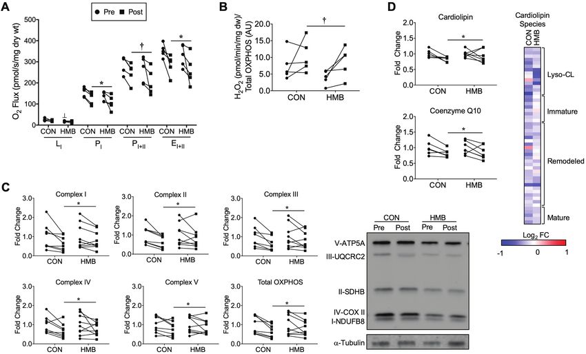
Figure 4. Skeletal muscle mitochondrial function, OXPHOS, cardiolipin, and coenzyme Q10 content are reduced, and H2O2 emission is elevated following
10 days of BR. (A) Mitochondrial respiratory capacity was measured in permeabilized fiber bundles and reported as O2 flux normalized to the dry weight of
the fiber bundles; n = 5 per group. (B) Mitochondrial H2O2 emission was measured in permeabilized fiber bundles and reported as H2O2 emission normalized
to total OXPHOS content; n = 5 per group. (C) Skeletal muscle mitochondria oxidative phosphorylation (OXPHOS) content pre- and post-10 days of BR and
representative Western blot images; n = 8–9 per group. Pre- and post-BR data are reported as fold change from pre-BR. (D) Skeletal muscle total cardiolipin and
coenzyme Q10 pre- and post-10 days of BR; n = 6–7 per group. ┴ Significant group effect for HMB group pre-BR (Group Effect: p = .008, Interaction: p = .44), *
Significant time effect for PI (Time Effect: p = .03, Interaction: 0.91) and EI+II (Time Effect: p = .01, Interaction: p = .96) to be reduced in both groups following
BR, and † trend for time effect for PI+II (Time Effect: p = .059, Interaction: p = .71) to be reduced in both groups following BR. † trend for time effect for H2O2
emission to be elevated in both groups following 10 days of BR (Time Effect p = .056, Interaction: p = .61). * Significant time effect for reductions in both groups
for total OXPHOS content (Time Effect: p = .002, Interaction: p = .34), complex I (Time Effect: p = .005, Interaction: p = .31), complex II (Time Effect: p = .008,
Interaction: p = .32), complex III (Time Effect: p = .004, Interaction: p = .28), complex IV (Time Effect: p = .017, Interaction: p = .76) and complex V (Time Effect:
p = .03, Interaction: p = .64). * Significant time effect for cardiolipin (Time Effect: p = .04, Interaction: 0.42) and † trend for time effect for coenzyme Q10 (Time
Effect: p = .0577, Interaction: p = .92) to be reduced following BR. The heat map represents the log2 fold change of all cardiolipin species during BR in the CON
and HMB groups. The cardiolipin species are grouped according to the maturation status of each species. n = 5 per group. Red (up-regulated) and blue (down
regulated) dots represent significantly changed metabolites during BR. Full color version is available within the online issue.
findings in a preclinical animal model of disuse atrophy in which sev- with lower muscle quality (32), slower walking speed (33,34), and
eral upstream regulators, for example, PGC-1α and ERRα, of mito- higher fatigability (35). Collectively, our study, supported by these
chondrial biogenesis and energetics were significantly affected (30). previous findings, suggests that reductions in mitochondrial function
Mahmassani et al. also found mitochondrial dysfunction and oxida- during a short period of disuse may be associated with a reduction
tive phosphorylation pathways were significantly regulated in both in physical function of older adults. The reductions in mitochondrial
young and old individuals following 5 days of BR (31). Collectively, respiration were likely driven by the reductions in mitochondrial
these studies highlight the potent effect of short-term disuse on skel- content (OXPHOS proteins and cardiolipin). Other investigations
etal muscle oxidative metabolism and mitochondrial transcriptome. have described reductions in mitochondrial content following 7 days
Down-regulation of transcripts associated with skeletal muscle BR in young men (16) and 10 days of HL unloading in mice (30).
metabolism and mitochondria were concomitant with an impact Indeed, the reduction in total cardiolipin content, respiration, and
on ex vivo mitochondrial respiration, markers of content, and ROS expression of mitochondrial biogenic transcription factors together
emission. The reductions in mitochondrial respiration with BR in demonstrate a profound reduction in mitochondrial content and
these older subjects are consistent with studies in younger adults (17) function following BR.
as well as in our recent findings in older mice following 10 days of Mitochondrial dysfunction during inactivity or immobilization
hindlimb (HL) unloading (30). The reductions in mitochondrial res- manifests through the release of proapoptotic factors (36,37), mor-
piration following BR could have significant ramifications for older phological alterations (fission, swelling), and energy stress (9) leading
adults during disuse atrophy and also when they become ambulatory to ROS production. ROS stimulates muscle atrophy by increasing
during recovery from illness or injury. Several studies have found that proteins involved in the proteasome system (38). We found older
lower mitochondrial oxidative capacity and efficiency are associated adults had higher ROS emission with no significant changes in skeletalJournals of Gerontology: MEDICAL SCIENCES, 2020, Vol. 75, No. 9 1751
muscle lipids following 10 days of BR (Figures 4B and Supplementary motion exercises to help prevent DVT. While there is evidence that
Figure 4). Dirks et al. found no changes in markers of oxidative stress stretching may activate mechanotransduction signaling pathways,
and skeletal muscle lipids following 7 days BR in young men (16). we do not believe the passive exercises would impact our results
Collectively, these findings suggest that younger individuals may not as they were conducted one time a day lasting ~10 minutes. We ac-
be as susceptible as older adults to disuse induced oxidative stress. knowledge the small number of subjects in this study and the subse-
Indeed, upon reloading after 10 days of HL unloading, older mice quent impact of smaller sample sizes for some of the analyses limits
still had elevated ROS emission while the ROS emission in younger our interpretation of our findings. Additional studies are needed to
mice returned to baseline (30). Older muscle may be more suscep- confirm the findings presented in this study.
tible to disuse induced ROS emission compared to younger adults In summary, our data implicate derangements in skeletal muscle
and may persist into recovery. These are similar findings to our pre- metabolism and mitochondria during BR-induced muscle atrophy
vious investigation after 10 days of BR in older adults (18) and in in older adults. HMB significantly attenuated or prevented some of
Downloaded from https://academic.oup.com/biomedgerontology/article/75/9/1744/5697344 by guest on 31 December 2020
young individuals (16). Further investigations could focus on earlier the negative consequences of disuse on expression of skeletal muscle
time points during BR to assess temporal sequence of changes that genes involved in mitochondrial biogenesis and energetics, fatty acid
occur in mitochondria and with the transcriptome prior to significant metabolism, ribosome pathways (protein synthesis), and collagen ac-
muscle atrophy. Finally, although we previously reported an increase cumulation. Future investigations are needed to determine whether
in TAG levels in older adults consuming HMB during BR (18); in the these effects are important for better recovery of muscle mass and
current investigation, we did not see any significant changes in TAG function following periods of disuse.
with BR or HMB supplementation.
Although no effect on skeletal muscle fiber CSA was observed,
HMB supplementation abrogated many of the transcriptomic Supplementary Material
changes during BR. This was particularly apparent with transcripts Supplementary data is available at The Journals of Gerontology,
associated with mitochondrial energy metabolism, ribosome, and Series A: Biological Sciences and Medical Sciences online.
extracellular matrix. In spite of the effects on the mitochondrial tran- Supplemental Figure 1. Representative histology images for both
scripts, respiration rates did not significantly differ between CON CON and HMB groups at pre-BR and post-BR.
and HMB, suggesting other mechanisms such as posttranslational Supplemental Figure 2. Gene ontology enrichment analysis using
modifications may also contribute to the mitochondrial dysfunc- DAVID. (A) All significantly (p < .05) down regulated transcripts
tion observed with BR. The observation that HMB counteracted during BR in the CON and HMB groups. (B) All significantly (p
the effect of BR on the ribosome transcripts is consistent with its < .05) up regulated transcripts during BR in the CON and HMB
effects to stimulate protein synthesis (39–41). Lastly, we found groups. The bars represent -log (p-value) of the top 10 most signifi-
the extracellular matrix and collagen pathways to be significantly cant gene ontology cellular component terms in each group; n = 5
up-regulated in the CON group with BR, which was attenuated in per group.
the HMB group. A recent investigation also found fibrotic pathways Supplemental Figure 3. Pathway enrichment analysis using
to be significantly up-regulated in older adults after 5 days of BR DAVID. (A) All significantly (p < .05) down regulated transcripts
(31). Up-regulation of the extracellular matrix and collagen proteins during BR in the CON and HMB groups. (B) All significantly (p
may have important implications for skeletal muscle function. Rats < .05) up regulated transcripts during BR in the CON and HMB
who were HL unloaded for 14 and 28 days had collagen remodeling groups. The bars represent -log (p-value) of the top 10 most signifi-
that resulted in muscle stiffness, which can impact muscle fatigability cant KEGG terms in each group; n = 5 per group.
(42). Taken together, these findings suggest that HMB has a major Supplemental Figure 4. Skeletal muscle lipids following 10 days
impact on skeletal muscle at the gene transcript level, which may of BR in the CON and HMB groups. Phosphatidylcholine (PC),
prove beneficial for the recovery following short-term disuse. A pre- phosphatidylethanolamine (PE), PC:PE ratio, sphingomyelin
vious study has shown older adults receiving HMB supplementation (SM), triacylglycerol (TAG), diacylglycerol (DAG), ceramides, and
during an 8-week exercise recovery program following 10 days of acylcarnitines (AC). n = 6–7 per group.
BR resulted in additional muscle mass and strength gains compared
to a control group (43). Combining HMB with an exercise regimen
following a period of disuse or hospitalization may have significant Funding
impact on clinical outcomes in sedentary older adults. Collectively, This work was supported by Abbott Nutrition, Abbott Laboratories
these data have important clinical implications as muscle mass and
function are impacted not only in natural aging but also conditions
such as sarcopenia and cancer cachexia. Future investigations are Acknowledgments
needed to explore the effect of HMB supplementation combined The authors gratefully appreciate the contributions of our study participants.
with exercise in these clinical populations. Author Contributions: P.M.C. and B.H.G. contributed by designing and
The goal of the present study was to take an unbiased and overseeing the clinical study. R.A.S., G.D., M.B.T., and R.B.V. were involved
in-depth approach to understanding the effects of BR on skeletal in conducting the experiments, acquiring, and analyzing the data. D.P.K. pro-
vided support and reagents for sample analysis. E.C., N.R.N., B.G., G.K.,
muscle metabolism, so we did not perform a biopsy following re-
V.V.T., and M.A.K. contributed by generating and analyzing the metabolomics
covery from BR. This limits our interpretation of the effects of
and lipisomics data. G.Y., R.B.V., and F.Q. provided bioinformatics support.
HMB’s effect on skeletal muscle recovery. Additionally, comparing
R.A.S. wrote the manuscript. R.A.S., P.M.C., and R.B.V. edited the manuscript.
these effects between older adults and younger subjects was also not
our objective. Moreover, while men have higher muscle mass and
strength than women, we observed no trends for gender-specific re- Conflict of Interest
sponses, and our study was not designed or powered to determine P.M.C. is a consultant for Astellas/Mitobridge, Inc; D.P.K. is a consultant for
gender differences. During BR, subjects underwent passive range of Pfizer, Amgen, and Janssen.1752 Journals of Gerontology: MEDICAL SCIENCES, 2020, Vol. 75, No. 9
References 19. He X, Duan Y, Yao K, Li F, Hou Y, Wu G, et al. beta-Hydroxy-
beta-methylbutyrate, mitochondrial biogenesis, and skeletal
1. Gibson JN, Halliday D, Morrison WL, et al. Decrease in human quadri-
muscle health. Amino Acids. 2016;48:653–664. doi:10.1007/
ceps muscle protein turnover consequent upon leg immobilization. Clin
s00726-015-2126-7
Sci (Lond). 1987;72:503–509. doi: 10.1042/cs0720503
20. Pinheiro CH, Gerlinger-Romero F, Guimarães-Ferreira L, et al. Metabolic
2. DeFrances CJ, Lucas CA, Buie VC, Golosinskiy A. National hospital dis-
and functional effects of beta-hydroxy-beta-methylbutyrate (HMB) sup-
charge survey. Natl Health Stat Report. 2006;2008:1–20. https://www.
plementation in skeletal muscle. Eur J Appl Physiol. 2012;112:2531–
cdc.gov/nchs/data/nhsr/nhsr005.pdf
2537. doi: 10.1007/s00421-011-2224-5
3. Kortebein P, Ferrando A, Lombeida J, Wolfe R, Evans WJ. Effect of
21. Pruchnic R, Katsiaras A, He J, Kelley DE, Winters C, Goodpaster BH.
10 days of bed rest on skeletal muscle in healthy older adults. JAMA.
Exercise training increases intramyocellular lipid and oxidative capacity
2007;297:1772–1774. doi: 10.1001/jama.297.16.1772-b
in older adults. Am J Physiol Endocrinol Metab. 2004;287:E857–E862.
4. Suetta C, Frandsen U, Mackey AL, et al. Ageing is associated with di-
doi: 10.1152/ajpendo.00459.2003
Downloaded from https://academic.oup.com/biomedgerontology/article/75/9/1744/5697344 by guest on 31 December 2020
minished muscle re-growth and myogenic precursor cell expansion early
22. Coen PM, Menshikova EV, Distefano G, et al. Exercise and weight loss
after immobility-induced atrophy in human skeletal muscle. J Physiol. improve muscle mitochondrial respiration, lipid partitioning, and insulin
2013;591:3789–3804. doi: 10.1113/jphysiol.2013.257121 sensitivity after gastric bypass surgery. Diabetes. 2015;64:3737–3750. doi:
5. Coker RH, Hays NP, Williams RH, Wolfe RR, Evans WJ. Bed rest pro- 10.2337/db15-0809
motes reductions in walking speed, functional parameters, and aerobic 23. Anderson EJ, Lustig ME, Boyle KE, et al. Mitochondrial H2O2 emission
fitness in older, healthy adults. J Gerontol A Biol Sci Med Sci. 2015;70:91– and cellular redox state link excess fat intake to insulin resistance in both
96. doi: 10.1093/gerona/glu123 rodents and humans. J Clin Invest. 2009;119:573–581. doi: 10.1172/
6. Fortinsky RH, Covinsky KE, Palmer RM, Landefeld CS. Effects of func- JCI37048
tional status changes before and during hospitalization on nursing home 24. Behan WM, Cossar DW, Madden HA, McKay IC. Validation of a simple,
admission of older adults. J Gerontol A Biol Sci Med Sci. 1999;54:M521– rapid, and economical technique for distinguishing type 1 and 2 fibres
M526. doi: 10.1093/gerona/54.10.m521 in fixed and frozen skeletal muscle. J Clin Pathol. 2002;55:375–380. doi:
7. Mithal A, Bonjour JP, Boonen S, et al.; IOF CSA Nutrition Working 10.1136/jcp.55.5.375
Group. Impact of nutrition on muscle mass, strength, and performance 25. Brocca L, Cannavino J, Coletto L, et al. The time course of the adaptations
in older adults. Osteoporos Int. 2013;24:1555–1566. doi: 10.1007/ of human muscle proteome to bed rest and the underlying mechanisms. J
s00198-012-2236-y Physiol. 2012;590:5211–5230. doi: 10.1113/jphysiol.2012.240267
8. Smuder AJ, Kavazis AN, Hudson MB, Nelson WB, Powers SK. 26. Kiebish MA, Bell R, Yang K, et al. Dynamic simulation of cardiolipin
Oxidation enhances myofibrillar protein degradation via calpain and remodeling: greasing the wheels for an interpretative approach
caspase-3. Free Radic Biol Med. 2010;49:1152–1160. doi: 10.1016/j. to lipidomics. J Lipid Res. 2010;51:2153–2170. doi: 10.1194/jlr.
freeradbiomed.2010.06.025 M004796
9. Romanello V, Guadagnin E, Gomes L, et al. Mitochondrial fission and re- 27. Simons B, Kauhanen D, Sylvänne T, Tarasov K, Duchoslav E, Ekroos K.
modelling contributes to muscle atrophy. EMBO J. 2010;29:1774–1785. Shotgun lipidomics by sequential precursor ion fragmentation on a hybrid
doi: 10.1038/emboj.2010.60 quadrupole time-of-flight mass spectrometer. Metabolites. 2012;2:195–
10. Cannavino J, Brocca L, Sandri M, Bottinelli R, Pellegrino MA. PGC1- 213. doi: 10.3390/metabo2010195
alpha over-expression prevents metabolic alterations and soleus muscle 28. Pennington ER, Funai K, Brown DA, Shaikh SR. The role of cardiolipin con-
atrophy in hindlimb unloaded mice. J Physiol. 2014;592:4575–4589. centration and acyl chain composition on mitochondrial inner membrane
doi:10.1113/jphysiol.2014.275545 molecular organization and function. Biochim Biophys Acta Mol Cell Biol
11. Coker RH, Hays NP, Williams RH, Xu L, Wolfe RR, Evans WJ. Bed rest Lipids. 2019;1864:1039–1052. doi: 10.1016/j.bbalip.2019.03.012
worsens impairments in fat and glucose metabolism in older, overweight 29. Pišot R, Marusic U, Biolo G, et al. Greater loss in muscle mass and func-
adults. J Gerontol A Biol Sci Med Sci. 2014;69:363–370. doi: 10.1093/ tion but smaller metabolic alterations in older compared with younger
gerona/glt100 men following 2 wk of bed rest and recovery. J Appl Physiol (1985).
12. Bergouignan A, Rudwill F, Simon C, Blanc S. Physical inactivity as the cul- 2016;120:922–929. doi: 10.1152/japplphysiol.00858.2015
prit of metabolic inflexibility: evidence from bed-rest studies. J Appl Physiol 30. Zhang X, Trevino MB, Wang M, et al. Impaired mitochondrial energetics
(1985). 2011;111:1201–1210. doi: 10.1152/japplphysiol.00698.2011 characterize poor early recovery of muscle mass following hind limb un-
13. Cree MG, Paddon-Jones D, Newcomer BR, et al. Twenty-eight-day bed loading in old mice. J Gerontol A Biol Sci Med Sci. 2018;73:1313–1322.
rest with hypercortisolemia induces peripheral insulin resistance and in- doi: 10.1093/gerona/gly051
creases intramuscular triglycerides. Metabolism. 2010;59:703–710. doi: 31. Mahmassani ZS, Reidy PT, McKenzie AI, Stubben C, Howard MT,
10.1016/j.metabol.2009.09.014 Drummond MJ. Age-dependent skeletal muscle transcriptome response
14. Paddon-Jones D, Sheffield-Moore M, Cree MG, et al. Atrophy and to bed rest-induced atrophy. J Appl Physiol (1985). 2019;126:894–902.
impaired muscle protein synthesis during prolonged inactivity and doi:10.1152/japplphysiol.00811.2018
stress. J Clin Endocrinol Metab. 2006;91:4836–4841. doi: 10.1210/ 32. Distefano G, Standley RA, Zhang X, et al. Physical activity unveils the re-
jc.2006-0651 lationship between mitochondrial energetics, muscle quality, and physical
15. Goodpaster BH. Mitochondrial deficiency is associated with insulin resist- function in older adults. J Cachexia Sarcopenia Muscle. 2018;9:279–294.
ance. Diabetes. 2013;62:1032–1035. doi: 10.2337/db12-1612 doi: 10.1002/jcsm.12272
16. Dirks ML, Wall BT, van de Valk B, et al. One week of bed rest leads to sub- 33. Coen PM, Jubrias SA, Distefano G, et al. Skeletal muscle mitochondrial en-
stantial muscle atrophy and induces whole-body insulin resistance in the ergetics are associated with maximal aerobic capacity and walking speed
absence of skeletal muscle lipid accumulation. Diabetes. 2016;65:2862– in older adults. J Gerontol A Biol Sci Med Sci. 2013;68:447–455. doi:
2875. doi: 10.2337/db15-1661 10.1093/gerona/gls196
17. Kenny HC, Rudwill F, Breen L, et al. Bed rest and resistive vibration exer- 34. Zane AC, Reiter DA, Shardell M, et al. Muscle strength mediates the re-
cise unveil novel links between skeletal muscle mitochondrial function lationship between mitochondrial energetics and walking performance.
and insulin resistance. Diabetologia. 2017;60:1491–1501. doi: 10.1007/ Aging Cell. 2017;16:461–468. doi: 10.1111/acel.12568
s00125-017-4298-z 35. Santanasto AJ, Glynn NW, Jubrias SA, et al. Skeletal muscle mitochondrial
18. Standley RA, Distefano G, Pereira SL, et al. Effects of beta-hydroxy-beta- function and fatigability in older adults. J Gerontol A Biol Sci Med Sci.
methylbutyrate (HMB) on skeletal muscle mitochondrial content and dy- 2015;70:1379–1385. doi: 10.1093/gerona/glu134
namics, and lipids after 10 days of bed rest in older adults. J Appl Physiol 36. Adhihetty PJ, O’Leary MF, Chabi B, Wicks KL, Hood DA.
(1985). 2017;123:1092–1100. doi:10.1152/japplphysiol.00192.2017 Effect of denervation on mitochondrially mediated apoptosis inJournals of Gerontology: MEDICAL SCIENCES, 2020, Vol. 75, No. 9 1753
skeletal muscle. J Appl Physiol (1985). 2007;102:1143–1151. doi: potent stimulation of protein synthesis in L6 rat myotubes. J Cachexia
10.1152/japplphysiol.00768.2006 Sarcopenia Muscle. 2016;7:68–78. doi: 10.1002/jcsm.12032
37. Max SR. Disuse atrophy of skeletal muscle: loss of functional activity of 41. Wheatley SM, El-Kadi SW, Suryawan A, et al. Protein synthesis in skeletal
mitochondria. Biochem Biophys Res Commun. 1972;46:1394–1398. doi: muscle of neonatal pigs is enhanced by administration of beta-hydroxy-
10.1016/s0006-291x(72)80130-x beta-methylbutyrate. Am J Physiol Endocrinol Metab. 2014;306:E91–99.
38. Reid MB. Response of the ubiquitin-proteasome pathway to doi:10.1152/ajpendo.00500.2013
changes in muscle activity. Am J Physiol Regul Integr Comp Physiol. 42. Miller TA, Lesniewski LA, Muller-Delp JM, Majors AK, Scalise D,
2005;288:R1423–R1431. doi: 10.1152/ajpregu.00545.2004 Delp MD. Hindlimb unloading induces a collagen isoform shift in
39. Wilkinson DJ, Hossain T, Hill DS, Phillips BE, Crossland H, the soleus muscle of the rat. Am J Physiol Regul Integr Comp Physiol.
Williams J, et al. Effects of leucine and its metabolite beta-hydroxy- 2001;281:R1710–R1717. doi: 10.1152/ajpregu.2001.281.5.R1710
beta-methylbutyrate on human skeletal muscle protein metabolism. J 43. Deutz NE, Pereira SL, Hays NP, et al. Effect of beta-hydroxy-beta-
Physiol. 2013;591:2911–2923. doi:10.1113/jphysiol.2013.253203 methylbutyrate (HMB) on lean body mass during 10 days of bed rest
Downloaded from https://academic.oup.com/biomedgerontology/article/75/9/1744/5697344 by guest on 31 December 2020
40. Girón MD, Vílchez JD, Salto R, et al. Conversion of leucine to β-hydroxy- in older adults. Clinical nutrition. 2013;32:704–712. doi:10.1016/j.
β-methylbutyrate by α-keto isocaproate dioxygenase is required for a clnu.2013.02.011You can also read