Functional skeletal muscle model derived from SOD1 mutant ALS patient ipScs recapitulates hallmarks of disease progression - Nature
←
→
Page content transcription
If your browser does not render page correctly, please read the page content below
www.nature.com/scientificreports
OPEN Functional skeletal muscle model
derived from SOD1‑mutant
ALS patient iPSCs recapitulates
hallmarks of disease progression
Agnes Badu‑Mensah1,2, Xiufang Guo1, Christopher W. McAleer3, John W. Rumsey3 &
James J. Hickman1,3*
Recent findings suggest a pathologic role of skeletal muscle in amyotrophic lateral sclerosis (ALS)
onset and progression. However, the exact mechanism by which this occurs remains elusive due to
limited human-based studies. To this end, phenotypic ALS skeletal muscle models were developed
from induced pluripotent stem cells (iPSCs) derived from healthy individuals (WT) and ALS patients
harboring mutations in the superoxide dismutase 1 (SOD1) gene. Although proliferative, SOD1
myoblasts demonstrated delayed and reduced fusion efficiency compared to WT. Additionally, SOD1
myotubes exhibited significantly reduced length and cross-section. Also, SOD1 myotubes had loosely
arranged myosin heavy chain and reduced acetylcholine receptor expression per immunocytochemical
analysis. Functional analysis indicated considerably reduced contractile force and synchrony in SOD1
myotubes. Mitochondrial assessment indicated reduced inner mitochondrial membrane potential
(ΔΨm) and metabolic plasticity in the SOD1-iPSC derived myotubes. This work presents the first well-
characterized in vitro iPSC-derived muscle model that demonstrates SOD1 toxicity effects on human
muscle regeneration, contractility and metabolic function in ALS. Current findings align with previous
ALS patient biopsy studies and suggest an active contribution of skeletal muscle in NMJ dysfunction.
Further, the results validate this model as a human-relevant platform for ALS research and drug
discovery studies.
Amyotrophic lateral sclerosis (ALS) is an aggressive, multi-factorial disease characterized by progressive moto-
neuron degeneration, muscle weakness and w asting1. It typically manifests between ages 40 and 70 and results in
death within 2–5 years of diagnosis. Ten percent of all reported ALS cases are familial (fALS) while the remain-
ing are sporadic (sALS). Currently, genetic mutations in over twenty genes including superoxide dismutase 1
(SOD1), chromosome 9 open reading frame 72 (C9orf72), fused in sarcoma (FUS), and TAR DNA binding
protein 43 (TDP43) are associated with A LS2. However, the pathogenesis of ALS remains largely unknown. It is
understood that disruption of the neuromuscular junction (NMJ) is an early event in ALS p athology3–6, how-
ever, the mechanisms that lead to NMJ dysfunction are still unresolved. Specifically, there is disagreement in
the literature regarding whether motoneuron (MN) dysfunction causes neuromuscular denervation, or skeletal
muscle dysfunction drives synaptic degradation, thereby causes retrograde MN dysfunction and death3,7,8. The
specific susceptibility of MNs in ALS led early researchers to adopt a “neurocentric” approach to investigating
the disease9,10. However, mounting evidence suggests the involvement of the skeletal muscle in ALS onset via
NMJ disruption11–13. Fischer and colleagues published evidence of disrupted NMJs without signs of MN death
in a spatiotemporal study performed in ALS murine m odels5,6,12. In the same report, they presented pathology
results from an ALS patient that demonstrated skeletal muscle atrophy without MN cell death5. In another,
the muscle-specific overexpression of mutant human SOD1 protein was stated to be sufficient to drive disease
symptoms in a transgenic mouse m odel14,15. Since then, many investigators have reported observations of skeletal
1
NanoScience Technology Center, University of Central Florida, 12424 Research Parkway, Suite 400, Orlando,
FL 32826, USA. 2College of Medicine, Burnett School of Biomedical Sciences, University of Central Florida,
Orlando, FL 32816, USA. 3Hesperos Inc., 12501 Research Pkwy, Suite 100, Orlando, FL 32826, USA. *email:
jhickman@ucf.edu
Scientific Reports | (2020) 10:14302 | https://doi.org/10.1038/s41598-020-70510-3 1
Vol.:(0123456789)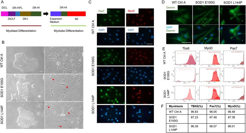
www.nature.com/scientificreports/
muscle metabolic dysregulation and atrophy before MN degeneration in ALS animal m odels16,17. In a study by
Deloach et al., they reported that ALS skeletal muscle secreted axonal chemorepellent molecules such as Nogo-A,
that stunt axonal growth18. Also, there are reports of altered morphology and regenerative capabilities of satellite
cells derived from patient skeletal muscle b iopsies19,20. Despite the above-mentioned findings, the role of skeletal
muscle dysfunction in ALS remains elusive due to the scarcity of human-based studies that substantiate the
phenomenon, the presence of other cell types that complicate the investigation of muscle pathology, and the lack
of models that can reproduce the evolvement of muscle pathology from the beginning of muscle development.
This study aimed to investigate the regenerative and functional deficits of the ALS skeletal muscle by devel-
oping a functional in vitro phenotypic skeletal muscle model from ALS patient-derived iPSCs (ALS-iPSCs)
harboring mutations in the SOD1 gene. This patient iPSC-derived muscle model is human-based, free of other
cell types such as motoneurons, and allows the investigation of muscle pathology from myogenesis to functional
muscle formation. With the iPSC-derived model, we demonstrated that ALS-iPSC myoblasts have deficits in
fusion despite their expression of appropriate myogenic markers. Additionally, significant morphological and
structural alterations were identified in iPSC-derived ALS myotubes that correlated with decreased contrac-
tile and metabolic function. Furthermore, subcellular investigation revealed ALS skeletal muscle had altered
mitochondrial function, which may negatively impact metabolic pathways and energy generation. Compared
to previous iPSC-derived muscle studies, this investigation provides a detailed and comprehensive view of the
morphological and structural deformity of ALS muscle and demonstrates for the first time their functional defects
in contractibility, as well as their metabolic dysregulation. The abnormalities revealed in these patient iPSC-
derived muscle models indicates that endogenous expression of the mutant SOD1 gene in muscle, independent
of the influence of motoneurons, has a toxic effect on skeletal muscle regeneration and function, which supports
the active role of muscle in NMJ degradation and ALS onset and/or progression.
Results
Characterization of WT and SOD1‑mutant iPSC myoblast lineage determination. Three iPSC
lines, wild type control A (WT Ctrl A), superoxide dismutase (SOD1) mutation with glutamate 100 to glycine
switch (E100G) and SOD1 with lysine 144 to proline switch (L144P) were differentiated into myogenic progeni-
tors (myoblasts) by the simultaneous activation of WNT signaling and inhibition of bone morphogenetic pro-
tein (BMP) pathways using a serum-free, small molecule-directed protocol adopted from Chal et al. (Fig. 1A)21.
Experiments that led to the modifications to Chal’s protocol are summarized in Supplementary Table S1. Cor-
rect iPSC to myogenic lineage progression was confirmed and quantified by evaluating the timely expression of
known differentiation markers including T-box transcription factor 6 (TBX6), myoblast determination protein
1 (MyoD) (proliferative satellite cell marker) and paired box protein 7 (Pax7) (satellite cell marker) via flow
cytometry (Fig. 1D). WT, SOD1 E100G and L144P cultures were found to have comparable percentages of cells
expressing each myogenic marker (Fig. 1E,F). At the end of the myogenic progenitor differentiation, myoblasts
from all three lines were seeded at low density for morphological assessment via phase-contrast and fluorescent
microscopy. While WT myoblasts were generally spindle-shaped, the SOD1 myoblasts were a mix of spindle-
shaped and flat-like, irregularly shaped cells (Fig. 1B). Regardless of morphology, immunocytochemistry results
indicated that WT and SOD1 myoblast cultures were MyoD and Pax7 positive confirming the flow cytometry
data (Fig. 1C). Together, these data indicated that myoblasts derived from healthy and ALS-mutant PD-iPSCs
were proliferative and expressed markers essential for correct myogenic progression differentiation.
Characterization of WT and SOD1‑mutant myogenic progenitor fusion. The ability of satellite
cells to initiate proliferation and progress through the myogenic differentiation program is essential to skeletal
muscle repair and mass maintenance after injury22. Thus, SOD1 myoblasts were tested for their ability to differ-
entiate into myotubes. While WT cultures had nascent myotube formation on day three after switching to differ-
entiation medium, none were observed in SOD1 cultures until the fifth day in differentiation medium (Fig. 2A).
Cultures were maintained for 14 days to accommodate the slower differentiation of the SOD1-mutant cultures.
On the 14th day of differentiation, the fusion indices of all cultures were quantified (Fig. 2B). Compared to WT,
SOD1-mutant myoblasts had a significantly lower fusion index (p < 0.001) (Fig. 2F). Additionally, the number
of myotubes per area in each culture was quantified as another measure of fusion efficiency. While WT cultures
were dense with myotubes, SOD1-mutant cultures had 3× less myotubes (Fig. 2G). To ensure that the decreased
fusion in the mutant cultures was not due to the presence of non-myogenic cells, the culture was stained with
the myogenic-specific marker, desmin. Both myotubes and unfused myoblasts in the cultures stained positive
for desmin (Fig. S1). The results confirmed that the observed reduced fusion in ALS cultures was not due to
the presence of contaminating cells, but the inability of the myoblasts to progress through the differentiation
program. Altogether, ALS-mutant cultures exhibited altered/decreased fusogenicity compared to WT cultures.
Morphometric assessment of WT and SOD1‑mutant myotubes. The phenotypic characteristics
and structural organization of SOD1 myotubes were evaluated to delineate morphometric alterations when
compared to WT. Measurements of the average width, thickness and length of myotubes from each cell line were
assessed. While SOD1 myotubes were slightly narrower than WT myotubes, they were significantly thinner and
shorter (p = 0.018, < 0.001 respectively) (Fig. 2C–E). The complex spatial organization of myogenic-proteins is
vital to their ability to accurately execute function23. Thus, the structural organization of Myosin Heavy Chain
(MyHC) within myotubes from each culture, as well as nicotinic acetylcholine receptor (nAChR) clustering,
were analyzed via immunocytochemistry. MyHC appeared densely arranged and compact in the WT myotubes.
Additionally, WT myotubes expressed nAChR clusters throughout the culture (Fig. 3). Conversely, SOD1 myo-
tubes had disorderly and loosely arranged MyHC that made striations within individual fibers appear discontin-
Scientific Reports | (2020) 10:14302 | https://doi.org/10.1038/s41598-020-70510-3 2
Vol:.(1234567890)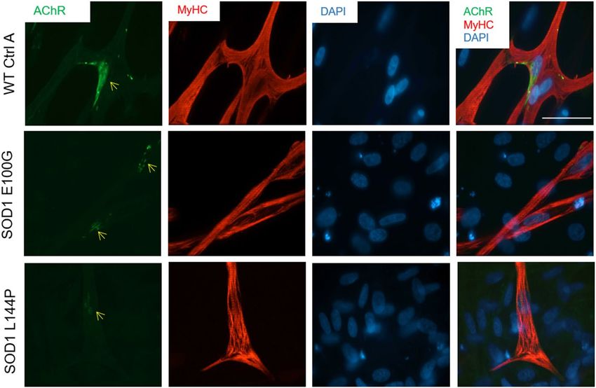
www.nature.com/scientificreports/
Figure 1. Myoblast derivation and myogenic characterization. (A) Illustration of the differentiation protocol
used in both myoblast and myotube phases of differentiation. (B) (left panel) Phase contrast images of iPSC
cultures of the various cell lines in maintenance medium (scale bar = 100 μm). (B) (right panel) Phase contrast
microscopy images of resultant myoblast cultures after replating in proliferation medium at the end of the
myoblast differentiation phase (scale bar = 50 μm). Red arrowheads directed at flat myoblasts in SOD1 cultures.
(C) Immunocytochemistry (ICC) staining of derived myoblasts for Pax7 and MyoD expression across all
cell lines (scale bar = 50 μm). (D) ICC staining for Desmin expression in WT and SOD1 cultures. (E) Flow
cytometry (FC) result plots showing TBX6 expression during the first week of differentiation (left panel) and
PAX7 and MyoD expression by the third week of myoblast differentiation (middle and right panels respectively).
Data analyzed with CytExpert software, version 2.1 (Beckman Coulter). (F) Tabulated percentage values of FC
results.
uous. Also, there were few to no nAChR clusters on SOD1 myotubes, and nAChR staining appeared diffuse and
faint (Fig. 3). Collectively, these results demonstrate that SOD1-mutant myotubes have altered morphological
and structural deficits that may affect function.
Functional evaluation of WT and SOD1‑mutant myotubes. Muscle weakness is an established ALS
hallmark and has been described in both patients and transgenic animal m odels24,25. From the neurocentric
viewpoint, muscle wasting and weakness are a result of motoneuron denervation. However, this opinion has
been challenged by recent findings that postulate there are pathological alterations in diseased skeletal muscle
function devoid of axonal retraction5,8,26. To test the latter hypothesis, individual myotubes from all lines were
subjected to stimulated contraction at frequencies of 0.3, 0.5, 1.2 and 4 Hz on day 14 of differentiation (Fig. 4A)
and the functional activity of WT myotubes was compared to that of SOD1 myotubes. WT myotubes con-
tracted consistently with increasing frequency, however both SOD1 E100G and L144P myotube twitches were
asynchronous and had comparatively weaker amplitudes (Fig. 4A,B). As expected, contractile force output for
SOD1 myotubes was also comparatively low (Fig. 5A,B). Additionally, to examine the dynamics of myofiber
contraction, time-to-peak (TTP) was analyzed by quantifying the duration from the initiation to the peak of
the contraction under electrical stimulation. As shown in Fig. 5C, there was significantly higher contraction
time-to-peak for both SOD1 E100G and L144P compared to WT, indicating a slowdown of contraction dynam-
ics. Because the SOD1 myotubes has been shown to mature at a slower pace, myotubes were tested at day 21 of
differentiation. This was done to determine if a longer maturation period could improve contractile synchrony
and amplitude in the SOD1 cultures. While the contractile synchrony in most SOD1 L144P myotubes improved
by day 21, the amplitude remained the same (Fig. 4A–C) (Supplemental Fig. S2). Conversely, SOD1 E100G
cultures indicated no improvement in either contraction synchrony or amplitude with increased time in culture
(Fig. 4A–C) (Supplemental Fig. S2). These results suggest that SOD1 myotubes have intrinsic defects that retard
maturation and contractile function independent of denervation.
Mitochondrial function analysis of WT and SOD1‑mutant myotubes. Studies have reported
changes in mitochondrial morphology, dynamics and function in ALS p athology4,24,27,28. Additionally, the pro-
cesses of muscle fusion and contraction are energy-intensive a ctivities29,30. Thus, the mitochondrial function of
Scientific Reports | (2020) 10:14302 | https://doi.org/10.1038/s41598-020-70510-3 3
Vol.:(0123456789)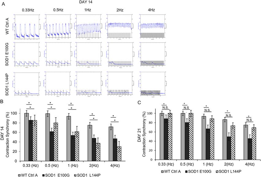
www.nature.com/scientificreports/
Figure 2. Phenotypic myotube characterization. (A) Phase contrast images showing myotube derivation from
myoblasts from the three cell lines. Scale bar = 100 μm. (B) ICC images of myotube cultures stained for MyHC
and DAPI. Note the unfused background cells in the DAPI+/MyHC SOD1 cultures. Imaged with Volocity
software, version 6.3.0 (PerkinElmer). Scale bar = 50 μm. (C) Bar graph showing slightly decreased myotube
cross sectional width compared in SOD1 cultures relative to WT (Error bars CI = 90%). (D) Average myotube
thickness **p value = 0.018. (E) Box and Whisker plots describing the lengths of WT and SOD1 myotubes. ***p
value < 0.001. One-way ANOVA, followed by Dunnett’s test. (F) Bar graph showing fewer fusion events in the
SOD1-mutant cultures compared to WT, evidenced by fusion index. (G) Bar graphs indicating lower average
myotube number per area in SOD1 cultures compared to WT. Error bars: SEM *p < 0.1; **p < 0.01; ***p < 0.001
by Student T-test, 2 tails, unequal variance.
Figure 3. Phenotypic myotube characterization. Myotubes stained for MyHC, nAChR and DAPI, depicting
diffuse to no AchR clusters in SOD1-mutant cultures. Yellow arrows point to nAChR clusters on SOD1
yHC−/
myotubes. Nuclei staining (DAPI) showing decreased fusion in myotube cultures, as evidenced by M
DAPI+ unfused myoblasts in the background. Imaged with Volocity software, version 6.3.0 (PerkinElmer). Scale
bar = 50 μm.
SOD1 cultures was investigated as a possible subcellular mechanism for the phenotypic observations in mus-
Scientific Reports | (2020) 10:14302 | https://doi.org/10.1038/s41598-020-70510-3 4
Vol:.(1234567890)www.nature.com/scientificreports/
Figure 4. Functional characterization of myotubes. (A) Contraction traces of individual myotubes from iPSC-
derived SOD1 E100G, SOD1 L144P and WT control cultures at 0.3, 0.5, 1, 2 and 4 Hz, on day 14 of myotube
differentiation. Electrical pulses were elicited at 1 V for 15 s. (B, C) Bar graphs showing the percentage of
contraction synchrony at days 14 and 21. p ≤ 0.05 ANOVA on ranks followed by Holm-Sidak method. NS Not
significant.
Figure 5. Functional characterization of myotubes. (A) Representative cantilever traces of WT and SOD1
myotubes. Traces extracted with Clampfit Version 10.7 (Axon Instruments). (B) Bar graph showing the
contractile force per area of the respective WT and SOD1 muscle lines. (C) Results depicting the time-to-peak
force of WT and SOD1 muscle. *p < 0.1, **p < 0.01, ***p < 0.001, One-way ANOVA, followed by Dunnett’s test.
cle regenerative and contractile function. The mitochondrial membrane potential (ΔΨm) of diseased and WT
myotubes was assessed because the ΔΨm has been shown to be vital to a myriad of cellular processes including
energy production, calcium handling and DNA r epair31,32. Results indicated significantly lower average ΔΨm
values for both SOD1 myotube lines compared to WT (Fig. 6A). Mitochondrial function of the SOD1 myotubes
was evaluated by measuring the rate of metabolism of various substrates. Overall, tricarboxylic acid (TCA) cycle
intermediates and fatty acids were metabolized at either a slightly faster or the same rate by SOD1 mitochon-
Scientific Reports | (2020) 10:14302 | https://doi.org/10.1038/s41598-020-70510-3 5
Vol.:(0123456789)www.nature.com/scientificreports/
Figure 6. Mitochondria fsunctional assessment. (A) TMRE assay measuring fluorescence intensity as a
measure of mitochondrial membrane potential at day 12 of differentiation. SOD1-mutant muscle exhibit
decreased mitochondrial membrane potential compared to WT Ctrl A. Error bars: SEM *p < 0.1; **p < 0.01;
***p < 0.001 by Student T-test, 2 tails, unequal variance. (B, C) Mitochondrial metabolic rate assessment of
SOD1 muscle for TCA cycle intermediates and glucose-derived substrates. (D–F) Mitochondrial metabolic rate
assessment of SOD1 muscle for amino acid-, fatty acid- and ketone-derived substrates respectively.
dria compared to WT (Fig. 6B,C). However, SOD1 cultures metabolized amino acids, ketones and glycogenic
substrates at a slower rate compared to WT (Fig. 6D–F). Among the assayed substrates, lactate metabolism was
particularly low in diseased cultures compared to WT (Fig. 6B). Together, these results suggest that mitochon-
drial dysfunction is a characteristic of SOD1 mutant skeletal muscle.
Discussion
The goal of this study was to create a phenotypic model of ALS muscle to investigate the inherent defects of
the ALS skeletal muscle in regeneration and function to better evaluate their contribution to NMJ disrup-
tion in ALS. To achieve this, functional in vitro biomimetic ALS skeletal muscle models were developed from
fALS patient-derived iPSCs. Two patient iPSCs lines harboring mutations in the SOD1 gene resulting in amino
acid substitutions at L144P and E100G, were differentiated into skeletal muscle myoblasts and subsequently
Scientific Reports | (2020) 10:14302 | https://doi.org/10.1038/s41598-020-70510-3 6
Vol:.(1234567890)www.nature.com/scientificreports/
myotubes. Systematic analysis of the ALS myoblasts revealed deficits in myogenesis, myotube morphology,
structure and function, as well as mitochondria properties and metabolism. Myotubes had notably decreased
acetylcholine receptor (AChR) clusters and loosely arranged myosin heavy chain proteins compared to WT
myotubes. BioMEMs multiplexed cantilever analysis indicated ALS-iPSC myotubes have significantly weaker
force output and reduced contraction synchrony compared to WT controls. These defects identified in iPSC-
derived ALS muscle, in the absence of other cell types, indicate muscle is an active contributor to the initiation
and/or progression of NMJ dysfunction in ALS.
ALS-iPSC myoblasts were found to be proliferative and expressed the appropriate stage-specific myogenic
markers at percentages comparable to WT. However, the flat and stretched morphology observed in the ALS myo-
blast cultures reiterates the senescent-like morphology reported in a patient myoblast study by Pradat et al.19.
We also demonstrated that ALS-iPSC myoblasts exhibited delayed and decreased fusibility when subjected to
differentiation. It has been established that correct myoblast activation and fusion are pertinent to skeletal muscle
mass maintenance and repair33. Thus, any impairment in the myogenic developmental program may have adverse
effects on downstream regeneration. Most SOD1 myoblasts were unable to fuse upon induction, suggesting there
may be defects in skeletal muscle regeneration in ALS pathology. The diminished fusibility of SOD1 myoblasts
observed in the current model could affect muscle mass maintenance and repair in patients, which may partly
explain observed muscle atrophy. This has been reported in previous patient skeletal muscle biopsy s tudies19,20.
Morphometry results showed that the mutant myotubes had narrower widths and shorter lengths compared
to WT controls. At a higher magnification, MyHC proteins appeared loosely arranged in SOD1 myotubes than
in WT. Contrastingly, Pradat et al., reported that ALS myotubes were long and thin, while the SOD1 myotubes
in our cultures were shorter and narrower than W T19,20. Notwithstanding, immunocytochemistry results from
both labs demonstrated lower expression level of MyHC in ALS myotubes. This could explain the loose arrange-
ment of MyHC and reduced thickness of myotubes observed in our culture system19,20.
Another notable observation was the reduced expression of AChR on SOD1 myotubes compared to WT
myotubes. AChR clusters expressed on ALS-iPSC myotubes were smaller and appeared diffuse compared to the
WT (Fig. 3). Robust expression and dense clustering of AChRs are necessary to receive action potential signals
from the presynaptic motoneuron. Reduced expression and clustering decrease the possibility for accurate trans-
mission of motoneuron signaling events. Abnormal AChR function has been described in both patient samples
and mice m odels34,35. Recently, Picchiarelli et al. reported decreased AChR clustering and endplate maturation
in FUS-mutant skeletal muscle generated from patient iPSCs as possible cause of NMJ d ysfunction36.
In addition, integration of ALS muscle with BioMEMs cantilevers revealed cell-autonomous deficits in con-
tractile force output and time-to-peak force. Although slowed contraction has been reported in the SOD1 G93A
transgenic murine model, this is the first human-based work to directly demonstrate defective contractile func-
tion of ALS muscle in a human-based s ystem37, although a previous researcher did extrapolate functional defects
by measuring the passive electrical properties of ALS skeletal muscle generated from patient iPSCs38. While skel-
etal muscle weakness is an established hallmark of ALS, the exact cause of this dysfunction is unclear. The gener-
ally accepted hypothesis is that muscle weakness in ALS is a result of MN denervation and axonal retraction39.
However, contrary to this view, muscle-centric studies suggest an intrinsic cause of muscle w eakness14,15,40. In the
current model, ALS-iPSC myotubes exhibited over 70% lower contractile force compared to WT. Evaluation of
the time-to-peak force (TTP) showed a significantly delayed TTP in SOD1 myotubes (Fig. 5C). This observation
is suggestive of irregularities in the excitation–contraction-coupling (ECC) machinery and/or calcium regulation.
It is noteworthy that while pathological ALS MNs could be an inevitable component of muscle weakness in vivo,
this study provides solid evidence confirming significant innate muscle deficits independent of motoneurons. By
reproducing human myogenesis from the stem cell stage, this systematic study revealed that ALS myotubes have
deficits in myotube regeneration, the capability of receiving innervation, contractibility upon excitation, and in
the force of contraction. It is conceivable that the combination of these muscle-derived deficits and those from
neuron-derived components result in the muscle weakness symptoms seen in ALS patients.
Furthermore, we identified mitochondrial dysregulation in these ALS muscle. First, TMRE staining indicated
a significantly decreased ΔΨm in SOD1 cultures compared to WT myotubes. ΔΨm is known to play a pivotal
role in oxidative phosphorylation, therefore, a reduced ΔΨm would imply the inability of ALS skeletal muscle
to provide energy support for its f unction31. More importantly, decreased ΔΨm interferes with C a2+ handling,
which is an important regulator of aerobic respiration31,32. Decreased ΔΨm has also been observed in the muscle
of SOD1 mice models in the absence of axonal retraction, suggesting skeletal muscle dysfunction could precede
NMJ disruption27. Additionally, altered mitochondrial morphology, dynamics and function have been reported
in ALS patient muscle studies and murine m odels27,28,41.
We also demonstrated that ALS-iPSC myotubes have reduced metabolic plasticity. With the provision of
citric acid cycle intermediates or fatty acids, the rate of metabolism in SOD1 cultures was either the same or
had a slightly faster rate compared to WT. However, metabolism of glycogen, lactate, amino acids, and ketones
were relatively slower in SOD1 cultures. The current findings are in line with previous studies that reported a
reduced preference for glycolytic metabolites and enhanced lipid metabolism in ALS skeletal m uscle42–44. In
addition to altered metabolism, researchers have also reported a shift toward an oxidative fiber type composi-
tion without apparent alterations in MyHC composition and e xpression6,42,44. The heterogeneity in metabolite
preference among skeletal muscle fiber types is a known phenomenon. For instance, fast twitch fibers typically
prefer the use of glucose for ATP production via anaerobic respiration. In contrast, slow twitch fibers tend to
utilize fatty acids for ATP s ynthesis45. The ability of diseased skeletal muscle to efficiently metabolize fatty acids
may explain the resilience of slow twitch fibers during ALS p athology6. Further investigation will be needed to
elucidate the relationship between fiber type and metabolism alterations during ALS progression, and this topic
can be pursued in future studies.
Scientific Reports | (2020) 10:14302 | https://doi.org/10.1038/s41598-020-70510-3 7
Vol.:(0123456789)www.nature.com/scientificreports/
The inability of ALS-iPSC muscle to metabolize lactate stood out among all glucogenic substrates. Once
thought to be a by-product, lactate has more recently been found to be major energy source for skeletal muscle.
Reduced lactate turnover has been reported in fALS patients and lactate metabolism was observed to significantly
decrease with disease p rogression46. Consistent reports concerning mitochondria defects in muscle phenotypes
from different ALS models suggest a mechanistic link between these two pathologies. While the mechanism of
the observed changes in ALS mitochondrial function is unclear, the results imply that provisions for selective
nutrient substrate utilization could partially circumvent the muscle metabolism obstacle and possibly alleviate
this pathology. This idea is supported by the effectiveness of a holistic therapy, the Deana Protocol, which contains
the citric acid cycle intermediates as a s upplement47.
While there is documented evidence of skeletal muscle dysfunction in ALS patients, exactly how the under-
lying ALS-associated genetic mutations induce dysfunction is poorly understood, which makes outlining the
pathogenic role of the muscle challenging. For instance the SOD1 mutants E100G and L144P, both with substi-
tutions distant from the metal-binding region of the protein, generated similar phenotypes but with significant
variation in the severity level of functional and metabolic defects43,48. Both mutations are missense substitutions
located in exons 4 (E100G) or 5 (L144P). However, it is hard to speculate further concerning the molecular
pathology due to the limited information available for these molecular changes due to these two mutations.
Genetic and phenotypic heterogeneity is well-known in SOD1 mutations. Over 180 different SOD1 mutations
have been identified throughout the five exons of the SOD1 gene. Of these, more than 160 have been reported to
be disease-associated. However, the precise shared and/or differing molecular and cellular pathological mecha-
nism of each SOD1 mutation has yet to be elucidated49,50. Additionally, the genetic background of the subjects
adds another significant variable19,20. The ALS-iPSC skeletal muscle models examined in this study demonstrates
deficits in the ALS skeletal muscle by directly measuring its regenerative, contractile and metabolic functions.
This phenotypic model, by performing systematic characterization of the functional changes in skeletal myofibers
containing patient-specific mutations, constitutes a valuable new platform for addressing this issue by providing
the gene-phenotype linkage.
Overall, we have developed a patient iPSC-derived skeletal system that recapitulates typical ALS pathology
in both phenotypes and also for subcellular mechanisms. Because this is a phenotypic model allowing for the
quantification of muscle function, it could be utilized in high content drug toxicological and efficacy screening.
In addition, when coupled with Human-on-a-chip and BioMEMs technologies, it may be used in engineer-
ing physiologically relevant multi-organ models for studying complex interactions among tissues during ALS
onset and progression. Compared to patient biopsy samples, sourcing from iPSCs eliminates the limitation
for the cell source availability and enables the possibility of generating multiple cell types from the same iPSC,
patient-specific models. Collectively, this system provides a valuable platform for ALS translational studies and
therapeutic development.
Conclusion
In conclusion, this work demonstrates that ALS patient-derived skeletal muscle has inherent defective regenera-
tive capabilities and function independent of motoneuron influence or denervation. This patient iPSC- derived
SOD1 skeletal muscle model recapitulates most ALS-diseased muscle characteristics reported in human clinical
and transgenic animal studies. Thus, this iPSC-derived muscle system is a good resource for etiological investiga-
tions and relevant high content drug testing.
Materials and methods
Cantilever fabrication and surface chemistry engineering. Cantilever microelectromechanical sys-
tems (MEMS) were made from 6-inch silicon-on-insulator wafers, polished with a 5 μm silicon layer and a
500 μm silicon dioxide layer on the front and back respectively, as previously described51. The cantilever pat-
terning was done using S1818 photoresist, followed by deep reactive ion etching. Glass coverslips were soaked
in 12 M hydrochloric acid and placed on a shaker overnight. The next day, the coverslips were thoroughly rinsed
with sterile water, and transferred to 100% ethanol. After 24 h, the coverslips were allowed to dry overnight and
stored in a desiccator.
IPSC‑derived myoblast differentiation. Human induced pluripotent stem cell (iPSCs) lines ND41865,
ND35662, ND39032 were obtained from the Coriell Institute for Medical Research. iPSCs were passaged a maxi-
mum of 12 times per recommended NIH passaging protocols. Cells from passages 6 to 12 were used in myoblast
differentiation experiments. Myoblast differentiation was performed strictly following a protocol published by
Chal et al.21. Briefly, iPSC cultures were maintained in mTESR1 (Cat# 85850, StemCell Technologies) or TESR-
E8 (Cat# 05990, StemCell Technologies) on matrigel-coated (Cat# 354230 Corning Life Sciences) surfaces to a
15–20% confluence before commencing myoblast differentiation. Cultures were maintained until 35–40 days of
differentiation and harvested. At the end of myoblast differentiation, the cultures were re-plated at 1:6 in MyoC-
ult serum free supplemented 1000 mg/L DMEM (Cat #05982; StemCell, Technologies) for myoblast expansion.
Medium was refreshed every other day until 50–60% confluence. Myogenic progenitors were harvested and
cryopreserved for downstream applications. All cultures were maintained in humidified air supplemented with
5% CO2, at 37 °C.
IPSC‑derived myotube maturation. Myoblasts were plated on acid-washed coverslips in Myocult
medium (1000 mg/L DMEM supplemented with MyoCult Serum Free nutrient cocktail) until confluent. Next,
cultures were switched to DK-HI medium (DMEM/F12 21041-025 ThermoFisher Scientific, 15%vol/vol Knock-
out Serum Replacement ThermoFisher Scientific 10828-028, 10 ng/ml hepatocyte growth factor (HGF) Pepro-
Scientific Reports | (2020) 10:14302 | https://doi.org/10.1038/s41598-020-70510-3 8
Vol:.(1234567890)www.nature.com/scientificreports/
tech 315-23, 2 ng/ml insulin-like growth factor (IGF), Sigma I1271, 0.1 mM β-mercaptoethanol, ThermoFisher
Scientific 31350010, 1% MEM Non-Essential Amino Acids Solution ThermoFisher Scientific 11140050 and
100 nM dexamethasone, Sigma D4902). After two days, cultures were switched to the terminal differentiation
medium, N2 (DMEM/F12 21041-025 ThermoFisher Scientific, 1% Insulin Transferrin Selenium (ITS) 41400045
ThermoFisher Scientific, 1% N2 supplement 17502048, ThermoFisher Scientific, 1% l-glutamine 20 mM
A2916801 ThermoFisher Scientific, 100 nM dexamethasone, Sigma D4902).
Myoblast myogenic characterization. Cultures, during the course of myoblast differentiation, were
subjected to flow cytometry (FC) to confirm temporal activation of the myogenesis gene program. Differentia-
tion markers assessed were the pre-somite marker, TBX-6 (Abcam ab170482), after 4–8 days of differentiation
and the myogenic progenitor markers, Pax7 (Invitrogen PA5-117P) and MyoD (Santa Cruzsc77460; LS Bio
LS C263888), after 18–21 days in differentiation. Differentiating cultures were lifted using TrypLE Express or
Trypsin–EDTA 0.05% (ThermoFischer Scientific12605010, 25300062) and centrifuged at 280 g for 5 min. The
resulting pellet was re-suspended in FC buffer (10% BSA). An equal volume of 4% paraformaldehyde, (PFA)
in 1X phosphate buffered saline (PBS) solution was added to the suspended culture and incubated at 4 °C for
15 min. Cultures were pelleted and rinsed twice with FACs buffer, followed by a 4 °C 15-min incubation with
0.2% saponin solution to permeabilize the cells. Cultures were centrifuged, rinsed twice and re-suspended in
FC buffer following the permeabilization step. Fc receptor blocker (Miltenyi Biotec, 130-059-901), was added at
a 1:5 dilution factor and incubated at 4 °C for 20 min. Antibodies were added at 0.1 to 10 μg/ml, incubated for
60 min at 4 °C, rinsed 2 × and re-suspended in FC buffer. The resultant cell solution was added to a 96-well plate
and analyzed using a CytoFlex LX (Beckman Coulter) flow cytometer. The data was analyzed using CytExpert
software, version 2.1 (Beckman Coulter).
Immunocytochemistry and confocal microscopy. Mutant and WT cultures on glass coverslips were
fixed with 4% PFA solution for 15 min and rinsed 3× with 1× PBS solution. Cells were then blocked with donkey
serum blocking buffer (2.5 mL Donkey Serum + 2.5 mL BSA + 40 mL sterile water + 5mLs PBS 10x) for 1 h after
a 15-min permeabilization period with 0.1% triton in PBS. Primary antibodies for MyoD, Pax7, desmin (Ther-
mofisher Scientific PA5-16705, MyHC DSHB, A4.1025-s), and Ryanodine receptor (RyR) (Millipore, AB9078),
dihydropyridine receptor (DHPR) (Thermofisher Scientific,MA3920) were added to blocked cultures and incu-
bated overnight at 4 °C. The secondary antibodies donkey anti-mouse and anti-rabbit (Thermofisher Scientific,
A10037, A-21206) were added the next day at a 1:250 dilution. Coverslips were rinsed 3× in 1× PBS mounted on
glass slides with Prolong gold mounting medium with DAPI (Life Technologies P36931) after a 2-h incubation
period for imaging. Images were taken with Axioskop 2 mot plus upright spinning disk confocal microscope
(Carl Zeiss) that had been connected to a XCite 120 Fluorescence Illumination system (EXFO) beam with multi-
spectral laser scanning software, Volocity version 6.3.0 (Perkin Elmer).
Myotube phenotypic characterization. Five representative phase-contrast images per coverslip were
taken from all myotube cultures with a Zeiss Axioscope 200. Images were loaded onto ImageJ software version
1.52 (NIH) and analyzed for average myotube number and myotube width using the cell counter plugin and
straight free hand tool. Myotube width measurements were taken from phase contrast images as described b y52.
Briefly horizontal lines were drawn across the middle of the images, depending on direction of fused myotubes.
Only widths of myotubes that crossed the line were measured using the ImageJ software. This was in effort to
eliminate subjective data selection. The average length of myotubes from each line was estimated using the Neu-
ronJ plugin of Image J. This ImageJ plugin has mostly been used for measuring axon lengths. Five randomized
images per coverslip were taken of myotubes stained for MyHC. Myotubes in each field view were traced from
one end to the other and measured. Myotube thickness was assessed by measuring the height of Z-stacks of
MyHC-stained myotubes, using Volocity software, version 6.3.0 (Perkin Elmer). Fusion index was quantified for
myotubes stained with MyHC and nuclear stain, 4′,6-diamidino-2-phenylindole, (DAPI). The number of nuclei
within myotubes was divided by the amount of total nuclei to obtain a number between 0 and 1, where 0 means
no fusion and 1 is fusion of all myoblasts53–56. Simply put,
number of DAPI − positive nuclei in myotubes in each field
Fusion index = .
total number of DAPI − positive nuclei in each field
Myotube contraction assessment. After 14 days in differentiation medium, mutant and WT myotubes
cultures in petri dishes were placed on a 37 °C heated stage (ThorLabs TC200) on an upright phase-contrast
microscope (Zeiss Hal 100). Myotube contraction was elicited using silver electrode-driven, 1 V broad field
stimulation at frequencies of 0.3, 0.5, 1.0, 2.0 and 4.0 Hz for 15 s. Myotube contractions were captured using a
high-speed acquisition digital camera (Hamamatsu model C8484-05G) from a selected region of interest (ROI).
This procedure is described in detail by Santhanam et al.57. Contraction synchrony was calculated by dividing
the number of contractions by the total number of expected contractions at a given frequency.
Myotube force and fatigability assessment. Mutant and WT iPSC-derived myoblasts were seeded at
400 cells/mm2 on collagen I-coated BioMEMs cantilever devices and assembled into engineered acrylic housings.
The cultures were maintained in proliferation medium for 5 days, switched to DK-HI medium for 2 days, and then
into terminal differentiation medium, N2, for 14 days. At the end of the differentiation period, the skeletal muscle
myotube cultures were tested for contraction. As previously described, the cultures were transferred to a heated
Scientific Reports | (2020) 10:14302 | https://doi.org/10.1038/s41598-020-70510-3 9
Vol.:(0123456789)www.nature.com/scientificreports/
stage, equipped with a light source (above), and photodetector and laser (below). System control and data collec-
tion were performed as described by Wilson et al.58. Skeletal muscle from both mutant and WT cultures tested
for their maximum contractile force output (MCF) with a frequency of 0.5 Hz and pulse width of 250 ms, at 5 V.
Mitochondrial membrane potential assessment. Mutant and WT iPSC-derived myoblasts were
plated at 50 cells/mm2 on Collagen I-coated 18 mm glass coverslips and differentiated into myotubes. At day 12
of differentiation, myotubes were incubated in 500 nM tetramethylrhodamine (TMRE) (Thermofisher Scientific,
Cat# T669) solution for 30 min and rinsed twice with 1× PBS. Fluorescence images of the myotube cultures were
taken with a Zeiss spinning disk confocal microscope (Axioskop 2 Mot Plus). Average fluorescence intensity per
image was measured via ImageJ software.
Mitochondrial metabolic profiling. Mitochondrial metabolism of mutant and WT iPSC-derived myo-
tubes was assessed by measuring the rate of electron flux through the electron transport chain (ETC) following
treatment with specific substrates. Assay solutions and preparation were done per manufacturer’s instructions
(BiOLOG, Cat#s 14105, 72303, 74353). Briefly, muscle cultures were harvested using TrypLE Express and cen-
trifuged at 280g for 5 min. The resulting pellet was re-suspended in 1× BiOLOG mitochondrial assay solution
(MAS). The cell suspension was added to a 96-well plate with the various substrates, incubated in BiOLOG assay
mix containing saponin, sterile water, MAS and BiOLOG’s redox dye MC. Data was collected over a two hour
period, with optical density measurements taken every 5 min by the HT Synergy plate reader at 590 nm. Initial
rate of each reaction of substrate was calculated from the resultant curves at the end of the experiment.
Statistical analysis. Morphometry results were analyzed by One-Way ANOVA followed by Dunnett’s test.
Functional data from the pixel subtraction apparatus was evaluated with ANOVA on ranks followed by the
Holm-Sidak method. Cantilever experimental results were analyzed with One-Way ANOVA followed by Dun-
nett’s test.
Data availability
The raw/processed data required to reproduce these findings cannot be shared at this time due to technical or
time limitations.
Received: 27 March 2020; Accepted: 30 July 2020
References
1. Tsitkanou, S., Della Gatta, P. A. & Russell, A. P. Skeletal muscle satellite cells, mitochondria, and microRNAs: their involvement
in the pathogenesis of ALS. Front. Physiol. 7, 403 (2016).
2. Pasinelli, P. & Brown, R. H. Molecular biology of amyotrophic lateral sclerosis: insights from genetics. Nat. Rev. Neurosci. 7, 710
(2006).
3. Cappello, V. & Francolini, M. Neuromuscular junction dismantling in amyotrophic lateral sclerosis. Int. J. Mol. Sci. 18, E2092.
https://doi.org/10.3390/ijms18102092 (2017).
4. Echaniz-Laguna, A. et al. Muscular mitochondrial function in amyotrophic lateral sclerosis is progressively altered as the disease
develops: a temporal study in man. Exp. Neurol. 198, 25–30 (2006).
5. Fischer, L. R. et al. Amyotrophic lateral sclerosis is a distal axonopathy: evidence in mice and man. Exp. Neurol. 185, 232–240
(2004).
6. Frey, D. et al. Early and selective loss of neuromuscular synapse subtypes with low sprouting competence in motoneuron diseases.
J. Neurosci. 20, 2534–2542 (2000).
7. Campanari, M. L., Bourefis, A. R. & Kabashi, E. Diagnostic challenge and neuromuscular junction contribution to ALS pathogen-
esis. Front. Neurol. 10, 68 (2019).
8. Martineau, E., Di Polo, A., Velde, C. V. & Robitaille, R. Dynamic neuromuscular remodeling precedes motor-unit loss in a mouse
model of ALS. Elife 7, e41973 (2018).
9. Gould, T. W. et al. Complete dissociation of motor neuron death from motor dysfunction by Bax deletion in a mouse model of
ALS. J. Neurosci. 26, 8774–8786 (2006).
10. Nijssen, J., Comley, L. H. & Hedlund, E. J. A. Motor neuron vulnerability and resistance in amyotrophic lateral sclerosis. Acta
Neuropathol. 133, 863–885 (2017).
11. Boillée, S. et al. Onset and progression in inherited ALS determined by motor neurons and microglia. Annu. Rev. Neurosci. 312,
1389–1392 (2006).
12. Loeffler, J. P., Picchiarelli, G., Dupuis, L. & Gonzalez De Aguilar, J. L. The role of skeletal muscle in amyotrophic lateral sclerosis.
Brain Pathol. 26, 227–236 (2016).
13. Yamanaka, K. et al. Astrocytes as determinants of disease progression in inherited amyotrophic lateral sclerosis. Nat. Neurosci. 11,
251 (2008).
14. Jensen, L., Jørgensen, L. H., Bech, R. D., Frandsen, U. & Schrøder, H. D. Skeletal muscle remodelling as a function of disease
progression in amyotrophic lateral sclerosis. BioMed. Res. Int. 2016, 5930621. https://doi.org/10.1155/2016/5930621 (2016).
15. Wong, M. & Martin, L. J. Skeletal muscle-restricted expression of human SOD1 causes motor neuron degeneration in transgenic
mice. Hum. Mol. Gen. 19, 2284–2302 (2010).
16. Brooks, K. J., Hill, M. D., Hockings, P. D. & Reid, D. G. MRI detects early hindlimb muscle atrophy in Gly93Ala superoxide
dismutase-1 (G93A SOD1) transgenic mice, an animal model of familial amyotrophic lateral sclerosis. NMR Biomed. 17, 28–32
(2004).
17. Marcuzzo, S. et al. Hind limb muscle atrophy precedes cerebral neuronal degeneration in G93A-SOD1 mouse model of amyotrophic
lateral sclerosis: a longitudinal MRI study. Exp. Neurol. 231, 30–37 (2011).
18. DeLoach, A., Cozart, M., Kiaei, A. & Kiaei, M. A retrospective review of the progress in amyotrophic lateral sclerosis drug discovery
over the last decade and a look at the latest strategies. Expert Opin. Drug Discov. 10, 1099–1118 (2015).
19. Pradat, P.-F. et al. Abnormalities of satellite cells function in amyotrophic lateral sclerosis. Amyotroph. Lateral Scler. 12, 264–271
(2011).
Scientific Reports | (2020) 10:14302 | https://doi.org/10.1038/s41598-020-70510-3 10
Vol:.(1234567890)www.nature.com/scientificreports/
20. Scaramozza, A. et al. Skeletal muscle satellite cells in amyotrophic lateral sclerosis. J. Ultrastruct. Pathol. 38, 295–302 (2014).
21. Chal, J. et al. Generation of human muscle fibers and satellite-like cells from human pluripotent stem cells in vitro. Nat. Protoc.
11, 1833 (2016).
22. Brack, A. S. & Rando, T. A. Tissue-specific stem cells: lessons from the skeletal muscle satellite cell. Cell Stem Cell 10, 504–514
(2012).
23. Craig, R. & Woodhead, J. L. Structure and function of myosin filaments. Curr. Opin. Struct. Biol. 16, 204–212 (2006).
24. Jean-Philippe, L., Picchiarelli, G., Dupuis, L. & Gonzalez De Aguilar, J.-L. The role of skeletal muscle in amyotrophic lateral sclerosis.
Brain Pathol. 26, 227–236 (2016).
25. Pansarasa, O., Rossi, D., Berardinelli, A. & Cereda, C. Amyotrophic lateral sclerosis and skeletal muscle: an update. Mol. Neurobiol.
49, 984–990 (2013).
26. Wong, M. & Martin, L. J. Skeletal muscle-restricted expression of human SOD1 causes motor neuron degeneration in transgenic
mice. Hum. Mol. Genet. 19, 2284–2302 (2010).
27. Luo, G. et al. Defective mitochondrial dynamics is an early event in skeletal muscle of an amyotrophic lateral sclerosis mouse
model. PLoS ONE 8, e82112 (2013).
28. Wiedemann, F. R. et al. Impairment of mitochondrial function in skeletal muscle of patients with amyotrophic lateral sclerosis. J.
Neurol Sci. 156, 65–72 (1998).
29. Bastow, E. L. et al. New links between sod1 and metabolic dysfunction from a yeast model of amyotrophic lateral sclerosis. J. Cell
Sci. 129, 4118–4129 (2016).
30. Tan, W., Pasinelli, P. & Trotti, D. Role of mitochondria in mutant sod1 linked amyotrophic lateral sclerosis. Biochim. Biophys. Acta.
1842, 1295–1301 (2014).
31. Hüttemann, M. et al. Regulation of oxidative phosphorylation, the mitochondrial membrane potential, and their role in human
disease. J. Bioenerg. Biomembr. 40, 445 (2008).
32. Zorova, L. et al. Functional significance of the mitochondrial membrane potential. Biochem. (Mosc) Suppl. Ser. A: Membr. Cell Biol.
12, 20–26 (2018).
33. Morgan, J. E. & Partridge, T. A. Muscle satellite cells. Int. J. Biochem. Cell Biol. 35, 1151–1156 (2003).
34. Palma, E. et al. Acetylcholine receptors from human muscle as pharmacological targets for ALS therapy. Proc. Natl. Acad. Sci. USA
113, 3060–3065 (2016).
35. Rocha, M. C., Pousinha, P. A., Correia, A. M., Sebastião, A. M. & Ribeiro, J. A. Early changes of neuromuscular transmission in
the SOD1 (G93A) mice model of ALS start long before motor symptoms onset. PLoS ONE 8, e73846 (2013).
36. Picchiarelli, G. et al. FUS-mediated regulation of acetylcholine receptor transcription at neuromuscular junctions is compromised
in amyotrophic lateral sclerosis. Nat. Neurosci. 22, 1793–1805 (2019).
37. Derave, W. et al. Skeletal muscle properties in a transgenic mouse model for amyotrophic lateral sclerosis: effects of creatine treat-
ment. Neurobiol. Dis. 13, 264–272 (2003).
38. Lenzi, J. et al. Differentiation of control and ALS mutant human iPSCs into functional skeletal muscle cells, a tool for the study of
neuromuscolar diseases. Stem Cell Res. 17, 140–147 (2016).
39. Shaw, C., Quinn, A. & Daniel, E. Amyotrophic lateral sclerosis/motor neurone disease. In Palliative Care in Amyotrophic Lateral
Sclerosis: From Diagnosis to Bereavement, Vol. 1 (eds Oliver, D. et al.) 1 (Oxford Scholarship Online, Oxford, 2014).
40. Dobrowolny, G. et al. Skeletal muscle is a primary target of sod1g93a-mediated toxicity. Cell Metab. 8, 425–436 (2008).
41. Chung, M. J. & Suh, Y. Ultrastructural changes of mitochondria in the skeletal muscle of patients with amyotrophic lateral sclerosis.
Ultrastruct. Pathol. 26, 3–7 (2002).
42. Dobrowolny, G. et al. Metabolic changes associated with muscle expression of SOD1G93A. Front. Physiol. 9, 831 (2018).
43. Tompa, D. R. & Kadhirvel, S. Far positioned ALS associated mutants of Cu/Zn SOD forms partially metallated, destabilized mis-
folding intermediates. Biochem. Biophys. Res. Commun. 516, 494–499 (2019).
44. Palamiuc, L. et al. A metabolic switch toward lipid use in glycolytic muscle is an early pathologic event in a mouse model of
amyotrophic lateral sclerosis. EMBO Mol. Med. 7, 526–546 (2015).
45. Baskin, K. K., Winders, B. R. & Olson, E. N. Muscle as a “mediator” of systemic metabolism. Cell Metab. 21, 237–248 (2015).
46. Zhang, Y.-J. & Fan, D. Elimination rate of serum lactate is correlated with amyotrophic lateral sclerosis progression. Chin. Med. J.
(Engl.) 129, 28 (2016).
47. Lavado, A. et al. Evaluation of holistic treatment for ALS reveals possible mechanism and therapeutic potential. Int. J. Pharm.
Pharm. Res. 11, 348–374 (2017).
48. Rodriguez, J. A. et al. Destabilization of apoprotein is insufficient to explain Cu, Zn-superoxide dismutase-linked ALS pathogenesis.
Proc. Natl. Acad. Sci. USA 102, 10516–10521 (2005).
49. Rakhit, R. & Chakrabartty, A. Structure, folding, and misfolding of Cu, Zn superoxide dismutase in amyotrophic lateral sclerosis.
Biochim. Biophys. Acta. 1762, 1025–1037 (2006).
50. Huai, J. & Zhang, Z. Structural properties and interaction partners of familial ALS-associated SOD1 mutants. Front. Neurol. 10,
527 (2019).
51. Smith, A. S. et al. Utilization of microscale silicon cantilevers to assess cellular contractile function in vitro. J. Vis. Exp. 92, e51866
(2014).
52. McAleer, C. W. et al. Mechanistic investigation of adult myotube response to exercise and drug treatment in vitro using a multi-
plexed functional assay system. J. Appl. Physiol. 1985(117), 1398–1405 (2014).
53. Nozaki, T., Nikai, S., Okabe, R., Nagahama, K. & Eto, N. A novel in vitro model of sarcopenia using BubR1 hypomorphic C2C12
myoblasts. Cytotechnology 68, 1705–1715 (2016).
54. Nishiyama, T., Kii, I. & Kudo, A. Inactivation of Rho/ROCK signaling is crucial for the nuclear accumulation of FKHR and myoblast
fusion. J. Biol. 279, 47311–47319 (2004).
55. Han, D.-S., Yang, W.-S. & Kao, T.-W. Dexamethasone treatment at the myoblast stage enhanced C2C12 myocyte differentiation.
Int. J. Med. Sci. 14, 434 (2017).
56. Périé, S. et al. Premature proliferative arrest of cricopharyngeal myoblasts in oculo-pharyngeal muscular dystrophy: therapeutic
perspectives of autologous myoblast transplantation. Neuromuscul. Disord. 16, 770–781 (2006).
57. Santhanam, N. et al. Stem cell derived phenotypic human neuromuscular junction model for dose response evaluation of thera-
peutics. Biomaterials 166, 64–78 (2018).
58. Wilson, K., Das, M., Wahl, K. J., Colton, R. J. & Hickman, J. J. Measurement of contractile stress generated by cultured muscle on
silicon cantilevers. PLoS ONE 5, e11042. https://doi.org/10.1371/journal.pone.0011042 (2010).
Author contributions
A.B.M.: Collection and/or assembly of data, Data analysis and interpretation, Manuscript writing. X.G.: Concep-
tion and design, Provision of study material or patients, Collection and/or assembly of data, Data analysis and
interpretation, Manuscript writing. C.W.M.: Collection and/or assembly of data, Data analysis and interpreta-
tion. J.W.R.: Data analysis and interpretation. J.J.H.: Conception and design, Data analysis and interpretation,
Manuscript writing, Final approval of manuscript.
Scientific Reports | (2020) 10:14302 | https://doi.org/10.1038/s41598-020-70510-3 11
Vol.:(0123456789)www.nature.com/scientificreports/
Funding
Funding for this research was provided by the National Institutes of Health Grant No. R01NS050452.
Competing interests
The authors confirm that competing financial interests exist but there has been no financial support for this
research that could have influenced its outcome. The only author with competing interest is J.J.H. who has
ownership interest and is Chief Scientist and member of the Board of Directors in a company that may benefit
financially as a result of the outcomes of the research or work reported in this publication.
Additional information
Supplementary information is available for this paper at https://doi.org/10.1038/s41598-020-70510-3.
Correspondence and requests for materials should be addressed to J.J.H.
Reprints and permissions information is available at www.nature.com/reprints.
Publisher’s note Springer Nature remains neutral with regard to jurisdictional claims in published maps and
institutional affiliations.
Open Access This article is licensed under a Creative Commons Attribution 4.0 International
License, which permits use, sharing, adaptation, distribution and reproduction in any medium or
format, as long as you give appropriate credit to the original author(s) and the source, provide a link to the
Creative Commons license, and indicate if changes were made. The images or other third party material in this
article are included in the article’s Creative Commons license, unless indicated otherwise in a credit line to the
material. If material is not included in the article’s Creative Commons license and your intended use is not
permitted by statutory regulation or exceeds the permitted use, you will need to obtain permission directly from
the copyright holder. To view a copy of this license, visit http://creativecommons.org/licenses/by/4.0/.
© The Author(s) 2020
Scientific Reports | (2020) 10:14302 | https://doi.org/10.1038/s41598-020-70510-3 12
Vol:.(1234567890)You can also read