HTCC AS A POLYMERIC INHIBITOR OF SARS-COV-2 AND MERS-COV - AMERICAN SOCIETY FOR MICROBIOLOGY
←
→
Page content transcription
If your browser does not render page correctly, please read the page content below
JVI Accepted Manuscript Posted Online 20 November 2020
J Virol doi:10.1128/JVI.01622-20
Copyright © 2020 American Society for Microbiology. All Rights Reserved.

            1   HTCC as a polymeric inhibitor of SARS-CoV-2 and
            2   MERS-CoV
            3

            4   Aleksandra Milewskaa,b$, Ying Chic, Artur Szczepanskia,b, Emilia Barreto-Durana, Agnieszka
            5   Dabrowskaa,b, Pawel Botwinaa,b, Magdalena Oblozad, Kevin Liue, Dan Liue, Xiling Guoc,
            6   Yiyue Gec, Jingxin Lic, Lunbiao Cuic, Marek Ochmanf, Maciej Urlikf, Sylwia Rodziewicz-
            7   Motowidlog, Fengcai Zhuc,h*, Krzysztof Szczubialkad*, Maria Nowakowskad*, Krzysztof
            8   Pyrca*$

                                                                                                             Downloaded from http://jvi.asm.org/ on December 29, 2020 by guest
            9
                a
           10     Virogenetics Laboratory of Virology, Malopolska Centre of Biotechnology, Jagiellonian
           11     University, Gronostajowa 7a, 30-387 Krakow, Poland
                b
           12     Microbiology Department, Faculty of Biochemistry, Biophysics and Biotechnology,
           13     Jagiellonian University, Gronostajowa 7, 30-387 Krakow, Poland
                c
           14     NHC Key Lab of Enteric Pathogenic Microbiology, Jiangsu Provincial Centre for Disease
           15     Control & Prevention, 172 Jiangsu Rd., Nanjing, Jiangsu, 210009, PR China
                d
           16     Department of Physical Chemistry, Faculty of Chemistry, Jagiellonian University,
           17     Gronostajowa 2, 30-387, Krakow, Poland
                e
           18     Nanjing Techboon Institute of Clinical Medicine, #1003, Tower B of Yangtze Sci. & Tech.
           19     Innovation Centre, 211 Pubing Rd., Nanjing, Jiangsu, 211800, PR China
                f
           20     Department of Cardiac, Vascular and Endovascular Surgery and Transplantology, Medical
           21     University of Silesia in Katowice, Silesian Centre for Heart Diseases, Zabrze, Poland
                g
           22     Department of Biomedical Chemistry, Faculty of Chemistry, University of Gdansk, Wita
           23     Stwosza 63, 80-308 Gdansk, Poland
                h
           24     Centre for Global Health, Nanjing Medical University, 18 Tianyuan Rd. E., Nanjing,
           25     Jiangsu, 210009, PR China
           26
           27   *Fengcai Zhu, Krzysztof Szczubialka, Maria Nowakowska, and Krzysztof Pyrc are co-senior
           28   authors.
           29
                $
           30    Corresponding authors: Krzysztof Pyrc (k.a.pyrc@uj.edu.pl) and Aleksandra Milewska
           31   (aleksandra.milewska@uj.edu.pl). www: http://virogenetics.info/.
           32
           33
           34
           35
           36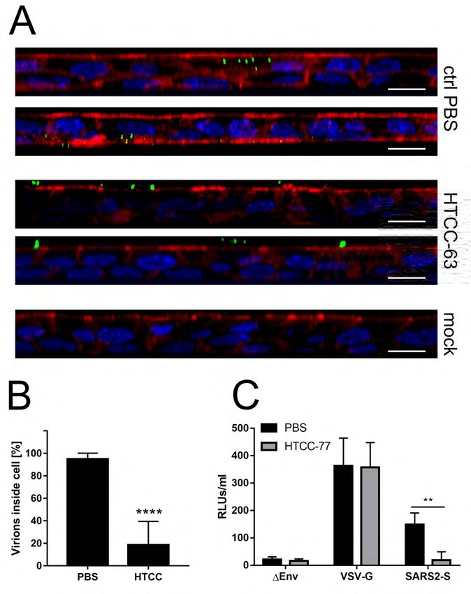
37   ABSTRACT

38   Among seven coronaviruses that infect humans, three (SARS-CoV, MERS-CoV, and the

39   newly identified SARS-CoV-2) are associated with a severe, life-threatening respiratory

40   infection and multiorgan failure. We previously proposed that the cationically modified

41   chitosan, N-(2-hydroxypropyl)-3-trimethylammonium chitosan chloride (HTCC) is a potent

     inhibitor of HCoV-NL63. Next, we demonstrated the broad-spectrum antiviral activity of the

                                                                                                      Downloaded from http://jvi.asm.org/ on December 29, 2020 by guest
42

43   compound, as it inhibited all low pathogenic human coronaviruses (HCoV-NL63, HCoV-

44   229E, HCoV-OC43, and HCoV-HKU1). Here, using in vitro and ex vivo model of human

45   airway epithelium, we show that HTCC effectively blocks MERS-CoV and SARS-CoV-2

46   infection. We also confirmed the mechanism of action for these two viruses, showing that the

47   polymer blocks the virus entry to the host cell by interaction with the S protein.

48

49   IMPORTANCE

50   The beginning of 2020 brought us information about the novel coronavirus emerging in

51   China. Rapid research resulted in the characterization of the pathogen, which appeared to be a

52   member of the SARS-like cluster, commonly seen in bats. Despite the global and local efforts,

53   the virus escaped the healthcare measures and rapidly spread in China and later globally,

54   officially causing a pandemic and global crisis in March 2020. At present, different scenarios

55   are being written to contain the virus, but the development of novel anticoronavirals for all

56   highly pathogenic coronaviruses remains the major challenge. Here, we describe the antiviral

57   activity of previously developed by us HTCC compound, which may be used as a potential

58   inhibitor of currently circulating highly pathogenic coronaviruses – SARS-CoV-2 and MERS-

59   CoV.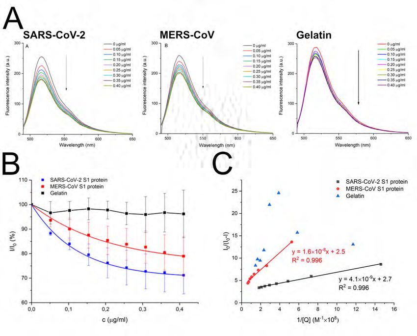
60   INTRODUCTION

61          Coronaviruses mainly cause respiratory and enteric diseases in humans, other

62   mammals, and birds. However, some species can cause more severe conditions such as

63   hepatitis, peritonitis, or neurological disease. Seven coronaviruses infect humans, four of

64   which (human coronavirus [HCoV]-229E, HCoV-NL63, HCoV-OC43, and HCoV-HKU1)

     cause relatively mild upper and lower respiratory tract disease. Other three zoonotic

                                                                                                        Downloaded from http://jvi.asm.org/ on December 29, 2020 by guest
65

66   coronaviruses - the severe acute respiratory syndrome coronaviruses (SARS-CoV and SARS-

67   CoV-2) and the Middle East respiratory syndrome coronavirus (MERS-CoV) are associated

68   with severe, life-threatening respiratory infections and multiorgan failure (1-7).

69          SARS-CoV-2 emerged in the Hubei province of China by the end of 2019 and caused

70   an epidemic that was partially contained in China by March 2020(8). However, the virus

71   rapidly spread globally and caused the pandemic. SARS-CoV-2 is a betacoronavirus and

72   belongs to a large cluster of SARS-like viruses in bats, classified in Sarbecovirus subgenus.

73   While bats are considered to be the original reservoir, it is believed that there is an

74   intermediate host, and pangolins were suggested as such (9). The virus is associated with a

75   respiratory illness that, in a proportion of cases, is severe. The mortality rate varies between

76   locations, but at present, is estimated to reach 3-4% globally. The virus infects primarily

77   ciliated cells and type II pneumocytes in human airways, hijacking the angiotensin-converting

78   enzyme 2 (ACE2) to enter the cell, similarly as SARS-CoV and HCoV-NL63.

79          MERS-CoV is related to SARS-CoV-2, but together with some bat viruses forms

80   a separate Merbecovirus subgenus. Bats are believed to serve as an original reservoir also in

81   this case (10), but camels were identified as the intermediate host (11). The virus never fully

82   crossed the species border, as the human-to-human transmission is limited, and the majority

83   of cases are associated with animal-to-human transmission. The entry receptor for MERS-

84   CoV is the dipeptidyl peptidase 4 (DPP4) (12, 13). In humans, MERS-CoV causes a
85 respiratory illness with severity varying from asymptomatic to potentially fatal acute
86 respiratory distress (14-16). To date, MERS-CoV infection was confirmed in 27 countries,
87 with over 2,000 cases and a mortality rate of ~35%.
88 Currently, there are no vaccines or drugs with proven efficacy to treat coronavirus
89 infection, and treatment is limited to supportive care. However, a range of therapeutics have
been experimentally used in clinic to treat SARS-CoV-2 and MERS-CoV-infected patients,
Downloaded from http://jvi.asm.org/ on December 29, 2020 by guest
90
91 and their use is based on the knowledge obtained in previous years. The most promising drug
92 candidates include broad-spectrum polymerase inhibitors (remdesivir) (17) and some re-
93 purposed drugs (e.g., HIV-1 protease inhibitors). However, until today none of these has
94 proven effective in randomized controlled trials. On the other hand, the antiviral potential of
95 several small molecules have been demonstrated in cell lines in vitro, but their effectiveness
96 in vivo have not been confirmed (18, 19). For the ones that reached animal models, some
97 promising drug candidates have been shown to exacerbate the disease (ribavirin,
98 mycophenolic acid) (20).
99 Drug development for SARS-CoV-2 and MERS-CoV is of great importance, but
100 considering the diversity of coronaviruses and the proven propensity to cross the species
101 barrier, the SARS-CoV-2 epidemic is probably not the last one. Warnings about the
102 possibility of another SARS-CoV epidemic have appeared in the scientific literature for a long
103 time (21). Consequently, broad-spectrum antivirals are essential in long-term perspective.
104 Previously, we have demonstrated an antiviral potential of the HTCC polymer (N-(2-
105 hydroxypropyl)-3-trimethylammonium chitosan chloride), which efficiently hampered
106 infection of all low pathogenic human coronaviruses in vitro and ex vivo (22) and several
107 animal coronaviruses (unpublished data). Furthermore, using several functional and structural
108 assays, we dissected the mechanism of the HTCC antiviral activity. We showed that the
109 polymer interacts with the coronaviral Spike (S) protein and blocks its interaction with the110 cellular receptor (22-24). Here, we analyzed the HTCC activity against SARS-CoV-2 and
111 MERS-CoV in vitro using permissive cell lines and ex vivo, using a model of human airway
112 epithelium (HAE). The study showed that the replication of both viruses was efficiently
113 hampered. Overall, our data show that HTCC polymers are potent broad-spectrum
114 anticoronavirals and may be very promising drug candidates for SARS-CoV-2 and MERS-
CoV.
Downloaded from http://jvi.asm.org/ on December 29, 2020 by guest
115
116
117 RESULTS AND DISCUSSION
118 HTCCs hamper MERS-CoV and SARS-CoV-2 replication in cell lines
119 Previously, we showed that HTCC with different degrees of substitution (DSs) is a
120 potent inhibitor of all four low pathogenic HCoVs (22). DSs are expressed as the fraction of
121 NH2 groups of gluocosamine units in chitosan substituted with glycidyltrimethylammonium
122 chloride (GTMAC). The DSs of the studied HTCC polymers varied between 57% and 77%;
123 thus, the polymers were named HTCC-57, HTCC-62, HTCC-63, HTCC-65, and HTCC-77
124 (Figure 1). The synthesis and characterization of polymers are described elsewhere (22, 23).
125 The analysis showed that HTCC-63 demonstrated the most significant inhibitory effect on
126 HCoV-NL63, HCoV-OC43, and HCoV-HKU1. On the other hand, HTCC-62 and HTCC-77
127 proved to be effective inhibitors of HCoV-229E infection. Further, HTCC-65 effectively
128 inhibited the replication of HCoV-NL63 and HCoV-OC43, while HTCC-62 showed a potent
129 antiviral effect on HCoV-HKU1 infection.
130 First, we determined the cytotoxicity of each HTCC on Vero and Vero E6 cells, which
131 showed that polymers may be used at a non-toxic concentration of 100 μg/ml (Figure 2A).
132 Next, the study on the MERS-CoV using the Vero cells revealed that all HTCC variants
133 inhibit virus replication to a similar extent (~-2-3-fold decrease in viral yields). The inhibition
134 of the SARS-CoV-2 infection in Vero E6 cells was even more pronounced, and all HTCC135 variants inhibited virus replication by ~10,000 times at non-toxic concentration (Figure 2B).
136 In this case, the HTCC-63 was arbitrarily selected for further studies on MERS-CoV, while
137 HTCC-77 was selected for SARS-CoV-2. Next, the dose-dependence was tested for the
138 HTCCs. The inhibitory activity of selected polymers was verified for three different
139 concentrations, and obtained data are shown in Figure 3. Based on the data obtained, the
basic parameters were calculated and are presented in Table 1. The parameters observed for
Downloaded from http://jvi.asm.org/ on December 29, 2020 by guest
140
141 the SARS-CoV-2 appear to be favorable with SI above 12. It is also worth noting that HTCC
142 was previously administered by inhalation in rats, and no adverse reactions were observed
143 (25). In that study, HTCC was used as a carrier for the active substance, and as such, was
144 reported to be promising for local sustained inhalation therapy of pulmonary diseases.
145 Furthermore, to prove the HTCC activity in vitro, Vero (MERS-CoV) and Vero E6
146 (SARS-CoV-2) cells were infected with a given virus in the presence of the compounds. After
147 24 h culture at 37°C, cells were collected, lysed, and subjected to SDS-PAGE. Western blot
148 detection of nucleocapsid proteins showed a dose-dependent reduction of each virus infection,
149 as N protein levels declined in the presence of the HTCCs (Figure 4).150
151 HTCC interacts with the coronaviral spike protein
152 First, we made an effort to delineate at which stage the HTCC interferes with the virus
153 replication. We have previously shown that HTCC blocks the interaction between the
154 coronaviral S protein and its cellular receptor (23). Here, using a set of functional assays, we
showed that pre-treatment of cells does not result in inhibition of viral replication, while the
Downloaded from http://jvi.asm.org/ on December 29, 2020 by guest
155
156 presence of HTCC during the infection blocks the process (Figure 5). The observed inhibition
157 of replication when the HTCC was present throughout the infection results most likely from
158 the multicycle infection process. However, additional inhibitory effect on the replication itself
159 must be considered.
160 Second, we aimed to verify whether the polymer interacts with the coronaviral S
161 protein, as previously shown for the HCoV-NL63(23). For this, we analyzed the fluorescence
162 spectra of HTCC-FITC in the presence of the SARS-CoV-2 or MERS-CoV spike proteins (S1
163 domains). Gelatin was used as a reference. The analysis showed that both proteins quenched
164 the fluorescence emission and that relative fluorescence of HTCC- FITC decreased with the
165 increase in SARS-CoV-2 and MERS-CoV S1 concentrations, but not gelatin, in a non-linear
166 manner (Figure 6A-B). The plot shape for that dependence indicated that HTCC-FITC is not
167 quenched by these proteins in a simple diffusion-controlled collisional process but that there
168 are specific interactions between these macromolecules (Figure 6B). Such dependence is
169 observed typically when the interaction between the chromophore and the quencher leads to a
170 complex formation (26, 27). To further verify that observation, experimental data were fitted
171 to modified Stern-Volmer equation(27):
172
173 I_0/(I_0-I)=1/([Q])∙1/f_a 1/K_a +1/f_a (1)
174175 where: I_0 is the fluorescence intensity of HTCC-FITC in the absence of S1, I is the
176 fluorescence intensity of FITC-HTCC in the presence of S1, [Q] is a concentration of S1, f_a
177 is a fraction of FITC chromophores in HTCC-FITC available for quenching by S1 and K_a is
178 a modified Stern-Volmer quenching constant for the accessible FITC residues. The results are
179 presented in Figure 6C. The analysis indicated that a fraction of polymeric chromophores
available for interaction with SARS-CoV-2 and MERS S1 proteins in both cases was
Downloaded from http://jvi.asm.org/ on December 29, 2020 by guest
180
181 ~f_a=0.4. This rather low value is understandable, as a specific orientation of both
182 macromolecules is necessary to allow their effective interaction within a complex. Strong
183 interaction between HTCC-FITC and S1 can be inferred based on the efficient quenching
184 process indicated by the high value of K_a, which was found to be equal to 6.59×10⁸ M-1 for
185 SARS-CoV-2 S1 and 1.56×10⁸ M-1 for MERS-CoV S1. The stronger interactions between
186 HTCC and SARS-CoV-2 S1 correspond with more efficient replication inhibition of that
187 virus in cell culture and confirms the proposed mechanism.
188
189 HTCCs hamper MERS-CoV and SARS-CoV-2 replication in human airway epithelium
190 While the Vero cells constitute a convenient model for antiviral research, it is of
191 utmost importance to verify whether the results obtained are not biased due to the artificial
192 system used. This is especially important for compounds, which activity is based on
193 electrostatic interaction. To verify whether the natural microenvironment, which is rich in
194 sugars and charged molecules, does not abrogate the effectiveness of the inhibitors, we
195 employed HAE cultures that mirror the fully differentiated layer lining the conductive
196 airways, as well as the site of coronavirus replication. Briefly, fully differentiated HAE
197 cultures were infected with a given virus (28) in the presence of previously selected HTCCs
198 (200 μg/ml) or control PBS. Following inoculation, apical lavage samples were collected
199 daily, and replication kinetics for each virus was investigated. The analysis revealed that the200 polymer efficiently hampered SARS-CoV-2 and MERS-CoV also in this model. For MERS-
201 CoV, the inhibitory effect was the most evident at 72 h p.i., while for SARS-CoV-2, the most
202 substantial decline of virus progeny was observed at 24 h p.i. (Figure 7). Whether such
203 kinetics will be reflected in vivo, it is to be investigated.
204
Downloaded from http://jvi.asm.org/ on December 29, 2020 by guest
205 HTCCs inhibits MERS-CoV and SARS-CoV-2 entry into susceptible cells
206 Our previous research showed that the HTCC-mediated inhibition of coronaviral
207 replication results from the electrostatic interaction between the polymer and the Spike
208 protein of coronaviruses. We hypothesize that the selectivity of the inhibitors yields from the
209 fitting charge distributions on the polymer and on the S proteins on the viral surface. While
210 the interaction of a single charged moiety is relatively weak, the concatemeric nature of the
211 virus and the polymer stabilizes the binding. Such structure-based interaction may be an
212 interesting entry point for further fine-tuning of the polymeric inhibitors of viral replication.
213 To ensure that the observed effect was a result of coronavirus entry inhibition by
214 HTCC, two experiments were performed. First, HAE cultures were inoculated with MERS-
215 CoV in the presence of HTCC-63 (200 μg/ml) or control PBS and incubated for 2 h at 37°C.
216 Next, cells were fixed, immunostained for MERS-CoV N protein, and actin. Virus entry was
217 analyzed with confocal microscopy. To visualize the effect, the signal attributed to
218 intracellular MERS-CoV was quantified, and the results show that the internalization of
219 MERS-CoV was drastically decreased (Figure 8A-B). Due to the limited availability of tools
220 for the SARS-CoV-2 at that time, we were not able to replicate the experiment. Here, we
221 employed a surrogate system based on lentiviral vectors pseudotyped with full-length Spike
222 protein of SARS-CoV-2. A549 cells overexpressing the ACE2 protein were incubated with
223 pseudovirions harboring SARS-CoV-2 Spike or control VSV-G protein in the presence of
224 HTCC-77 (100 μg/ml) or control PBS for 2 h at 37°C. After 72 h p.i. cells were lysed, and225 pseudovirus entry was quantified by measurement of the reporter luciferase protein. The
226 analysis showed a significant reduction in SARS-CoV-2 Spike pseudoviruses internalization
227 in the presence of the polymer, while no inhibition was observed for the control VSV-G
228 (Figure 8C).
229 Next, to verify whether the mechanism of action for the highly pathogenic
betacoronaviruses is similar to that observed for alphacoronaviruses (22), and is based on
Downloaded from http://jvi.asm.org/ on December 29, 2020 by guest
230
231 blocking the interaction between the virus and the entry receptor, we analysed MERS-CoV
232 co-localization with its entry receptor, DPP4 in the presence or absence of the HTCC. For
233 this, human cell line Huh7 was inoculated with the virus or mock in the presence of HTCC-63
234 (100 μg/ml) or control PBS and incubated for 2 h at 4°C. Subsequently, cells were fixed and
235 immunostained for the DPP4 and MERS-CoV N using specific antibodies. Virus
236 colocalization with its receptor was examined using confocal microscopy. Obtained results
237 demonstrated that in the control samples, the signal originating from the N protein co-
238 localizes with the DPP4 protein, while in the presence of the polymer, this interaction is
239 blocked (Figure 9).
240 We show here that the previously developed and described polymeric HTCC
241 anticoronaviral HTCC compounds can efficiently inhibit infection with emerging
242 coronaviruses. We believe that the HTCC can be fine-tuned to target any coronavirus, and this
243 interaction is specific to viruses that belong to the Coronaviridae family. One may speculate
244 that the inhibition results from the concatemeric nature of the virus surface and the fact that
245 the polymer with appropriate charge distribution can interact with multiple sites on this
246 surface. While the interaction of the monomer is relatively weak, and no inhibition is
247 observable for monomers, the sum of interactions stabilizes the binding and specific inhibition
248 is observed. Considering that the extended chain length for the HTCC used is ~700 nm this
249 scenario seems realistic (29). If that would be true, HTCC would constitute a first structure-250 specific inhibitor of viral replication. The major disadvantage of the HTCC is that, at present,
251 it is not registered for use in humans. However, previous experience with HTCC in different
252 laboratories shows that it may be delivered topically to the lungs, it is not associated with
253 toxicity, and it does not worsen the lung function (25). We believe that HTCC is a promising
254 drug candidate that should be further studied, as it provides a ready-to-use solution for
SARS-CoV-2 and future emerging coronaviruses.
Downloaded from http://jvi.asm.org/ on December 29, 2020 by guest
255
256
257 MATERIALS AND METHODS
258 Materials
259 Chitosan (low molecular weight, Mv = 50-190 kDa, degree of deacetylation DDA =
260 73% based on elemental analysis, Sigma-Aldrich), glycidyltrimethylammonium chloride
261 (GTMAC, Sigma-Aldrich), fluorescein isothiocyanate isomer 1 (FITC, Sigma-Aldrich),
262 glacial acetic acid (CH3COOH, 99.5% pure p.a., CHEMPUR), acetone (Sigma-Aldrich),
263 ethanol (Stanlab), AgNO3 (Sigma-ALdrich), DMSO (POCh) and pyridine (Sigma Aldrich)
264 were used as received. Recombinant SARS-CoV-2 Spike Protein, (S1 subunit derived from
265 E.coli, MW = 75 kDa) and MERS (S1 fragment (aa 56-296) derived from E.coli, MW = 27
266 kDa) were purchased from RayBiotech. Gelatin from bovine skin (type B) was purchased
267 from Sigma Aldrich. Water was distilled twice and deionized using the Milipore Simplicity
268 system.
269
270 The active compound
271 The HTCC was prepared in the same manner as previously described (22, 23, 30).
272 Briefly, 30 ml of GTMAC was added to the chitosan solution in dilute acetic acid (3 g of
273 chitosan in 150 ml of 1% v/v acetic acid solution in water). The solution was stirred at 70°C
274 for 24 h, and then centrifuged at 4000 rpm for 10 min to remove unreacted chitosan. The275 product was precipitated from the supernatant with a cold mixture of acetone and ethanol (4:1,
276 v/v). The solution was decanted, and the resulting precipitate was dissolved in distilled water.
277 The purification process was repeated, resulting pellet was washed with ethanol three times
278 and purified HTCC was dried in a vacuum oven for 24 h. Degree of substitution (DS) of NH2
279 groups of the aminoglucose units with GTMAC was found to be 69 ± 2% as determined via
conductometric titration of the chloride anion with 0.017 M solution of AgNO3.
Downloaded from http://jvi.asm.org/ on December 29, 2020 by guest
280
281
282 Synthesis of fluorescein-labeled HTCC (HTCC-FITC)
283 250 mg of HTCC was suspended in a mixture of DMSO and pyridine (21 ml, 20:1,
284 v/v). 15 mg of fluorescein isothiocyanate (FITC) was dissolved in 5 ml of DMSO and added
285 to vigorously stirred suspension of HTCC. The reaction mixture was stirred for 24 h in 95oC
286 in the dark. The crude product was dialyzed against water and freeze-dried. In its absorption
287 spectra, a band was present with a maximum at λmax = 494 nm in water confirming the
288 substitution of HTCC with FITC. The degree of substitution of HTCC with FITC, defined as
289 the number of FITC groups per aminoglucose group of HTCC and calculated based on the
290 UV-Vis spectra, was equal to 2.3%.
291
292 Plasmid constructs
293 The codon-optimized full-length SARS-CoV-2 S gene was designed and purchased
294 from GeneArt (Thermo Fisher Scientific, Poland). The gene was cloned into pCAGGS vector
295 sequence verified that was a gift from Xingchuan Huang. psPAX (Addgene plasmid # 12260)
296 and pMD2G (Addgene plasmid # 12259) was a gift from Didier Trono. pRRL Luciferase
297 vector (Addgene plasmid #120798)was a gift from Paul Khavari.
298
299 Cell culture300 Vero and Vero E6 (Cercopithecus aethiops; kidney epithelial; ATCC: CCL-81 and CRL-
301 1586), Huh7 (Homo sapiens; hepatocellular carcinoma; ECACC: 01042712) and A549 cells
302 with ACE2 overexpression (A549ACE2+)(31) were cultured in Dulbecco’s MEM (Thermo
303 Fisher Scientific, Poland) supplemented with 3% fetal bovine serum (heat-inactivated;
304 Thermo Fisher Scientific, Poland) and antibiotics: penicillin (100 U/ml), streptomycin
(100 μg/ml), and ciprofloxacin (5 μg/ml). Cells were maintained at 37°C under 5% CO2.
Downloaded from http://jvi.asm.org/ on December 29, 2020 by guest
305
306
307 Human airway epithelium (HAE) cultures
308 Human airway epithelial cells were isolated from conductive airways resected from
309 transplant patients. The study was approved by the Bioethical Committee of the Medical
310 University of Silesia in Katowice, Poland (approval no: KNW/0022/KB1/17/10 dated
311 16.02.2010). Written consent was obtained from all patients. Cells were dislodged by protease
312 treatment, and later mechanically detached from the connective tissue. Further, cells were
313 trypsinized and transferred onto permeable Transwell insert supports ( = 6.5 mm). Cell
314 differentiation was stimulated by the media additives and removal of media from the apical
315 side after the cells reached confluence. Cells were cultured for 4-6 weeks to form well-
316 differentiated, pseudostratified mucociliary epithelium. All experiments were performed in
317 accordance with relevant guidelines and regulations.
318
319 Cell viability assay
320 HAE cultures were prepared as described above. Cell viability assay was performed by
321 using the XTT Cell Viability Assay (Biological Industries, Israel) according to the
322 manufacturer’s instructions. On the day of the assay, 100 μl of the 1 × PBS with the 50 μl of
323 the activated XTT solution was added to each well/culture insert. Following 2 h incubation at
324 37°C, the solution was transferred onto a 96-well plate, and the signal was measured at325 λ = 490 nm using the colorimeter (Spectra MAX 250, Molecular Devices). The obtained
326 results were further normalized to the control sample, where cell viability was set to 100%.
327
328 Virus preparation and titration
329 MERS-CoV stock (isolate England 1, 1409231v, National Collection of Pathogenic
Viruses, Public Health England, United Kingdom) was generated by infecting monolayers of
Downloaded from http://jvi.asm.org/ on December 29, 2020 by guest
330
331 Vero cells. SARS-CoV-2 stock (isolate 026V-03883; kindly granted by Christian Drosten,
332 Charité – Universitätsmedizin Berlin, Germany by the European Virus Archive - Global
333 (EVAg); https://www.european-virus-archive.com/) was generated by infecting monolayers of
334 Vero E6 cells. The virus-containing liquid was collected at day 3 post-infection (p.i.),
335 aliquoted and stored at −80°C. Control Vero or Vero E6 cell lysate from mock-infected cells
336 was prepared in the same manner. Virus yield was assessed by titration on fully confluent
337 Vero or Vero E6 cells in 96-well plates, according to the method of Reed and Muench. Plates
338 were incubated at 37°C for 3 days and the cytopathic effect (CPE) was scored by observation
339 under an inverted microscope.
340
341 Virus infection
342 In in vitro experiments, fully confluent Vero, Vero E6, or Huh7 cells in 96-well plates
343 (TPP) were exposed to MERS-CoV, SARS-CoV-2 or mock at a TCID50 of 400 per ml in the
344 presence of tested polymer or control medium. Following a 2 h incubation at 37°C, unbound
345 virions were removed by washing with 100 μl of 1 × PBS and fresh medium containing
346 dissolved respective polymer was added to each well. Samples of cell culture supernatant
347 were collected at day 3 p.i. and analyzed using RT-qPCR.
348 For the ex vivo study, fully differentiated human airway epithelium (HAE) cultures were
349 exposed to the tested polymer or control PBS for 30 min at 37°C, following inoculation with350 MERS-CoV or SARS-CoV-2 at a TCID50 of 400 per ml in the presence of the polymer or
351 control PBS. Following 2 h incubation at 37°C, unbound virions were removed by washing
352 with 200 μl of 1 × PBS, and HAE cultures were maintained at air-liquid interphase for the rest
353 of the experiment. To analyze virus replication kinetics, each day p.i., 100 μl of 1 × PBS was
354 applied at the apical surface of HAE and collected following the 10 min incubation at 32°C.
All samples were stored at −80°C and analyzed using RT-qPCR.
Downloaded from http://jvi.asm.org/ on December 29, 2020 by guest
355
356
357 Functional assays
358 To delineate the mechanism of action for the HTCC a set of functional assays was carried
359 out. Fully confluent Vero or Vero E6 in 48-well plates were exposed to MERS-CoV or
360 SARS-CoV-2, respectively, at 400 TCID50 per ml. HTCC-63 was used for MERS-CoV and
361 HTCC-77 for SARS-CoV-2. First, the ability of polymers to interact with a cell to prevent
362 infection was examined (Assay I-pre). Briefly, cells were overlaid with the test compound or
363 control PBS and incubated for 1 h at 37°C. Subsequently, the polymer was removed by a
364 triple wash with 1 × PBS, and a virus or mock was added. After 2 h infection at 37°C, cells
365 were washed and further cultured in a fresh medium for 48 h at 37°C. Next, the influence of
366 polymers on virus-cell interactions was inspected (Assay II-during). Cells were infected in the
367 presence of compounds or control PBS and incubated at 37°C. After 2 h, cells were washed
368 twice with 1 × PBS, fresh culture medium with polymers was applied, and cells were cultured
369 for 48 h at 37°C. In the third assay (Assay III-post), the effects of compounds on virus
370 replication, assembly, and egress were evaluated. Cells were infected and incubated for 2 h at
371 37°C. Subsequently, unbound virions were removed with two washes with 1 × PBS, and
372 media supplemented with polymers or control PBS were applied. Cells were then were
373 cultured for 48 h at 37°C. Samples of supernatant were collected at day 2 p.i. and analyzed
374 using qPCR and plaque assay.375
376 Plaque assay
377 Vero E6 cells were seeded in 24-well and grown to 90% confluency. Tenfold serial
378 dilutions of previously collected samples in culture medium were prepared and placed on
379 cells. Following 1 h incubation in 37°C, an equal volume of 2 × overlay media (DMEM
supplemented with 4% fetal bovine serum, penicillin (100 U/ml), streptomycin (100 μg/ml)
Downloaded from http://jvi.asm.org/ on December 29, 2020 by guest
380
381 and 0.1% agarose) were added to inoculum and cultures were incubated for 3 days at 37°C
382 without moving. Next, cells were fixed with 4% paraformaldehyde, stained using 0.1% crystal
383 violet solution, washed with tap water, and plaques were counted.
384
385 Isolation of nucleic acids and reverse transcription (RT)
386 Viral DNA/RNA Kit (A&A Biotechnology, Poland) was used for nucleic acid isolation
387 from cell culture supernatants, according to the manufacturer’s instructions. cDNA samples
388 were prepared with a High Capacity cDNA Reverse Transcription Kit (Thermo Fisher
389 Scientific, Poland), according to the manufacturer’s instructions.
390
391 Quantitative PCR (qPCR)
392 Viral RNA yield was assessed using real-time PCR (7500 Fast Real-Time PCR; Life
393 Technologies, Poland). cDNA was amplified in a reaction mixture containing 1 × qPCR
394 Master Mix (A&A Biotechnology, Poland), in the presence of probe (100 nM) and primers
395 (450 nM each) (Table 2). The reaction was carried out according to the scheme: 2 min at
396 50°C and 10 min at 92°C, followed by 40 cycles of 15 s at 92°C and 1 min at 60°C. In order
397 to assess the copy number for N gene, DNA standards were prepared, as described before
398 (28).
399400 Immunostaining and confocal imaging
401 Fixed cells were permeabilized with 0.1% Triton X-100 in 1 × PBS and incubated
402 overnight at 4°C in 1× PBS supplemented with 5% bovine serum albumin (BSA) and 0.5%
403 Tween 20. To visualize MERS-CoV particles, cells were incubated for 2 h at room
404 temperature with mouse anti-MERS-CoV N IgGs (1:000 dilution, Sino Biological, China),
followed by 1 h of incubation with Alexa Fluor 488-labeled goat anti-mouse IgG (2.5 µg/ml;
Downloaded from http://jvi.asm.org/ on December 29, 2020 by guest
405
406 Thermo Fisher Scientific, Poland). Actin filaments were stained using phalloidin coupled with
407 Alexa Fluor 633 (0.2 U/ml; Thermo Fisher Scientific, Poland). Nuclear DNA was stained
408 with DAPI (4’,6’-diamidino-2-phenylindole) (0.1 µg/ml; Sigma-Aldrich, Poland).
409 Immunostained cultures were mounted on glass slides in ProLong Gold antifade medium
410 (Thermo Fisher Scientific, Poland). Fluorescent images were acquired under a Leica TCS SP5
411 II confocal microscope (Leica Microsystems GmbH, Mannheim, Germany) and a Zeiss LSM
412 710 confocal microscope (Carl Zeiss Microscopy GmbH). Images were acquired using Leica
413 Application Suite Advanced Fluorescence LAS AF v. 2.2.1 (Leica Microsystems CMS
414 GmbH) or ZEN 2012 SP1 software (Carl Zeiss Microscopy GmbH) deconvolved with
415 Huygens Essential package version 4.4 (Scientific Volume Imaging B.V., The Netherlands)
416 and processed using ImageJ 1.47v (National Institutes of Health, Bethesda, MD, USA). At the
417 time of the study, no antibodies specific to SARS-CoV-2 were available to us.
418
419 Pseudovirus production and transduction
420 293T cells were seeded on 10 cm2 dishes, cultured for 24 h at 37°C with 5% CO2 and
421 transfected using polyethyleneimine (Sigma-Aldrich, Poland) with the lentiviral packaging
422 plasmid (psPAX), the VSV-G envelope plasmid (pMD2G) or SARS-CoV-2 S glycoprotein
423 (pCAGGS-SARS-CoV-2-S) and third plasmid encoding firefly luciferase protein (pRRL424 Luciferase). Cells were further cultured for 72 h at 37°C with 5% CO2, and pseudoviruses
425 were collected every 24 h and stored at 4°C.
426 A549ACE2+ cells were seeded in 48-wells plates, cultured for 24 h at 37°C with 5%
427 CO2 and transduced with pseudoviruses harboring VSV-G or S-SARS-CoV-2 proteins or
428 lacking the fusion protein (ΔEnv) in the presence of polybrene (4 µg/ml; Sigma-Aldrich,
Poland) and HTCC-77 (100 μg/ml) or control PBS. After 4 h incubation at 37°C unbound
Downloaded from http://jvi.asm.org/ on December 29, 2020 by guest
429
430 virions were removed by washing thrice with 1 × PBS, and cells were further cultured for
431 72 h at 37°C with 5% CO2. Cells were lysed in Bright-Glo™ Luciferase Assay buffer
432 (Promega, Poland)and transferred onto white 96-wells plates. Luminescence levels were
433 measured on a microplate reader Gemini EM (Molecular Devices, UK).
434
435 Western blot analysis
436 Cells were trypsinized, centrifuged and resuspended in RIPA buffer (50 mM Tris,150
437 mM NaCl, 1% Nonidet P-40, 0.5% sodium deoxycholate, 0.1% SDS, pH 7.5) followed by
438 lysis for 30 min on ice. Subsequently, samples were centrifuged (10 min at 12,000 × g), and
439 the pelleted cell debris was discarded. Resulting supernatants were mixed with sample buffer
440 (0.5 M Tris pH 6.8, 10% SDS, 50 mg/ml DTT), boiled for 5 min, cooled on ice, and separated
441 on 10% polyacrylamide gels alongside dual-color Page Ruler Pre-stained Protein size markers
442 (Thermo Fisher Scientific, Poland). The separated proteins were then transferred onto a
443 Westran S PVDF membrane (GE Healthcare, Poland) by wet blotting (Bio-Rad, Poland) for 1
444 h, 100 Volts in transfer buffer: 25 mM Tris, 192 mM glycine, 20% methanol at 4 °C. The
445 membranes were then blocked by overnight incubation at 4 °C in TBS-Tween (0.1%) buffer
446 supplemented with 5% skimmed milk (BioShop, Canada). Mouse anti-MERS-CoV N IgGs
447 (1:000 dilution, Sino Biological, China) and horseradish peroxidase-labeled rabbit anti-mouse
448 IgG (65 ng/ml; Dako, Denmark) were used to detect MERS-CoV nucleocapsid protein.449 Mouse anti-SARS-CoV-2 N IgGs (1:000 dilution, Bioss Antibodies, USA) and horseradish
450 peroxidase-labeled rabbit anti-mouse IgG (65 ng/ml; Dako, Denmark) were used to detect
451 SARS-CoV-2 nucleocapsid protein. A rabbit anti-GAPDH antibody (1:5000 dilution; Cell
452 Signalling Technology, Poland) and horseradish peroxidase-labeled goat anti-rabbit IgG (0.35
453 μg/ml; Sigma-Aldrich, Poland) were used to detect the GAPDH in cell lysates. All antibodies
were diluted in 1% skimmed milk/TBS-Tween (0.1%). The signal was developed using the
Downloaded from http://jvi.asm.org/ on December 29, 2020 by guest
454
455 Immobilon Western Chemiluminescent HRP Substrate (Millipore, USA).
456
457 Interaction between HTCC and SARS-CoV-2 spike protein
458 The fluorescence spectra of HTCC labeled with FITC (cHTCC-FITC = 0.5 µg/ml) in
459 water were measured in the absence and in the presence of various concentrations of SARS-
460 CoV-2 and MERS-CoV spike proteins S1 domains ranging from 0.05 µg/ml to 0.4 µg/ml).
461 NMR spectra were measured in deuterium oxide (D2O) using Bruker Advance II 600 MHz
462 spectrometer. Elemental analysis was performed using a Vario Micro CHNS elemental
463 analyzer. UV-Vis absorption spectra were recorded using a Varian Cary 50 UV-Vis
464 spectrometer in 1-cm quartz cuvettes. Fluorescence spectra of HTCC-FITC were measured at
465 λex=470 nm using a HITACHI F-7000 spectrofluorimeter at room temperature.
466 Conductometric titrations were performed using an Elmetron CX-741 multifunction computer
467 meter.
468
469 Statistical analysis.
470 All the experiments were performed in triplicate, and the results are presented as mean
471 ± standard deviation (SD). To determine the significance of the obtained results, Student t-test
472 was carried out. P values of < 0.05 were considered significant.
473474 ACKNOWLEDGMENTS
475 This work was supported by the subsidy from the Polish Ministry of Science and Higher
476 Education for the research on the SARS-CoV-2, a grant from the National Science Center
477 UMO-2017/27/B/NZ6/02488 to KP, and EU-Horizon2020 ITN OrganoVir grant 812673.
478 The funders had no role in study design, data collection, and analysis, the decision to
publish, or preparation of the manuscript.
Downloaded from http://jvi.asm.org/ on December 29, 2020 by guest
479
480 The technology is owned by the Jagiellonian University (Krakow, Poland) and
481 protected by patent no. WO2013172725A1 and associated documents.
482 A.M., Y.C., A.S., E. B. D., M.Ob., X.G., Y.G., J.L., L.C., A.D., P.B. conducted the
483 experiments. M.O., M.U., and S.R.M provided materials and methods for the study. A.M.,
484 K.P. designed the study and experiments, analyzed the data, and wrote the manuscript. K.L.,
485 D.L., F.Z., M.N., K.S. analyzed the data. K.P. supervised the study. All authors reviewed the
486 manuscript and approved the submitted version. All authors agreed to be personally
487 accountable for their own contributions and to ensure that questions related to the accuracy or
488 integrity of any part of the work are appropriately investigated, resolved, and the resolution
489 documented in the literature.
490 The authors declare no competing financial interests.
491
492
493
494
495496 REFERENCES
497 1. Fields BN, Knipe DM, Howley PM. 2013. Fields virology, 6th ed. Wolters Kluwer
498 Health/Lippincott Williams & Wilkins, Philadelphia.
499 2. Peiris JS, Yuen KY, Osterhaus AD, Stöhr K. 2003. The severe acute respiratory syndrome. N
500 Engl J Med 349:2431-41.
501 3. de Groot RJ, Baker SC, Baric RS, Brown CS, Drosten C, Enjuanes L, Fouchier RA, Galiano M,
502 Gorbalenya AE, Memish ZA, Perlman S, Poon LL, Snijder EJ, Stephens GM, Woo PC, Zaki AM,
503 Zambon M, Ziebuhr J. 2013. Middle East respiratory syndrome coronavirus (MERS-CoV):
504 announcement of the Coronavirus Study Group. J Virol 87:7790-2.
505 4. Zaki AM, van Boheemen S, Bestebroer TM, Osterhaus AD, Fouchier RA. 2012. Isolation of a
Downloaded from http://jvi.asm.org/ on December 29, 2020 by guest
506 novel coronavirus from a man with pneumonia in Saudi Arabia. N Engl J Med 367:1814-20.
507 5. van der Hoek L, Pyrc K, Jebbink MF, Vermeulen-Oost W, Berkhout RJ, Wolthers KC,
508 Wertheim-van Dillen PM, Kaandorp J, Spaargaren J, Berkhout B. 2004. Identification of a new
509 human coronavirus. Nat Med 10:368-73.
510 6. van der Hoek L, Sure K, Ihorst G, Stang A, Pyrc K, Jebbink MF, Petersen G, Forster J, Berkhout
511 B, Uberla K. 2005. Croup is associated with the novel coronavirus NL63. PLoS Med 2:e240.
512 7. Zhu N, Zhang D, Wang W, Li X, Yang B, Song J, Zhao X, Huang B, Shi W, Lu R, Niu P, Zhan F, Ma
513 X, Wang D, Xu W, Wu G, Gao GF, Tan W, Team CNCIaR. 2020. A Novel Coronavirus from
514 Patients with Pneumonia in China, 2019. N Engl J Med 382:727-733.
515 8. Yang J, Chen X, Deng X, Chen Z, Gong H, Yan H, Wu Q, Shi H, Lai S, Ajelli M, Viboud C, Yu PH.
516 2020. Disease burden and clinical severity of the first pandemic wave of COVID-19 in Wuhan,
517 China. Nature Communications 11:5411.
518 9. Andersen KG, Rambaut A, Lipkin WI. 2020. The proximal origin of SARS-CoV-2. Nature
519 Medicine doi:https://doi.org/10.1038/s41591-020-0820-9.
520 10. Corman VM, Ithete NL, Richards LR, Schoeman MC, Preiser W, Drosten C, Drexler JF. 2014.
521 Rooting the phylogenetic tree of middle East respiratory syndrome coronavirus by
522 characterization of a conspecific virus from an African bat. J Virol 88:11297-303.
523 11. Zhang Z, Shen L, Gu X. 2016. Evolutionary Dynamics of MERS-CoV: Potential Recombination,
524 Positive Selection and Transmission. Sci Rep 6:25049.
525 12. Lu G, Hu Y, Wang Q, Qi J, Gao F, Li Y, Zhang Y, Zhang W, Yuan Y, Bao J, Zhang B, Shi Y, Yan J,
526 Gao GF. 2013. Molecular basis of binding between novel human coronavirus MERS-CoV and
527 its receptor CD26. Nature 500:227-31.
528 13. Raj VS, Mou H, Smits SL, Dekkers DH, Müller MA, Dijkman R, Muth D, Demmers JA, Zaki A,
529 Fouchier RA, Thiel V, Drosten C, Rottier PJ, Osterhaus AD, Bosch BJ, Haagmans BL. 2013.
530 Dipeptidyl peptidase 4 is a functional receptor for the emerging human coronavirus-EMC.
531 Nature 495:251-4.
532 14. Mackay IM, Arden KE. 2015. MERS coronavirus: diagnostics, epidemiology and transmission.
533 Virol J 12:222.
534 15. Milne-Price S, Miazgowicz KL, Munster VJ. 2014. The emergence of the Middle East
535 respiratory syndrome coronavirus. Pathog Dis 71:121-36.
536 16. Chan RW, Hemida MG, Kayali G, Chu DK, Poon LL, Alnaeem A, Ali MA, Tao KP, Ng HY, Chan
537 MC, Guan Y, Nicholls JM, Peiris JS. 2014. Tropism and replication of Middle East respiratory
538 syndrome coronavirus from dromedary camels in the human respiratory tract: an in-vitro
539 and ex-vivo study. Lancet Respir Med 2:813-22.
540 17. Agostini ML, Andres EL, Sims AC, Graham RL, Sheahan TP, Lu X, Smith EC, Case JB, Feng JY,
541 Jordan R, Ray AS, Cihlar T, Siegel D, Mackman RL, Clarke MO, Baric RS, Denison MR. 2018.
542 Coronavirus Susceptibility to the Antiviral Remdesivir (GS-5734) Is Mediated by the Viral
543 Polymerase and the Proofreading Exoribonuclease. mBio 9.
544 18. Agnihothram S, Yount BL, Donaldson EF, Huynh J, Menachery VD, Gralinski LE, Graham RL,
545 Becker MM, Tomar S, Scobey TD, Osswald HL, Whitmore A, Gopal R, Ghosh AK, Mesecar A,546 Zambon M, Heise M, Denison MR, Baric RS. 2014. A mouse model for Betacoronavirus
547 subgroup 2c using a bat coronavirus strain HKU5 variant. MBio 5:e00047-14.
548 19. Ratia K, Pegan S, Takayama J, Sleeman K, Coughlin M, Baliji S, Chaudhuri R, Fu W, Prabhakar
549 BS, Johnson ME, Baker SC, Ghosh AK, Mesecar AD. 2008. A noncovalent class of papain-like
550 protease/deubiquitinase inhibitors blocks SARS virus replication. Proc Natl Acad Sci U S A
551 105:16119-24.
552 20. Barnard DL, Day CW, Bailey K, Heiner M, Montgomery R, Lauridsen L, Winslow S, Hoopes J, Li
553 JK, Lee J, Carson DA, Cottam HB, Sidwell RW. 2006. Enhancement of the infectivity of SARS-
554 CoV in BALB/c mice by IMP dehydrogenase inhibitors, including ribavirin. Antiviral Res 71:53-
555 63.
556 21. Cheng VC, Lau SK, Woo PC, Yuen KY. 2007. Severe acute respiratory syndrome coronavirus as
Downloaded from http://jvi.asm.org/ on December 29, 2020 by guest
557 an agent of emerging and reemerging infection. Clin Microbiol Rev 20:660-94.
558 22. Milewska A, Kaminski K, Ciejka J, Kosowicz K, Zeglen S, Wojarski J, Nowakowska M,
559 Szczubiałka K, Pyrc K. 2016. HTCC: Broad Range Inhibitor of Coronavirus Entry. PLoS One
560 11:e0156552.
561 23. Milewska A, Ciejka J, Kaminski K, Karewicz A, Bielska D, Zeglen S, Karolak W, Nowakowska M,
562 Potempa J, Bosch BJ, Pyrc K, Szczubialka K. 2013. Novel polymeric inhibitors of HCoV-NL63.
563 Antiviral Res 97:112-21.
564 24. KAMIL K, ALEKSANDRA M, MARIA N, KRZYSZTOF P, KRZYSZTOF S. THE USE OF CHITOSAN
565 POLYMER IN THE TREATMENT AND PREVENTION OF INFECTIONS CAUSED BY
566 CORONAVIRUSES.
567 25. Yang TT, Wen BF, Liu K, Qin M, Gao YY, Ding DJ, Li WT, Zhang YX, Zhang WF. 2018.
568 Cyclosporine A/porous quaternized chitosan microspheres as a novel pulmonary drug
569 delivery system. Artif Cells Nanomed Biotechnol 46:552-564.
570 26. Samworth CM, Degli Esposti M, Lenaz G. 1988. Quenching of the intrinsic tryptophan
571 fluorescence of mitochondrial ubiquinol--cytochrome-c reductase by the binding of
572 ubiquinone. Eur J Biochem 171:81-6.
573 27. Lakowicz JR. 1988. Principles of frequency-domain fluorescence spectroscopy and
574 applications to cell membranes. Subcell Biochem 13:89-126.
575 28. A M, A K-P, J W, A S, A S, A D, K O, M O, T S, Z R, P L, W B, K P, View ORCID Profil Anna Kula-
576 Pacurar JW, Agnieszka Suder , Artur Szczepanski , Agnieszka Dabrowska ,
577 Katarzyna Owczarek , Marek Ochman , Tomasz Stacel View ORCID Profile.
578 Replication of SARS-CoV-2 in human respiratory epithelium
579 doi:https://doi.org/10.1101/2020.03.20.999029.
580 29. Kühtreiber WM, P. LR, Chick WL, View ORCID Profil Anna Kula-Pacurar JW, Agnieszka
581 Suder , Artur Szczepanski , Agnieszka Dabrowska , Katarzyna Owczarek , Marek
582 Ochman , Tomasz Stacel View ORCID Profile. 1999. Cell Encapsulation Technology
583 and Therapeutics.
584 30. Ciejka J, Wolski K, Nowakowska M, Pyrc K, Szczubiałka K. 2017. Biopolymeric
585 nano/microspheres for selective and reversible adsorption of coronaviruses. Mater Sci Eng C
586 Mater Biol Appl 76:735-742.
587 31. Milewska A, Zarebski M, Nowak P, Stozek K, Potempa J, Pyrc K. 2014. Human coronavirus
588 NL63 utilizes heparan sulfate proteoglycans for attachment to target cells. J Virol 88:13221-
589 30.
590
591592 Figure Legends
593 Figure 1. Different HTCC polymers varying in the degree of substitution (DS). The
594 effective compounds range is 60-70% degree of substitution of the chitosan chain.
595
596 Figure 2. In vitro inhibition of MERS-CoV and SARS-CoV-2 by HTCC at non-toxic
concentration. (A) Cytotoxicity of HTCCs with a degree of substitution ranging from 57% to
Downloaded from http://jvi.asm.org/ on December 29, 2020 by guest
597
598 77% in vitro using Vero and Vero E6 cells. Cell viability was assessed with XTT assay. Data
599 on the y-axis represent the percentage of values obtained for the untreated reference samples.
600 All assays were performed in triplicate, and average values obtained for both cell lines with
601 standard errors are presented. (B) Vero cells were infected with MERS-CoV, and Vero E6
602 cells were infected with SARS-CoV-2. Briefly, cultures were inoculated with a given virus in
603 the presence of HTCC (100 μg/ml) or control PBS. Replication of viruses was evaluated at
604 48 h post-inoculation using RT-qPCR. The data are presented as the number of viral RNA
605 copies per ml. The assay was performed in triplicate, and average values with standard errors
606 are shown.
607
608 Figure 3. Dose-dependent inhibition of MERS-CoV and SARS-CoV-2 replication. (A)
609 Vero (MERS-CoV) or (B) Vero E6 cells (SARS-CoV-2) were inoculated with a given virus
610 in the presence of different concentrations of HTCC. Replication of viruses was evaluated at
611 48 h post-inoculation using RT-qPCR. The data are presented as the number of viral RNA
612 copies per ml (left panel) and as Log Removal Value (LRV) compared to the untreated
613 sample (right panel). The assay was performed in triplicate, and average values with standard
614 errors are displayed. All the results are statistically significant, as determined using the
615 Student’s t-test.
616617 Figure 4. Dose-dependent inhibition of MERS-CoV and SARS-CoV-2 infection. Vero
618 (MERS-CoV) or Vero E6 cells (SARS-CoV-2) were inoculated with a given virus in the
619 presence of different concentrations of HTCC (HTCC-63 for MERS-CoV; HTCC-77 for
620 SARS-CoV-2). Following incubation for 24 h at 37 °C, cells were lysed in RIPA buffer.
621 Nucleocapsid proteins and control GAPDH protein were detected by western blotting using
specific antibodies.
Downloaded from http://jvi.asm.org/ on December 29, 2020 by guest
622
623
624 Figure 5. HTCC hampers interaction between the virus and the cell. Vero (MERS-CoV)
625 or Vero E6 cells (SARS-CoV-2) were inoculated with a virus in the presence of HTCC
626 (HTCC-63 for MERS-CoV; HTCC-77 for SARS-CoV-2). Polymers were applied in three
627 different manners: assay I-pre – cells were pretreated with compounds before infection,
628 assay II-during – polymers were added alongside the virus, and were present throughout the
629 incubation, assay III – post – polymers were added 1 h after the infection. Replication of
630 viruses was evaluated at 48 h post-infection using plaque assay (A) and RT-qPCR (B).
631
632 Figure 6. Interaction between HTCC-FITC and SARS-CoV-2 S1 domain. (A)
633 Fluorescence spectra of HTCC-FITC (λex= 470 nm, cHTCC-FITC = 0.5 µg/ml) in the
634 absence and in the presence of SARS-CoV-2, MERS-CoV S1 or gelatin proteins at various
635 concentrations. (B) Dependence of relative fluorescence intensity (I/I0) of HTCC-FITC (λem=
636 517 nm) on the concentration of SARS-CoV-2, MERS-CoV S1, and gelatin. (C) Dependence
637 of relative fluorescence I0/(I0-I) of HTCC-FITC on 1/[Q] for SARS-CoV-2 S1 protein and
638 MERS-CoV S1 protein.
639
640 Figure 7. Ex vivo inhibition of MERS-CoV and SARS-CoV-2 by HTCC in human
641 airway epithelium cultures. HAE cultures were exposed to MERS-CoV (A) or SARS-CoV-642 2 (B) in the presence of HTCC-63 (for MERS-CoV) or HTCC-77 (for SARS-CoV-2) at 200
643 μg/ml or control PBS. To analyze virus replication kinetics, each day post-infection, 100 μl of
644 1 × PBS was applied to the apical surface of HAE cultures and collected after 10 min of
645 incubation at 37°C. Replication of viruses was evaluated using quantitative RT-qPCR. The
646 data are presented as the number of viral RNA copies per ml (left panel) and as Log Removal
Value (LRV) compared to the untreated sample (right panel). The assay was performed in
Downloaded from http://jvi.asm.org/ on December 29, 2020 by guest
647
648 triplicate, and average values with standard errors are shown. All the results are statistically
649 significant, as determined using the Student’s t-test.
650 Figure 8. Coronavirus internalization into susceptible cells is hampered by HTCC. (A-B)
651 Pre-cooled HAE cultures were incubated with ice-cold MERS-CoV suspension in the
652 presence or absence of HTCC-63 (200 μg/ml) for 2 h at 37°C. Next, cells were fixed in PFA
653 and immunostained for MERS-CoV N protein and actin. Virus entry was analyzed with
654 confocal microscopy. The data shown are representative of three independent experiments,
655 each performed in triplicate. Mann Whitney test, **** P < 0.0001. (C) A549 cells
656 overexpressing ACE2 were incubated with lentiviral particles bearing the firefly luciferase
657 reporter gene, pseudotyped with SARS-CoV-2 Spike (S-SARS-CoV-2), VSV control G
658 protein (VSV-G) or particles without an envelope protein (ΔEnv) in the presence of HTCC-77
659 or control PBS. After 2 h at 37°C, cells were washed with PBS and overlaid with fresh
660 medium. Following 72 h incubation, cells were lysed, and luminescence signal was measured
661 using a spectrophotometer. Pseudovirus entry is presented as Relative Luminescence Units
662 per ml. The assay was performed in triplicate, and average values with standard errors are
663 presented. Student’s t-test, ** P < 0.005.
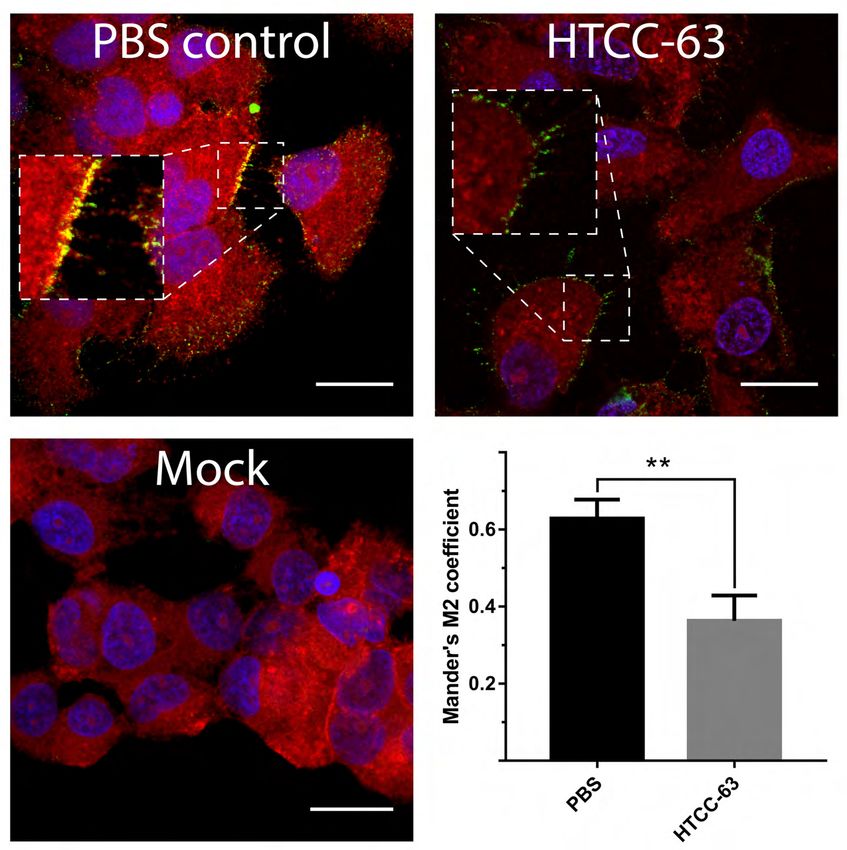
664 Figure 9. HTCC blocks the interaction between the virus and its entry receptor. Pre-
665 cooled Huh7 cells were incubated for 3 h at 4°C with ice-cold MERS-CoV or mock in the
666 presence or absence of HTCC-63 (100 μg/ml). Next, cells were fixed with PFA and667 immunostained for MERS-CoV-N (green), DPP4 (red), and nuclear DNA (blue). MERS-CoV
668 interaction with the DPP4 protein was analyzed with confocal microscopy. Co-localisation of
669 DPP4 with MERS-CoV-N was determined by confocal microscopy and presented as
670 Manders’ M2 coefficient after excluding nonspecific nuclear signal. Colocalization analysis
671 was carried out with ImageJ JACoP (Just Another Colocalization Plugin) plugin. The
decrease in colocalization was statistically significant (P < 0.0005). Each image represents a
Downloaded from http://jvi.asm.org/ on December 29, 2020 by guest
672
673 single axial plane. Representative images are shown. Bar 20 µm.
674
675676 Tables
677
678 Table 1. 50% Cytotoxic concentration (CC50), 50% inhibitory concentration (IC50), and the
679 selectivity index (SI) of two most effective HTCCs: HTCC-63 (for MERS-CoV) and HTCC-
680 77 (for SARS-CoV-2).
Downloaded from http://jvi.asm.org/ on December 29, 2020 by guest
CC50 [µg/ml] IC50 [µg/ml] SI [CC50/IC50]
MERS-CoV 161.0 62.8 2.6
SARS-CoV-2 158.0 12.5 12.6
681
682
683 Table 2. Primers and probes.
MERS-CoV SARS-CoV-2
5’ primer GGG TGT ACC TCT TAA TGC CAA CAC ATT GGC ACC CGC AAT C
TTC
3’ primer TCT GTC CTG TCT CCG CCA AT GAG GAA CGA GAA GAG GCT TG
probe ACC CCT GCG CAA AAT GCT GGG ACT TCC TCA AGG AAC AAC ATT GCC A
(FAM / TAMRA) (FAM / BHQ1)
684Downloaded from http://jvi.asm.org/ on December 29, 2020 by guest
Downloaded from http://jvi.asm.org/ on December 29, 2020 by guest
Downloaded from http://jvi.asm.org/ on December 29, 2020 by guest
Downloaded from http://jvi.asm.org/ on December 29, 2020 by guest
Downloaded from http://jvi.asm.org/ on December 29, 2020 by guest
Downloaded from http://jvi.asm.org/ on December 29, 2020 by guest
Downloaded from http://jvi.asm.org/ on December 29, 2020 by guest
Downloaded from http://jvi.asm.org/ on December 29, 2020 by guest
Downloaded from http://jvi.asm.org/ on December 29, 2020 by guest
You can also read