Iron-Regulated Expression of Alginate Production, Mucoid Phenotype, and Biofilm Formation by Pseudomonas aeruginosa
←
→
Page content transcription
If your browser does not render page correctly, please read the page content below
Downloaded from mbio.asm.org on March 20, 2015 - Published by mbio.asm.org
RESEARCH ARTICLE
Iron-Regulated Expression of Alginate Production, Mucoid Phenotype,
and Biofilm Formation by Pseudomonas aeruginosa
Jacinta R. Wiens,a,b Adriana I. Vasil,b Michael J. Schurr,b Michael L. Vasila,b
Program in Molecular Biology, University of Colorado School of Medicine, Aurora, Colorado, USAa; Department of Microbiology, University of Colorado School of
Medicine, Aurora, Colorado, USAb
ABSTRACT Pseudomonas aeruginosa strains of non-cystic fibrosis (non-CF) origin do not produce significant amounts of extra-
cellular alginate and are nonmucoid. In CF, such isolates can become mucoid through mutation of one of the genes (mucA,
mucB, mucC, or mucD) that produce regulatory factors that sequester AlgU, required for increased expression of alginate genes.
Mutation of the muc genes in the nonmucoid PAO1, PA14, PAKS-1, and Ps388 strains led to increased levels of extracellular algi-
nate and an obvious mucoid phenotype, but only under iron-limiting growth conditions (10 M). In contrast, >50% of P. aeruginosa isolates from chronic CF pulmonary infections expressed increased levels
of alginate and mucoidy both under iron-limiting and iron-replete conditions (i.e., iron-constitutive phenotype). No single iron
regulatory factor (e.g., Fur, PvdS) was associated with this loss of iron-regulated alginate expression and mucoidy in these CF
isolates. However, the loss of only pyoverdine production, or its uptake, abrogated the ability of P. aeruginosa to produce a ro-
bust biofilm that represents the Psl-type of biofilm. In contrast, we show that mutation of the pyoverdine and pyochelin biosyn-
thesis genes and the pyoverdine receptor (FpvA) lead to iron-constitutive expression of the key alginate biosynthesis gene, algD,
and an explicitly mucoid phenotype in both iron-limiting and iron-replete conditions. These data indicate that alginate produc-
tion and mucoidy, in contrast to other types of biofilms produced by P. aeruginosa, are substantially enhanced under iron limi-
tation. These results also have compelling implications in relation to the use of iron chelators in the treatment of P. aeruginosa
CF infections.
IMPORTANCE Pseudomonas aeruginosa is a leading model for the investigation of biofilms. While data have been generated about
the role of iron in alginate-independent (Psl/Pel) biofilm development, there is a paucity of data regarding the role of iron in al-
ginate production and its associated mucoid phenotype. We demonstrate that biologically relevant levels of iron that exist in the
airway mucus of cystic fibrosis (CF) patients have a substantial influence on production of alginate and the overt mucoid pheno-
type, pathognomonic of P. aeruginosa infections in CF. Mucoid mutants of non-CF P. aeruginosa isolates are mucoid only un-
der iron limitation and do not express increased levels of alginate under iron-replete growth conditions. However, a significant
number of long-term CF isolates lost their iron-regulated expression of increased alginate production and mucoidy and became
iron constitutive for these properties. In contrast to the formation of Psl-type biofilms, increasing iron limitation ultimately
leads to an iron-constitutive expression of alginate and mucoidy.
Received 26 November 2013 Accepted 19 December 2013 Published 4 February 2014
Citation Wiens JR, Vasil A, Schurr MJ, Vasil ML. 2014. Iron-regulated expression of alginate production, mucoid phenotype, and biofilm formation by Pseudomonas aeruginosa.
mBio 5(1):e01010-13. doi:10.1128/mBio.01010-13.
Editor Gerald Pier, Harvard Medical School
Copyright © 2014 Wiens et al. This is an open-access article distributed under the terms of the Creative Commons Attribution-Noncommercial-ShareAlike 3.0 Unported
license, which permits unrestricted noncommercial use, distribution, and reproduction in any medium, provided the original author and source are credited.
Address correspondence to Michael L. Vasil, mike.vasil@ucdenver.edu.
T hroughout the past decade, the ability of microbes to form
complex communities called biofilms has become an exceed-
ingly intense and worthy area of investigation in environmental
lates recovered from the lungs of chronically infected CF patients,
but rarely from other types of infections (4–6). The Psl (polysac-
charide synthesis locus) exopolysaccharide and its associated bio-
and medical microbiology. Pseudomonas aeruginosa has become a film were uncovered by examining P. aeruginosa ⌬algD mutants
leading paradigm based on its proclivity to form biofilms in di- that were unable to produce alginate yet were still capable of form-
verse environments (e.g., pipelines, heart valves, bone), yet also in ing biofilms on glass or plastic surfaces (6, 7). The third exopoly-
highly specialized situations, such as the bronchioles of cystic fi- saccharide, Pel (named Pel for pellicle) was associated with
brosis (CF) patients (1–3). Exceptional research efforts during this P. aeruginosa biofilms at an air-liquid interface (8, 9). While it is
time have revealed that P. aeruginosa is able to produce as many as not yet entirely clear which aspects of P. aeruginosa infection (e.g.,
three distinct exopolysaccharides, each of which is associated with colonization, survival) the Psl- and Pel-associated biofilms might
specific types of biofilms and conditions under which they are contribute, particularly in CF patients, the long-term presence of
formed. Overexpression of the alginate exopolysaccharide was highly mucoid strains, in the airways of CF patients, continues to
first identified as being associated with P. aeruginosa mucoid iso- provide a compelling rationale for further investigation into the
January/February 2014 Volume 5 Issue 1 e01010-13 ®
mbio.asm.org 1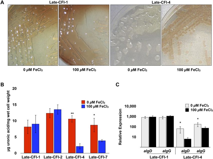
Downloaded from mbio.asm.org on March 20, 2015 - Published by mbio.asm.org
Wiens et al.
environmental conditions and mechanisms that govern the pro- P. aeruginosa infections, especially in the context of the CF pul-
duction of alginate and the ensuing mucoid phenotype (1, 4, 10). monary airways.
There is substantial knowledge relating to the biochemistry
and molecular genetics of alginate biosynthesis, as well as the mu- RESULTS
coid phenotype of P. aeruginosa. Additionally, during the past two Iron influences alginate production and the mucoid phenotype
decades, considerable insight has been garnered relating to how of P. aeruginosa muc mutants. Human lungs, including those of
iron influences the expression of an array of P. aeruginosa viru- CF patients, have historically been considered iron-limited envi-
lence factors (exotoxins, proteases, and siderophores), basic met- ronments. However, recent comprehensive studies indicate that
abolic processes (quorum sensing, intermediary metabolism, and there can be diverse microenvironments in the lung and that iron
resistance to redox stress) and even how this particular metal af- levels may show considerable variation and are not necessarily as
fects the formation of some types of P. aeruginosa biofilms (Psl and limited by iron as previously thought. Reid et al. suggested that
Pel) (11–17). In 2005, Banin et al. presented a comprehensive Fe3⫹ ions (13 to 134 M) and ferritin (15 to 300 g/liter) are
examination of the molecular and biochemical processes by which major sources of iron in the lungs of CF patients and that patients
iron can affect biofilm formation by P. aeruginosa (11). However, with chronic obstructive pulmonary disease (COPD) commonly
the parental strain (PAO1) used in that study did not carry any have much higher levels of elemental iron and ferritin (21–24).
muc mutations, and it did not produce significant levels of algi- Additionally, recent findings show that Fe2⫹ ions are present in
nate. Moreover, the biofilms were grown on glass surfaces using a sputum samples from CF patients (25). In order to directly inves-
flowthrough system, thereby most likely representing a Psl type of tigate the impact of iron on alginate biosynthesis, we constructed
biofilm. Nevertheless, it was demonstrated that strains with mu- ⌬mucA, ⌬mucB, ⌬mucC, or ⌬mucD isogenic mutants of P. aerugi-
tations in pyoverdine biosynthesis (⌬pvdS and ⌬pvdA) and the nosa PAO1 or mucA and mucB insertion mutants of P. aeruginosa
cognate receptor (⌬fpvA) were all unable to form robust biofilms. PA14, PAKS-1, and Ps388 and examined their production of ex-
In contrast, a strain carrying a missense mutation (Ala10Gly) in tracellular alginate and mucoid phenotypes in the presence of
the ferric uptake regulator (Fur) that constitutively expresses Fur- variable iron levels (ⱕ5 M to 100 M). These strains are all
repressed iron acquisition systems retained the ability to form nonmucoid, non-CF-related isolates originating from widely dif-
structured biofilms both in the presence and absence of lactofer- fering types of infections and geographic regions. PAO1 is a
wound isolate from Melbourne, Australia. PA14 is a burn isolate
rin, an iron-binding protein that otherwise inhibits nonalginate
from Boston, MA. PAKS-1 is a urine isolate from Stockholm, Swe-
biofilm formation by sequestering available iron. Taken together,
den. Ps388 is a blood isolate from Seattle, WA, USA. The strains
sufficient levels of iron and intact P. aeruginosa iron acquisition
were grown on various concentrations of iron reported to be pres-
systems (pyoverdine, pyochelin, and ferric dicitrate) were re-
ent in the sputa of CF patients (⬍5 M to ⬎100 M) and were
quired for the formation of structured nonalginate biofilms.
evaluated for alginate production by measurement of extracellular
The impact of iron and iron acquisition systems on alginate
uronic acid levels (see Materials and Methods). Unlike all of their
production has not been nearly as extensively examined. Boyce
parental strains, these strains carry single gene deletions in mucA,
and Miller (18) presented the earliest investigation of the potential
mucB, mucC, or mucD (PAO1) or insertion mutations in mucA or
influence of iron on the mucoid phenotype. In cultures of mucoid mucB (PA14, PAKS-1, or Ps388), are phenotypically mucoid, and
CF isolates grown in the presence of iron, nonmucoid revertants produce substantially increased amounts of extracellular alginate
accumulated to ⬎80% of the population after 72 h (18). In a when grown under iron limitation (ⱕ5 M) than when grown
subsequent study, those observations were extended to demon- under more iron-replete conditions (ⱖ10 M) (Fig. 1A and B and
strate that the mucoid phenotype of a single CF isolate was more 2). No alginate was detected from any of the parental strains
stable under iron-limited growth than under iron-replete growth grown under either iron-limited or iron-replete conditions (data
(19). Subsequently, Terry et al. reported that ~2% of colonies not shown).
isolated from strain PAO1 became mucoid after growing under Moreover, when a mucB insertion mutation was introduced
iron limitation in a chemostat for at least 6 days (20). Since 1992, into a nonmucoid CF isolate, it clearly displayed a mucoid pheno-
there have been no additional published reports that directly ad- type and regulated its alginate production in response to iron, as
dress how iron levels might differentially and specifically affect did the non-CF isolates (PAO1, PA14, PAKS-1, and Ps388) (see
alginate production or its associated mucoid phenotype. Table S2 in the supplemental material).
Herein, we examined whether iron has a regulatory effect on Iron is present in various forms in the CF lungs; therefore, we
alginate production and mucoidy in non-CF and CF isolates. Our wanted to know whether iron sources other than iron salts (FeCl3)
data illustrate that iron profoundly influences the production of would result in the repression of alginate production. P. aerugi-
alginate and the expression of the mucoid phenotype in strains nosa PAO1 and ⌬mucA and ⌬mucB mutants were cultured in the
with newly acquired muc mutations and that this regulation may presence of alternative iron sources, such as ferric dicitrate or he-
be lost after prolonged survival in the CF airways. Further exam- min. Significant decreases in extracellular alginate levels in these
ination of the molecular mechanisms associated with the observed mutants were observed compared to mutants grown without the
iron-regulated alginate production revealed that strains with mu- addition of these iron sources (Fig. 3). It should also be noted that
tations in iron acquisition systems lost the ability to regulate algi- corresponding increases in the concentrations of other metals
nate production in response to iron. Taken together, this report (Zn2⫹, Cu2⫹, Ca2⫹, and Mg2⫹) that are similar to the concentra-
describes the first in-depth assessment of the influence of iron on tions used for iron (ⱕ5 M to 300 M) failed to cause parallel
the expression of alginate, an important virulence factor. It also decreases in alginate levels in any of the muc mutants examined
raises significant questions about the design and administration of (see Fig. S1 in the supplemental material; also data not shown).
therapeutics targeted at iron homeostasis to treat chronic The data described above indicated that iron exhibits a selec-
2 ®
mbio.asm.org January/February 2014 Volume 5 Issue 1 e01010-13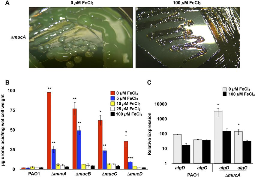
Downloaded from mbio.asm.org on March 20, 2015 - Published by mbio.asm.org
Iron Repression of Alginate Production
FIG 1 Effect of iron on alginate production by muc mutants of P. aeruginosa PAO1. (A) P. aeruginosa PAO1 ⌬mucA mutant was grown on DTSA with or without
100 M FeCl3 and imaged after 48 h. (B) The PAO1 strain and ⌬mucA, ⌬mucB, ⌬mucC, and ⌬mucD mutants were grown on DTSA containing 5, 10, 25, or
100 M FeCl3 for 48 h, and the levels of extracellular alginate were evaluated using a uronic acid assay (see Materials and Methods). Values that are significantly
different from the value for PAO1 (wild type [WT]) by Student’s t test are indicated by asterisks as follows: ***, P ⬍ 0.0005; **, P ⬍ 0.005; *, P ⬍ 0.05. (C) Relative
expression of algD and algG in wild-type PAO1 and PAO1 ⌬mucA mutant was analyzed using qRT-PCR after 24 h of growth on DTSA with and without 100 M
FeCl3. Values that are significantly different from the value for PAO1 (WT) by Student’s t test are indicated by an asterisk.
tive regulatory effect on alginate biosynthesis. The regulation, bio- biosynthesis. In addition, various proteases (e.g., AlgW and
synthesis, and export of the alginate exopolysaccharide are com- MucP) function in posttranscriptional regulation by liberating
plex processes involving numerous adjacently located structural AlgU/T from the MucA/B/C/D sequestration complex at the
genes (PA3540 to PA3551) and an assortment of genes encoding membrane, thereby leading to the constitutive transcription of
regulatory factors (e.g., algR, algC, algB, and kinB) scattered algD and the alginate biosynthetic operon (29). Quantitative re-
throughout the P. aeruginosa genome (26–29). The biosynthesis verse transcription-PCR (qRT-PCR) was used to validate the ex-
and export of this exopolysaccharide are initiated at the algD pression of algD and algG (encoding alginate-c5-mannuronan-
(PA3540) promoter, which encodes the first enzyme (GDP- epimerase in the alginate biosynthesis operon) in the PAO1
mannose 6-dehydrogenase) in the pathway leading to alginate ⌬mucA mutant grown under iron limitation and iron-replete
FIG 2 Effects of iron levels on alginate production in ⌬mucA and ⌬mucB FIG 3 Influences of various iron sources on alginate production by P. aerugi-
mutants of P. aeruginosa PA14, PAKS-1, and Ps388. Alginate production was nosa PAO1 and ⌬mucA and ⌬mucB mutants. The strains were grown on DTSA
evaluated by measurement of extracellular uronic acid levels produced by supplemented with various sources and concentrations of iron-carrying com-
PA14, PAKS-1, and Ps388 with insertion mutations in either mucA or mucB pounds. Extracellular alginate production was measured by uronic acid assay
after 48 h of growth on DTSA with and without 100 M FeCl3. Values that are after 48 h of growth. Values that are significantly different from the value for
significantly different from the value found with 100 M FeCl3 by Student’s PAO1 (WT) by Student’s t test are indicated by asterisks as follows: ***, P ⬍
t test are indicated by asterisks as follows: **, P ⬍ 0.005; *, P ⬍ 0.05. 0.0005; **, P ⬍ 0.005.
January/February 2014 Volume 5 Issue 1 e01010-13 ®
mbio.asm.org 3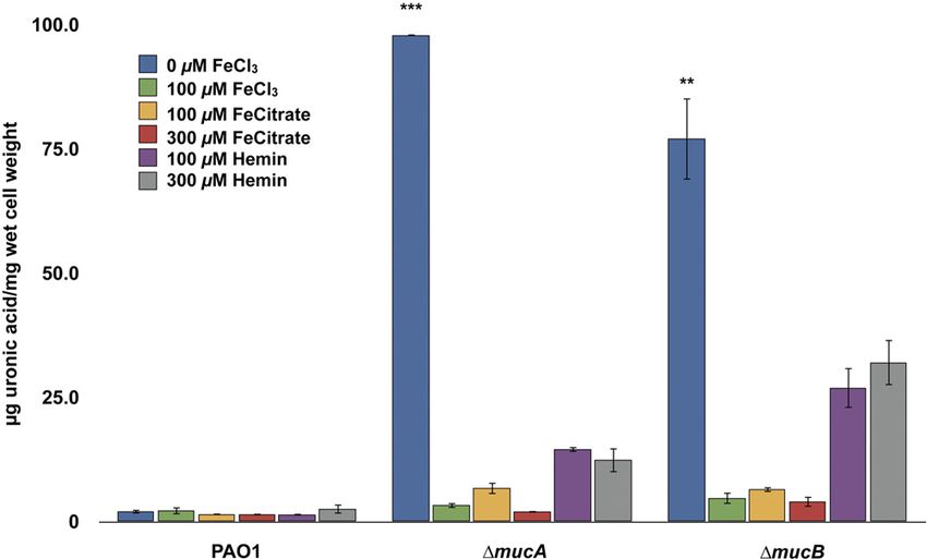
Downloaded from mbio.asm.org on March 20, 2015 - Published by mbio.asm.org
Wiens et al.
TABLE 1 EPS carbohydrate composition profiles for P. aeruginosa PAO1 and mutants
Carbohydrate composition (g/107 CFU ml⫺1) for the following strains grown under the indicated condition:
PAO1 ⌬mucA mutant ⌬mucB mutant
Carbohydrate 0 M FeCl3 100 M FeCl3 0 M FeCl3 100 M FeCl3 0 M FeCl3 100 M FeCl3
Mannuronic acid 0 0 88.2 12.1 176 33.7
Glucose 45.8 25.6 18.6 13.6 34.56 1.4
Rhamnose 9.0 9.5 6.6 4.0 8.96 0.8
Mannose 0.7 0.4 1.8 0.5 3.84 0.4
Ribose 0.5 1.6 1.8 1.4 2.56 0.6
Xylose 0.5 0.1 1.2 0.2 3.84 0.4
KDO 1.0 3.6 0 0.7 0 0
N-Acetylglucosamine 1.6 1.6 0.6 0.9 0.64 0.1
conditions. Triplicate cultures of P. aeruginosa wild type PAO1 composition of Pel biofilms is mainly glucose, rhamnose, and
and the ⌬mucA mutant were grown on DTSA solid medium with mannose, whereas the extracellular polysaccharide composition
and without 100 M FeCl3, and cells were harvested after 24 h. of Psl biofilms is mainly galactose and mannose (6, 30). These
Relative expression analysis by qRT-PCR showed that the tran- results are consistent with previous findings that mannuronic acid
script levels of both algD and algG in the ⌬mucA mutant were is the predominant extracellular polysaccharide of alginate bio-
significantly higher than the levels in the wild-type PAO1 strain films and mucoidy and confirmed the results of uronic acid anal-
under iron limitation (Fig. 1C). As predicted from the uronic acid ysis (6).
analysis shown in Fig. 1A and B, the expression of both algD and Global transcriptional analysis of wild-type PAO1 and PAO1
algG transcripts in the ⌬mucA mutant decreased to the same levels ⌬mucA mutant under iron limitation. While there have been a
as those for the wild type under iron-replete conditions (Fig. 1C). considerable number of transcriptional studies of P. aeruginosa
These data showed that iron levels of ⱖ10 M result in a substan- grown under different levels of iron, there have not been any com-
tial reduction in the transcription of the alginate biosynthetic parable assessments of transcriptional responses to variable levels
operon corresponding to the decrease in the amount of alginate of this biologically consequential metal for isogenic mucoid vari-
measured by the uronic acid analysis described above. ants and their nonmucoid parents (14, 15). Based on the observa-
Taken together, these data provide the first evidence that bio- tions described above, it was deemed worthwhile to examine the
logically significant levels of iron can have substantial effects on global transcriptional responses of P. aeruginosa PAO1 (wild type)
the ability of newly derived muc mutants from a very diverse group compared to those of a ⌬mucA mutant under iron-limited and
of P. aeruginosa strains to express high levels of extracellular algi- iron-replete conditions using GeneChip microarray analysis (see
nate and manifest mucoid phenotypes. Materials and Methods for growth conditions used for these mi-
Chemical analysis of P. aeruginosa ⌬mucA and ⌬mucB ex-
croarray experiments). Table S1 in the supplemental material pro-
tracellular polymeric substance (EPS). The data described above
vides a complete list of the genes and operons of PAO1, the wild-
indicate that iron strongly influences the expression of the alginate
type parent, and its isogenic ⌬mucA mutant, which exhibited the
exopolysaccharide. To ensure that the increased levels of uronic
most robust responses in iron-limited growth compared to iron-
acids measured in our uronic acid analysis under iron-limiting
replete growth. As with most microarray-based approaches, a sur-
conditions accurately mirror those of alginate [(1– 4)-linked -D-
feit of data was generated. Nevertheless, there are several interest-
mannuronate and ␣-L-guluronate polymer], extracellular poly-
saccharides extracted from P. aeruginosa PAO1 ⌬mucA and ing outcomes based on these experiments that warrant specific
⌬mucB mutants were evaluated by size exclusion chromatography comments.
and glycosyl composition analysis with combined gas There were a number of genes and operons with increased
chromatography-mass spectrometry (GC-MS). Because it was expression under iron limitation in the ⌬mucA mutant that had
possible that other biofilm-associated exopolysaccharides (Psl or significantly lower expression or were not expressed in the PAO1
Pel) could be produced by these strains, particularly from cells parent under iron limitation (Table 2; see Table S1 in the supple-
harvested from iron-replete cultures, exopolysaccharides were ex- mental material). Genes with increased expression in the ⌬mucA
tracted from PAO1, the wild-type parent, and ⌬mucA and ⌬mucB mutant may provide insights into the basic metabolism of mucoid
mutants grown in both iron-limited and iron-replete conditions strains and the stresses related to increased production and secre-
and evaluated (see Materials and Methods). The most abundant tion of extracellular alginate under iron limitation. The most
carbohydrate detected in the ⌬mucA and ⌬mucB mutants grown striking and obvious examples in this regard are the increased
under iron limitations was mannuronic acid (Table 1). In con- transcriptional responses of genes involved in alginate biosynthe-
trast, the carbohydrates detected from samples from wild-type sis and export (algD-algA) in the ⌬mucA mutant under iron lim-
PAO1 were glucose and rhamnose, with a small amount of itation, but not in the PAO1 parent (Table 2). These results and
3-deoxy-D-manno-octulosonate (KDO), which was not detected the fact that we did not observe corresponding increases in the
in the ⌬mucA or ⌬mucB samples. The samples from the ⌬mucA expression of genes involved in the biosynthesis of the Psl (pslA-
and ⌬mucB mutants grown under iron limitation contained sig- pslO; PA2231-PA2245) or the Pel (pelA-pelG; PA3058-PA3064)
nificantly lower levels of glucose and rhamnose than those from exopolysaccharides strongly reinforce the idea that iron limitation
the iron-limited parent, PAO1. The extracellular polysaccharide selectively influences the increased production of alginate, in con-
4 ®
mbio.asm.org January/February 2014 Volume 5 Issue 1 e01010-13Downloaded from mbio.asm.org on March 20, 2015 - Published by mbio.asm.org
Iron Repression of Alginate Production
TABLE 2 Select genes with increased gene expression under iron limitation versus iron-replete conditions in P. aeruginosa PAO1 and ⌬mucA
mutant
Change in gene expressiona
Locus tag Gene PAO1 ⌬mucA mutant Description/functionb
PA0509 nirN NC 3 Probable c-type cytochrome
PA0510 NC 3 Uroporphyrin III c-methyltransferase
PA0511 nirJ NC 4 Heme d1 biosynthesis protein
PA0512 NC 3 Conserved hypothetical protein
PA0513 NC 3 Probable transcriptional regulator
PA0514 nirL NC 4 Heme d1 biosynthesis protein
PA0515 NC 4 Probable transcriptional regulator
PA0516 nirF NC 4 Heme d1 biosynthesis protein
PA0517 nirC NC 5 Probable c-type cytochrome precursor
PA0518 nirM NC 6 Cytochrome c-551 precursor
PA0519 nirS NC 7 Nitrite reductase precursor
PA0520 nirQ NC 3 Regulatory protein; central metabolism
PA0523 norC NC 11 Nitric oxide reductase subunit C
PA0524 norB 2 11 Nitric oxide reductase subunit B
PA0525 norD 4 7 Probable dinitrification protein
PA2147 katE NC 13 Heme-binding catalase
PA2185 katN 4 2 Mn-containing catalase
PA3540 algD 2 23 GDP-mannose 6-dehydrogenase
PA3541 alg8 NC 7 Alginate biosynthesis
PA3542 alg44 NC 4 Alginate biosynthesis
PA3543 algK NC 60 Scaffold protein
PA3544 algE NC 6 Outer membrane protein
PA3545 algG NC 11 Alginate-C5-mannuronan-epimerase
PA3546 algX NC 4 Alginate biosynthesis
PA3547 algL NC 4 Alginate lyase
PA3548 algI NC 8 Acetylase
PA3549 algJ D 5 Alginate O-acetyltransferase
PA3550 algF NC 5 Alginate O-acetyltransferase
PA3551 algA 2 11 Phosphomannose isomerase
PA5097 hutT 3 6 ␥-Aminobutyrate permease
PA5098 hutH 2 14 Histidine ammonia-lyase
PA5099 NC 47 Nucleoside transporter family
PA5100 hutU 5 23 Urocanase
a Change in gene expression for P. pseudomonas PAO1 or the ⌬mucA mutant grown in 0 M FeCl3 versus 100 M FeCl3. The numbers are the fold increase in gene expression.
Abbreviations: NC, no change; D, decreased expression.
b Descriptions were obtained from the Pseudomonas Genome Database (www.pseudomonas.com).
trast to either of the other biofilm-associated extracellular poly- CF-associated infections. For this purpose, two groups of CF pul-
saccharides. monary isolates were examined. The first group, early CF isolates
One additional salient outcome from these experiments is the (Early-CFI) consisted of 8 isolates derived from individual chil-
considerably increased levels of expression of PA5097 (hutT), dren with CF who were ⬍3 years of age (yoa) at the time of sputum
PA5098 (hutH), and PA5099 (Table 2). Transcription of these collection (see Table S2 in the supplemental material). The second
genes, which comprise an operon, was significantly increased in group, late CF isolates (Late-CFI), consisted of 17 isolates origi-
the PAO1 ⌬mucA mutant under iron limitation, but to a substan- nating from older CF patients (ⱖ10 yoa) who had been colonized
tially lesser extent, or not at all in PAO1 (wild type) under iron with P. aeruginosa for at least 5 years (Table S3). While both the
limitation. Increased expression of these genes in P. aeruginosa Early-CFI and Late-CFI groups contained isolates with mucoid
PAO1, which are involved in histidine utilization, leads to de- phenotypes, there were some appreciable differences between
creased cytotoxicity and the expression of at least one of the type these groups.
III secreted cytotoxins (ExoS) (31). Taken together, such results Only 50% (4 of 8) of the Early-CFI intrinsically produced algi-
could provide insights into why a substantial number of mucoid nate along with an obvious mucoid phenotype, while most (16/17
CF isolates display diminished type III cytotoxin-mediated cyto- [94%]) of the Late-CFI produced significant levels of extracellular
toxicity (32). alginate, along with an obvious mucoid phenotype (see Table S2
Do CF pulmonary isolates regulate alginate production in and Table S3 in the supplemental material). Of the mucoid Early-
response to iron levels? Mucoidy and alginate production by CFI examined, 1 out of 4 showed constitutive expression of algi-
P. aeruginosa are commonly associated with chronic CF pulmo- nate levels and a mucoid phenotype when grown under iron-
nary infections. Therefore, it was of interest to ascertain whether limited and iron-replete conditions, while 56% (9/16) of the
variable levels of iron (ⱕ5 M to 100 M) known to exist in the mucoid Late-CFI were constitutive for alginate expression and
sputa of CF patients can influence the expression of alginate and mucoidy, even with the highest level of iron (100 M) tested
mucoidy in alginate-producing isolates obtained directly from (Fig. 4A and B and Table S2 and Table S3). Figure 4 illustrates the
January/February 2014 Volume 5 Issue 1 e01010-13 ®
mbio.asm.org 5Downloaded from mbio.asm.org on March 20, 2015 - Published by mbio.asm.org
Wiens et al.
FIG 4 Alginate production and mucoidy of late CF isolates (Late-CFI) in response to variable iron levels. (A) Late-CFI were grown on DSTA with and without
100 M FeCl3 and imaged at 48 h. (B) The level of extracellular alginate produced by various Late-CFI grown on DSTA with and without 100 M FeCl3 was
measured by uronic acid assay at 48 h. Values that are significantly different from the value for late CF isolate 4 (Late-CFI-4) or Late-CFI-7 with 100 M FeCl3
by Student’s t test are indicated by asterisks as follows: **, P ⬍ 0.005; *, P ⬍ 0.05. (C) Relative expression of algD and algG of Late-CFI-1 and Late-CFI-4 was
analyzed using qRT-PCR after 24 h of growth on DSTA with and without 100 M FeCl3. Values that are significantly different (P ⬍ 0.05) from the value for
Late-CFI-4 with 100 M FeCl3 by Student’s t test are indicated by an asterisk.
differences between a select group of constitutively producing al- S3 in the supplemental material). For example, late CF isolate 1
ginate isolates and those that regulate alginate production in re- (Late-CFI-1), Late-CFI-2, Late-CFI-12, and Late-CFI-16, which
sponse to iron levels as shown by uronic acid analysis of extracel- all exhibit iron-constitutive alginate production produce very low
lular alginate and qRT-PCR analysis of algD and algG. levels of siderophores (pyoverdine and pyochelin) on both iron-
These results demonstrate the robust variations that occur in limited and iron-replete CAS agar plates (Table S3). However, no
the expression of a highly relevant virulence determinant (alginate compelling correlation could be discerned between this and the
production and mucoidy) as the consequence of the fluctuating loss of iron-regulated alginate production. There are a variety of
iron levels that are known to occur during P. aeruginosa CF pul- ways that a constitutive mucoid phenotype, most often associated
monary infections. Although the number of CF isolates examined with Late-CFI, can occur (see Discussion below).
in this study is relatively small, taken together, our results do sup- In the Banin et al. study, the impact of iron and iron limitation
port the idea that over time (years to decades), an initially vigorous on the formation of Psl/Pel-type biofilms was examined in an
regulatory influence of iron on the expression of alginate and mu- assortment of mutants altered in their ability to (i) express key
coidy can be lost, resulting in alginate production that is indiffer- iron-related regulatory factors (Fur and PvdS), (ii) produce en-
ent to the variable levels of iron known to exist in the lungs of CF zymes involved in siderophore biosynthesis (PvdA), or (iii) ex-
patients. press receptors for specific siderophores (FpvA) (11). Based on
How does iron initially control the increased production of those data, it was clear that interfering with such iron regulatory or
extracellular alginate and mucoidy and how can this regulation iron acquisition systems resulted in a significantly diminished
be lost? Based on the high percentage of Late-CFI that had lost ability of wild-type PAO1 to produce robust Psl-type biofilms
iron-regulated alginate production, it was of interest to examine under the conditions used in that study.
whether these Late-CFI were capable of acquiring iron. Chrome In the present study, a similar approach was used. However,
Azurol S (CAS) agar plate assay is a technique used to study sid- mucoid strains were created by introducing either mucA or mucB
erophore production in bacteria (12). Using this technique, re- mutations into a variety of single, double, and triple mutants of
duced siderophore production was observed in several CFI that the PAO1 strain carrying deletions in genes encoding (i) an iron
exhibit constitutive alginate production (see Table S2 and Table regulatory factor (pvdS), (ii) assorted pyoverdine (pvdA and
6 ®
mbio.asm.org January/February 2014 Volume 5 Issue 1 e01010-13Downloaded from mbio.asm.org on March 20, 2015 - Published by mbio.asm.org
Iron Repression of Alginate Production
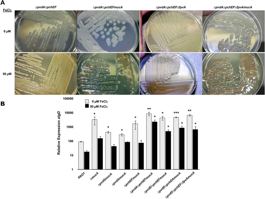
FIG 5 Investigation of alginate production in iron acquisition mutants of P. aeruginosa PAO1. (A) P. aeruginosa PAO1 and several isogenic iron acquisition
mutants were grown on DTSA with and without 50 M FeCl3 and imaged after 48 h. (B) Relative expression of algD in PAO1 (wild type) and several isogenic iron
acquisition mutants were analyzed using qRT-PCR after 24 h of growth on DTSA with and without 50 M FeCl3. Values that are significantly different from the
value for PAO1 (WT) by Student’s t test are indicated by asterisks as follows: ***, P ⬍ 0.0005; **, P ⬍ 0.005; *, P ⬍ 0.05.
pvdD) and pyochelin (pchEF and pchDA) siderophore biosynthe- regulate alginate production in response to iron and because mu-
sis enzymes, or (iii) the pyoverdine receptor (fpvA). The single, tations in both siderophore iron acquisition systems resulted in
double, and triple iron acquisition mutants of PAO1, which also constitutive alginate expression regardless of iron levels, it is pos-
carry mucA or mucB insertion mutations, were grown on dialyzed sible that either mutations in Fur or a change in the expression of
tryptic soy agar (DTSA) with or without 50 M FeCl3 for 24 h or Fur contributes to constitutive alginate expression in some of the
48 h and then examined for the mucoid phenotype and the relative Late-CFI. Fur is the master regulator of iron homeostasis, and a
expression of algD by qRT-PCR (Fig. 5A and B). None of the mutation in Fur would result in the derepression of a large num-
single, double, or even triple iron acquisition mutations caused ber of regulatory and structural genes, including those that might
significant decreases in the expression of the algD transcript under be involved in alginate production (33). While Fur is essential in
iron limitation (Fig. 5A and B). Notably, however, the following P. aeruginosa, strains with point mutations that partially affect Fur
mutants, ⌬pvdA ⌬pchEF mucA mutant, ⌬pvdD ⌬pchEF mucA function or reduce Fur levels show a constitutive phenotype with
mutant, ⌬pvdD ⌬pchDA mucA mutant, and ⌬pvdD ⌬pchEF regard to the expression of a plethora of iron-regulated factors,
⌬fpvA mucA mutant produced alginate and were mucoid under including two siderophores (pyoverdine and pyochelin) and exo-
iron-replete conditions at the same levels observed under iron toxin A (12). It is possible that CF isolates, which exhibit a consti-
limitation (Fig. 5A and data not shown). Further, the relative ex- tutive phenotype in terms of an increased expression of alginate
pression of algD remained unchanged in these mutants regardless and mucoidy under both iron-limited and iron-replete condi-
of the iron concentration (Fig. 5B). Interestingly, in stark contrast tions, could have acquired point mutations that either affect Fur
to previous data (22) regarding the requirement of PvdS for the function or reduce its levels (12). The involvement of Fur was
formation of structured Psl/Pel-type biofilms, PvdS was not re- examined in two ways. First, the sequence of the fur gene, includ-
quired for the production of alginate and was not involved in ing its regulatory region, was examined in both Early-CFI and
iron-regulated alginate production (Fig. 5B). Taken together, the Late-CFI, including those showing an iron-regulated phenotype
present data suggest that strains with mutations in multiple iron and an iron-constitutive phenotype with regard to the production
acquisition systems (pyoverdine and pyochelin), which likely re- of alginate. No base changes in the structural and regulatory re-
sult in the reduced acquisition of iron, lose the ability to regulate gion of fur were detected in any of these isolates (data not shown).
alginate in response to iron. Second, the levels of Fur protein and fur transcript, as detected by
Due to the decreased ability of the majority of Late-CFI to Western blotting and qRT-PCR were examined in CFI that exhibit
January/February 2014 Volume 5 Issue 1 e01010-13 ®
mbio.asm.org 7Downloaded from mbio.asm.org on March 20, 2015 - Published by mbio.asm.org
Wiens et al.
FIG 6 Investigation of the role of Fur in alginate production. (A) P. aeruginosa PAO1, ⌬mucA mutant, Early-CFI-1, Late-CFI-1, and Late-CFI-4 were grown on
DTSA with and without 100 M FeCl3. The relative expression of fur was measured by qRT-PCR after 24 h of growth. (B) Strain PAO1, ⌬mucA mutant,
Early-CFI-1, Late-CFI-1, Late-CFI-2, and Late-CFI-4 were grown on DTSA with and without 100 M FeCl3. Fur protein levels (shown as relative intensities
within parentheses below the blot) were analyzed by Western blotting and quantified as described in Materials and Methods.
both an iron-regulated phenotype and an iron-constitutive phe- lead to rapidly fulminating infections (5, 21, 22, 24, 35). During an
notype with regard to alginate production (Fig. 6A and B). There infectious disease, the level of free iron can vary from one that is
were no differences in the levels of Fur protein or fur transcript extremely restrictive to one where free iron or a readily available
produced under iron-limited or iron-replete conditions that source of iron may become more accessible as a consequence of
could be associated with either an iron-regulated or iron- infection-related pathology (hemorrhage, inflammation, proteol-
constitutive phenotype in terms of increased alginate production ysis of host proteins) (21, 22, 24, 36). For instance, the levels of free
or mucoidy. Fur in P. aeruginosa is not autoregulated. Conse- iron in the airway mucus of CF patients during acute exacerba-
quently, we did not expect to find differences between iron- tions are known to fluctuate between very low, growth-limiting
limiting and iron-replete growth conditions in the amount of pro- levels (⬍1 M) to more than 100 times these concentrations
tein or transcript if mutations were not present. However, while (⬎100 M) (22–24). Notably, however, such low levels of free
we did not find Fur mutations or expression deviations, the indi- iron are those that are typically responsible for an increase in the in
rect involvement of Fur cannot be ruled out. It is still possible that vitro expression of iron-regulated genes encoding virulence deter-
Fur represses an activator of alginate biosynthesis or indirectly
minants (e.g., exotoxins, proteases, and siderophores), while
controls a repressor involved in the iron-regulated alginate phe-
higher levels most often suppress their expression, favor increased
notype.
growth rates, and in certain cases, favor the eventual formation of
DISCUSSION particular types of biofilms (11, 15, 33, 37).
Iron, probably more so than any other metal, has been shown to As previously discussed, Banin et al. demonstrated that the
have profound effects on the expression of bacterial virulence de- robust formation of a wild-type P. aeruginosa nonalginate biofilm
terminants, host-pathogen interactions, and the ensuing out- grown on glass slides requires either the biosynthesis of pyover-
comes of bacterial infectious diseases. While the levels of free iron dine or the expression of its cognate receptor (FpvA) (11). The
in mammalian hosts are ordinarily exceedingly limited, most pro- diminution of biofilm formation by these mutants could be over-
karyotes have at their disposal multiple strategies (siderophores, come by providing alternative sources of iron (1 M ferric dici-
heme uptake, utilization of host iron-binding proteins) that can trate or 1.5 M desferrioxamine). Furthermore, in those studies,
overcome the paucity of free iron they might encounter upon nonalginate biofilm formation was significantly inhibited by ex-
colonization of a eukaryotic host (34). Yet, an abnormal increase posing the bacteria to the iron-binding protein lactoferrin (20 g/
in the levels of free iron in sera or tissues of mammalian hosts can ml). Overall, the results from that study provided cogent support
8 ®
mbio.asm.org January/February 2014 Volume 5 Issue 1 e01010-13Downloaded from mbio.asm.org on March 20, 2015 - Published by mbio.asm.org
Iron Repression of Alginate Production
for the view that the normal ability to form a robust nonalginate (Late-CFI-3, -4, -5, -11, and -13 [Table S3]) exhibited decreased
biofilm by P. aeruginosa can be affected merely by disruption of siderophore production in the CAS assay. Perhaps, as suggested in
only one of its iron acquisition systems (pyoverdine biosynthesis the review by Poole and McKay entitled “Iron acquisition and its
or uptake) or by exposing it to an exogenous competitor for iron control in Pseudomonas aeruginosa: many roads lead to Rome”
(lactoferrin). The bottom line is that it is probably the ability of (39) or in a more recent report by Konings et al. (25), there are
P. aeruginosa to acquire sufficient levels of cytoplasmic iron that is multiple ways for this pathogen to strategically acquire iron. At
crucial to the formation of robust nonalginate biofilms. It should this point, it has not yet been discerned whether alginate expres-
be noted, however, that in the Banin et al. studies, the actual pro- sion is controlled by specific iron-responsive regulatory factors or
duction of a biofilm-associated exopolysaccharide (Psl or Pel) was whether generally low cytoplasmic iron levels generate further
never directly examined. However, it could be argued that that the stresses leading to increased alginate expression, even in an iron-
striking mucoid presentation on a solid agar surface as shown in replete environment. Nevertheless, we demonstrated that none of
the figures from the present study (Fig. 1, 4, and 5) could at least in the examined iron-constitutive alginate-producing CFI carried
some manner, be akin to alginate-related biofilms as they exist in mutations that could alter the function or level of Fur, the master
mucus-plugged CF airways. regulator of iron homeostasis. Likewise, a possible role for the
In the present study, we investigated whether biologically rel- Fur-regulated sigma factor PvdS was eliminated. These observa-
evant levels of iron (ⱕ5 M to ⱖ100 M) have an influence on tions, however, do not as of yet exclude the possibility that muta-
the production of alginate and the mucoid phenotype by P. aerugi- tion of an unidentified Fur-regulated gene that controls the ex-
nosa. Since wild-type P. aeruginosa does not normally produce pression of one or more alginate biosynthesis genes (e.g., algD and
alginate, we constructed muc mutants from a variety of non-CF algR) occurs. A mutation in a Fur-regulated gene could in some
strains from diverse geographic regions and determined whether cases be the cause of the iron-constitutive mucoid phenotype seen
these levels of iron in any way affected alginate production and the in the majority of the Late-CFI. Along these lines, it is of interest
ensuing mucoid phenotype. Similarly, a nonmucoid isolate from that recently two genes of Pseudomonas syringae associated with
an early CF patient (Early-CFI) was converted to the mucoid phe- alginate biosynthesis (algJ and algF) were identified as being Fur
notype by introducing an insertion mutation into mucB. All of regulated (40). In P. aeruginosa, AlgJ and AlgF seem to be involved
these newly constructed muc mutants produced increased levels of in the acetylation of alginate (41), rather than in its biosynthesis.
alginate under iron-limiting (ⱕ5 M) growth and significantly These genes should be further examined. While unlikely, it is also
reduced levels of alginate under iron-replete (ⱖ10 M) growth. possible that some of the Late-CFI in this study exhibit an iron-
In contrast, in a group of CF isolates that intrinsically produce constitutive phenotype because they have acquired mutations in
alginate by acquisition of mutations within the CF lung, a signif- other alginate regulatory genes (e.g., KinB and type IV pili) instead
icant proportion (56%) had lost iron-regulated control over algi- of the commonly acquired muc mutations most often seen in CF
nate production and are mucoid irrespective of the levels of iron to isolates (42, 43).
which they were exposed. Finally, iron homeostasis, particularly in the context of biofilm
These observations posed additional salient questions about formation, has become an attractive target for therapeutic inter-
iron, the expression of alginate, and the overtly mucoid pheno- ventions in non-CF and CF P. aeruginosa infection models (44–
type. (i) How does iron initially exhibit its tight control over algi- 46). In some cases (i.e., gallium nitrate), they are the focus of
nate production? (ii) How can this regulatory influence be nulli- current ongoing clinical trials (see study NCT01093521 at Clini-
fied during the course of chronic pulmonary disease? To address calTrials.gov). Gallium (Ga3⫹) is a FDA-approved ferric (Fe3⫹)
these questions, we considered whether alteration of one of more iron analog and while it is not an iron chelator, it is thought to
known iron regulatory factors (Fur or PvdS) or iron acquisition interfere with how iron is processed by P. aeruginosa (45, 47). Even
systems (pyoverdine or pyochelin) could specifically affect the though Ga3⫹ can kill P. aeruginosa in vitro, including mucoid
iron-regulated expression of alginate production and mucoidy, as strains and those growing in biofilms, there is a significant popu-
they did in relation to the production of nonalginate biofilms (22). lation that shows resistance to this agent. There are some proper-
Our results demonstrated in a variety of ways that abrogation of ties of Ga3⫹ that, very likely, would not allow it to affect all the
multiple acquisition systems (pyoverdine or pyochelin) failed to systems that P. aeruginosa possesses in its armamentarium for
diminish the expression of alginate under iron limitation. Inter- scavenging iron and maintaining iron homeostasis. For example,
estingly, strains with mutations in two or more iron acquisition unlike iron, Ga3⫹ cannot be reduced under physiological condi-
systems exhibited enhanced production of extracellular alginate tions, and therefore, it cannot substitute for ferrous (Fe2⫹) iron,
under iron-limiting and iron-replete conditions, similar to what which P. aeruginosa can acquire through its ferrous (Feo) uptake
was observed with more than half (56%) of the mucoid Late-CFI system (15). What is more, gallium cannot interfere with heme
examined. uptake by P. aeruginosa, because iron is in the ferrous form (i.e.,
While at this time it is not clear whether our observations with Fe2⫹) in heme.
the PAO1 muc mutants carrying multiple mutations in iron ac- Another approach that targets iron homeostasis to interfere
quisition (e.g., ⌬pvdD ⌬pchEF ⌬fpvA mucA mutant) directly re- with biofilm formation involves the use of FDA-approved iron
late to the iron-constitutive phenotypes of the Late-CFI, several of chelators deferoxamine (DFO) and deferasirox (DSX) (Exjade),
the Late-CFI did exhibit reduced levels of siderophores (Late- along with tobramycin, a primary antibiotic used to treat CF lung
CFI-1, -2, -12, and -16 [see Table S3 in the supplemental mate- infections (48, 49). Investigators have examined P. aeruginosa bio-
rial]). Importantly, a significant number of P. aeruginosa isolates film formation on human CF airway epithelial cells and reported
from CF patients have been shown to be unable to produce de- that tobramycin and DFO together do not reduce biofilm forma-
tectable levels of pyoverdine (38). On the other hand, not all of the tion to a greater extent than tobramycin alone. However, tobra-
Late-CFI with an iron-constitutive alginate-expressing phenotype mycin and DSX together significantly decreased the ability of
January/February 2014 Volume 5 Issue 1 e01010-13 ®
mbio.asm.org 9Downloaded from mbio.asm.org on March 20, 2015 - Published by mbio.asm.org
Wiens et al.
P. aeruginosa PAO1 to form biofilms on human CF airway epithe- ysis was performed by combined gas chromatography-mass spectrometry
lial cells compared with tobramycin alone. These investigators (GC-MS) of the per-O-trimethylsilyl (TMS) derivatives of the monosac-
used wild-type PAO1, which does not produce alginate, as their charide methylglycosides produced from the sample by acidic methanoly-
model organism. It is also worth mentioning that P. aeruginosa has sis. The samples were placed into test tubes, and 20 g of inositol was
added to each test tube. Methylglycosides were then prepared from the dry
a receptor for DFO, and it is not yet known whether PAO1 has a
samples by methanolysis in 1 M HCl in methanol at 80°C for 18 h, fol-
DSX receptor. lowed by reacetylation (N-acetylation) with pyridine and acetic anhydride
Consequently, studies that have investigated the use of thera- in methanol. The samples were then per-O-trimethylsilylated by treat-
peutics targeting iron homeostasis and biofilm formation used ment with Tri-Sil (Pierce) at 80°C for 0.5 h. GC-MS analysis of the TMS
only P. aeruginosa strains that, while capable of biofilm formation, methylglycosides was performed on an Agilent 6890 N GC interfaced to a
do not express high levels of extracellular alginate or exhibit a 5975B MSD, using an Agilent DB-1 fused silica capillary column (30 m BY
notable mucoid phenotype. Further, thus far, iron-targeted ther- 0.25 mm inner diameter [ID]).
apeutics have frequently focused on the ability of this pathogen to qRT-PCR and microarray analysis. Total RNA was isolated using
acquire iron. It would be predicted, based on the observations Qiagen RNeasy minicolumns and DNase treated with RNase-free DNase
presented in this report that reduction of iron acquisition by this I (NEB). ImPromII reverse transcription system (Promega) was used for
cDNA synthesis. Each cDNA reaction mixture contained 500 ng of RNA.
opportunist would actually lead to increased alginate production
Quantitative reverse transcription-PCR (qRT-PCR) experiments were
and the maintenance of the mucoid phenotype. Such a strategy, in carried out in the Roche LightCycler 480 system using LightCycler 480
reality, is potentially detrimental in some cases, especially in the probes master and gene-specific primers and probes. Relative transcript
context of CF lung infections where alginate-producing strains levels were determined by the comparative standard curve method. All
dominate the population. samples were normalized to the constitutively produced omlA transcript
(14).
MATERIALS AND METHODS Western blot analysis. Western blotting was performed on whole-cell
Strains, mutant construction, and culture conditions. P. aeruginosa extracts of bacteria grown for 24 h at 37°C on DTSA medium with either
PAO1, PA14, PAKS-1, and Ps388 originated from various types of non-CF 0 M or 100 M FeCl3. A total of 3 ⫻ 109 cells (optical density at 590 nm
infections and have been continuously maintained at ⫺80°C. Jane Burns, [OD590] of 1.0) were harvested from plates, washed with 0.85% NaCl, and
Seattle Children’s Hospital and Department of Pediatrics, University of then pelleted by centrifugation. Cell pellets were suspended in 500 l of
Washington School of Medicine, provided the Early CF isolates (Early- phosphate-buffered saline (PBS) and sonicated three times for 30 s each
CFI). Benjamin Staudinger, Pulmonary and Critical Care Medicine, Uni- time at an output of 0.5 Watts. Western blotting was performed as previ-
versity of Washington School of Medicine, provided the Late CF isolates ously described (12). The intensity of the bands was determined using
(Late-CFI). P. aeruginosa PAO1 ⌬mucA, ⌬mucB, ⌬mucC, and ⌬mucD Image Lab software (Bio-Rad) and then normalized to the OmlA protein.
mutants were constructed as previously described (11–17). The mucA and
mucB mutations of PA14, PAKS-1, and Ps388 strains and the mucB mu- SUPPLEMENTAL MATERIAL
tation of the nonmucoid Early-CFI were constructed by insertional mu- Supplemental material for this article may be found at http://mbio.asm.org
tagenesis. Briefly, internal fragments from mucA or mucB genes were am- /lookup/suppl/doi:10.1128/mBio.01010-13/-/DCSupplemental.
plified by PCR and ligated into pCR 2.1-TOPO (Invitrogen). The Figure S1, TIF file, 0.3 MB.
fragments were excised from the cloning vector and ligated into pSUP203, Table S1, DOCX file, 0.1 MB.
which is transferred to P. aeruginosa by conjugation, but cannot autono- Table S2, DOC file, 0.1 MB.
mously replicate in this organism (50). This approach was also used to Table S3, DOCX file, 0.1 MB.
create mucA mutations in previously constructed PAO1 ⌬pvdS, ⌬pvdA, Text S1, DOCX file, 0.1 MB.
⌬pchEF, ⌬pvdA ⌬pchEF, ⌬pvdD ⌬pchED, ⌬pvdD ⌬pchDA, and ⌬pvdD
⌬pchEF ⌬fpvA mutants. ACKNOWLEDGMENTS
Chelex-treated and dialyzed tryptic soy broth (DTSB) or agar (DTSA) This work was supported by the NIH National Institute of Allergy and
supplemented with 1% glycerol and 50 mM glutamate was used as an Infectious Diseases grant R37-AI15940-33 (M.L.V.) and by a National
iron-limited base medium and iron-replete conditions were created by Institute of Allergy and Infectious Diseases predoctoral fellowship grant
the addition of various concentrations of FeCl3, ferric dicitrate, or bovine T32-AI0520660 (J.R.W.).
hemin (Sigma). Brain heart infusion (BHI) agar or broth was used for the We thank Pradeep Singh and Benjamin Staudinger at the University of
general maintenance of cultures. Washington and Jane Burns at Seattle Children’s Hospital for providing
Uronic acid assay. P. aeruginosa cultures were grown at 37°C on DTSA CF isolates.
with or without supplementation. All biological material was removed
from the surface of each agar plate with a sterile petri dish cell scraper and REFERENCES
suspended in 0.85% NaCl. The suspension was spun at 10,000 ⫻ g to pellet
1. Boyd A, Chakrabarty AM. 1995. Pseudomonas aeruginosa biofilms: role
cells, and the supernatant fraction was removed. The levels of uronic acids of the alginate exopolysaccharide. J. Ind. Microbiol. 15:162–168. http://
were measured using a carbazole assay previously described by Knutson dx.doi.org/10.1007/BF01569821.
and Jeanes (51), with slight modifications. Alginate was quantified using a 2. Costerton JW, Stewart PS, Greenberg EP. 1999. Bacterial biofilms: a
standard curve made from Macrocystis pyrifera alginate (Sigma) and re- common cause of persistent infections. Science 284:1318 –1322. http://
ported as micrograms of uronic acid per milligram of cell weight (wet dx.doi.org/10.1126/science.284.5418.1318.
weight). 3. Moreau-Marquis S, Stanton BA, O’Toole GA. 2008. Pseudomonas
Glycosyl composition analysis of extracellular polysaccharides. aeruginosa biofilm formation in the cystic fibrosis airway. Pulm. Pharma-
P. aeruginosa PAO1 (wild type) and ⌬mucA and ⌬mucB mutants were col. Ther. 21:595–599. http://dx.doi.org/10.1016/j.pupt.2007.12.001.
4. Lyczak JB, Cannon CL, Pier GB. 2002. Lung infections associated with
grown at 37°C for 24 h on DTSA with and without 100 M FeCl3 covered
cystic fibrosis. Clin. Microbiol. Rev. 15:194 –222. http://dx.doi.org/
with 12,000- to 14,000-molecular-weight-cutoff (MWCO) membrane 10.1128/CMR.15.2.194-222.2002.
(Life Science Products, Inc.). Total exopolysaccharides were isolated as 5. Murray TS, Egan M, Kazmierczak BI. 2007. Pseudomonas aeruginosa
previously described with slight modifications (52). Glycosyl composition chronic colonization in cystic fibrosis patients. Curr. Opin. Pediatr. 19:
analysis was performed at the Complex Carbohydrate Research Center, 83– 88. http://dx.doi.org/10.1097/MOP.0b013e3280123a5d.
University of Georgia, under the supervision of Parastoo Azadi. This anal- 6. Wozniak DJ, Wyckoff TJ, Starkey M, Keyser R, Azadi P, O’Toole GA,
10 ®
mbio.asm.org January/February 2014 Volume 5 Issue 1 e01010-13Downloaded from mbio.asm.org on March 20, 2015 - Published by mbio.asm.org
Iron Repression of Alginate Production
Parsek MR. 2003. Alginate is not a significant component of the extracel- 25. Konings AF, Martin LW, Sharples KJ, Roddam LF, Latham R, Reid
lular polysaccharide matrix of PA14 and PAO1 Pseudomonas aeruginosa DW, Lamont IL. 2013. Pseudomonas aeruginosa uses multiple pathways
biofilms. Proc. Natl. Acad. Sci. U. S. A. 100:7907–7912. http://dx.doi.org/ to acquire iron during chronic infection in cystic fibrosis lungs. Infect.
10.1073/pnas.1231792100. Immun. 81:2697–2704. http://dx.doi.org/10.1128/IAI.00418-13.
7. Jackson KD, Starkey M, Kremer S, Parsek MR, Wozniak DJ. 2004. 26. Deretic V, Gill JF, Chakrabarty AM. 1987. Gene algD coding for GDP-
Identification of psl, a locus encoding a potential exopolysaccharide that is mannose dehydrogenase is transcriptionally activated in mucoid Pseu-
essential for Pseudomonas aeruginosa PAO1 biofilm formation. J. Bacte- domonas aeruginosa. J. Bacteriol. 169:351–358.
riol. 186:4466 – 4475. http://dx.doi.org/10.1128/JB.186.14.4466- 27. Franklin MJ, Nivens DE, Weadge JT, Howell PL. 2011. Biosynthesis of
4475.2004. the Pseudomonas aeruginosa extracellular polysaccharides, alginate, Pel,
8. Yang L, Hu Y, Liu Y, Zhang J, Ulstrup J, Molin S. 2011. Distinct roles and Psl. Front. Microbiol. 2:167. http://dx.doi.org/10.3389/
of extracellular polymeric substances in Pseudomonas aeruginosa biofilm fmicb.2011.00167.
development. Environ. Microbiol. 13:1705–1717. http://dx.doi.org/ 28. Hershberger CD, Ye RW, Parsek MR, Xie ZD, Chakrabarty AM. 1995.
10.1111/j.1462-2920.2011.02503.x. The algT (algU) gene of Pseudomonas aeruginosa, a key regulator involved
9. Li Z, Chen JH, Hao Y, Nair SK. 2012. Structures of the PelD cyclic in alginate biosynthesis, encodes an alternative sigma factor (sigma E).
diguanylate effector involved in pellicle formation in Pseudomonas aerugi- Proc. Natl. Acad. Sci. U. S. A. 92:7941–7945. http://dx.doi.org/10.1073/
nosa PAO1. J. Biol. Chem. 287:30191–30204. http://dx.doi.org/10.1074/ pnas.92.17.7941.
jbc.M112.378273. 29. Qiu D, Eisinger VM, Rowen DW, Yu HD. 2007. Regulated proteolysis
10. Govan JR, Deretic V. 1996. Microbial pathogenesis in cystic fibrosis: controls mucoid conversion in Pseudomonas aeruginosa. Proc. Natl. Acad.
mucoid Pseudomonas aeruginosa and Burkholderia cepacia. Microbiol. Sci. U. S. A. 104:8107– 8112. http://dx.doi.org/10.1073/pnas.0702660104.
Rev. 60:539 –574. 30. Ma L, Lu H, Sprinkle A, Parsek MR, Wozniak DJ. 2007. Pseudomonas
11. Banin E, Vasil ML, Greenberg EP. 2005. Iron and Pseudomonas aerugi- aeruginosa Psl is a galactose- and mannose-rich exopolysaccharide. J. Bac-
nosa biofilm formation. Proc. Natl. Acad. Sci. U. S. A. 102:11076 –11081. teriol. 189:8353– 8356. http://dx.doi.org/10.1128/JB.00620-07.
http://dx.doi.org/10.1073/pnas.0504266102. 31. Rietsch A, Wolfgang MC, Mekalanos JJ. 2004. Effect of metabolic im-
12. Barton HA, Johnson Z, Cox CD, Vasil AI, Vasil ML. 1996. Ferric uptake balance on expression of type III secretion genes in Pseudomonas aerugi-
regulator mutants of Pseudomonas aeruginosa with distinct alterations in nosa. Infect. Immun. 72:1383–1390. http://dx.doi.org/10.1128/
the iron-dependent repression of exotoxin A and siderophores in aerobic IAI.72.3.1383-1390.2004.
and microaerobic environments. Mol. Microbiol. 21:1001–1017. http:// 32. Jain M, Bar-Meir M, McColley S, Cullina J, Potter E, Powers C, Prickett
dx.doi.org/10.1046/j.1365-2958.1996.381426.x. M, Seshadri R, Jovanovic B, Petrocheilou A, King JD, Hauser AR. 2008.
13. Ochsner UA, Johnson Z, Lamont IL, Cunliffe HE, Vasil ML. 1996. Evolution of Pseudomonas aeruginosa type III secretion in cystic fibrosis: a
Exotoxin A production in Pseudomonas aeruginosa requires the iron- paradigm of chronic infection. Transl. Res. 152:257–264. http://
regulated pvdS gene encoding an alternative sigma factor. Mol. Microbiol. dx.doi.org/10.1016/j.trsl.2008.10.003.
21:1019 –1028. http://dx.doi.org/10.1046/j.1365-2958.1996.481425.x. 33. Vasil ML. 2007. How we learnt about iron acquisition in Pseudomonas
14. Ochsner UA, Vasil ML. 1996. Gene repression by the ferric uptake regu- aeruginosa: a series of very fortunate events. Biometals 20:587– 601.
lator in Pseudomonas aeruginosa: cycle selection of iron-regulated genes. 34. Nairz M, Schroll A, Sonnweber T, Weiss G. 2010. The struggle for
Proc. Natl. Acad. Sci. U. S. A. 93:4409 – 4414. http://dx.doi.org/10.1073/ iron—a metal at the host-pathogen interface. Cell. Microbiol. 12:
pnas.93.9.4409. 1691–1702. http://dx.doi.org/10.1111/j.1462-5822.2010.01529.x.
15. Ochsner UA, Wilderman PJ, Vasil AI, Vasil ML. 2002. GeneChip ex- 35. Fischer R, Simmerlein R, Huber RM, Schiffl H, Lang SM. 2007. Lung
pression analysis of the iron starvation response in Pseudomonas disease severity, chronic inflammation, iron deficiency, and erythropoie-
aeruginosa: identification of novel pyoverdine biosynthesis genes. Mol. tin response in adults with cystic fibrosis. Pediatr. Pulmonol. 42:
Microbiol. 45:1277–1287. http://dx.doi.org/10.1046/j.1365- 1193–1197. http://dx.doi.org/10.1002/ppul.20717.
2958.2002.03084.x. 36. Ward RJ, Crichton RR, Taylor DL, Della Corte L, Srai SK, Dexter DT.
16. Wilderman PJ, Sowa NA, FitzGerald DJ, FitzGerald PC, Gottesman S, 2011. Iron and the immune system. J. Neural Transm. 118:315–328.
Ochsner UA, Vasil ML. 2004. Identification of tandem duplicate regula- http://dx.doi.org/10.1007/s00702-010-0479-3.
tory small RNAs in Pseudomonas aeruginosa involved in iron homeostasis. 37. Vasil ML, Ochsner UA. 1999. The response of Pseudomonas aeruginosa to
Proc. Natl. Acad. Sci. U. S. A. 101:9792–9797. http://dx.doi.org/10.1073/ iron: genetics, biochemistry and virulence. Mol. Microbiol. 34:399 – 413.
pnas.0403423101. http://dx.doi.org/10.1046/j.1365-2958.1999.01586.x.
17. Wilderman PJ, Vasil AI, Johnson Z, Wilson MJ, Cunliffe HE, Lamont 38. De Vos D, De Chial M, Cochez C, Jansen S, Tümmler B, Meyer JM,
IL, Vasil ML. 2001. Characterization of an endoprotease (PrpL) encoded Cornelis P. 2001. Study of pyoverdine type and production by Pseudomo-
by a PvdS-regulated gene in Pseudomonas aeruginosa. Infect. Immun. 69: nas aeruginosa isolated from cystic fibrosis patients: prevalence of type II
5385–5394. http://dx.doi.org/10.1128/IAI.69.9.5385-5394.2001. pyoverdine isolates and accumulation of pyoverdine-negative mutations.
18. Boyce JR, Miller RV. 1980. Effects of cations on stability of cystic fibrosis Arch. Microbiol. 175:384 –388. http://dx.doi.org/10.1007/
associated mucoid Pseudomonas. Lancet 2:268 –269. s002030100278.
19. Boyce JR, Miller RV. 1982. Selection of nonmucoid derivatives of mucoid 39. Poole K, McKay GA. 2003. Iron acquisition and its control in Pseudomo-
Pseudomonas aeruginosa is strongly influenced by the level of iron in the nas aeruginosa: many roads lead to Rome. Front. Biosci. 8:d661– d686.
culture medium. Infect. Immun. 37:695–701. http://dx.doi.org/10.2741/1051.
20. Terry JM, Piña SE, Mattingly SJ. 1992. Role of energy metabolism in 40. Butcher BG, Bronstein PA, Myers CR, Stodghill PV, Bolton JJ, Markel
conversion of nonmucoid Pseudomonas aeruginosa to the mucoid pheno- EJ, Filiatrault MJ, Swingle B, Gaballa A, Helmann JD, Schneider DJ,
type. Infect. Immun. 60:1329 –1335. Cartinhour SW. 2011. Characterization of the Fur regulon in Pseudomo-
21. Reid DW, Anderson GJ, Lamont IL. 2009. Role of lung iron in deter- nas syringae pv. tomato DC3000. J. Bacteriol. 193:4598 – 4611. http://
mining the bacterial and host struggle in cystic fibrosis. Am. J. Physiol. dx.doi.org/10.1128/JB.00340-11.
Lung Cell. Mol. Physiol. 297:L795–L802. http://dx.doi.org/10.1152/ 41. Franklin MJ, Ohman DE. 2002. Mutant analysis and cellular localization
ajplung.00132.2009. of the AlgI, AlgJ, and AlgF proteins required for O acetylation of alginate in
22. Reid DW, Carroll V, O’May C, Champion A, Kirov SM. 2007. Increased Pseudomonas aeruginosa. J. Bacteriol. 184:3000 –3007. http://dx.doi.org/
airway iron as a potential factor in the persistence of Pseudomonas aerugi- 10.1128/JB.184.11.3000-3007.2002.
nosa infection in cystic fibrosis. Eur. Respir. J. 30:286 –292. http:// 42. Damron FH, Qiu D, Yu HD. 2009. The Pseudomonas aeruginosa sensor
dx.doi.org/10.1183/09031936.00154006. kinase KinB negatively controls alginate production through AlgW-
23. Reid DW, Lam QT, Schneider H, Walters EH. 2004. Airway iron and dependent MucA proteolysis. J. Bacteriol. 191:2285–2295. http://
iron-regulatory cytokines in cystic fibrosis. Eur. Respir. J. 24:286 –291. dx.doi.org/10.1128/JB.01490-08.
http://dx.doi.org/10.1183/09031936.04.00104803. 43. Ryan Withers T, Heath Damron F, Yin Y, Yu HD. 2013. Truncation of
24. Reid DW, Withers NJ, Francis L, Wilson JW, Kotsimbos TC. 2002. Iron type IV pilin induces mucoidy in Pseudomonas aeruginosa strain PAO579.
deficiency in cystic fibrosis: relationship to lung disease severity and MicrobiologyOpen 2:459 – 470. http://dx.doi.org/10.1002/mbo3.86.
chronic Pseudomonas aeruginosa infection. Chest 121:48 –54. http:// 44. Imperi F, Massai F, Facchini M, Frangipani E, Visaggio D, Leoni L,
dx.doi.org/10.1378/chest.121.1.48. Bragonzi A, Visca P. 2013. Repurposing the antimycotic drug flucytosine
January/February 2014 Volume 5 Issue 1 e01010-13 ®
mbio.asm.org 11You can also read